6
Angiogenesis and Metastasis
C.B. Wyss
G. Lorusso
C. Rüegg
Laboratory of Experimental and Translational Oncology,
Division of Pathology, Department of Medicine, Faculty of Science, University of Fribourg, Switzerland
Introduction
Tumour initiation and progression are not simple cell-autonomous events limited to malignant cells, but rather complex conditions involv-ing reciprocal and dynamic heterotypic interactions between cancer cells and normal cells present in their immediate vicinity. Hence, the concepts of tumour microenvironment and tumour–host interactions were intro-duced to denote this complexity. The tumour microenvironment contains many different cell types, including endothelial cells, carcinoma-asso-ciated fibroblasts, and immune/inflammatory cells, either derived from pre-existing resident cells or recruited from the bone marrow. Changes in the tumour microenvironment are largely orchestrated by the cancer cells themselves. In some circumstances, however, they can be initi-ated and maintained by the microenvironment directly, for example as part of chronic inflammatory or tissue remodelling processes induced by infections, chemical or physical damage preceding or accompany-ing tumourigenesis. Collectively, reciprocal heterotypic interactions in the tumour microenvironment dynamically contribute to promote cancer cell survival, proliferation, motility, invasion, and metastasis. Thereby, they determine local tumour progression, distant metastasis formation, and response (or resistance) to therapy and eventually disease outcome.
Tumour angiogenesis, invasion, and metastasis involve profound and complex tumour–host interactions. While tumour angiogenesis has been therapeutically targeted to provide some survival benefits, tumour inva-sion and metastasis are conditions still lacking valid treatments. Recent advances in metastasis research, however, have shed new light on mech-anisms that may open unprecedented opportunities for clinical therapies. In this chapter we will summarise the essentials of tumour angiogenesis and metastasis and highlight open questions and new opportunities for future therapies.
Physiological and Pathological Angiogenesis (Figure 1)
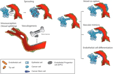
Angiogenesis is the process of new blood vessel formation from pre- existing vessels. It is broadly involved in tissue and organ generation during development, while in the adult it is limited to a few physiological events such as wound healing, skeletal morphogenesis, reproductive cycle, and pregnancy. Angiogenesis, however, occurs in several pathological condi-tions including chronic inflammation and cancer. The development of new vascular structures is crucial to support growing tissues or tumours with oxygen and nutrients and to allow for recirculation of immune surveillance cells. Angiogenesis occurs mostly by sprouting, migration, and proliferation of endothelial cells from pre-existing blood vessels (Figure 1). Several crucial pro-angiogenic factors have been iden-tified such as vascular endothelial growth factor A (VEGF-A), placental growth factor (PlGF), hepatocyte growth factor (HGF), fibroblast growth factors (FGFs), angiopoietins, ephrins, semaphorins, cytokines (interleukin [IL] 6), and chemokines (e.g. IL-8, stromal cell-derived factor 1 [SDF-1]) (Table 1). Endogenous inhibitors of angiogenesis have also been reported, including thrombospondin, tissue inhibitors of metalloproteinases, angiostatin, or endostatin (Table 1). The switch to the angiogenic phenotype in specific physiologi-cal and pathologiphysiologi-cal conditions is determined by the balance between pro- and anti-angiogenic factors. The angiogenic switch is a hallmark of malignant tumour progression.Figure 1 Mechanisms of vascularisation. Several mechanisms of vessel formation in normal tissues and tumours have been described. Angiogenesis, the sprouting of endothelial cells from pre-existing vessels. Vasculogenesis, driven by EPCs mobilised from the bone marrow in response to tumour-derived chemoattractants and differentiating into endothelial cells. Intussusception, the splitting of a preformed vessel into two daughter vessels. Vessel co-option, the appropriation of a pre-existing blood vessel, especially at the invading front. Vascular mimicry, the replacement of the vascular endothelial cell lining by tumour cells. Endothelial cell differentiation from putative cancer stem cells. The first three modes of vascularisation occur in both physiological and pathological conditions, whereas the other three are only observed in tumours.
Cancer cell Epithelial cell Endothelial cell
Cancer Stem cell Pericyte Endothelial Progenitor cell (EPC) Tip cell Angiogenic factors Sprouting Vessel co-option Vascular mimicry Intussusception
(Vessel splitting) Vasculogenesis
Bone marrow
Endothelial cell differentiation
Cancer cell Epithelial cell Endothelial cell
Cancer Stem cell Pericyte Endothelial Progenitor cell (EPC) Tip cell Angiogenic factors Sprouting Vessel co-option Vascular mimicry Intussusception
(Vessel splitting) Vasculogenesis
Bone marrow
Endothelial cell differentiation
Cancer cell Epithelial cell Endothelial cell
Cancer Stem cell Pericyte Endothelial Progenitor cell (EPC) Tip cell Angiogenic factors Sprouting Vessel co-option Vascular mimicry Intussusception
(Vessel splitting) Vasculogenesis
Bone marrow
Endothelial cell differentiation
Cancer cell Epithelial cell Endothelial cell
Cancer Stem cell Pericyte Endothelial Progenitor cell (EPC) Tip cell Angiogenic factors Sprouting Vessel co-option Vascular mimicry Intussusception
(Vessel splitting) Vasculogenesis
Bone marrow
Endothelial cell differentiation
Cancer cell Epithelial cell Endothelial cell
Cancer Stem cell Pericyte Endothelial Progenitor cell (EPC) Tip cell Angiogenic factors Sprouting Vessel co-option Vascular mimicry Intussusception
(Vessel splitting) Vasculogenesis
Bone marrow
Endothelial cell differentiation
Tumour-associated Vasculature
Tumour angiogenesis is key for tumour growth, invasion, and metastasis. In contrast to physiological angiogenesis, angiogenic tumour vessels are structurally and functionally abnormal and do not evolve to form fully mature vessels. Tumour vessels are very heterogeneous and chaotic with great variation in lumen size, excessive branching, and tortuous patterns. Tumour endothelial cells are poorly interconnected, leaky, and occa-sionally multilayered. Pericyte coverage and the basement membrane are also abnormal. As a consequence of these structural abnormalities,
tumour vessel function is severely compromised. Irregular perfusion and leakiness are two main functional consequences, resulting in heteroge-neous and insufficient delivery of oxygen and nutrients, increased inter-stitial pressure, facilitated intravasation, and the escape of tumour cells. The deficient and abnormal functionality of the tumour vasculature has three important consequences. Firstly, it induces a vicious cycle of angio- genesis, whereby starved tumour regions further promote angiogenesis to attract oxygen and nutrients in a compensatory self-sustaining man-ner. Secondly, hypoxic regions of the tumour microenvironment select hypoxia-resistant tumour cells that are also more aggressive and resistant to therapy. Thirdly, high interstitial pressure severely limits the delivery and diffusion of drugs, including chemotherapy, into the tumour, thus facilitating the selection of therapy-resistant cancer cells.
Tumours can also exploit pre-existing, normal blood vessels, particularly at the tumour periphery, in a process referred to as vascular co-option. Besides endothelial cell sprouting, additional modes of tumour vascularisation have been described. They include vascular mimicry, replacing the endothe-lial cells in the vessel wall with tumour cells, differentiation of tumour stem cells into endothelial cells, vasculogenesis, the de-novo formation of endothelial cells from recruited bone marrow-derived cells (BMDCs), endothelial progenitor cells (EPCs), and intussusception, the splitting of preformed vessels into two daughter vessels (Figure 1). Additional angio- genic signals originating from cells of the tumour microenvironment,
Table 1 Principal Pro- and Anti-angiogenesis Factors.
Pro-angiogenic factors Anti-angiogenic factors
Angiopoietin-1 Angiotensin
Epidermal growth factor Endostatin Fibroblast growth factor Thrombospondin
Hepatocyte growth factor Tissue inhibitors of metalloproteinases Insulin-like growth factor Vasostatin
Placental growth factor Platelet-derived growth factor Pleitropin
Transforming growth factors (α and β) Vascular endothelial growth factor
mainly from cancer-associated fibroblasts (CAFs) producing SDF-1/ CXCL12 and infiltrating inflammatory cells secreting a variety of fac-tors, further contribute to angiogenesis. Angiogenic inflammatory cells comprise a wide variety of BMDCs, including Tie2-expressing mono-cytes (TEMs), M2-polarised tumour-associated macrophages (TAMs), or
CD11b+ Gr1+ myeloid-derived suppressor cells which release soluble
fac-tors such as PlGF, Bv8, granulocyte-colony stimulating factor (G-CSF), and S100A8/9.
While physiological angiogenesis largely depends on VEGF, tumour angiogenesis can be induced by other factors such as FGFs, HGF, and SDF1. This explains, at least in part, the intrinsic or acquired resistance to VEGF-blockade therapy.
Anti-angiogenic Therapy in Cancer
Angiogenesis has been long considered an attractive therapeutic target in anti-cancer therapy. Traditionally, anti-angiogenic therapy has been designed to inhibit the formation of new blood vessels and to destroy existing ones with the purpose to starve the tumour. More recently, the notion of vascular normalisation, i.e. the reversal of the abnormal struc-tural and functional features described above, has been introduced with the aim to improve delivery of chemotherapy.
The prominent role of VEGF in promoting tumour angiogenesis made it an appealing target for therapeutic interventions. The anti-VEGF antibody bevacizumab has been the first United States Food and Drug Administration-approved anti-angiogenic drug for clinical use to treat several advanced, metastatic cancers (e.g. colorectal, breast, ovary, non-small cell lung [NSCLC], kidney). Anti-VEGF therapy prolongs pro-gression-free survival (PFS) and (only minimally) overall survival (OS). However, these benefits are rather limited (months). Multi-targeted pan-VEGF receptor tyrosine kinase inhibitors such as sunitinib, pazopanib, sorafenib, or vandetanib have been subsequently approved for various metastatic cancers (Table 2). VEGF blockade decreases vascular branch-ing, reduces homing of BMDCs, enhances endothelial cell sensitivity to chemotherapeutic drugs, and reduces leakiness and interstitial pressure.
Except for a few cancers, most notably liver cancer, kidney cancer, and glioblastoma, anti-angiogenic therapy provides benefit only in combi-nation with chemotherapy. It has been proposed that anti-angiogenic therapy may act by facilitating the activity of chemotherapy rather than having anti-tumour effects itself. The mechanism for this co-operation between anti-angiogenic and chemotherapy drugs is not fully elucidated and may include increased drug delivery, sensitisation of cancer cells to genotoxic drugs, or elimination of cancer stem cells. As some tumour cells express VEGF receptors (VEGFR) and use VEGF as a survival fac-tor, anti-VEGF therapy may also directly affect tumour cells.
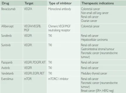
Table 2 Therapeutic Indications for Angiogenesis Inhibitors Currently Used in the Clinic.
Drug Target Type of inhibitor Therapeutic indications Bevacizumab VEGFA Monoclonal antibody Colorectal cancer
Non-small cell lung cancer Renal cell cancer Ovarian cancer Aflibercept VEGFA/VEGFB,
PIGF Chimeric VEGF/PIGFneutralising receptor Colorectal cancer Sorafenib VEGFR TKI Renal cell cancer
Hepatocellular carcinoma Sunitinib VEGFR TKI Renal cell cancer
Gastrointestinal stromal tumour Pancreatic cancer (neuroendocrine tumour)
Pazopanib VEGFR, PDGFR, KIT TKI Renal cell cancer
Axitinib VEGFR TKI Renal cell cancer
Vandetanib VEGFR, EGFR, RET TKI Medullary thyroid cancer Everolimus mTOR mTORC1 inhibitor Renal cell cancer
Pancreatic cancer (neuroendocrine tumour)
Breast cancer (ER+, HER2 neg) EGFR, Epidermal growth factor receptor; ER, oestrogen receptor; HER2, human epidermal growth factor 2 receptor; mTOR, mammalian target of rapamycin; PDGFR, platelet-derived growth factor receptor; PlGF, placental growth factor; TKI, tyrosine kinase inhibitor; VEGF, vascular endothelial growth factor.
Resistance to Anti-angiogenic Therapy
Despite the initial breakthrough of inhibiting VEGF/VEGFR in con-trolling tumour progression, with time it has become evident that not all patients respond to this therapy. Tumours can be either resistant to anti-angiogenics at start (intrinsic resistance) or become resistant during treatment (acquired resistance). Recent clinical trials with anti-angio-genic drugs in adjuvant settings showed that the initial benefit in PFS did not correlate with improved OS of patients, consistent with the patients’ development of acquired resistance to these drugs.
The mechanisms of resistance to anti-VEGF/VEGFR therapy are likely to be multiple and complex. One proposed mechanism is that tumours, besides VEGF, produce additional pro-angiogenic molecules such as PlGF, FGFs, or IL-8, thereby escaping VEGF/VEGFR blockade. Fur-thermore, tumours can use modes of vascularisation other than sprouting, such as co-option or intussusception, which are not necessarily depend-ent on VEGF. Further, anti-VEGF therapy can increase tumour hypoxia, thereby selecting hypoxia-resistant, aggressive cancer cell clones. Alter-natively, tumours can adapt to metabolic starvation by rearranging their energy metabolism, for example by switching to anaerobic metabolism in hypoxic areas, or by importing intermediate metabolites such as pyruvate or lactate produced in hypoxic regions to generate high amounts of aden-osine triphosphate (ATP) in oxygenated regions (metabolic symbiosis). Many strategies to overcome resistance to anti-angiogenic therapy have been proposed. In a recent preclinical model named “vascular promo-tion therapy”, the chemotherapeutic agent gemcitabine was used in combination with the anti-angiogenic drug cilengitide and the calcium channel blocker verapamil to increase vessel dilatation and blood flow. This resulted in improved chemotherapy delivery and activity-inhibiting tumour growth and distant metastasis formation, which was more effec-tive compared to gemcitabine treatment alone. This study raises the ques-tion of whether promoting rather than inhibiting tumour perfusion may have better therapeutic effects. Similarly, researchers have shown that increased vascular permeability induced by cilengitide, tumour necrosis factor-alpha (TNF-α), or histamine enhances chemotherapy delivery to the tumour, resulting in increased therapeutic efficacy.
Biomarkers of Tumour Angiogenesis
No validated biomarkers to monitor tumour angiogenesis, to prospectively identify responding patients, and to monitor the efficacy of anti-angiogenic drugs exist for clinical use. Availability of such a biomarker would allow the stratification of cancer patients into responders and non-responders and to possibly predict the development of drug resistance. Many molecules, in particular angiogenic factors including VEGF, have been investigated, but none has turned out to be clinically useful. In alternative to
circulat-ing molecules, mobilised CD45--circulating EPCs have been considered as
potential biomarkers, but with no better success. Other BMDCs, however, should be further explored. Recently, it has been reported that a bone
mar-row-derived immature B-cell population, CD45dim, CD31low, IgM+, IgD-,
acts as a surrogate marker for response to multiple anti-angiogenic drugs
in preclinical cancer models. We have shown that circulating CD11b+ cells
in breast cancer patients, but not in healthy donors, are pro-angiogenic. Circulating monocytes should be further considered as potential candidate biomarkers or a source of biomarkers.
Invasion and Metastasis
The main cause of death among cancer patients is metastatic colonisa-tion of distant sites leading to organ dysfunccolonisa-tion and failure. The lack of effective therapies against metastatic disease represents the greatest challenge to efficiently treat patients with advanced cancers. Therefore, in order to improve a cancer patient’s survival, innovative and effective strategies are needed to prevent and treat the disseminated disease. Recent advances in translational cancer research have greatly contributed to the understanding of the molecular and cellular mechanisms occurring in the tumour and in its microenvironment that promote metastasis. Deeper insights into these processes and translation to the clinic may raise unprec-edented opportunities in the management of metastatic disease.
The Metastatic Cascade
From a biologist’s point of view, metastases are the end product of an evolutionary process involving a sequence of discrete steps overcoming
the physical boundaries of the primary tumour and allowing formation of colonies at distant organ sites. This process is extremely inefficient (<0.01%). It is driven by a multitude of genetic and epigenetic altera-tions within the cancer cell, followed by selection steps in line with the concept of Darwinian evolution. Genomic instability, as promoted by the inactivation of DNA repair mechanisms, is key to the acquisition of alterations necessary to gain metastatic capacity. The tumour-suppressor protein p53, which normally acts as a gatekeeper of genomic integrity by inducing cell cycle arrest or apoptosis in response to DNA damage, is lost in about 50% of all cancers. Inactivation of this oncosuppressor increases the risk of acquiring metastatic capacity. In addition, loss of p53 promotes the angiogenic switch and favours cancer cell resistance to genotoxic drugs. The tumour microenvironment, in particular inflamma-tory cells, plays multiple critical roles in promoting metastasis.
The main events of the metastatic cascade are described below (Figure 2). 1. Local tissue invasion
The first step of the metastatic cascade is local invasion. Inflammatory cells and activated stromal fibroblasts produce matrix metalloproteinases, chemokines, and growth factors that activate migratory programmes in subsets of cancer cells. Those cells undergo drastic epigenetic repro-gramming by switching from a static epithelial phenotype to a highly motile mesenchymal phenotype through a programme known as epithe-lial–mesenchymal transition (EMT). EMT can be induced by several factors (e.g. transforming growth factor-beta [TGF-β], epidermal growth factor [EGF], and FGFs), pathways (e.g. Wnt/β-catenin, Notch, HIF-1α, and Ras), and activating transcription factors such as Slug, Snail, Twist, and Zeb.
2. Intravasation and circulation
After breaching the basement membrane, tumour cells penetrate hae-matic or lymphatic vessels. Once in the blood or lymphatic circulation, they rapidly redistribute to distant secondary host tissues. To withstand mechanical shear stress and immunological attack during the travel within the circulatory system, circulating tumour cells (CTCs) can circu-late as small clusters, camouflaged by pcircu-latelets sticking to their surface.
3. Arrest at distant sites
The CTCs arrest at foreign organs through mechanical trapping in the capillaries due to vessel size restriction or by cell surface receptor inter-actions with endothelial cells lining the target organ vasculature. 4. Extravasation and seeding
Upon arrival at their final destination, the tumour cells engage molecular mechanisms to extravasate and invade the target organ. This involves the secretion of matrix metalloproteinases, degradation of the basal mem-brane and the extracellular matrix, and integrin-dependent migration.
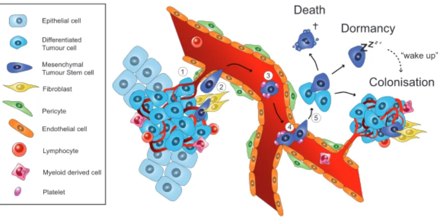
Figure 2 Cellular and molecular mechanisms in metastatic cancer progression. 1. At the site of the primary tumour, cancer cells interact with the stroma where various cell types are found, including fibroblasts, lymphocytes, and myeloid cells. Heterotypic interactions between the tumour and the stroma induce an epithelial–mesenchymal transition (EMT), which promotes tumour cell invasion into the adjacent tissues. 2. Gain of motility results in nearby vessel invasion, intravasation, and mesenchymal tumour cell shedding into the host circulation. 3. Survival in the blood stream is augmented by platelets covering the surface of the circulating tumour cells. 4. Arrest at a distant organ site can be mediated by homing molecules or may occur mechanically due to vessel size restriction. 5. Extravasation across the endothelial layer, local tissue invasion, and subsequent adaptation to the foreign environment are mandatory steps to avoid cell death. Alternatively, metastasis-initiating cells may reside in a dormant state for extended periods of time before environmental factors initiate mesenchymal–epithelial transition, proliferation, and colonisation resulting in the formation of clinically relevant metastases.
ZZZZ Death Dormancy Colonisation 1 2 3 4 5 “wake up” Fibroblast Platelet Endothelial cell Differentiated Tumour cell Epithelial cell Mesenchymal Tumour Stem cell
Pericyte
Myeloid derived cell Lymphocyte ZZZZ Death Dormancy Colonisation 1 2 3 4 5 “wake up” Fibroblast Platelet Endothelial cell Differentiated Tumour cell Epithelial cell Mesenchymal Tumour Stem cell
Pericyte
Myeloid derived cell Lymphocyte
5. Colonisation
In order to give rise to clinically detectable metastases, additional changes must occur in tumour cells. The reversion of EMT in a process known as mesenchymal–epithelial transition is necessary in order to regain prolif-eration after migration. The non-permissive foreign environment of the target organ challenges the survival of tumour cells. Local immune cells initiate mechanisms to eliminate invading tumour cells, which have to adapt to the foreign tissue by co-opting the colonised tissue stroma and modulating the environment to their own advantage. The suppression of the anti-tumour immune response, the initiation of angiogenesis, and the release of growth and survival factors are strategies that metastatic cells may adopt in order to successfully colonise the secondary organ.
Metastatic Organotropism
The propensity of a metastatic tumour cell to colonise a certain organ is called “organotropism”. Breast cancers, for instance, form metastases at many sites including the lung, bone, and liver, whereas colon and prostate cancers preferentially metastasise to liver and bone, respectively. This can-not be explained by the vascular anatomy alone. The “seed and soil hypoth-esis” proposed by Stephen Paget hypothesised that the intrinsic ability of tumour cells (“seeds”) to grow in a particular permissive organ (“soil”) determines the pattern of tumour spread. In analogy, the nature of the seed and its compatibility with the soil determine the outcome of the planting. In recent years, gene expression profiling derived from human tumour samples and preclinical models of organ-specific metastasis have helped researchers to better understand the mechanisms of organotropism. It has become clear that metastatic cells are distinct from the primary tumour on both genetic and epigenetic levels. Genome-wide transcriptome stud-ies have identified genes that mediate metastasis to various organs, in particular to the bone, the lung, and the brain. Analysis of pathways involved with these targets elucidated their role in metastatic progres-sion. Research has shown that most of the genetic and epigenetic altera-tions required for metastasis formation are acquired within the primary tumour; only a few subsequent modifications appear to be necessary to unlock optimal metastatic capacity.
These recent discoveries have enabled researchers to take more inno-vative approaches to preventing metastatic colonisation or treating established metastases. Further translational clinical trials are needed to validate targets and to test drugs in cancer patients with an invasive or aggressive tumour or metastatic disease.
Metastatic Dormancy
Clinical evidence shows that the timeframe in which metastases develop differs greatly. For example, patients with pancreatic and small cell lung cancers often have clinically manifest metastases at the time of diagnosis and may die within months. In contrast, patients with breast and prostate cancers and melanoma develop clinically relevant metastases years or even decades after resection of the primary tumour. This extended latent phase suggests that tumour cells undergo long periods of dormancy. Metastatic dormancy can be defined as the ability of individual or small clusters of disseminated cancer cells to remain viable over prolonged periods of time without evidence of productive growth. Dormant tumour cells represent a major problem in the management of metastatic disease, as they are virtually undetectable (with the exception of cancer cells disseminated in the bone marrow) and resistant to adjuvant chemo- therapies designed to target rapidly proliferating cells.
Three mechanisms of dormancy have been proposed: (i) cellular
dor-mancy, i.e. the cancer cells reside in G0 cell cycle phase and do not
prolif-erate; (ii) immunological dormancy, i.e. cancer cells are kept in check by the immune system; (iii) angiogenic dormancy, i.e. cancer cell prolifera-tion is counterbalanced by cell death due to the inability to induce angio-genesis. Lack of integrin engagement with extracellular matrix anchorage and subsequent lack of activation of kinases (e.g. focal adhesion kinase) prevents the transition from a quiescent to a proliferative state. Tumour cells may also be exposed to stroma-derived growth-suppressive signals. For instance, expression of bone morphogenetic protein antagonist by dormant tumour cells was shown to terminate the quiescent state and initiate metastatic outgrowth in the lung.
Concluding Remarks and Outlook
The discovery and understanding that cancer progression are not fully cell-autonomous events but rather depend on dynamic, reciprocal inter-actions with the surrounding normal host cells/tissue has changed the way we understand cancer biology nowadays, and has created oppor-tunities for novel diagnostic and therapeutic modalities. Anti-angiogen-esis was the first therapeutic approach to demonstrate in patients that targeting microenvironmental cues can lead to clinical benefits. Lack of biomarkers for patients’ stratification, limited survival benefits, and development of resistance are the main challenges in the field of anti-angiogenic therapy. Impinging on metabolic adaptation should be con-sidered as a strategy to counterbalance the effects of starvation on the tumour cells induced by anti-angiogenic drugs. Targeting the recruitment of inflammatory cells into the tumour microenvironment might be envis-aged to break the vicious circle of compensatory angiogenesis following therapy-induced hypoxia and necrosis. Lastly, strategies increasing ves-sel permeability should be explored in combination with chemotherapy. The development of effective therapies to prevent and treat metastasis is a top priority in experimental, translational, and clinical cancer research. The understanding of metastasis is progressing at an unprecedented pace. Mechanisms have been unravelled and many potential targets identified. Cancer immunotherapy is providing impressive benefits in patients with advanced metastatic disease. Immunomodulatory therapy may be applied to patients at risk for metastatic progression before overt metastases appear. Although researchers have not yet found a cure for cancer, important advances in treatment strategies developed in preclinical, clinical, and translational research studies may hopefully continue to improve the overall outcome for cancer patients and cancer survivors.
Declaration of Interest:
Professor Rüegg has declared that he is a founder and stockholder of Diagnoplex and Novigenix.
Dr Wyss has reported no conflicts of interest. Dr Lorusso has reported no conflicts of interest.
Further Reading
Aguirre-Ghiso JA. Models, mechanisms and clinical evidence for cancer dor-mancy. Nat Rev Cancer 2007; 7:834–846.
Carmeliet P, Jain RK. Molecular mechanisms and clinical applications of angio-genesis. Nature 2011; 473:298–307.
Chung AS, Ferrara N. Developmental and pathological angiogenesis. Annu Rev Cell Dev Biol 2011; 27:563–584.
Fidler IJ. The pathogenesis of cancer metastasis: the ‘seed and soil’ hypothesis revisited. Nat Rev Cancer 2003; 3:453–458.
Lorusso G, Rüegg C. New insights into the mechanisms of organ-specific breast cancer metastasis. Semin Cancer Biol 2012; 22:226–233.
Oskarsson T, Batlle E, Massagué J. Metastatic stem cells: sources, niches, and vital pathways. Cell Stem Cell 2014; 14:306–321.
Pantel K, Speicher MR. The biology of circulating tumor cells. Oncogene 2015; Jun 8 [Epub ahead of print].
Potente M, Gerhardt H, Carmeliet P. Basic and therapeutic aspects of angiogen-esis. Cell 2011; 146:873–887.
Sennino B, McDonald DM. Controlling escape from angiogenesis inhibitors. Nat Rev Cancer 2012; 12:699–709.
Sessa C, Guibal A, Del Conte GL, et al. Biomarkers of angiogenesis for the development of antiangiogenic therapies in oncology: tools or decorations? Nat Clin Pract Oncol 2008; 5:378–391.
Valastyan S, Weinberg RA. Tumor metastasis: molecular insights and evolving paradigms. Cell 2011; 147:275–292.
Welti J, Loges S, Dimmeler S, et al. Recent molecular discoveries in angiogenesis and antiangiogenic therapies in cancer. J Clin Invest 2013; 123:3190–3200.