TRANSLATIONAL RESEARCH
Volatile anaesthetics reduce neutrophil inflammatory
response by interfering with CXC receptor-2 signalling
B. Mu¨ller-Edenborn
1,2, R. Frick
1,2, T. Piegeler
1,2,3, M. Schla¨pfer
1,2, B. Roth-Z’graggen
1,2, A. Schlicker
1and B. Beck-Schimmer
1,2*
1
Institute of Anaesthesiology, University Hospital Zurich, Zu¨rich, Switzerland
2Institute of Physiology, Zurich Center of Integrative Human Physiology, University of Zurich, Zu¨rich, Switzerland 3
Department of Anesthesiology, University of Illinois Hospital & Health Sciences Center, Chicago, IL, USA * Corresponding author. E-mail: [email protected]
Editor’s key points
† Accumulation of
neutrophils contributes to ischaemia-reperfusion injury.
† The role of CXC signalling in the mechanism of sevoflurane- and desflurane-mediated protection was studied in human neutrophils. † Sevoflurane and
desflurane interfered with CXC receptor-2 signalling.
Background. Growing evidence suggests a protective effect of volatile anaesthetics in ischaemia-reperfusion (I/R)-injury, and the accumulation of neutrophils is a crucial event. Pro-inflammatory cytokines carrying the C-X-C-motif including interleukin-8 (IL-8) and CXC-ligand 1 (CXCL1) activate CXC receptor-1 (CXCR1; stimulated by IL-8), CXC receptor-2 (CXCR2; stimulated by IL-8 and CXCL1), or both to induce CD11b-dependent neutrophil transmigration. Inhibition of CXCR1, CXCR2, or both reduces I/R-injury by preventing neutrophil accumulation. We hypothesized that interference with CXCR1/CXCR2 signalling contributes to the well-established beneficial effect of volatile anaesthetics in I/R-injury.
Methods.Isolated human neutrophils were stimulated with IL-8 or CXCL1 and exposed to volatile anaesthetics (sevoflurane/desflurane). Neutrophil migration was assessed using an adapted Boyden chamber. Expression of CD11b, CXCR1, and CXCR2 was measured by flow cytometry. Blocking antibodies against CXCR1/CXCR2/CD11b and phorbol myristate acetate were used to investigate specific pathways.
Results.Volatile anaesthetics reduced CD11b-dependent neutrophil transmigration induced by IL-8 by .30% and CD11b expression by 18 and 27% with sevoflurane/desflurane, respectively. This effect was independent of CXCR1/CXCR2 expression and CXCR1/CXCR2 endocytosis. Inhibition of CXCR1 signalling did not affect downregulation of CD11b with volatile anaesthetics. Blocking of CXCR2-signalling neutralized effects by volatile anaesthetics on CD11b expression. Specific stimulation of CXCR2 with CXCL1 was sufficient to induce upregulation of CD11b, which was impaired with volatile anaesthetics. No effect of volatile anaesthetics was observed with direct stimulation of protein kinase C located downstream of CXCR1/CXCR2.
Conclusion.Volatile anaesthetics attenuate neutrophil inflammatory responses elicited by CXC cytokines through interference with CXCR2 signalling. This might contribute to the beneficial effect of volatile anaesthetics in I/R-injury.
Keywords: anaesthetics, inhalation, desflurane; anaesthetics, inhalation, sevoflurane; receptors, chemoreceptors
Accepted for publication: 12 February 2014
Ischaemia-reperfusion injury (I/R-injury) during anaesthesia occurs as an unforeseen event, for example, transient myocar-dial ischaemia or as a planned step in surgery such as the Pringle manoeuvre during liver resection. Volatile anaesthetics protect different organs from I/R-injury such as liver, lung, and heart.1–3However, the mechanism of this protective effect
remains unclear.
The restoration of blood flow after ischaemia activates innate and adaptive immune responses leading to an
accumulation of neutrophils in the reperfused organ and sub-sequent tissue damage.4Consequently, inhibition of neutrophil
invasion during reperfusion was demonstrated to reduce the extent of tissue damage.5–7
The accumulation of neutrophils in I/R-injury is the result of a three-step process: first, circulating neutrophils establish a low-affinity adhesive interaction with the endothelium called rolling.8This rolling is mediated byL-selectin (CD62L), which
surface that binds endothelial carbohydrate determinants. In a second step, the neutrophil adheres firmly to the endothelium through adhesion between integrins on the neutrophil surface and endothelial intercellular adhesion molecules. The most prominent integrin during this process is CD11b, which binds to endothelial intercellular adhesion molecule-1. The third and final step, which involves various adhesive glycoproteins including CD62L and CD11b, is transmigration of the neutrophil through the endothelium.
The family of CXC cytokines is defined by two N-terminal cystines separated by one amino acid (hence C-X-C). A subgroup of these CXC cytokines carries a Glu-Leu-Arg tripeptide (ELR) motif at the NH2-end and is termed ELR+ CXC cytokines. This subgroup includes interleukin-8 (IL-8) and promotes the recruitment of neutrophils into inflamed tissues.9ELR+ cyto-kines bind to two G protein-coupled receptors on the neutrophil surface, CXC receptor-1 (CXCR1) and CXC receptor-2 (CXCR2).10
11 Signalling through these receptors is important during I/
R-injury and pharmacological inhibition was demonstrated to reduce neutrophil infiltration and subsequent tissue damage.12 We hypothesized that the beneficial effects of volatile anaes-thetics during I/R-injury are due to direct or indirect effects on CXCR1 and CXCR2 signalling that might alter the process of neu-trophil accumulation. We chose IL-8 as a representative ELR+ cytokine as it binds to both CXCR1 and CXCR2 with high affinity and is released by leucocytes and stromal cells such as fibro-blasts in high concentrations during reperfusion.13–16
Methods
The study protocol was approved by the ethics committee for studies on humans of the University Hospital Zurich (KEK-ZH 2012-0274) and written informed consent was obtained. Twelve healthy volunteers (7 males/5 females, aged 19–52
yrs) were recruited from the Institute of Physiology in Zurich and blood samples (5 ml) were obtained from an antecubital vein into citrate tubes. Exclusion criteria were acute disease in the last 14 days or chronic disease with or without medical treatment of any type. Oral contraceptives were accepted for female donors. Red blood cells were lysed and neutrophils were isolated using Ficoll-Histopaque 1077 (Sigma-Aldrich, Buchs, Switzerland) as described previously.17The isolated neu-trophils were resuspended at a concentration of 2×10⁶ml21in Ham’s F-12 medium supplemented with 10% heat-inactivated fetal bovine serum, 5% penicillin/streptomycin (10 000 U litre21), and 5% 4-(2-hydroxyethyl)-1-piperazineethanesulfonic acid (all from Invitrogen/Life Technologies, Zug, Switzerland). All steps were performed at 48C to prevent neutrophil activation.
Fifty microlitres of neutrophil cell suspension (105 neutro-phils) were placed into sterile 96-well plates and exposed to 10 nM IL-8 (recombinant human IL-8, BD Pharmingen, Allschwil, Switzerland) or 200 ng ml21CXC-ligand 1 (CXCL1) (re-combinant human CXCL1, R&D systems, Wiesbaden, Germany). Phorbol myristate acetate at 10 nM (Sigma-Aldrich) was used to activate protein kinase C in the corresponding experiments. These concentrations are commonly used and known to induce profound neutrophil activation and migration. To study the effect of I/R in our setting, human lung microvascu-lar endothelial cells were exposed to 12 h of hypoxia (0.2% O2),
followed by 12 h of reoxygenation at 21% O2. Neutrophils were
then stimulated with the harvested supernatants at 1:1 dilution. In some experiments, neutrophils were incubated for 15 min before stimulation with 10 mg ml21of anti-human CXCR1 and anti-human CXCR2 (Abcam, Cambridge, UK) or against the acti-vation epitope of CD11b (anti-human CBRM1/5, Biolegend, Lucerne, Switzerland). Plates were then put in humified airtight chambers (Oxoid anaerobic jar; Oxoid AG, Basel, Switzerland).
Anesthetic IL-8 (10 nM) Air – 0 20 40 P
ercentage of total cell count
60 80
Viable (Annexin–/7AAD–)
Apoptotic (Annexin+/7AAD–)
Necrotic (Annexin+/7AAD+)
100 Sevo – Des – Air + Sevo + Des +
Fig 1Viability of neutrophils exposed to IL-8 with and without sevoflurane or desflurane. Viable neutrophils are negative for annexin and 7-aminoactinomycin (7AAD, Annexin2
/7AAD2
, blue). Apoptotic neutrophils are positive for annexin and negative for 7AAD (Annexin+/7AAD2 , green). Necrotic cells are positive for annexin and 7AAD (Annexin+/7AAD+, pink). Median, inter-quartile and full range shown (n¼6).
Chambers were flushed with air/5% CO2containing 2.2 vol%
sevoflurane (Sevoranew; Abbott AG, Baar, Switzerland,
corre-sponding vaporizer: Sevotec5w; Abbott AG) or 6% vol%
desflur-ane (Forenew, Baxter, Switzerland; corresponding vaporizer:
Tec6, Carbamed, Switzerland). Control cells were exposed to air/5% CO2only. Neutrophils from each donor were
investi-gated under all the three conditions. Concentrations of volatile anaesthetics were measured with the Ohmeda 5330 Agent Monitor (Abbott AG). After reaching the described concentra-tions, the chambers were sealed and kept in an incubator at 378C (Bioblock, Ittingen, Switzerland). Volatile anaesthetic con-centrations were checked again at the end of the incubation period to ensure that there was no loss of anaesthetic from in-sufficient sealing or evaporation.17
A 96-well migration plate with 3-mm pores (Millipore-Merck, Zug, Switzerland) was used to investigate neutrophil migration. Fifty microlitres of neutrophil suspension (105cells) was added to the upper compartment of the plate. Medium supplemented with 10 nM IL-8 was added to the lower compartment. Plates were exposed to volatile anaesthetics as described above im-mediately after assembly of the plate and neutrophils were allowed to migrate for 1 h. Migrated neutrophils were quanti-fied using an automated optical cell counter (TC-10, Biorad, Cressier, Switzerland).
Surface expression of CXCR1, CXCR2, CD11b, and CD11b activation epitope was quantified using flow cytometry. Neu-trophils were kept on ice and fixed with 2% paraformaldehyde to prevent changes in receptor expression during staining. Cells were stained with specific antibodies for 30 min at 48C, washed twice, and measured on a FACS Canto II (BD Pharmingen). Flow cytometry raw data were analysed using the FlowJo-Software for Macintosh (version 8.8.6, Tree Star Inc., Ashland, OR, USA). The following mouse anti-human antibodies were used (all antibodies from BD Pharmingen): APC-Cy7-conjugated CD11b (final staining concentration 2 mg ml21), APC-conjugated CXCR1 (1.25 mg ml21), and FITC-conjugated CXCR2 (0.25 mg ml21). FITC-conjugated CBRM1/5 (0.2 mg ml21) was obtained from Biolegend. Appropriate isotype-control antibodies were used to quantify nonspecific binding.
Neutrophil viability was quantified by staining the cells with annexin-V and 7-aminoactinomycin (PE Annexin V Apoptosis Detection Kit I, BD Pharmingen). IL-8 concentrations in super-natants were quantified using an enzyme-linked immunosorb-ent assay according to the manufacturer’s protocol (R&D systems). This kit has a sensitivity range from 31.2 to 2000 pg ml21and all samples were diluted to achieve a concentration in the linear range.
Distribution of the data was assessed using the Kolmo-gorov– Smirnov test. Normally distributed data were analysed using a one-way ANOVA with Bonferroni post hoc test. Non-parametric data were analysed using Kruskal–Wallis and Dunn’s multiple comparison tests. All experiments were per-formed with blood from three or more different donors with conditions repeated in duplicates. A P-value of ,0.05 was con-sidered statistically significant. Statistical analyses and graph creation were executed with Graphpad Prism 6 for Mac (Graph-pad Software, La Jolla, CA, USA).
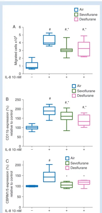
– + + + 0 IL-8 10 nM 1 2 Mig rated cells x10 4 3 4 5 6 A # #,* #,* Air Sevoflurane Desflurane – + + + 0 IL-8 10 nM 50 100 CD11b e xpression (%) relativ e to control 150 200 250 B # #,* #,* Air Sevoflurane Desflurane – + + + 0 IL-8 10 nM 50 100 CBRM1/5 e xpression (%) relativ e to control 150 200 C # * * Air Sevoflurane Desflurane
Fig 2 (A) Neutrophil transmigration in response to IL-8 in the presence of sevoflurane or desflurane for 1 h. #P,0.0001 vs control; *P,0.05 vs IL-8 stimulated neutrophils exposed to air/5% CO2during transmigration. n¼9. (B) Neutrophil CD11b expression after 1 h exposure to IL-8 with and without sevoflur-ane or desflursevoflur-ane. Mean fluorescence of CD11b relative to control neutrophils not exposed to IL-8 shown. #P,0.0001 vs control neutrophils without IL-8; *P,0.05 vs IL-8 treated neutrophils without anaesthetic. (n¼12). (C) Neutrophil activation-epitope of CD11b-specific CBRM1/5 expression. Mean fluorescence of the antibody clone relative to control neutrophils not exposed to IL-8 shown.#P,0.001 vs control neutrophils without IL-8; *P,0.05 vs IL-8 treated neutrophils without anaesthetic. Median, inter-quartile, and full range shown (n¼6).
Results
Our isolation procedure yielded neutrophils with a purity of .94% and a viability of .99% (data not shown). Exposure to volatile anaesthetics did not reduce neutrophil viability (Fig.1). Exposure to IL-8 induced transmigration of 43% of neutro-phils within 1 h; exposure to sevoflurane or desflurane reduced migration by 36 and 32% (Fig.2A; P¼0.012 for
sevo-flurane, P¼0.032 for desflurane). Neutrophil transmigration in this setting was dependent upon CD11b and abrogated by CD11b-blocking antibodies (Supplementary Fig. S1). Accord-ingly, upregulation of CD11b after stimulation with IL-8 was attenuated by both sevoflurane and desflurane (Fig. 2B;
P¼0.013 for sevoflurane, P¼0.001 for desflurane). Staining for the activation-epitope of CD11b revealed a similar pattern with lower levels of activated CD11b in neutrophils exposed to sevoflur-ane and desflursevoflur-ane (Fig.2C; P¼0.002 for sevoflurane, P¼0.02 for
desflurane). Reduction of CD11b expression with volatile anaes-thetics was also observed when neutrophils were stimulated with supernatants derived from hypoxia/reoxygenation-exposed endothelial cells (Supplementary Fig. S2).
Stimulation of neutrophils with IL-8 resulted in marked endocytosis and therefore reduction of surface expression of CXCR1 by 47% and CXCR2 by 81% (Fig.3A; P,0.0001). Neither
sevoflurane nor desflurane affected this process. Also, baseline CXC receptor expression in resting neutrophils was not affected by volatile anaesthetics (data not shown). The extent of endo-cytosis of the ligand–receptor complexes formed by IL-8 and CXCR1 or CXCR2 resulted in consumption and hence decrease of free IL-8 in the medium during incubation (Fig. 3B,
P¼0.008). Again, this was not influenced by sevoflurane or desflurane (Fig.3B).
Direct stimulation of protein kinase C with phorbol myristate acetate resulted in upregulation of CD11b. No effect of sevo-flurane and dessevo-flurane was observed in this setting (data not shown).
Blocking antibodies against CXCR1 and CXCR2 inhibited the upregulation of CD11b in response to IL-8 (Fig.4A; P,0.001 and
P¼0.01, respectively). However, blocking of CXCR2 before stimulation neutralized the effect of volatile anaesthetics, while blocking of CXCR1 resulted in preserved attenuation of CD11b with sevoflurane and desflurane (Fig.4B; P,0.05 for
both sevoflurane and desflurane). In addition, stimulation with CXCL1, to activate CXCR2, reproduced the effect of volatile anaesthetics (Fig. 4C; P,0.01 for sevoflurane, P,0.001 for
desflurane).
Conclusions
Our study demonstrates that volatile anaesthetics interfere with neutrophil inflammatory pathways, leading to decreased neutrophil migration and reduced expression of b2-integrin
CD11b. We locate the site of action of volatile anaesthetics to be downstream of CXCR2, a target receptor of ELR+CXC cyto-kines that are involved in I/R-injury.
We focused the current study on the specific impact of vola-tile anaesthetics on neutrophil signalling through CXCR1 and CXCR2. The importance of the ELR+family of cytokines that signals through these receptors is well established and their in-hibition emerges as a new therapeutic option in I/R-injury.9 IL-8 is a suitable cytokine to represent the ELR+ family because of its high affinity to both of the ELR+target receptors, CXCR1 and CXCR2.18In addition, high levels of IL-8 are released during I/R-injury, and neutralization of IL-8 was found to attenuate tissue damage during I/R-injury.16
The accumulation of neutrophils in the reperfused organ after ischaemia is a multi-step process eventually leading to local tissue damage.4Inhibition of this event through various
means such as depletion of neutrophils or antibodies against neutrophil adhesion molecules decreases tissue injury.7 19
The ability of neutrophils to transmigrate through the endothe-lium is pivotal to their accumulation in the reperfused organ.
– + 0 IL-8 10 nM PMN present IL-8 10 nM 50 Expression (%) relativ e to control IL-8 concentr ation in medium (ng ml –1 ) 100 150 CXCR1 CXCR2 A B # # # # # # – + + + + + Air Sevoflurane Desflurane + + 0 40 20 50 80 100 n.s. n.s. + + + +
Fig 3(A) CXCR1 (left panel) and CXCR2 (right panel) expression in neutrophils exposed to IL-8 for 1 h with and without sevoflurane or desflurane. Mean fluorescence relative to control neutrophils without IL-8 shown.#P,0.001 vs control neutrophils without IL-8 (n¼6). (B) IL-8 concentrations in medium from neutrophils polymorphonuclear leucocyte (PMN) cultured with IL-8 for 1 h with and without sevoflurane or desflurane. Dashed line shows measured IL-8 concentration in the absence of neutrophils (expected concentration 80 ng ml21). No effect of sevoflurane or desflurane in IL-8 treated neutrophils. Median, inter-quartile, and full range shown (n¼8).
We found that sevoflurane and desflurane reduced IL-8 induced transmigration of neutrophils.
The process of transmigration is mediated by several neu-trophil and endothelial adhesive proteins. However, inhibition of the neutrophil– endothelial interaction between CD11b on the neutrophil surface and endothelial intercellular adhesion molecule-1 proved to be most important.20CD11b is critical to establish tight adherence of neutrophils to the endothelium, and inhibition of CD11b using antibodies blocks neutrophil recruitment and reduces tissue damage both in vitro and in vivo.21 22 In our setting, neutrophil transmigration was dependent on CD11b expression and both sevoflurane and desflurane attenuated the upregulation of CD11b in response to IL-8. These findings concur with previous reports demon-strating the inhibition of neutrophil –endothelial interactions with isoflurane and sevoflurane.23 24
To elucidate the site of action of volatile anaesthetics, expres-sion of the ELR+receptors CXCR1 and CXCR2 which are activated by IL-8 was determined. These receptors undergo endocytosis upon ligand binding followed by a recycling step back to the membrane, which presumably serves to reduce neutrophil ac-tivity in response to high chemokine concentrations at inflam-matory sites.25We found that IL-8 decreased the expression of CXCR1 and CXCR2. Exposure to volatile anaesthetics did not influence this effect, nor did it lead to a more pronounced down-regulation in resting neutrophils. We further investigated the effect of endocytosis of the CXCR1 and CXCR2 ligand–receptor-complex on free IL-8 levels. The marked decrease in membrane CXCR1 and CXCR2, and hence high endocytosis of receptor-bound IL-8, resulted in diminished levels of IL-8 in the medium and volatile anaesthetics did not affect this. These observations suggest that volatile anaesthetics do not induce changes in CXCR1 or CXCR2 surface expression and endocytosis, which might have altered neutrophil activity.
Ligand binding to the G protein-coupled receptors CXCR1 and CXCR2 leads to dissemination of the G protein into the GTP-bound Gai and the Gbg subunit. Gai then increases the ac-tivity of phosphatidyl-inositide-3 kinase while Gbg activates phospholipase C. This leads to an increase of diacylglycerol that activates protein kinase C, which in turn induces CD11b ex-pression.26We found that phorbol myristate acetate, an ana-logue of diacylglycerol that directly activates protein kinase C without involvement of surface receptors, upregulates CD11b. However, no effect of volatile anaesthetics was observed in this setting. This suggests that volatile anaesthetics alter the neutrophil ELR+pathway at a site upstream of protein kinase C. We next investigated whether the effects of volatile anaes-thetics were attributable to downstream pathways specific to CXCR1 or CXCR2. Indeed, blocking of CXCR2 abrogated the impact of both sevoflurane and desflurane on CD11b expres-sion. However, blocking of CXCR1 preserved the effects of vola-tile anaesthetics, although at a lower level of CD11b expression. We then used CXCL1, a cytokine of the same family as IL-8, but which binds only to CXCR2. In line with the above findings, we found that stimulation with CXCL1 reproduced neutrophil in-hibition by volatile anaesthetics. Sevoflurane and desflurane
– – – + – – + + – + – + 0 IL-8 10 nM Anti-CXCR1 Ab Anti-CXCR2 Ab 50 CD11b e xpression (%) relativ e to control 100 150 200 250 A # * * Air – – – + – – + + – + – + 0 IL-8 10 nM Anti-CXCR1 Ab Anti-CXCR2 Ab 50 CD11b e xpression (%) relativ e to control 100 150 200 250 B # # # * * Air Sevoflurane Desflurane – + + + 0 CXCL1 200 ng ml–1 CD11b e xpression (%) relativ e to control 100 200 300 400 C # * * Air Sevoflurane Desflurane
Fig 4 (A) Effect of anti-CXCR1- or anti-CXCR2-antibodies on CD11b expression in neutrophils with and without IL-8. Mean fluorescence of CD11b relative to control neutrophils without IL-8 shown. #
P,0.0001 vs control, *P,0.05 vs IL-8 alone (n¼8). (B) Effect of anti-CXCR1- or anti-CXCR2-antibodies on CD11b expression in neu-trophils with and without exposure to sevoflurane or of desflurane. Mean fluorescence of CD11b relative to control neutrophils without IL-8 shown. #P,0.0001 vs untreated control, *P,0.05 vs IL-8 without anaesthetics (n¼6). (C) Effect of CXCL1 on CD11b expres-sion on neutrophils exposed to sevoflurane or desflurane. Mean fluorescence of CD11b relative to neutrophils without CXCL1 shown. #P,0.05 vs neutrophils without CXCL1, *P,0.01 vs without anaesthetic. Median, inter-quartile, and full range shown (n¼6).
therefore seem to affect IL-8-induced neutrophil activation downstream of CXCR2. A particular role of CXCR2 during I/R-injury was also reported by Tarzami and colleagues who investigated the outcome of experimental myocardial infarction in CXCR22/2(knockout) mice: not only were infarct sizes smaller, suggesting decreased tissue damage but also the number of infiltrating neutrophils was reduced in the infarcted area.27In line with our finding, this also suggests that CXCR2 signalling is important for leucocyte recruitment in I/R-injury.
Our experimental setup uses an in vitro system of isolated neutrophils and as such is limited as possible interactions of neutrophils with other cell types involved in I/R-injury in vivo cannot be quantified. We chose this approach as it allows us to investigate pathways and inflammatory responses specific to neutrophils while avoiding expected or unanticipated cell– cell signalling.
Our results provide insight into how volatile anaesthetics can influence the accumulation of neutrophils in I/R-injury through inhibition of neutrophil migration and b2-integrin expression.
Both sevoflurane and desflurane had comparable effects and seem to interfere within the CXCR2 signalling pathway at a site of action upstream of protein kinase C and downstream of CXCR2. Inhibition of CXCR2 signalling might therefore contribute to the well-established anti-inflammatory effect of volatile anaesthetics for the development of I/R-injury in vivo.
Authors’ contributions
B.M.-E. designed and conducted the study, analysed the data, and drafted the manuscript; R.F. contributed to study design and conduct, data analysis, and manuscript preparation; T.P. contributed to data analysis and manuscript preparation; M.S. contributed to conducting the study and revision of the manuscript; B.R.-Z. and A.S. contributed to study design and conduct; B.B.-S. contributed to study design, data analysis, and manuscript preparation.
Supplementary material
Supplementary material is available at British Journal of Anaesthesia online.
Acknowledgements
We thank Livia Reyes and Christa Booy of the Institute of Physiology, Zurich Center for Integrative Human Physiology for their excellent technical assistance and constant support.
Declarations of interest
B.M.-E., R.F., T.P., M.S., B.R.-Z., and A.S. have no conflicts of inter-est to declare. B.B.-S. has received honoraria for advisory board meetings and research grants from Abbott, Switzerland, and Baxter Switzerland, but not for the current study.
Funding
This study was funded by Swiss National Science Foundation, Berne, Switzerland, Grant No. 320030_141216.
References
1 Beck-Schimmer B, Breitenstein S, Urech S, et al. A randomized controlled trial on pharmacological preconditioning in liver surgery using a volatile anesthetic. Ann Surg 2008; 248: 909 – 18
2 De Conno E, Steurer M, Wittlinger M, et al. Anesthetic-induced im-provement of the inflammatory response to one-lung ventilation. Anesthesiology 2009; 110: 1316–26
3 Nader ND, Li CM, Khadra WZ, Reedy R, Panos AL. Anesthetic myocar-dial protection with sevoflurane. J Cardiothorac Vasc Anesth 2004; 18: 269 –74
4 Eltzschig HK, Eckle T. Ischaemia and reperfusion—from mechanism to translation. Nat med 2011; 17: 1391– 401
5 Kempf T, Zarbock A, Widera C, et al. GDF-15 is an inhibitor of leuco-cyte integrin activation required for survival after myocardial infarc-tion in mice. Nat Med 2011; 17: 581–8
6 Block H, Herter JM, Rossaint J, et al. Crucial role of SLP-76 and ADAP for neutrophil recruitment in mouse kidney ischaemia-reperfusion injury. J Exp Med 2012; 209: 407 –21
7 Kohtani T, Abe Y, Sato M, Miyauchi K, Kawachi K. Protective effects of anti-neutrophil antibody against myocardial ischaemia/reperfu-sion injury in rats. Eur Surg Res 2002; 34: 313 –20
8 Butcher EC. Leucocyte-endothelial cell recognition: three (or more) steps to specificity and diversity. Cell 1991; 67: 1033–6
9 Bizzarri C, Beccari A, Bertini R, Cavicchia M, Giorgini S, Allegretti M. ELR+CXC chemokines and their receptors (CXC chemokine receptor 1 and CXC chemokine receptor 2) as new therapeutic targets. Phar-macol Ther 2006; 112: 139– 49
10 Murphy PM, Tiffany HL. Cloning of complementary DNA encoding a functional human interleukin-8 receptor. Science 1991; 253: 1280– 3
11 Holmes WE, Lee J, Kuang WJ, Rice GC, Wood WI. Structure and func-tional expression of a human interleukin-8 receptor. Science 1991; 253: 1278–80
12 Bertini R, Allegretti M, Bizzarri C, et al. Noncompetitive allosteric inhibitors of the inflammatory chemokine receptors CXCR1 and CXCR2: prevention of reperfusion injury. Proc Natl Acad Sci USA 2004; 101: 11791– 6
13 Gravante G, Ong SL, Metcalfe MS, et al. Cytokine response to ischae-mia/reperfusion injury in an ex vivo perfused porcine liver model. Transplant Proc 2009; 41: 1107– 12
14 Kukielka GL, Smith CW, LaRosa GJ, et al. Interleukin-8 gene induction in the myocardium after ischaemia and reperfusion in vivo. J Clin Invest 1995; 95: 89–103
15 Mukaida N. Pathophysiological roles of interleukin-8/CXCL8 in pul-monary diseases. Am J Physiol Lung Cell Mol Physiol 2003; 284: L566– 77
16 Sekido N, Mukaida N, Harada A, Nakanishi I, Watanabe Y, Matsushima K. Prevention of lung reperfusion injury in rabbits by a monoclonal antibody against interleukin-8. Nature 1993; 365: 654– 7
17 Mu¨ller-Edenborn B, Roth-Zgraggen B, Bartnicka K, et al. Volatile anesthetics reduce invasion of colorectal cancer cells through down-regulation of matrix metalloproteinase-9. Anesthesiology 2012; 117: 293–301
18 Stillie R, Farooq SM, Gordon JR, Stadnyk AW. The functional signifi-cance behind expressing two IL-8 receptor types on PMN. J Leukoc Biol 2009; 86: 529– 43
19 Panes J, Perry M, Granger DN. Leucocyte-endothelial cell adhesion: avenues for therapeutic intervention. Br J Pharmacol 1999; 126: 537– 50
20 Kurose I, Anderson DC, Miyasaka M, et al. Molecular determinants of reperfusion-induced leucocyte adhesion and vascular protein leakage. Circ Res 1994; 74: 336–43
21 Faxon DP, Gibbons RJ, Chronos NA, Gurbel PA, Sheehan F. The effect of blockade of the CD11/CD18 integrin receptor on infarct size in patients with acute myocardial infarction treated with direct angio-plasty: the results of the HALT-MI study. J Am Coll Cardiol 2002; 40: 1199– 204
22 Lefer DJ, Shandelya SM, Serrano CV Jr., et al. Cardioprotective actions of a monoclonal antibody against CD-18 in myocardial ischaemia-reperfusion injury. Circulation 1993; 88: 1779 – 87 23 Hu G, Salem MR, Crystal GJ. Isoflurane and sevoflurane precondition
against neutrophil-induced contractile dysfunction in isolated rat hearts. Anesthesiology 2004; 100: 489 –97
24 Hu G, Vinten-Johansen J, Salem MR, Zhao ZQ, Crystal GJ. Iso-flurane inhibits neutrophil-endothelium interactions in the
coronary circulation: lack of a role for adenosine triphos-phate-sensitive potassium channels. Anesth Anal 2002; 94: 849 – 56
25 Rose JJ, Foley JF, Murphy PM, Venkatesan S. On the mechanism and significance of ligand-induced internalization of human neutrophil chemokine receptors CXCR1 and CXCR2. J Biol Chem 2004; 279: 24372– 86
26 Takami M, Terry V, Petruzzelli L. Signaling pathways involved in IL-8-dependent activation of adhesion through Mac-1. J Immunol 2002; 168: 4559– 66
27 Tarzami ST, Miao W, Mani K, et al. Opposing effects mediated by the chemokine receptor CXCR2 on myocardial ischaemia-reperfusion injury: recruitment of potentially damaging neutro-phils and direct myocardial protection. Circulation 2003; 108: 2387 – 92