Polymer-based nanoparticles loaded with a TLR7 ligand to target the lymph
node for immunostimulation
Jérôme Widmer
a, Cédric Thauvin
b, Inès Mottas
a,b, Van Nga Nguyen
b, Florence Delie
b,
Eric Allémann
b,⁎, Carole Bourquin
a,b,⁎⁎aDepartment of Medicine, Faculty of Science, University of Fribourg, Chemin Du Musée 5, 1700 Fribourg, Switzerland
bSchool of Pharmaceutical Sciences, University of Geneva, University of Lausanne, Rue Michel-Servet 1, 1211 Geneva, Switzerland
A R T I C L E I N F O
Keywords: Nanoparticle Immunostimulation Resiquimod TLR7A B S T R A C T
Small-molecule agonists for the Toll-like receptors (TLR) 7 and 8 are effective for the immunotherapy of skin cancer when used as topical agents. Their systemic use has however been largely unsuccessful due to dose-limiting toxicity. We propose a polymer-based nanodelivery system to target resiquimod, a TLR7 ligand, to the lymph node in order to focus the immunostimulatory activity and to prevent a generalized inflammatory re-sponse. We demonstrate successful encapsulation of resiquimod in methoxypoly(ethylene glycol)-b-poly(DL -lactic acid) (mPEG-PLA) and mixed poly(DL-lactic-co-glycolic acid) (PLGA)/mPEG-PLA nanoparticles. We show that these particles are taken up mainly by dendritic cells and macrophages, which are the prime initiators of anticancer immune responses. Nanoparticles loaded with resiquimod activate these cells, demonstrating the availability of the immune-stimulating cargo. The unloaded particles are non-inflammatory and do not have cytotoxic activity on immune cells. Following subcutaneous injection in mice, mPEG-PLA and PLGA/mPEG-PLA nanoparticles are detected in dendritic cells and macrophages in the draining lymph nodes, demonstrating the targeting potential of these particles. Thus, polymer-based nanoparticles represent a promising delivery system that allows lymph node targeting for small-molecule TLR7 agonists in the context of systemic cancer im-munotherapy.
1. Introduction
Chemotherapy, radiotherapy and surgery have long formed the mainstay of cancer treatment, but new therapies such as im-munotherapy are emerging. This strategy, which is based on the sti-mulation of the patient’s own immune system against tumor tissues, has proven highly successful in different types of cancer. The initiation of an anticancer immune response is orchestrated by cells of the immune system, in particular macrophages and dendritic cells. These cell types are activated through triggering of pattern-recognition receptors such as the family of Toll-like receptors (TLRs), which are pivotal for the generation of many types of immune responses. TLRs play a key role in immunotherapy, as they participate in the induction of an innate im-mune response that controls the development of antitumoral immunity (Iwasaki and Medzhitov, 2015).
Imiquimod and resiquimod (R848), two synthetic imidazoquino-lines, activate signaling of TLR7 and TLR8 (Heil et al., 2003). These receptors are mainly expressed by monocytes, macrophages and
dendritic cells and are localized intracellularly in the endosomal membranes (Nishiya and DeFranco, 2004). Activation of TLR7 leads to development of NK-cell and cytotoxic T-cell responses, which are in-strumental for anticancer immunity, and at the same time blocks the function of immunosuppressive cells such as regulatory T cells and myeloid-derived suppressor cells (Anz et al., 2010; Bourquin et al., 2009; Dumitru et al., 2009; Hotz et al., 2016; Spinetti et al., 2016).
Due to their proven clinical efficacy for the topical treatment of skin tumors (Papakostas and Stockfleth, 2015), imidazoquinolines have been attractive candidates as immunostimulating agents for systemic use in the treatment of non-skin cancers. Unfortunately, systemic ad-ministration has met with limited success in clinical trials so far (Iribarren et al., 2016). Part of the lack of efficacy may lie in a
phe-nomenon of tolerance that leads to immune unresponsiveness upon repetitive applications of these TLR7 ligands (Bourquin et al., 2011). In addition, upon systemic application in clinical studies, dose-limiting toxicity restricted the therapeutic potential of these drugs (Kobold et al., 2014). The lack of solubility of these molecules in aqueous media
⁎Corresponding author.
⁎⁎Corresponding author at: School of Pharmaceutical Sciences, University of Geneva, University of Lausanne, Rue Michel-Servet 1, 1211 Geneva, Switzerland.
E-mail addresses:[email protected](E. Allémann),[email protected](C. Bourquin).
http://doc.rero.ch
Published in "International Journal of Pharmaceutics 535(1): 444–451, 2018"
which should be cited to refer to this work.
may also represent a hurdle for their development as systemic treat-ments (Allémann et al., 1993; Fahr and Liu, 2007).
The development of polymer nanoparticles encapsulating TLR7 li-gands may bypass their limitations for systemic application. The tar-geting of the immune activating substances by particulate delivery di-rectly to the lymph nodes, where the anticancer immune response is initiated, could help reduce unspecific adverse effects (Kranz et al., 2016). Furthermore, encapsulation in a polymer matrix allows the formulation of poorly hydrophilic drugs as injectable suspensions and may provide a delayed release to avoid tolerance (Leroux et al., 1996; Zeisser-Labouèbe et al., 2006). To date, a limited number of studies have reported the use of nanocarriers in imiquimod or resiquimod de-livery for cancer immunotherapy. Poly(DL-lactic-co-glycolic acid) (PLGA) particles containing a TLR7 agonist combined with a vascular disruptive agent were administered intratumorally with good results (Seth et al., 2017). In a clinical study, imiquimod and a TLR9 agonist coupled with a peptide antigen were loaded into virus-like nano-particles and administered as intralymph node injection (Goldinger et al., 2012). The co-encapsulation of TLR3 and TLR7 ligands and an-tigen in PLGA nanoparticles has been reported as vaccine design and enhances the development of B cell responses (Kasturi et al., 2011).
An ideal system in nanomedicine should improve the activity of drugs while decreasing adverse effects by a precise delivery to target tissues (Riehemann et al., 2009). New delivery systems with optimized properties that include biocompatibility, favorable biodistribution, prolonged circulation time, optimal pharmacokinetics, and high loading capacity are thus currently under investigation. In the present report, resiquimod was encapsulated into pegylated polymer-based nanoparticles. Poly(DL-lactic acid) (PLA) and PLGA derivative polymers have been chosen for the matrix of the particles based on their well-known biocompatibility when used in implants or microspheres for injectable slow release delivery systems. These polymers have been approved by the FDA and numerous formulations are already on the market, for example Nutropin Depot®, Decapeptyl®, Trelstar™ Depot as microparticles or Zoladex®, as implant. Methoxypoly(ethylene glycol) (mPEG) residues have been added to avoid excessive and fast opsoni-zation of the particles upon administration leading to a rapid elimina-tion. The therapeutic aim for the resiquimod-loaded nanoparticles is to target the drug to dendritic cells and macrophages in the lymph node, since these cells are critical for the initiation of an antitumoral immune response. This directed delivery should support the development of anticancer immunity while at the same time reducing unwanted sys-temic proinflammatory reactions. This proof of concept study aims at demonstrating the possibility of encapsulating R848 in nanoparticles made of two polymers without losing or shielding the pharmacological efficiency of the active compound.
2. Materials and methods 2.1. Materials for NP synthesis
PLGA (RG 504-H) and mPEG-PLA polymers were either obtained from Boehringer Ingelheim or synthesized by ring-opening poly-merization, respectively. 3,3′Dioctadecyl-oxacarbocyanine perchlorate (DiO), stannous octoate, methoxypolyethylene glycol 2000 (mPEG2000), sucrose and polyvinyl alcohol (PVAL) (Mowiol 4-88, 26000 Da, 88% hydrolysis) were all purchased from Sigma-Aldrich.DL
-Lactide was purchased from Polysciences. Resiquimod was purchased from Enzo Life Sciences. All materials were used as received. The water wasfiltered through a Millipak 40 filter by a Millipore system. 2.2. Cell line and mice
The J774A.1 macrophage cell line (ATCC) was plated in complete medium: high glucose (4.5 g/l) DMEM (Biowest), 10% FCS (Biological industries) 2 mmol/ml L-glutamine (PAA), 1 nM sodium pyruvate
(PAA), 0.5% ciproxin (Bayer) inflat-bottom 96-well plates at a con-centration of 5 × 10⁵cells/ml (100μl/well in a 96-well plate) unless
indicated otherwise, and cultured at 37 °C in 5% CO2.
C57BL/6 mice were purchased from Janvier Labs (Le Genest Saint Isle, France) and maintained in SPF conditions. Experiments were performed at 6–12 weeks of age. All experiments were performed in accordance with Swiss regulations on animal experimentation.
2.3. Synthesis of mPEG-PLA
After three successive vacuum-argon cycles a mixture of mPEG2000 (1.0 g, 0.5 mmol), DL-lactide (4.6 g, 40 mmol), and stannous octoate (7 mg) were stirred at 115 °C for 20 h under an argon atmosphere. The resulting mixture was then diluted in dry CHCl3and precipitated in cold
and dry petroleum ether. Afterfiltration, the crude polymer was dried under vacuum to give mPEG-PLA without any further purification. The chemical structure and molecular composition of mPEG-PLA diblock copolymer was determined by 1H NMR spectroscopy. 1H NMR (300 MHz, CDCl3)δ ppm 5.29–5.09 (m, 100 H), 3.64 (s, 146 H), 3.38 (s,
3 H), 1.63–1.44 (m, 300 H). 2.4. Nanoparticle preparation
A solution of 150 mg of polymer (PLGA/mPEG-PLA 67/33 w/w: NPa or mPEG-PLA: NPb) in 1 ml of dichloromethane was emulsified in 2 ml of a 2.5% PVAL solution by ultra-sonication with a Branson Digital Sonifier (Branson Ultrasonics, Danbury, USA) for 40 s and with ampli-tude of 50%. The freshly prepared emulsion was then added dropwise to 50 ml of water and stirred 1 h at 1300 rpm with an Ultra-Turrax Eurostar digital Euros ST D (IKA-Werke GmbH & Co., Staufen, Germany) in an ice bath. The stirring speed was then reduced to 600 rpm and the solution was stirred for 2 h at room temperature to remove the solvent. 3 cycles of washing and centrifugation using an Avanti 30 Centrifuge (Beckman, USA) equipped with a F1010 Fixed-Angle Rotor (Beckman, USA), each time for 15 min at 34,000 × g, were finally done to obtain 10 ml of a nanoparticle suspension. The latter was then divided infive fractions of 2 ml in which 1 ml of a 15% w/w su-crose aqueous solution was added. The samples were freeze dried using a Christ Alpha 2-4 LD plus freeze dryer (Christ, Osterode am Harz, Germany). In this study, one cycle of 48 h was done at a pressure of 0.6 mBar and a temperature of−85 °C, the final heating temperature was set to 22 °C. In the case of loaded nanoparticles, R848 (5 mg) or DiO (22.5μg) were solubilized in dichloromethane and added to the polymer solution before mixing it with the PVA solution. Different types of NP were prepared, as presented inTable 1.
Table 1
Composition and characteristics of the different batches of NP.
NP PLGA mPEG-PLA Loading Size (PDI) Drug loadingμg/mg
NP R848-NPa 67% 33% R848 278 nm (0.19) 8.36 R848-NPb 0 100% R848 154 nm (0.14) 0.74 u-NPa 67% 33% Ø 254 nm (0.10) / u-NPb 0 100% Ø 176 nm (0.17) / DiO-NPa 67% 33% DiO 230 nm (0.14) / DiO-NPb 0 100% DiO 201 nm (0.16) /
http://doc.rero.ch
2.5. Characterization of NP
NP size and ζ-potential were determined with a Zetasizer 3000 (Malvern Instruments, Ltd., U.K.) after dilution of nanoparticle sus-pensions in Milli-Q filtered water according to the supplier’s re-commendations: ca. 0.1 mg/ml to obtain a slightly opalescent suspen-sion. NP morphology was determined by scanning electron microscopy (SEM) using a JSM-7001FA microscope (JEOL, Tokyo, Japan). A drop of nanoparticle suspension in water was placed onto stubs, dried under vacuum and covered with a 15–20 nm layer of gold. The stubs were then examined at emission 5.0 kV and working distance between 9.3 nm and 12.0 nm.
NP were dissolved in HPLC grade DMSO at a concentration of 3 mg/
ml. The loaded amount of R848 was quantified by a U-HPLC separation performed by a Thermo Scientific Accela LC system equipped with a Thermo Scientific Accela PDA (Thermo Fischer Scientific, Waltham, USA). The column used was a C18 Hypersil Gold (50 × 2.1 mm, 1.9μm particle size, 175 Å pore size; Thermo Fischer Scientific, Waltham, USA). The chromatographic separation was carried out with a linear gradient mode starting from 98% of water containing 0.1% of formic acid (1.8–4 min) to 98% of acetonitrile containing 0.1% of formic acid (for two minutes) with aflow rate of 400 μl/min. R848 was detected at 320 nm wavelength. The injection volume was 10μl.
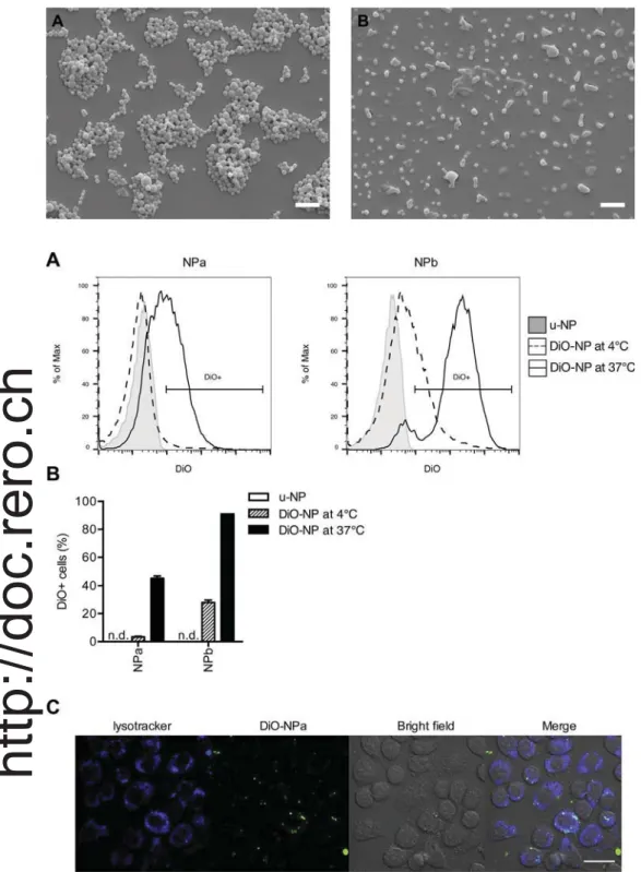
Fig. 1. Scanning electron microscopy images of (A)
NPa and (B) NPb (scale bar = 1μm).
Fig. 2. NPa and NPb are efficiently taken up by macrophages.
(A–B) J774 macrophages were exposed for 24 h to DiO-NPa or DiO-NPb at 4 °C or 37 °C and the uptake
was assessed byflow cytometry. (A) Representative
histograms showing the intensity of DiO labeling. (B) Percentage of DiO-positive cells. Each bar represents mean ± SEM of one experiment performed in tri-plicate. Data are representative of 4 independent experiments. (C) J774 macrophages were exposed for 2 h to DiO-NPa and analyzed by confocal micro-scopy. Green: DiO-NPa; blue: Lysotracker. Pictures are representative of 5 independent experiments
(scale bar = 33μm). (For interpretation of the
re-ferences to colour in thisfigure legend, the reader is
referred to the web version of this article.)
2.6. NP dilution for cell culture
Lyophilized NP were suspended in apyrogenic water at a con-centration of 20μg/ml R848 (200× working concentration). The sus-pension was diluted in complete medium to reach a 2× working con-centration and added to the J774 macrophages 1:1 (final R848 working concentration 0.1μg/ml; final NP concentration: 15.8 μg/ml NPa, 82.6μg/ml NPb). u-NP (unloaded) and DiO-NP were used at the same concentrations as the respective R848-NP unless indicated otherwise.
2.7. Cytotoxicity assay
J774 macrophages (5 × 10⁴cells/well) were exposed for 24 h to u-NPs or R848-NP. Staurosporine (0.1μm) was used as a positive control. Cells were stained with annexin V in annexin V buffer (both from Biolegend). Propidium iodide (PI; 0.5 mg/ml, Sigma) was added auto-matically at 1:100 dilution before acquisition by the MACSquant Analyzer 10 flow cytometer (Miltenyi Biotec). Data were analyzed using FlowJo software.
2.8. NP uptake in J774 cells
Forflow cytometry analysis, J774 macrophages (5 × 10⁴cells/well)
were exposed for 24 h to DiO-NP at 4 °C or 37 °C and resuspended in FACS buffer (PBS, 0.5% bovine serum albumin (BSA) and 5 mM EDTA, PAA). Zombie violet (Biolegend) was used to exclude dead cells. For confocal microscopy, J774 cells (5 × 10⁵cells/ml) were incubated in 300μl with 4 μg/ml NP for 10 h in complete medium. 75 nM LysoTracker DND22 (blue; Invitrogen) were used for lysosomal staining according to manufacturer’s protocol. Cells were visualized using a confocal microscope (Zeiss 710 Meta, 63× oil immersion objective). 2.9. Immune activation
J774 macrophages (5 × 10⁴cells/well) were incubated with NP-R848 (0.1μg/ml of R848) for 24 h and resuspended in FACS buffer. Free R848 was used as positive control. Cells were stained with Zombie violet and incubated with Fc block True Stain fcX CD16/32 (Biolegend) for 15 min before addition of the following antibodies: MHCII (APC-Cy7, clone M5/114.15.2), CD86 (PE, clone GL-1), CD11b (PE-(APC-Cy7, clone M1/70) (all Biolegend). Data was acquired by MACSquant Analyzer and analyzed using FlowJo software. Interleukin-6 (IL-6) was determined in the cell culture supernatant using Mouse IL-6 ELISA MAX kit (Biolegend) according to the manufacturer’s protocol. Absorbance at 570 nm was measured and subtracted from the absorbance at 450 nm by Infinite 200 PRO plate-reader (TECAN).
2.10. Uptake into splenocytes
Splenocytes were isolated from C57BL/6 mice and red blood cell lysis was performed (BD Pharm Lyse, BD). Cells were resuspended in T-cell medium: RPMI1640 (PAA), 2 mmol/mlL-glutamine (PAA), 1 IU/ml Penicillin (PAA), 100 mg/ml streptomycin (PAA), 0.1 mm non-essential amino acid (PAA), 1 mm sodium pyruvate and 25μM beta-mercapto ethanol (both life technologies). Splenocytes (106cells/well in a 24-well plate) were incubated for 2 h with DiO-NP at the standard working concentration (15.8μg/ml NPa, 82.6 μg/ml NPb) or at 10× working concentration. This short incubation time was selected due to the short half-life in culture of some primary cells. Cells were washed twice with PBS beforeflow cytometry staining with Zombie violet, incubation with Fc block for 15 min before addition of the following antibodies: CD11b (APC, clone M1/70), CD19 (APC-Cy7, clone 6D5), CD3 (PB, clone 17A2) and CD11c (PE, clone N418) (all from Biolegend). Data was acquired byflow cytometry as described above.
2.11. Biodistribution
500μg of DiO-NP in 100 μl PBS were injected s.c. close to the in-guinal space in C57BL/6 mice (Janvier Labs). 24 h after injection, spleen and lymph nodes were isolated and single cell suspensions were prepared. The inguinal lymph nodes ipsilateral to the injection (draining lymph nodes), and inguinal lymph nodes contralateral to the injection (non-draining lymph nodes) were isolated. Lymph nodes were cut into 3–4 pieces before passing through a 40 μm cell strainer. After centrifugation, single cell suspensions were resuspended in medium and counted. Erythrocyte lysis was performed for spleen (BD Pharm Lyse). Cells were stained with Zombie violet and incubated with Fc block for 15 min before addition of the following antibodies: CD11b (Pe-Cy7, Clone M1/70), B220 (PB, Clone RA3-6B2), CD3 (PerCP, clone 17A2) and CD11c (APC-Cy7, clone N418) (all from Biolegend) beforeflow cytometry analysis.
3. Results
3.1. NP synthesis and characterization
While the PLGA polymer was commercially available, mPEG-PLA was synthesized under bulk conditions via a ring-opening polymeriza-tion (ROP) ofDL-lactide, using mPEG 2000 as the initiator and stannous octoate as the catalyst. By1H NMR spectroscopy it was possible to
as-sess the molecular weight of the synthesized polymer by calculating the ratio of protons corresponding to mPEG moiety vs protons corre-sponding to polylactic acid moiety. A molecular weight of 8 kDA was
Fig. 3. NP do not affect cell viability.
J774 macrophages were exposed for 24 h to u-NP or R848-NP and viability
was assessed byflow cytometry. (A and B) Representative dot plots, (C)
percentage of Annexin V/PI double-negative cells (live cells). Medium: incubation condition without NP, without R848 (white bar) or with free R848 (black bar). Each bar represents mean ± SEM of 6 independent experiments performed in quadruplicate.
determined.
Batches of NP constituted of a 67/33 mass blend of PLGA and mPEG-PLA (NPa) or 100% mPEG-PLA (NPb) were then synthesized by a solvent-evaporation method. These NP were either loaded with R848 as the active molecule (R848-NP), DiO as afluorescent probe (DiO-NP) or unloaded (u-NP). Particles had a mean size ranging from 154 to
278 nm. Their zeta potential ranged from−10 to −25 mV. Table 1
summarizes the principal characteristics of the different batches. Scanning electron microscopy showed spherical and homogeneous shape of the particles both for NPa and NPb (Fig. 1). However, although NPb are well dispersed in the aqueous medium, they appear as merging entities when deposited on the slabs (Fig. 1B) due to the high percen-tage of hydrophilic PEG moiety.
3.2. NP are taken up by a macrophage cell line
To examine whether NP were internalized by antigen-presenting cells, we used the well-characterized macrophage cell line J774. J774 cells were exposed to DiO-NPa and DiO-NPb for 24 h and NP uptake was assessed byflow cytometry. Both NPa and NPb were efficiently taken up by macrophages at 37 °C, as we measured over 40% DiO-po-sitive cells for NPa and approximately 90% DiO-poDiO-po-sitive cells for NPb (Fig. 2A and B). NP were also exposed to J774 macrophages for 24 h at 4 °C, a temperature at which many active intracellular processes are decreased (Sokolova et al., 2013). At this temperature, the amount of DiO-positive cells was strongly reduced for both NPa and NPb, in-dicating that a major part of the NP is actively taken up at 37 °C. J774 cells incubated with DiO-NP were also examined by confocal micro-scopy. DiO-NP (green) were observed to accumulate in vesicles inside the cells and colocalized with LysoTracker, an endosomal marker (blue) (Fig. 2C). Thus, our results show that NPa and NPb are readily taken up by macrophages.
3.3. NP do not affect macrophage viability
The impact of NPa and NPb on the viability of J774 macrophages was examined byflow cytometry with annexin V/PI staining. These NP did not affect cell viability after 24 h of exposure, as there was no sig-nificant change in the proportion of live annexin V/PI double-negative cells after incubation (Fig. 3). The impact on cell viability was also assessed for R848-NPa and R848-NPb (Fig. 3C). No decrease in cell viability was observed after incubation with these particles. Thus, neither NPa nor NPb affect the viability of macrophages, both with and without R848 cargo.
3.4. R848-loaded NP activate macrophages
The imidazoquinoline R848 is a pharmacological immunostimulant that activates the Toll-like receptor 7, which is present in the endosome of myeloid immune cells such as macrophages (Heil et al., 2003). To assess the immune-activating effect of R848 delivered by NP, we de-termined the amount of the pro-inflammatory cytokine interleukin 6 (IL-6) secreted by J774 macrophages after exposure to R848-NP. When J774 macrophages were exposed to u-NP, no IL-6 was produced, in-dicating that these NP are not pro-inflammatory by themselves. In contrast, J774 cells exposed to free R848 produced high amounts of IL-6, as expected (Fig. 4A). J774 exposed to R848-NP produced IL-6 in high amounts, similar to the J774 cells exposed to free R848. Fur-thermore, activation markers were measured on J774 cells by flow cytometry. R848-NPa and R848-NPb enhanced the surface expression of the activation markers MHC-II, CD11b and CD86 to a similar extent as free R848 (Fig. 4B). In contrast, u-NP did not significantly enhance the expression of these markers. Thus, unloaded NP do not cause acti-vation of macrophages, whereas R848-NP lead to a strong actiacti-vation of macrophages.
3.5. NP are taken up by primary immune cells
The expression of the Toll-like receptor 7 in the organism is mainly restricted to immune cells of the myeloid lineage, including macro-phages, monocytes and dendritic cells, and to B lymphocytes. To de-termine whether these primary cells take up NPa and NPb, we exposed
Fig. 4. R848 released by NP efficiently activates macrophages.
J774 macrophages were exposed for 24 h to u-NP or R848-NP. (A) IL-6 levels in super-natant were measured by ELISA. Each bar represents mean ± SEM of 4 independent experiments performed in quadruplicate. (B) Cell surface expression of MHCII, CD11b
and CD86 was measured byflow cytometry. Medium: incubation condition without NP,
without R848 (white bar) or with free R848 (black bar). Each bar represents mean ± SD of one experiment performed in triplicate. Data are representative of 3 independent ex-periments.
freshly isolated murine splenocytes to DiO- NP for 2 h. The spleen contains a variety of immune cells, in particular B and T lymphocytes and a mixture of myeloid cells. Our results confirm that also freshly isolated CD11b-positive monocytes and macrophages took up NPa and NPb. Indeed, at the higher concentration of 1μg/ml NP, approximately 70% and 90% of monocytes/macrophages were positive for DiO-NPa and DiO-NPb, respectively (Fig. 5A). Approximately 50% of den-dritic cells (DC) took up both types of NP. We observed that NP are also taken up by lymphocytes, with 70–90% of DiO-positive B cells and 10–20% of DiO-positive T cells. We further examined the mean fluor-escence intensity (MFI) of the different cell populations (Fig. 5B). The cells with the highest MFI were the monocytes/macrophages and the B cells, confirming that these populations take up the most NP. 3.6. NP are targeted to the lymph nodes
Since the initiation of adaptive immune responses occurs in the lymph nodes, it is of great importance that NP serving as delivery system for immune activators should be transported to these organs. To examine whether this was the case for NPa and NPb, we injected DiO-NP subcutaneously in mice and isolated spleen and draining and non-draining lymph nodes 24 h after injection. We found DiO-positive cells mainly in the draining lymph nodes, very few DiO-positive cells in non-draining lymph nodes and none in the spleen (Fig. 6A). We further investigated which cell types took up NPs within the draining lymph nodes. Both DiO-NPa and DiO-NPb were mainly taken up by dendritic cells (DC) and the monocyte/macrophage CD11b+ population (Figs. 6B and S1). These cell types are antigen-presenting cells and are instrumental for initiating immune responses. In addition, both these cell types express the Toll-like receptor 7. Thus, NP are targeted to the draining lymph nodes following s.c. application.
4. Discussion
Imidazoquinolines including R848 can generate highly effective
Fig. 5. NP are efficiently taken up by primary an-tigen-presenting cells.
Freshly isolated splenocytes were exposed for 2 h to
DiO-NP before flow cytometry analysis. (A)
Percentage of DiO-positive cells within the indicated cell populations: monocytes/macrophages (mono/ Mϕ) (CD11b + CD11c−), dendritic cells (DC) (CD11b + CD11c+), B lymphocytes (CD19+), T
cells (CD3+) and (B) Meanfluorescence intensity
(MFI) of DiO in the indicated cell populations. Data are representative of 2 independent experiments.
Fig. 6. In vivo, NP accumulate in antigen-presenting cells of the draining lymph nodes.
500μg DiO-NP were injected s.c. into the flank of C57BL/6 mice (n = 3). PBS (no NP) was
used as negative control (n = 1). 24 h after injection, cells from the spleen, the non-draining lymph nodes (non-DLN) and the non-draining lymph nodes (DLN) were isolated for flow cytometry analysis. (A) Number of DiO-positive cells in the indicated organs. (B) Percentage of DiO-positive cells in DLN within the indicated cell populations: Monocytes/ macrophages (mono/Mϕ) (CD11b + CD11c−), dendritic cells (DC) (CD11b+CD11c+), B lymphocytes (CD19+), T cells (CD3+). Data are representative of 2 independent ex-periments.
antitumoral responses (Kobold et al., 2014). However, these small molecules also induce inflammatory responses with high serum levels of proinflammatory cytokines when applied systemically. Indeed, even subcutaneous application of R848 leads to a rapid increase of the cy-tokines IFNα and IL-6 in the serum (Bourquin et al., 2011). Since these unspecific and generalized inflammatory responses can result in dose-limiting toxicity (Kobold et al., 2014), it is of great therapeutic interest to develop particulate delivery systems that may focus the immune-activating effect of these molecules to the target tissues.
Here, we successfully encapsulated R848 in either mPEG-PLA or PLGA/mPEG-PLA nanoparticles. Interestingly, the encapsulation rate was higher when PLGA was present in the formulation (8.36μg/mg of NP vs 0.74μg/mg of NP). The exclusive presence of the PEG hydrophilic moieties in R848-NPb batches resulted in lower affinity of the drug to the polymer matrix leading therefore to lower encapsulation rates. In the literature, several studies relate the encapsulation of R848 or ana-logues in polymer nanoparticles either alone or co-encapsulated with antigens or adjuvants (Cruz et al., 2012; Ilyinskii et al., 2014; Kasturi et al., 2017, 2011; Seth et al., 2017; Tacken et al., 2011; Tel et al., 2013). When encapsulation rates are given by the authors, the values are ranging from 1.2 to 6.4μg/mg of NP. However, in these specific cases, co-encapsulation of resiquimod with antigens or adjuvants in NP made of RG502H as PLGA is discussed. Only Seth et al. report an en-capsulation rate of 11.1μg/mg while loading a resiquimod analogue, gardiquimod, alone in PLGA NP. Comparison with our data is difficult as preparation methods and materials are different. It is well known that the molecular weight and the PLA/PLGA ratio are crucial factors in the encapsulation efficiency. However, we assume that the encapsula-tion efficiencies observed herein are in the same order of magnitude as those observed in the literature. Pure mPEG-PLA nanoparticles (NPb) were smaller than mixed polymer nanoparticles (NPa). This observation is likely related to the combined action of 1) the more amphiphilic nature of the pegylated polymer and 2) its lower molecular weight. Indeed, we observed that by fixing the molecular weight of the polymer, the only addition of an mPEG moiety does not lead to a change in the size of the nanoparticles.
Both TLR7 ligand R848-loaded mPEG-PLA NP and PLGA/mPEG-PLA NP were readily taken up by antigen-presenting cells such as macrophages and dendritic cells both in vitro and in vivo (Figs. 2, 5 and 6). Importantly, the R848 cargo could induce immune activation of these cells, demonstrating its accessibility to the immune system (Fig. 3). We observed similar levels of activation in cell culture with R848-loaded NP and with the same amount of free R848. Depending on the type of NP used as carrier, others have seen similar results or even reduced activation in vitro with NP-delivered R848 (Tacken et al., 2011; Cruz et al., 2012; Ilyinskii et al, 2014). The addition of molecules to the surface of the NP that specifically target dendritic cells may further improve the uptake of the particles and the immunostimulatory activity of R848 cargo, as shown with polymer-based NP decorated with anti-DC-SIGN antibodies (Cruz et al., 2012). The unloaded particles did not show any unspecific proinflammatory effects, demonstrating the ab-sence of relevant levels of endotoxin, and none of the NP tested showed cytotoxicity, highlighting their biocompatibility.
The use of mPEG-PLA or PLGA/mPEG-PLA NP allows delivery of R848 specifically to antigen-presenting cells. In vitro, these NP are taken up by B cells, dendritic cells, monocytes and macrophages, whereby monocytes/macrophages seem to take up larger amounts of particles (Amoozgar and Goldberg, 2015). Following subcutaneous injection, the NP are found within the draining lymph nodes both in dendritic cells and in macrophages. These cells are responsible for initiating the T cell-mediated immunity that is crucial for anti-tumor efficacy, and are therefore the prime therapeutic target (Chen and Mellman, 2013). Considering the size of the particles (> 150 nm), it is probable that the NP were taken up by the antigen-presenting cells at the site of injection and carried to the lymph nodes, rather than reaching the nodes by passive diffusion through the lymph, as would be the case for smaller
NP (Irvine et al., 2013). Little uptake is seen by B cells, which do not reside in subcutaneous areas.
Targeting TLR7 agonists directly to the lymph nodes offers several advantages, since this can focus immune activation to the injection site and the draining lymph node. This targeting has been shown to enhance cytotoxic T-cell responses when the TLR7 agonist is applied together with antigen in the context of vaccination (Jewell et al., 2011; Nuhn et al., 2016). In addition, lymph node targeting of TLR ligands can re-duce systemic levels of proinflammatory cytokines (Bourquin et al., 2008). We propose that mPEG-PLA and PLGA/mPEG-PLA nanoparticles represent an attractive and safe delivery system to target small mole-cule TLR7 agonists to lymph nodes, allowing a focused immune re-sponse that may be of benefit for anticancer immunotherapy. Acknowledgements
This study was supported by the National Center of Competence in Research (NCCR) for Bio-Inspired Materials and the Swiss National Science Foundation grants 310030_156871 and 310030_156372. Appendix A. Supplementary data
Supplementary material related to this article can be found, in the online version, at doi:https://doi.org/10.1016/j.ijpharm.2017.11.031
References
Allémann, E., Gurny, R., Doelker, E., 1993. Drug-loaded nanoparticles: preparation methods and drug targeting issues. Eur. J. Pharm. Biopharm. 39, 173–191.
Amoozgar, Z., Goldberg, M.S., 2015. Targeting myeloid cells using nanoparticles to im-prove cancer immunotherapy. Adv. Drug Deliv. Rev. 91, 38–51.
Anz, D., Koelzer, V.H., Moder, S., Thaler, R., Schwerd, T., Lahl, K., Sparwasser, T., Besch, R., Poeck, H., Hornung, V., Hartmann, G., Rothenfusser, S., Bourquin, C., Endres, S., 2010. Immunostimulatory RNA blocks suppression by regulatory T cells. J. Immunol. 184, 939–946.
Bourquin, C., Anz, D., Zwiorek, K., Lanz, A.L., Fuchs, S., Weigel, S., Wurzenberger, C., von der Borch, P., Golic, M., Moder, S., Winter, G., Coester, C., Endres, S., 2008. Targeting CpG oligonucleotides to the lymph node by nanoparticles elicits efficient antitumoral immunity. J. Immunol. 181, 2990–2998.
Bourquin, C., Hotz, C., Noerenberg, D., Voelkl, A., Heidegger, S., Roetzer, L.C., Storch, B., Sandholzer, N., Wurzenberger, C., Anz, D., Endres, S., 2011. Systemic cancer therapy with a small molecule agonist of toll-like receptor 7 can be improved by cir-cumventing TLR tolerance. Cancer Res. 71, 5123–5133.
Bourquin, C., Schmidt, L., Lanz, A.L., Storch, B., Wurzenberger, C., Anz, D., Sandholzer, N., Mocikat, R., Berger, M., Poeck, H., Hartmann, G., Hornung, V., Endres, S., 2009. Immunostimulatory RNA oligonucleotides induce an effective antitumoral NK cell response through the TLR7. J. Immunol. 183, 6078–6086.
Chen, D.S., Mellman, I., 2013. Oncology meets immunology: the cancer-immunity cycle. Immunity 39, 1–10.
Cruz, L.J., Tacken, P.J., Pots, J.M., Torensma, R., Buschow, S.I., Figdor, C.G., 2012. Comparison of antibodies and carbohydrates to target vaccines to human dendritic cells via DC-SIGN. Biomaterials 33, 4229–4239.
Dumitru, C.D., Antonysamy, M.A., Gorski, K.S., Johnson, D.D., Reddy, L.G., Lutterman, J.L., Piri, M.M., Proksch, J., McGurran, S.M., Egging, E.A., Cochran, F.R., Lipson, K.E., Tomai, M.A., Gullikson, G.W., 2009. NK1.1+ cells mediate the antitumor eVects of a dual toll-like receptor 7/8 agonist in the disseminated B16-F10 melanoma model. Cancer Immunol. Immunother. 58, 575–587.
Fahr, A., Liu, X., 2007. Drug delivery strategies for poorly water-soluble drugs. Expert Opin. Drug Deliv. 4, 403–416.
Goldinger, S.M., Dummer, R., Baumgaertner, P., Mihic-Probst, D., Schwarz, K., Hammann-Haenni, A., Willers, J., Geldhof, C., Prior, J.O., Kundig, T.M., Michielin, O., Bachmann, M.F., Speiser, D.E., 2012. Nano-particle vaccination combined with TLR-7 and -9 ligands triggers memory and effector CD8(+) T-cell responses in melanoma patients. Eur. J. Immunol. 42, 3049–3061.
Heil, F., Ahmad-Nejad, P., Hemmi, H., Hochrein, H., Ampenberger, F., Gellert, T., Dietrich, H., Lipford, G., Takeda, K., Akira, S., Wagner, H., Bauer, S., 2003. The Toll-like receptor 7 (TLR7)-specific stimulus loxoribine uncovers a strong relationship within the TLR7, 8 and 9 subfamily. Eur. J. Immunol. 33, 2987–2997.
Hotz, C., Treinies, M., Mottas, I., Rotzer, L.C., Oberson, A., Spagnuolo, L., Perdicchio, M., Spinetti, T., Herbst, T., Bourquin, C., 2016. Reprogramming of TLR7 signaling en-hances antitumor NK and cytotoxic T cell responses. Oncoimmunology 5, e1232219.
Ilyinskii, P.O., Roy, C.J., O'Neil, C.P., Browning, E.A., Pittet, L.A., Altreuter, D.H., Alexis, F., Tonti, E., Shi, J., Basto, P.A., Iannacone, M., Radovic-Moreno, A.F., Langer, R.S., Farokhzad, O.C., von Andrian, U.H., Johnston, L.P., Kishimoto, T.K., 2014. Adjuvant-carrying synthetic vaccine particles augment the immune response to encapsulated antigen and exhibit strong local immune activation without inducing systemic cy-tokine release. Vaccine 32, 2882–2895.
Iribarren, K., Bloy, N., Buque, A., Cremer, I., Eggermont, A., Fridman, W.H., Fucikova, J., Galon, J., Spisek, R., Zitvogel, L., Kroemer, G., Galluzzi, L., 2016. Trial watch: im-munostimulation with toll-like receptor agonists in cancer therapy. Oncoimmunology 5, e1088631.
Irvine, D.J., Swartz, M.A., Szeto, G.L., 2013. Engineering synthetic vaccines using cues from natural immunity. Nat. Mater. 12, 978–990.
Iwasaki, A., Medzhitov, R., 2015. Control of adaptive immunity by the innate immune system. Nat. Immunol. 16, 343–353.
Jewell, C.M., Lopez, S.C., Irvine, D.J., 2011. In situ engineering of the lymph node mi-croenvironment via intranodal injection of adjuvant-releasing polymer particles. Proc. Natl. Acad. Sci. U. S. A. 108, 15745–15750.
Kasturi, S.P., Kozlowski, P.A., Nakaya, H.I., Burger, M.C., Russo, P., Pham, M., Kovalenkov, Y., Silveira, E.L., Havenar-Daughton, C., Burton, S.L., Kilgore, K.M., Johnson, M.J., Nabi, R., Legere, T., Sher, Z.J., Chen, X., Amara, R.R., Hunter, E., Bosinger, S.E., Spearman, P., Crotty, S., Villinger, F., Derdeyn, C.A., Wrammert, J., Pulendran, B., 2017. Adjuvanting a simian immunodeficiency virus vaccine with toll-like receptor ligands encapsulated in nanoparticles induces persistent antibody re-sponses and enhanced protection in TRIM5alpha restrictive Macaques. J. Virol. 91 e01844-16.
Kasturi, S.P., Skountzou, I., Albrecht, R.A., Koutsonanos, D., Hua, T., Nakaya, H.I., Ravindran, R., Stewart, S., Alam, M., Kwissa, M., Villinger, F., Murthy, N., Steel, J., Jacob, J., Hogan, R.J., Garcia-Sastre, A., Compans, R., Pulendran, B., 2011. Programming the magnitude and persistence of antibody responses with innate im-munity. Nature 470, 543–547.
Kobold, S., Wiedemann, G., Rothenfusser, S., Endres, S., 2014. Modes of action of TLR7 agonists in cancer therapy. Immunotherapy 6, 1085–1095.
Kranz, L.M., Diken, M., Haas, H., Kreiter, S., Loquai, C., Reuter, K.C., Meng, M., Fritz, D., Vascotto, F., Hefesha, H., Grunwitz, C., Vormehr, M., Husemann, Y., Selmi, A., Kuhn, A.N., Buck, J., Derhovanessian, E., Rae, R., Attig, S., Diekmann, J., Jabulowsky, R.A., Heesch, S., Hassel, J., Langguth, P., Grabbe, S., Huber, C., Tureci, O., Sahin, U., 2016. Systemic RNA delivery to dendritic cells exploits antiviral defence for cancer im-munotherapy. Nature 534, 396–401.
Leroux, J.-C., Allémann, E., De Jaeghere, F., Doelker, E., Gurny, R., 1996. Biodegradable
nanoparticles—from sustained release formulations to improved site specific drug delivery. J. Control. Release 39, 339–350.
Nishiya, T., DeFranco, A.L., 2004. Ligand-regulated chimeric receptor approach reveals distinctive subcellular localization and signaling properties of the Toll-like receptors. J. Biol. Chem. 279, 19008–19017.
Nuhn, L., Vanparijs, N., De Beuckelaer, A., Lybaert, L., Verstraete, G., Deswarte, K., Lienenklaus, S., Shukla, N.M., Salyer, A.C.D., Lambrecht, B.N., Grooten, J., David, S.A., De Koker, S., De Geest, B.G., 2016. pH-degradable imidazoquinoline-ligated nanogels for lymph node-focused immune activation. Proc. Natl. Acad. Sci. U. S. A. 113, 8098–8103.
Papakostas, D., Stockfleth, E., 2015. Topical treatment of basal cell carcinoma with the immune response modifier imiquimod. Future Oncol. 11, 2985–2990.
Riehemann, K., Schneider, S.W., Luger, T.A., Godin, B., Ferrari, M., Fuchs, H., 2009. Nanomedicine–challenge and perspectives. Angew. Chem. Int. Ed. Engl. 48, 872–897.
Seth, A., Lee, H., Cho, M.Y., Park, C., Korm, S., Lee, J.-Y., Choi, I., Lim, Y.T., Hong, K.S., 2017. Combining vasculature disrupting agent and toll-like receptor 7/8 agonist for cancer therapy. Oncotarget 8, 5371–5381.
Sokolova, V., Kozlova, D., Knuschke, T., Buer, J., Westendorf, A.M., Epple, M., 2013. Mechanism of the uptake of cationic and anionic calcium phosphate nanoparticles by cells. Acta Biomater. 9, 7527–7535.
Spinetti, T., Spagnuolo, L., Mottas, I., Secondini, C., Treinies, M., Ruegg, C., Hotz, C., Bourquin, C., 2016. TLR7-based cancer immunotherapy decreases intratumoral myeloid-derived suppressor cells and blocks their immunosuppressive function. Oncoimmunology 5, e1230578.
Tacken, P.J., Zeelenberg, I.S., Cruz, L.J., van Hout-Kuijer, M.A., van de Glind, G., Fokkink, R.G., Lambeck, A.J., Figdor, C.G., 2011. Targeted delivery of TLR ligands to human and mouse dendritic cells strongly enhances adjuvanticity. Blood 118, 6836–6844.
Tel, J., Sittig, S.P., Blom, R.A., Cruz, L.J., Schreibelt, G., Figdor, C.G., de Vries, I.J., 2013. Targeting uptake receptors on human plasmacytoid dendritic cells triggers antigen cross-presentation and robust type I IFN secretion. J. Immunol. 191, 5005–5012.
Zeisser-Labouèbe, M., Lange, N., Gurny, R., Delie, F., 2006. Hypericin-loaded nano-particles for the photodynamic treatment of ovarian cancer. Int. J. Pharm. 326, 174–181.