971
Mode of Inoculation of the Lyme Disease Agent
Borrelia burgdorferi Influences
Infection and Immune Responses in Inbred Strains of Mice
Lise Gern, Ulrich E. Schaible, and Markus M. Simon Institut deZoologie. Universite de Neuchdtel, Switzerland: Max-Planck-Lnstitutfiir Inununbiologie, Freiburg. Germany
Mice were infected with Borrelia burgdorferi by infection via Ixodes ricinus and experimental inoculation to determine whether transmission rates of spirochetes and antibody responses are influenced. Mice infected by the natural route were substantially more infective for ticks; two- to sixfold more tick larvae were positive for B. burgdorferi than those fed on experimentally inocu-lated mice. In natural infection, spirochetemia may be greater or spirochetes may be more accessi-ble for transmission. Thus, this form of xenodiagnosis could be used to determine levels of spirochetes in the vertebrate host. Similar levels of antibody were present in all mice; however, those infected by the natural route lacked antibodies to outer surface proteins (Osp) A and B. The small antigen dose given through a tick bite may not have been sufficient to induce rapid OspA or OspB antibodies, thereby allowing the later development of higher levels of spirochetemia.
Since the discovery of Borrelia burgdorferi as the cause of Lyme borreliosis, most information on the pathogenesis of the disease and the immune responses to B. burgdorferi de-rives from laboratory models in which animals were experi-mentally inoculated with viable spirochetes. In mice, ham-sters, and white-footed mice (Perornyscus leucopus), the first antibodies detected were those to the outer surface proteins (Osp) A and B, to a 39-kDa protein, and to the 41-kDa fla-gellin (fla) [1-3].Itwas subsequently shown that antibodies specific for OspA and OspB are protective and able to pre-vent B. burgdorferi-induced disease in immunodeficient SCID [4, 5] and immunocompetent C3H/HeJ mice [6, 7].
In contrast to results in experimentally inoculated mice, the first antibodies to be detected in patients with Lyme borreliosis recognize the 41-kDa flagellin or the 39-kDa anti-gen (or both) but not OspA or OspB [8]. The differential immune responses of experimentally inoculated mice versus patients with Lyme disease may be related to the mode of inoculation of spirochetes, as suggested by a study in dogs [9]; only experimentally but not naturally infected dogs ex-pressed antibodies to OspA.
The present study was undertaken to investigate the influ-ence ofthe route of inoculation of B. burgdorferi on infection and antibody response in 3 inbred strains of mice.
Materials and Methods
B. burgdorferi. The European strain ZS7 used in this study was cultivated and quantified as described previously [10].
Received 1 May 1992; revised 30 October 1992.
Grant support: Swiss National Science Foundation (32-29964.90) and Bundesministerium fllr Forschung und Technologie (Kl 8909/8).
Reprints or correspondence: Dr. Lise Gem, Institut de Zoologie. Chante-merle 22. CH-2000 Neuchatel, Switzerland.
The Journal of Infectious Diseases 1993;167:971-5
© 1993 by The University of Chicago. All rights reserved. 0022-1899/93/6704-0029$01.00
Recombinant OspA. Recombinant OspA from strain ZS7 was expressed from the puEX I expression vector in Escherichia
coli.The protein was extracted and purified by affinity chroma-tography using an OspA-specific monoclonal antibody as de-scribed previously [5].
Tick colony. The larval and nymphal I. ricinus ticks used in this study were derived from a B. burgdorferi-free laboratory colony maintained at the Institute of Zoology (Neuchatel).
Animals. Adult mice of strains AKR/N (H-2k
) , C3H/HeJ
(H-2k
) ,and DBA/2 (H-2d)were bred at the Institute ofZoology.
Females 8-10 weeks old were used,
Injection procedures. Unfed I. ricinus nymphs were infected using the modified capillary method [I I]. Strain ZS7 at a con-centration of I X 108 cells/rnl. BSK II medium was used to
infect the ticks. Ticks took up --1.5 p,L (--1.5 X 10⁵
spiro-chetes). Each experimental group of mice included 2 AKR/N, 2 DBA/2, and 2 C3H/HeJ mice,
For group I, 6 mice were inoculated subcutaneously (sc) in the tail with 0.2 mL of BSK II medium containing 2X 10⁷
B. burgdorferiZS7 organisms.
For group II, 6 mice were inoculated with a suspension of infected I. ricinus nymphs. Immediately after the infection by capillary, 42 infected nymphs were triturated in 1.5 mL ofBSK II medium. Spirochetes were counted and adjusted to 3 X 10⁶
spirochetes/ml.; 0.2 mL of the suspension (--6 X 10⁵
spiro-chetes/mouse) was inoculated sc in the tail of each mouse. For group III, 6 mice were exposed to 7 artificially infectedI. ricinus nymphs. Nymphs were fed in two hollow plastic caps fixed bilaterally on the back of the mouse, and a collar was placed around the neck to prevent grooming. Engorged ticks (4-6) were collected from each mouse.
Xenodiagnosis and spirochete detection in ticks. To deter-mine the level of spirochetemia, infected mice were exposed to uninfected I. ridnus larvae at 14 (xenodiagnosis I, 80 larvae/ mouse) and 100 days (xenodiagnosis II, 60 larvae/mouse) after infection. Larvae were put on the head of the mouse, and a collar was placed around the neck to prevent grooming. Mice were kept in separate cages over trays of water, and engorged ticks that had dropped into water were collected daily, placed into vials, and stored at room temperature and 95% humidity.
972 Concise Communications lID1993;167(April)
Ticks were evaluated for B. burgdorferi after molting using a
direct immunofluorescence antibody test [12].
Western blot and ELISA. Sera derived from infected mice were analyzed by Western blots of spirochetal lysates (strain
ZS7) and by ELISA with either soluble B. burgdorferi (ZS7)
antigen or recombinant OspA as described [5].
Results
Inbred mice of strains AKR/N (H-2k
) , C3H/HeJ (H-2k) ,
and OBA/2 (H-2d
)were infected withB. burgdorferiby
exper-imental inoculation with in vitro-propagated spirochetes (group I), experimental inoculation with a tissue suspension of infected ticks (group II), and natural infection via tick bites (group III). The numbers of spirochetes transferred to
mice via syringe were r -2 X 10⁷for group I andr--6 X 10⁵for
group II. The numbers of spirochetes transmitted via tick
bites can only be estimated and were r-:1 X 10⁶ at most.
At 14 and 100 days after infection, mice were exposed to
uninfectedI. ricinuslarvae to asses their capacity to transmit
spirochetes. Mice from each of the 3 inbred strains previ-ously infected via tick bites (group III) were shown to infect,
on average, about two- to fourfold more I. ricinus larvae
(42.5% at day 14 and 88% at day 100) than mice infected via
syringe with eitherB. burgdorferiorganisms (group I, 6% on
day 14 and 37.5% at day 100) or a tissue suspension of in-fected ticks (group II, 10%at day 14 and 33.3% at day 100).
In all 3 strains of mice, infection rates ofI. ricinuslarvae were
significantly increased (two- to sixfold) at day 100 compared with those at day 14 independent of the inoculation proto-col, reaching 88% (twofold increase) after tick infection (group III) and between 37.5% (sixfold increase, group I) and 33.3% (threefold increase, group II) after experimental inocu-lation.
Serum samples were taken from mice at days 7, 14,26,40, 63, 77, and 110 after infection and analyzed individually for
B. burgdorftri-specificantibodies by ELISA. All mice, inde-pendent of strain or inoculation protocol, expressed similar amounts of specific antibodies as assessed on total cell
ly-sates ofB. burgdorferistrain ZS7 (figure lA). However, when
assayed on purified recombinant OspA [5] alone, specific antibodies were found only in experimentally inoculated (group I, II) but not in tick-infected mice (group III, figure 1B).
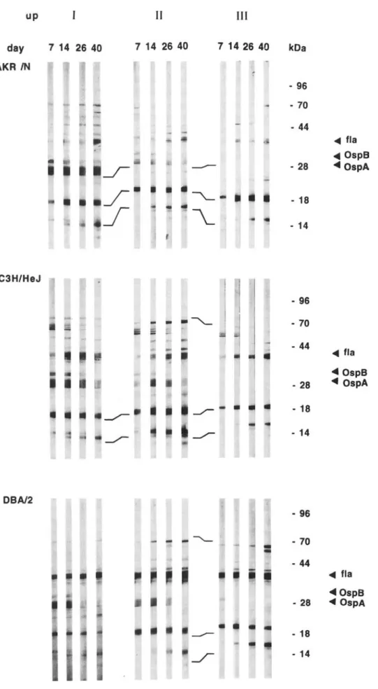
To verify these results, individual sera were analyzed for antibody specificities by Western blot. Figure 2 depicts repre-sentative banding patterns obtained with sera from AKR/N, C3H/HeJ, and OBA/2 mice at days 7, 14,26, and 40 after inoculation. Sera from mice experimentally inoculated with either spirochetes (group I) or lysates derived from infected ticks (group II) contained antibodies to OspA (31 kOa) and
OspB (34 kOa) in addition to various other B. burgdorferi
structures, including ftagellin (41-kOa) and antigens with approximate molecular masses of 14, 18,22,26, 39,65, and 70 kOa. In contrast, sera from mice inoculated via infected
B
---e-----
..
-.
CB-1761Vftick---
..
-.
3Vftick---
..
--
lIV ftick---
..
--
3m/tick---0---
6Dftick-..-
171V-..-
7IV ~ 81V -+-- 14IV ~ 12IV 0,5 ci ci 0 0 1,0 o,~ 0,3 0,5 0,04-...--.----..--...--.__..._..._...--.--...--...-...-1 1:100 1:200 1:400 1:800 1:1600 1:3200 1:6400 1:12800 1:100 1:200 1:400 1:800 1:1600 1:3200 1:64001:12800 dil dllFigure 1. Antibody responses of mice inoculated with 2X 10⁷B. burgdorferi ( - - , mouse 14 and 17). tissue lysatcs derived from
experimentally infected ticks (--, 6X 10⁵spirochetes/mouse, mouse 7, 8, and 12), or via experimentally infected ticks (----, mouse 1,3,
and 6) by ELISAon wholeB. burgdorferiproteins (A) and on recombinant (rec) outer surface protein (Osp) A (B). Sera from mouse strains
AKR/N(mouse 1,7,8, and 14), DBA/2 (mouse 6, 12, and 17),and C3H/HeJ (mouse 3) were tested. Sera were taken 14 (11),26 (111),40
(IV), or 63(V)days after infection. Pooled immune serum derived from 5 CB17 micewas used as control (64 p.L/mLspecificimmunoglobu-lin). OD, optical density; dil, dilution.
JID 1993:167(April) Concise Communications 973 up
II
III
day AKR IN 7 14 26 40 7 14 26 40 7 1426 40 kDa - 96 - 70 - 44 ... fla ... OspS.r:
----
- 28 "'OspAJ
~- - 18J
~
- 14 C3H/HeJ - 96-
-
.
""""-
- 70 - 44 - 28 ~•
- 18 ~..
---/""" . 14 -.../""" ... fla ... OspS ... OspAFigure 2. Western blot ana lysis
ofsera taken fromindividu almice of strainsAKR/N,C3H/HeJ ,and DBA/2 at indicated days afte r in
-fection.Micewereinoc ula ted with
2 X 10⁷
B. burgdorferi spirochetes
in BSKII(I).tissuelysatesfromex
-perim entally infected ticks (6 X
105/mou se. II). orvia experime n-tallyinfectedticks (III).Repr
esen-tati ve data from I mouse ofeach
group are shown. Osp, outer s
ur-face protein ;fla,flagellin.
DBA/2 - 96
-
-
---
- 70 - 44 ... fla "'OspS - 28 ... OspA-
- 18-.r
- 14974 Concise Communications JID 1993;167 (April)
ticks (group III) contained no or few antibodies to OspA and OspB but had a banding pattern similar to that of the sera from groups I and II. Similar results were obtained with sera taken at days 63, 77, and I 10 (data not shown).
Discussion
This investigation showed that the rate of transmission of spirochetes to noninfected tick larvae is much higher in mice infected via tick bites than in those experimentally inocu-lated by needle injection and that antibodies to OspA and OspB are generated only in response to experimentally but not to tick-delivered organisms.
At first glance, the finding that naturally infected mice in turn infect more ticks than mice experimentally inoculated with cultured spirochetes is surprising. since the number of spirochetes transfered by needle inoculation was at least two to three orders of magnitude higher. This discrepancy may be related to the site of infection, the immune reaction of the host, the number of spirochetes transferred (antigenic load), or the phenotypic changes of the spirochetes within the vec-tor or host or both.
It is possible that skin areas chosen by ticks for blood
meals are more heavily infested with spirochetes whereas others are relatively free of the organism. In that case, more naive ticks would become infected when feeding on a recipi-ent that was itself infected by tick bites. At presrecipi-ent, detailed information on that point is lacking and requires more
inten-sive studies.Itis more likely that the discrepancy of
transmis-sion rates observed in naturally versus experimentally in-fected mice is due to qualitative differences in their immune responses. In fact, sera from all mice infected by any of the three protocols contained similar quantities of spirochete-specific antibodies independent of the greatly differing
num-bers of spirochetes transferred
«
106by tick bites, 6 X 10⁵-2X 10⁷ by needle infection); however, antibodies to OspA or
OspB were observed only in mice experimentally inoculated by needle injection and not in those infected via tick bites.
The finding that the transmission ofB. burgdorferiorganisms
to ticks correlated inversely with the presence of antibodies to OspA and OspB in mice therefore suggests that these anti-bodies are responsible for the elimination of spirochetes and consequently for the reduction in the rate of transmission. This assumption is also supported by previous studies show-ing that OspA- and OspB-specific antibodies protect SCID [4, 5] and C3H/HeJ mice [6, 7] against infection and devel-opment of arthritis after experimental and tick-mediated
in-fection withB. burgdorferi,when these antibodies are present
at the time of inoculation [13, 14].
The finding that the humoral immune response to tick-de-livered spirochetes did not include recognition of OspA and OspB is analogous to the situation in patients with Lyme disease, who develop these antibodies, if at all, only during
later stages of the infection [8]. Itis therefore possible that
the immune response of the host is influenced by inoculation route. This hypothesis is substantiated by previous studies demonstrating that experimentally infected dogs [9] and inbred [I, 3, 13] and wild mice (Kurtenbach K, personal communication) express antibodies to OspA and OspB, whereas naturally infected animals do not. The host immune system may indeed be affected by the tick itself, which has been shown before to secrete immunomodulatory constitu-ents during feeding [15]. However, such a mechanism is hard to reconcile with the fact that the suppression seen affects antibodies to OspA and OspB but not to other spirochetal antigens. including flagellin.
It is more likely that the inability of naturally infected
mice to produce antibodies to OspA and OspB is due to sub-optimal doses of spirochetes delivered during the tick's blood meal. This assumption is strongly supported by recent stud-ies showing that mice experimentally inoculated with
differ-ent numbers of spirochetes (101-108
) express similar
quanti-ties of antibodies to B. burgdorferi but that those with
specificities for OspA and OspB are seen only in animals
receiving> 104 organisms (unpublished data). These results
not only indicate that the number of spirochetes transmitted by ticks during a blood meal is rather low but also suggest that the qualitative differences of humoral immune re-sponses observed are due to differential immunogenicity of individual antigens, such as OspA, OspB, and flagellin, in the inoculum. Experiments to test this hypothesis are un-derway.
The possibility that phenotypic changes of the spirochetes occurring in the tick or mouse environment (or both) as dem-onstrated previously for certain isolates [16] may contribute to the suppression of antibody responses to OspA and OspB cannot be formally excluded; however, this seems less likely, since similar phenotypic patterns were obtained with the in vitro-propagated strain ZS7 and its reisolates from either the vector or the murine host. Compared with the first xenodiag-nosis at 14 days after infection, the percentage of infected ticks increased from two- to sixfold in all experimental groups of mice at day 100 after infection. These data suggest that spirochetes may survive or multiply in mice even
ifpro-tective antibodies are generated early during disease. It is
possible that the kinetics of appearance of anti-OspA and anti-OspB antibodies and/or their serum level only controls the development of disease, as demonstrated previously [4, 13], but are insufficient to eradicate the spirochetes from the host. Further studies will be directed toward the develop-ment of immunization protocols that allow the
establish-ment of sterile immunity against B. burgdorferiinfection in
this animal model.
Acknowledgments
We thank Olivier Rais and Gabi Nerz for technical assistance, Jean Langhorne for critically reading the manuscript, and Rose Brugger and Gabi Prosch for preparing the manuscript.
JIO 1993;167 (April) Concise Communications 975
References
I. Benach JL, Coleman JL, Garcia-Monco JCG, Deponte Pc. Biological activity of Borrelia burgdorferi antigens. Ann NY Acad Sci 1988;539: 115-25.
2. Schwan TG, Kime KK, Schrumpf ME, Coe JE, Simpson WJ. Antibody response in white-footed mice (Peromyscus leucopus) experimentally infected with the Lyme disease spirochete (Borrelia burgdorferi). In-fect Immun 1989;57:3445-51.
3. Schaible UE, Kramer MD, WallichR,Tran T, Simon MM. Experimen-tal Borrelia burgdorferi infection in inbred mouse strains: antibody response and association ofH-2 genes with resistance and susceptibil-ity to development of arthritis. Eur J Immunol 1991;21 :2397-405. 4. Schaible UE, Kramer MD, Eichmann K.Modolell M, MuseteanuC, Simon MM. Monoclonal antibodies specific for the outer surface protein A (OspA) of Borreliaburgdorferiprevent Lyme borre1iosis in severe combined immunodeficiency (scid) mice. Proc Natl Acad Sci USA 1990;87:3768-72.
5. Simon M, Schaible UE, Kramer MD, et al. Recombinant outer surface protein A from Borrelia burgdorferi induces antibodies protective against spirochetal infection in mice. J Infect Dis 1991; 164: 123-32. 6. Fikrig E, Barthold SW, Kantor FS, Flawell RA. Protection of mice against the Lyme disease agent by immunizing with recombinant OspA. Science 1991;250:553-6.
7. Fikrig E, Barthold SW, Marcantonio N, Deponte K, Kantor FS, Flavell RA. Roles of OspA, OspB, and f1agellin in protective immunity to Lyme borreliosis in laboratory mice. Infect Immun 1992;60:657-61.
8. Steere AC. Lyme disease. N Engl J Med 1989;321: 586-96.
9. Greene RT, Walker RL, Nicholson WL, et al. Immunoblot analysis of immunoglobulin G response to the Lyme disease agent (Borrelia
burgdOlferi)in experimentally and naturally infected dogs. J Clin Microbiol 1988;26:648-53.
10. Schaible UE, Kramer MD. MuseteanuC, Zimmer G, Mossmann H, Simon MM. The severe combined immunodeficiency (scid) mouse: a laboratory model for the analysis of Lyme arthritis and carditis. J Exp Med 1989;170: 1427-32.
II. Gern L, Zhu Z. Aeschlimann A. Development of Borrelia burgdorferi in
Ixodes ricinus females during blood feeding. Ann Parasitol Hum Comp 1990;65:89-93.
12. Gern L. Toutoungi LN, Hu CM, Aeschlimann A. Ixodes (Pholeoix-odes) hexagonus, an efficient vector of Borrelia burgdorferi in the laboratory. Med Vet EntomoI1991;5:431-5.
13. Schaible UE, Wallich R. Kramer MD. et al. The role of the immune response in Lyme disease: lessons from the mouse model. Curr Comm Cell Mol BioI 1992;6:243-62.
14. Fikrig E, Telford SR III, Barthold SW, Kantor FS. Spielman A, Flavell RA. Elimination of Borrelia burgdorferi from vector ticks feeding on OspA-immunized mice. Proc Natl Acad Sci USA 1990;89:5418-21. 15. Ribeiro JMC. Role of saliva in tick/host interactions. Exp Appl Acarol
1989;7: 15-20.
16. Hu CM, Gern L, Aeschlimann A. Changes in protein profile and anti-genicity of Borrelia burgdorferi after reintroduction to Ixodes ricinus ticks. Parasite Immunol 1992; 14:415-28.
DNA Fragment Length Polymorphism Analysis of
Mycobacterium tuberculosis
Isolates by Arbitrarily Primed Polymerase Chain Reaction
Prasit Palittapongarnpim, Sylvia Chomyc,Anne Fanning, and Dennis Kunimoto
Department of Medical Microbiology and Infectious Diseases. Provincial Laboratory of Public Health for Northern Alberta; Department of Medicine, University ofAlberta, Edmonton, Canada
Strain identification of Mycobacterium tuberculosis would prove whether transmission had occurred between individuals. A method to characterize strains of M. tuberculosis has been devel-oped utilizing polymerase chain reaction (PCR). Purified chromosomal DNA of cultured clinical samples of M. tuberculosis were subjected to PCR using short(10-12nucleotide) oligonucleotide primers. PCR products visualized after agarose gel electrophoresis and ethidium bromide stain-ing demonstrated that different strains of M. tuberculosis give different bandstain-ing patterns. This technique was used to confirm the relationship between cases of tuberculosis in several clusters, prove the lack of relationship between 2 isolates with the same antibiotic-resistance pattern, confirm a suspected mislabeling event, and suggest the source of infection in a case of tuberculous meningitis. This method is rapid and simple and does not require radioactive probes.
Tuberculosis control in developed countries is based mainly on the interruption of the disease transmission by case finding and treatment. Strategies for case finding
gener-Received 13 July 1992; revised 5 November 1992.
Financial support: Canadian International Development Agency, Al-berta Lung Association. and AlAl-berta Heritage Foundation (O.K. is a foun-dation clinical investigator).
Reprints or correspondence: Dr. D. Y. Kunimoto, Dept. of Medical Mi-crobiology and Infectious Diseases. 1-41 Medical Sciences Bldg., University of Alberta, Edmonton. Alberta, Canada T6G 2H7.
The Journal ofInfectious Diseases 1993;167:975-8
© 1993 by The University of Chicago. All rights reserved. 0022-1899/93/6704-0030$01.00
ally include case tracing from an index patient with the as-sumption of airborne spread to individuals in close contact. Direct proof of transmission by strain identification of the organisms is rarely done since all strains ofMycobacterium tuberculosis are serologically homogeneous [I]. Until re-cently, strain identification could be done only by phage typ-ing, which is time-consuming and technically demanding. Analysis of the resistance profile is oflimited value since the number of drugs is finite and an identical resistance profile does not prove that the strains are identical.
Restriction fragment length polymorphism (RFLP) can differentiate strains ofM. tuberculosis. RFLP could be ana-lyzed after ethidium bromide staining[2]or after Southern