HAL Id: hal-01923290
https://hal.archives-ouvertes.fr/hal-01923290
Submitted on 8 Dec 2020
HAL is a multi-disciplinary open access
archive for the deposit and dissemination of
sci-entific research documents, whether they are
pub-lished or not. The documents may come from
teaching and research institutions in France or
abroad, or from public or private research centers.
L’archive ouverte pluridisciplinaire HAL, est
destinée au dépôt et à la diffusion de documents
scientifiques de niveau recherche, publiés ou non,
émanant des établissements d’enseignement et de
recherche français ou étrangers, des laboratoires
publics ou privés.
Distributed under a Creative Commons Attribution| 4.0 International License
families and reveals an adaptive component to
attachment organ morphology of this parasite genus
Françoise Messu Mandeng, Charles Bilong Bilong, Antoine Pariselle, Maarten
Vanhove, Arnold Bitja Nyom, Jean-François Agnèse
To cite this version:
Françoise Messu Mandeng, Charles Bilong Bilong, Antoine Pariselle, Maarten Vanhove, Arnold Bitja
Nyom, et al.. A phylogeny of Cichlidogyrus spp. (Monogenea, Dactylogyridea) clarifies a host-switch
between fish families and reveals an adaptive component to attachment organ morphology of this
parasite genus. Parasites and Vectors, BioMed Central, 2015, 8 (1), �10.1186/s13071-015-1181-y�.
�hal-01923290�
R E S E A R C H
Open Access
A phylogeny of Cichlidogyrus spp.
(Monogenea, Dactylogyridea) clarifies a
host-switch between fish families and
reveals an adaptive component to
attachment organ morphology of this
parasite genus
Françoise D. Messu Mandeng
1,7, Charles F. Bilong Bilong
1, Antoine Pariselle
2,8*, Maarten P. M. Vanhove
3,4,5,9,
Arnold R. Bitja Nyom
6and Jean-François Agnèse
2Abstract
Background: Parasite switches to new host species are of fundamental scientific interest and may be considered an important speciation mechanism. For numerous monogenean fish parasites, infecting different hosts is associated with morphological adaptations, in particular of the attachment organ (haptor). However, haptoral morphology in Cichlidogyrus spp. (Monogenea, Dactylogyridea), parasites of African cichlids, has been mainly linked to phylogenetic rather than to host constraints. Here we determined the position of Cichlidogyrus amieti, a parasite of species of Aphyosemion (Cyprinodontiformes, Nothobranchiidae) in the phylogeny of its congeners in order to infer its origin and assess the morphological changes associated with host-switching events.
Methods: The DNA of specimens of C. amieti isolated from Aphyosemion cameronense in Cameroon was sequenced and analyzed together with that of Cichlidogyrus spp. from cichlid hosts. In order to highlight the influence of the lateral transfer of C. amieti on the haptoral sclerotised parts we performed a Principal Component Analysis (PCA) to compare the attachment organ structure of C. amieti to that of congeners infecting cichlids.
Results: Cichlidogyrus amieti was found to be nested within a strongly supported clade of species described from Hemichromis spp. (i.e. C. longicirrus and C. dracolemma). This clade is located at a derived position of the tree, suggesting that C. amieti transferred from cichlids to Cyprinodontiformes and not inversely. The morphological similarity between features of their copulatory organs suggested that C. amieti shares a recent ancestor with C. dracolemma. It also indicates that in this case, these organs do not seem subjected to strong divergent selection pressure. On the other hand, there are substantial differences in haptoral morphology between C. amieti and all of its closely related congeners described from Hemichromis spp..
(Continued on next page)
* Correspondence:antoine.pariselle@ird.fr
2
Institut des Sciences de l’Évolution, IRD UMR 226, CNRS UMR 5554, Université de Montpellier, CC 63, Place Eugène Bataillon, 34095 Montpellier Cedex 05, France
8Present address: IRD, BP 1857, Yaoundé, Cameroon
Full list of author information is available at the end of the article
© 2015 Messu Mandeng et al. Open Access This article is distributed under the terms of the Creative Commons Attribution 4.0 International License (http://creativecommons.org/licenses/by/4.0/), which permits unrestricted use, distribution, and reproduction in any medium, provided you give appropriate credit to the original author(s) and the source, provide a link to the Creative Commons license, and indicate if changes were made. The Creative Commons Public Domain Dedication waiver (http://creativecommons.org/publicdomain/zero/1.0/) applies to the data made available in this article, unless otherwise stated.
(Continued from previous page)
Conclusions: Our study provides new evidence supporting the hypothesis of the adaptive nature of haptor morphology. It demonstrates this adaptive component for the first time within Cichlidogyrus, the attachment organs of which were usually considered to be mainly phylogenetically constrained.
Keywords: Phylogeny, Lateral transfer, Cichlidogyrus amieti, Aphyosemion, Nothobranchiidae, Cichlidae, Cameroon, Africa
Background
Teleost fishes of the order Cyprinodontiformes, com-monly called cyprinodonts, or rivulines, livebearers and killifishes [1–3], are well known ornamental fishes. American representatives like xiphos (Xiphophorus Heckel, 1848) and guppies (Poecilia Bloch & Schneider, 1801) have been adopted as model species featuring in an increasing number of laboratory studies [4–6]. This is also the case for some African representatives, such as species belonging to Nothobranchius Peters, 1868 [7–10]. They are also established models in ecology and evolu-tionary biology [11–16] and parasitology [17, 18]. Evolutionary-parasitological research on these fishes often deals with monogeneans, a species-rich clade of mostly ectoparasitic flatworms. Fish-monogenean systems are established models to study the evolution of host-parasite interactions (e.g. [19, 20]). A diverse fauna of gyrodactylid monogeneans has been described from cyprinodontiform hosts in both the Neotropics [21] and Africa [22]. The first dactylogyridean monogenean parasites from African cyprinodonts were described by Birgi and Euzet [23] on the gills of some species of Aphyosemion Myers, 1924 sampled in different localities [Kala, Zamakoe and Yaoundé (Central Region)] in Cameroon. Members of this fish genus in general inhabit narrow, shallow and slowly-flowing forest streams [3, 24]. One of these killifish mono-genean species, Cichlidogyrus amieti Birgi & Euzet [23], was isolated from the gills of Aphyosemion cameronense (Boulenger, 1903) and Aphyosemion obscurum (Ahl, 1924), two related species [2]. This discovery raised ques-tions regarding the specificity of species belonging to Cichlidogyrus Paperna [25]. Indeed, no representative of Cichlidogyrushad, at that time, ever been collected from a fish not belonging to Cichlidae [26]. Birgi and Euzet [23] therefore hypothesized that the presence of C. amieti on the above mentioned two African cyprinodonts was prob-ably the result of a lateral transfer from cichlid fishes. Switches to new host species represent a substantial risk to, e.g., aquaculture and fisheries [27, 28]. They are also of fundamental scientific interest [20], e.g. in understanding disease transmission [29], host biogeography [30, 31] and the relationship between niche specialization and host range [32]. Several analyses on phylogeny and evolution of host specificity of the monogenean gill parasites of African cichlids have been conducted [33–36]. However,
congeners infecting non‐cichlids such as Cichlidogyrus amieti have not yet been included in these analyses. Hence the aspect of host-switching over larger phylo-genetic distances was not looked into. Moreover, Pari-selle et al. [19, 31] raised the question of the origin of Cichlidogyrus spp. described from cichlid hosts in Africa. Based on fossil, genetic and parasitic evidence, the authors hypothesized that cichlids may have origi-nated from Madagascar [37, 38] after the Gondwanan split and subsequently dispersed over Africa, Central and South America, India and the Middle East across various marine pathways [31, 38–40]. In this case, these teleosts would have encountered salinities that resulted in the loss of their ectoparasitic monogeneans (probably representatives of Malagasy Insulacleidus Rakotofiringa & Euzet [41] or one of their ancestors) which show a poor tolerance to salinity and osmotic variations [31]. It is then likely that cichlids, after reaching the African continent, have been newly colonized by an ancestor species of Cichlidogyrus, presumably transferred from a cur-rently unspecified African fish. From there, the ances-tor of Cichlidogyrus evolved and specialized on members of Cichlidae [26], and became host-specific (i.e. oioxenous [42]). As C. amieti is known to infect representatives of Cyprinodontiformes, it could be possible that these fish represent the origin of the first host-switch to cichlids from which the present-day species-rich assemblage of Cichlidogyrus spp. on old world cichlids arose. Indeed, similar radiation epi-sodes following a switch to a new host family have been identified in monogeneans, for example in Gyro-dactylus [43]. In gyrodactylids, host-switching is even considered an important speciation mechanism [44]. It has been suggested for a range of monogeneans that colonization of different hosts is associated with morphological adaptations, in particular to the attach-ment organ ([45] and references therein). However, morphological analysis linked the structure of hap-toral hard parts in Cichlidogyrus to phylogenetic ra-ther than to host-related constraints [46]. Parasites belonging to Cichlidogyrus infecting non-cichlid hosts have never been taken into account in this context. Therefore, the influence of phylogenetically distant host-switches on haptoral morphology and speciation of Cichlidogyrus remains to be tested.
This paper therefore aims at determining the position of C. amieti in the phylogenetic tree of Cichlidogyrus spp. using molecular analyses. This will allow testing whether the putative switch between cyprinodonts and cichlids happened early in the history of Cichlidogyrus, seeding its radiation, or whether it rather represents a more recent event. If C. amieti is phylogenetically close to the species that first host-switched onto a cichlid, it should be situated close to the root of the tree of Cichli-dogyrus spp. If C. amieti (or its ancestor) originated from a lateral transfer from a cichlid species, it should be closely related to a species of Cichlidogyrus found on that cichlid.
Determining the origin of C. amieti will also allow us to compare it morphologically to its closely related con-geners, hence assessing the changes associated with a host-switch between fish families.
Methods
Sample collection and PCR amplification
Specimens of Aphyosemion spp. from some forest streams of the central and southern plateau and the littoral plain of Cameroon were caught using a dipnet of 2 mm x 2 mm mesh size, and immediately transferred into an empty container for freezing or into 96° alcohol for fixation and conservation. In the laboratory, fishes were dissected; gills from both sides were removed, placed in glass Petri dishes and examined under a Wild dissecting microscope. Fish identifications were done following Amiet [2] and Sonnenberg [47]. The studied specimens of Cichlidogyrus amieti were collected from the gills of A. obscurum captured in the locality of Mba-lelon (03°33’54”N, 011°22’07”E, 695 m), A. cameronense from the localities of Oman II (03°37’45”N, 011°27’40”E, 720 m), Nkol Ngbwa (02°56’53”N, 011°50’07”E, 693 m) and Nkong (03°32’58”N, 011°25’00”E, 700 m) and A. exiguumfrom Nkong. They were individually placed in-between slide and coverslip, in a drop of water and ex-amined under a Leica DM2500 microscope equipped with a LEICA DFC425 video camera. Parasite identifica-tion was performed using the morphology and size of sclerotized parts of the attachment apparatus (haptor) and that of the genitalia (vagina and male copulatory or-gans) following the original description of Birgi and Euzet [23]. While some individuals were fixed and mounted in a mixture of glycerin and ammonium pic-rate [48] for further morphological study, three adult specimens (fixed alive together with the host and pre-served in alcohol) were prepared for PCR amplification following the protocol of Marchiori et al. [49], i.e., dir-ectly without DNA extraction. Standard PCR was per-formed with two primers specific to the D1-D2 domain of the large subunit region (LSU) of the 28S ribosomal gene: C1 (forward; 5’-ACCCGCTGAATTTAAGCAT-3’)
and D2 (reverse; 5’-TGGTCCGTGTTTCAAGAC-3’) [50]. The amplification protocol began with 2 min at 93 °C for initial denaturation followed by 40 cycles of 30 s at 93 °C, 30 s at 56 °C for annealing, 1 min 30 s at 72 °C for extension, with a final 5 min extension step at 72 °C. The different reagents’ final concentra-tions were as follows: GoTaq Flexibuffer (Promega) 1x, MgCl2 2.5 mM, PCR nucleotide mix, 0.2 nM of
each DNTP, forward and reverse primers 1 μM each, GoTaq (Promega) DNA polymerase 2 U, template DNA 0.2 μg (between 1.6and 3 μl depending on the DNA extract concentration), nuclease-free water to 20 μl. Sequencing was performed using the same primers as in initial PCR amplification: C1 and D2. Purification was performed with an Agencourt® AMPure® PCR purification kit following the manufac-turer’s recommendations.
Sequence analyses
Sequences were aligned and improved manually using BioEdit version 5.09 [51]. Additional sequences ob-tained from GenBank were also included in the ana-lysis (Table 1). Aligned sequences were analysed using Maximum Likelihood (ML), Maximum Parsimony (MP) and Minimum Evolution (ME) using MEGA (Molecular Evolutionary Genetics Analysis) version 5.1 [52]. Prior to analysis, an evolutionary model for ML and ME was selected by MEGA 5.1 using the Bayesian information criterion (BIC) [53]. Models with the lowest BIC scores are considered to describe the substitution pattern the best. Support for inferred clades was obtained in all three methods through non-parametric bootstrap [54] with 2000 replicates.
Principal Component Analysis (PCA)
A PCA, using Statistica 9, was performed with “stan-dardised” measurements to avoid morphometrical dif-ferences possibly due to developmental stage of the examined parasite or the influence of temperature on the size of the sclerites [55, 56]: i.e. the length of all sclerotized haptoral parts were divided by that of uncinuli pair II (= pair V sensu Mizelle [57]), which is supposed to keep its larval size (see [58]). The fol-lowing characters were used in this analysis: total length of uncinuli I [I], III [VI], IV [VII], V [IV], VI [III], VII [II]; dorsal transverse bar: total length, max-imum width, distance between auricles and auricle length; ventral transverse bar: branch total length and maximum width; total length of (ventral and dorsal) anchor, and the length of their blade, shaft, guard and point. Ten specimens of each of the following species of Cichlidogyrus were considered: C. cf. bychowskii (Markevich [59]) (see remark below) collected on the gills of an Hemichromis bimaculatus Gill, 1862
(MRAC 74155-63 voucher specimen) from the Congo River at Bokalakala (2°08'00"S, 16°22'00"E) in the Democratic Republic of Congo; C. euzeti Dossou & Birgi [60] and C. longicirrus Paperna [61] on H. cf. elongatus from a small stream near Idenao (4° 13’24”N, 8°59’18”E) (both) and Soo River on the road between Abang and Adjap (3°19’21”N, 11°28’55”E) and Ossa Lake near Dizangué (3°46’43”N, 10°00’02”E) (respectively) in Cameroon; C. falcifer Dossou & Birgi [60] on H. fasciatus Peters, 1852 from Banjul on the Casamance River in the Gambia (13°26’51”N, 16° 35’09”W); C. sanseoi Pariselle & Euzet [62] and C. teugelsi Pariselle & Euzet [62] both on H. fasciatus from a small stream near Kounoukou (4°49'37"N, 6° 24'04"W) (misspelled Kounougou in the original de-scription) in Ivory Coast. The voucher specimen of C. amieti we deposited in the invertebrate collection of the Royal Museum for Central Africa (Tervuren, Belgium) (MRAC 37784, host: A. cameronense, local-ity: Nkol Ngbwa) was used for supplementary observations.
Ethical approval
Fish were handled in respect with the Cameroon National Ethical Committee Reg. Num. FWAIRD 0001954.
Remark
Paperna [61] found and re-described on Hemichromis bimaculatus in southern Ghana, a species of Cichlido-gyrus he named C. bychowskii only based on haptoral sclerotized parts morphology. Due to the fact that this was the only species already described on this cichlid, that Paperna did not know the morphology of its copu-latory organ (no drawing in the original description and description done in Russian [59]), that the haptoral sclerotized parts are quite similar in all Cichlidogyrus spp. from hosts belonging to Hemichromis, and accord-ing to Řehulková et al. [63], we think that Paperna [61] confused the species of Cichlidogyrus living in Africa (Ghana) on H. bimaculatus with C. bychowskii described from a dead fish from the Leningrad aquarium [59]. The
Table 1 List of monogenean species used in this study including their host species and accession numbers for the LSU 28S rDNA sequences
Parasite Species Host Species GenBank Accession Number Cichlidogyrus aegypticus Ergens, 1981 [73] Tilapia guineensis (Günther, 1862) HQ010021
Cichlidogyrus amieti Birgi & Euzet, 1983 [23] Aphyosemion cameronense (Boulenger, 1903) KT945076 Cichlidogyrus amphoratus Pariselle & Euzet, 1996 [74] Tilapia guineensis (Bleeker, 1862) HE792772 Cichlidogyrus arthracanthus Paperna, 1960 [25] Tilapia guineensis (Günther, 1862) HQ010022 Cichlidogyrus cirratus Paperna, 1964 [76] Oreochromis niloticus (Linnaeus, 1758) HE792773 Cichlidogyrus cubitus Dossou, 1982 [71] Tilapia guineensis (Günther, 1862) HQ010037 Cichlidogyrus digitatus Dossou, 1982 [71] Tilapia guineensis (Günther, 1862) HQ010023 Cichlidogyrus douellouae Pariselle, Bilong & Euzet, 2003 [72] Sarotherodon galilaeus (Linnaeus, 1758) HE792774 Cichlidogyrus dracolemmaŘehulková, Mendlová & Šimková, 2013 [63] Hemichromis letourneuxi Sauvage, 1880 HQ010027 Cichlidogyrus ergensi Dossou, 1982 [71] Tilapia guineensis (Günther, 1862) HQ010038 Cichlidogyrus falcifer Dossou & Birgi, 1984 [60] Hemichromis fasciatus Peters, 1857 HQ010024 Cichlidogyrus halli (Price & Kirk, 1967) [77] Sarotherodon galilaeus (Linnaeus, 1758) HQ010025 Cichlidogyrus longicirrus Paperna, 1965 [61] Hemichromis fasciatus Peters, 1857 HQ010026 Cichlidogyrus njinei Pariselle, Bilong Bilong & Euzet, 2003 [72] Sarotherodon galilaeus (Linnaeus, 1758) HE792775 Cichlidogyrus pouyaudi Pariselle & Euzet, 1994 [70] Tylochromis intermedius (Boulenger, 1916) HQ010039 Cichlidogyrus sclerosus Paperna & Thurston, 1969 [75] Oreochromis niloticus (Linnaeus, 1758) DQ157660 Cichlidogyrus tiberianusPaperna, 1960 [25] Tilapia guineensis (Bleeker, 1862) HE792776 Cichlidogyrus yanni Pariselle & Euzet, 1996 [74] Tilapia guineensis (Bleeker, 1862) HE792777 Haliotrema cromileptis Young, 1968 [64] Epinephelus coioides (Hamilton, 1822) EU523146.1 Haliotrema johnstoni Bychowsky & Nagibina, 1970 [65] Upeneus luzonius Jordan & Seale, 1907 DQ157664.1 Ligophorus chabaudi Euzet & Suriano, 1977 [66] Mugil cephalus Linnaeus, 1758 JN996833.1 Ligophorus cephali Rubtsova et al., 2006 [67] Mugil cephalus Linnaeus, 1758 JN996830.1 Thaparocleidus asoti (Yamaguti, 1937 [68]) Parasilurus asotus (Linnaeus, 1758) DQ157669.1 Tetrancistrum sp. Siganus fuscescens (Houttuyn, 1782) AF026114
latter parasite, which possesses a spirally coiled copula-tory organ [63], has never been recovered from H. bima-culatus nor on the closely related H. letourneuxi in the wild in Africa. Then we consider that either Markevich’ identification of the host was wrong, or the parasite he described was laterally transferred from another cichlid host present in that aquarium. Consequently, C. bychowskii, of which neither type nor voucher specimens have been deposited in any museum, should be consid-ered as a numen nudum. In this study the parasite spe-cies collected from H. bimaculatus, although morphologically related to C. dracolemma Řehulková et al., [63], does not necessarily belong to the latter parasite species which was described from H. letourneuxi. Pend-ing genetic comparison, we therefore used C. cf. bychowskii to designate the parasites we collected from H. bimaculatusfrom the Congo River basin.
Results
Eleven species of Aphyosemion Myers, 1924 (Cyprino-dontiformes, Nothobranchiidae) were captured: Aphyosemion loennbergii (Boulenger, 1903) (266 mens), A. koungueense (Sonnenberg, 2007) (5 speci-mens), A. omega (Sonnenberg, 2007) (85 specispeci-mens), A. riggenbachi (Ahl, 1924) (18 specimens), A. ahli Myers, 1933 (86 specimens), A. raddai Scheel, 1975 (83 specimens), A. exiguum (Boulenger, 1911) (100
specimens), A. amoenum Radda & Pürzl, 1976 (71 specimens), A. obscurum (46 specimens), A. camero-nense (133 specimens) and A. batesii (Boulenger, 1911) (61 specimens). The parasite Cichlidogyrus amieti was recovered from the gills of only three of them: A. obscurum captured in the locality of Mbale-lon (2 worms), A. cameronense from the localities of Oman II (2 worms) and Nkol Ngbwa (23 worms), and A. exiguum from Nkong (3 worms). This is the first record of C. amieti on A. exiguum.
Phylogenetic analysis
A 827 base pair alignment for the 28S rDNA region of the nuclear genome was obtained after trimming the ends of each sequence. The three newly sequenced specimens of C. amieti have the same haplotype (GenBank accession number KT945076). This unique sequence was then aligned and compared to 17 other Cichlidogyrus sequences available in GenBank (Table 1). Sequences from other dactylogyridean representatives, namely Tetrancis-trumsp., Haliotrema cromileptis Young [64], H. johnstoni Bychowsky & Nagibina [65], Ligophorus chabaudi Euzet & Suriano [66], L. cephali Rubtsova et al. [67] and Thaparo-cleidus asoti (Yamaguti [68]) (Table 1), were used to root the tree.
A total of 445 variable sites were identified in the data-set, 327 of which were parsimony informative (i.e. shared
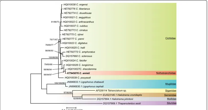
Fig. 1 Consensus tree obtained with Maximum Likelihood analysis. Bootstrap values correspond to ME/MP/ML values respectively after 2000 iterations. Only values≥ 50 have been indicated. Species newly sequenced for this study is in bold. Species belonging to Ligophorus, Tetrancistrum, Haliotrema and Thaparocleidus were used as outgroups. GenBank sequence ID precedes species name
by at least two different sequences). The optimal model of sequence evolution was TN93 + G [69]. The G param-eter indicates that non-uniformity of evolutionary rates among sites is modeled by using a discrete Gamma distribution. This model was used for the subsequent analysis. The three different methods used gave congruent results summarized in Fig. 1.
Relative to the outgroup taxa, all the species of Cichli-dogyrusappeared grouped in a monophyletic assemblage supported by high bootstrap values (100, 86 and 99 % for ME, MP and ML respectively). Cichlidogyrus pouyaudi Pariselle & Euzet [70] occupied a basal pos-ition in this group (bootstrap values, 75, 71 and 67 %) being the sister species of all the other species of Cichli-dogyrusas already observed by Mendlová et al. [35].
Four clusters with high bootstrap support were appar-ent. One cluster was made up of C. ergensi Dossou [71], C. tiberianus Paperna [25], C. douellouae Pariselle, Bilong & Euzet, [72], C. aegypticus Ergens [73], C. arthracanthus Paperna [25] and C. cubitus Dossou [71] (bootstrap values 81, 88 and 73 %). Another cluster was made up of C. yanni Pariselle & Euzet [74] and C. digi-tatus Dossou [71] (78, 79 and 77 %), a third one of C. amphoratus Pariselle & Euzet [74] and C. sclerosus Paperna & Thurston, [75] (98, 96 and 93 %) and the last
one of C. falcifer, C. longicirrus, C. dracolemma and C. amieti. Within this last cluster, C. falcifer was the sister species of C. longicirrus, C. dracolemma and C. amieti (99, 96 and 94 %) while C. longicirrus was sister to C. dracolemmaand C. amieti (98, 94 and 97 %). These four clusters were not supported by high bootstrap values. Three other species: C. cirratus Paperna [76], C. njinei Pariselle, Bilong & Euzet [72] and C. halli Price & Kirk [77] did not appear related to any group or species.
Principal Component Analysis (PCA)
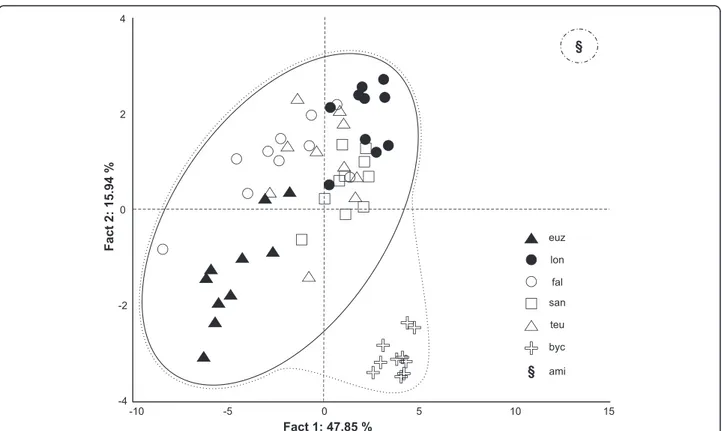
The PCA analysis shows a well-defined clustering (64 % of variance on axes 1 and 2) of parasite indi-viduals according to their respective host species (Fig. 2). The specimens of C. cf. bychowskii from H. bimaculatus are closer to those from H. fasciatus s. l. (C. euzeti, C. falcifer, C. longicirrus, C. sanseoi and C. teugelsi) than to the one collected from Aphyosemion cameronense (C. amieti), the latter been set apart re-garding the two axes. The most represented variables and their coordinates on axis 1 are DA a (–0.95), DA b (–0.93), VA a (–0.93), VB x (–0.92) and I (–0.87); and VII [II] (–0.82), VI [III] (–0.75), III [VI] (–0.70) on factor axis 2 (Table 2).
Fig. 2 Principal component analysis scatterplot of 10 Cichlidogyrus specimens of each of the following species. (euz) C. euzeti and (lon) C. longicirrus both from Hemichromis cf. fasciatus in Cameroun; (fal) C. falcifer, (san) C. sanseoi and (teu) C. teugelsi all from H. fasciatus in Senegal (fal) or Ivory Coast (san and teu); (byc) C. cf. bychowskii from H. bimaculatus in DRC; (ami) one specimen of C. amieti from Aphyosemion cameronense in Cameroon was used for supplementary observations
Discussion
Origin and host range of Cichlidogyrus amieti
Cichlidogyrus is the most species-rich ectoparasitic dactylogyridean monogenean genus known to parasitize African cichlid fishes. Species are distributed among a wide range of cichlid hosts [33, 58, 78]. The description of C. amieti from the gills of representatives of Cyprino-dontiformes by Birgi and Euzet [23] raises the question whether a species from this fish order could have been the source host at the origin of the Cichlidogyrus radi-ation in cichlids (see theories on cichlid biogeography above). An alternative explanation is lateral parasite transfer from a cichlid to a killifish host.
Our phylogenetic reconstruction indicates that C. amieti is phylogenetically nested within the parasites from species of Hemichromis Peters, 1857 at a derived position of the tree. Although we cannot rule out incomplete taxon coverage of Central West African
Cichlidogyrus, the present results suggest that C. amieti results from a recent transfer from cichlids to nothobranchiids. That is in accordance with the Birgi and Euzet [23] hypothesis. Such lateral transfer or host-switch can occur between related host species [31, 33, 79], but even between phylogenetically distant host species, both in artificial and natural conditions [19, 20, 80–84].
Aphyosemion spp. inhabit small forest streams [2, 3] where they live in sympatry with Hemichromis spp.. Bilong Bilong [85], based on morphological features, already hypothesized that C. amieti could derive from Hemichromis’ monogeneans.
Birgi and Euzet [23] reported that C. amieti was re-stricted to A. cameronense and A. obscurum, two species belonging to the same lineage (i.e. the A. cameronense group), but differing from one another by their biology and the fact that they are never found together in the same biotope [2]. In this study, C. amieti was also col-lected from A. exiguum, a species that does not belong to the A. cameronense group. This new host record can be explained by the sympatry of A. exiguum and A. cameronense or A. obscurum and by the relative phylo-genetic proximity of these fish species (compared to the phylogenetic distance between species of Aphyosemion and Hemichromis).
Influence of host-switching on haptoral and reproductive morphology
While the morphology and size of the sclerotized parts of the haptor and copulatory organs of species of Dacty-logyrus Diesing, 1850 [86], Anacanthorus Mizelle & Price, 1965 [87] or other genera are subject to distinct selective constraints [88–90], for Cichlidogyrus spp. these sclerotized parts seem to be mostly shaped by phylogenetic constraints [33, 35, 46]. In this case, the haptoral sclerite morphology is more suitable for infer-ring phylogenetic relationships, while the morphology of the reproductive organs is more useful for species-level identification, probably because of its faster evolutionary change [33, 35, 46]. In fact, for a given host species, the constraints on the haptoral sclerites aim to harmonize their morphologies (adapted to attach to the specific host’s gills), when those on the reproductive organs aim to make their morphologies mechanically incompatible, so profoundly different (leading to their reproductive isolation) (see Figs. 3 and 4).
Working on species of Dactylogyrus,Šimková et al. [91] stated that congeneric monogenean species occupying similar niches tend to have similar attachment organs. This resemblance is due to the fact that they are subject to the same considerable selective pressure imposed by the microhabitat within the host, possibly the gill morphology
Table 2 Loadings and explained variance of the first two PC of the PCA conducted on the“standardized” size of sclerites
Fact. 1 Fact. 2 Variance (%) 47.85 15.94 I (I) −0.873132 −0.389889 III (VI) −0.535295 −0.709933 IV (VII) −0.352651 −0.638280 V (IV) 0.032798 −0.186986 VI (III) −0.242397 −0.753181 VII (II) −0.383708 −0.825743 DB L −0.805421 −0.199626 DB y −0.705113 −0.021445 DB w −0.838653 0.044545 DB h −0.797425 0.231792 DA a −0.954211 0.212423 b −0.935548 0.165900 c −0.700350 0.015457 d −0.864610 0.324455 e −0.416138 0.547829 VB x −0.926706 0.077698 VB w −0.790515 0.033730 VA a −0.931174 −0.004236 b −0.933118 0.063475 c −0.532477 −0.117323 d −0.720919 0.190484 e −0.586523 0.488463
(I) [I], (III) [VI], (IV) [VII], (V) [IV], (VI) [III], (VII) [II] total length of uncinuli [Mizelle [57] nomenclature]; dorsal transverse bar: (DB L) total length, (DB y) distance between auricles, (DB w) maximum width, (DB h) auricle length; (DA a) total length of dorsal anchor:, (b) blade length, (c) shaft length, (d) guard length, (e) point length; ventral transverse bar: (VB a) branch total length, (VB x) maximum width; (VA a) ventral anchor total length, (b) blade length, (c) shaft length, (d) guard length, (e) point length
[92–96]. When these parasites occur on different host species, their attachment organs tend to differ from each other in their morphology and/or size, because different host species may have different gill structures. As pointed out byŠimková et al. [91], the morphology of the haptor is therefore an important adaptation of parasites to their hosts (host specificity) and to specific sites within their hosts (niche preference).
The phylogenetic tree obtained in this study (Fig. 1) suggests that C. amieti clusters within the monophyletic group already proposed by Mendlová et al. [34] and Řehulková et al. [63], made up of C. longicirrus, C. dracolemma and C. falcifer, all of them parasitizing Hemichromis spp.. The Cichlidogyrus spp. that para-sitize Hemichromis spp. have a highly homogenous
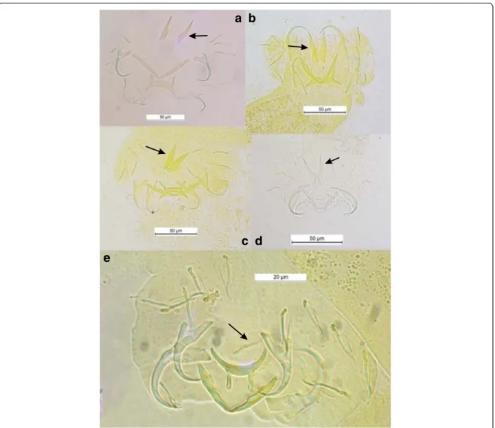
configuration of their haptoral sclerotized parts (group B in Vignon et al. [46]): very large first pair and small pairs III to VII of marginal hooks (= pairs II-III-IV and VI-VII sensu Mizelle [57]) combined with short auricles that are continuous with the dorsal surface of the dorsal transverse bar (Fig. 3a, b, c and d); this morphological relationship is also well supported by the PCA analysis (Fig. 2). In contrast, in C. amieti all marginal hooks are of similar small size including pair I (group A in Vignon et al. [46]) (Fig. 3e); this difference is also highlighted by our PCA results where this species is set apart regarding the two axes from all the other ones parasitizing Hemi-chromis spp. (Fig. 2). Therefore we hypothesize that, as soon as the ancestor of C. amieti (with group B morph-ology of its haptoral sclerites) colonized a species of
a b
c
e
d
Fig. 3 Haptoral sclerotized parts of some Cichlidogyrus spp. parasitizing Hemichromis spp. and C. amieti Birgi & Euzet [23] from Aphyosemion cameronense Boulenger, 1903. (a) C. longicirrus Dossou & Birgi [60]; (b) C. euzeti Dossou & Birgi [60]; (c) C. falcifer Dossou & Birgi [60]; (d) C. cf. bychowskii (Markevich [59]); (e) C. amieti Birgi & Euzet [23]. Arrow indicates uncinuli pair I [I]
Aphyosemion, selective pressures lead to a substantial morphological change in the haptoral sclerites, the most visible being the drastic reduction of the size of marginal hook pair I (Fig. 3 arrows). Vignon et al. [46], focusing on the same monogenean genus, did not find any evidence of host-related adaptation of the haptor morphology. However, these authors only considered Cichlidogyrus spp. infecting cichlids. The present study, considering also a more distant host-switch, provides new evidence supporting the hypothesis of the adaptive
nature of haptor morphology also within Cichlidogyrus in accordance with studies on other monogeneans by Morand et al. [97, 98], Huyse and Volckaert [99] and Bush et al. [100].
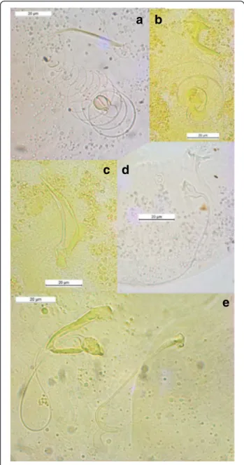
Rohde and Hobbs [101] and Šimková et al. [91] showed that congeneric parasite species living in the same niche presented differences in the morphology or size of their reproductive organs, as a result of random differentiation, which made possible their coexistence according to the hypothesis of reinforcement of repro-ductive barriers by mate discrimination [102–104]. This is the case for Cichlidogyrus spp. harbored by Hemichro-mis spp., which are well differentiated from each other by the morphology or size of their reproductive organs (Fig. 4a, b, c and d). Regarding the male copulatory organ (MCO) of C. amieti, we notice that it presents a tubular filiform single-looped penis without swollen por-tion and with a well-developed heel, and a sharply curved accessory piece with rounded ending [23, 58] (Fig. 4e). It resembles C. dracolemma (Fig. 4e) as pointed out by Řehulková et al. [63]. Therefore we may assume that C. dracolemma or a close relative was transferred from a species of Hemichromis to an Aphyosemion. This suggestion is strongly supported by the close phylogenetic relationship between these two parasite species (Fig. 1). Finally, the specialization of these two parasite species on phylogenetically dis-tant hosts (i.e. cichlid and killifish species) prevented their hybridization, thus explaining why their MCO morphologies have not been affected by selective pressure and thus did not substantially diverge.
Conclusion
Phylogenetic analysis suggests that C. amieti results from a recent host-switch from a cichlid species belonging to Hemichromis. The fact that the haptoral hard parts of C. amieti are of a different morphotype than those of its closely related congeners infecting Hemichromis spp., is the first proof, within Cichlidogyrus, of an adaptive compo-nent to haptoral morphology influenced by transfer to a new host. Previously, haptoral morphology of Cichlido-gyrus was considered to be mainly phylogenetically constrained. The changes in the haptoral elements after the host-switching event are in stark contrast to the similarity in male genital morphology to the parasites of representatives of Hemichromis. As genital differentiation between monogenean species is thought to be linked to reinforcement of parasite genetic isolation within the same host species, we suggest this similarity is a consequence of C. amieti having speciated as a result of host-switching. This study underscores the potential of Cichlidogyrus as a model to test the influence of ecology and evolution on parasite speciation [19, 78]. The fact that the adaptive com-ponent of haptoral morphology of Cichlidogyrus was not
a b
c d
e
Fig. 4 Male copulatory organs of some Cichlidogyrus spp. parasitizing Hemichromis spp. and C. amieti Birgi & Euzet [23] from Aphyosemion cameronense Boulenger, 1903. (a) C. longicirrus Dossou & Birgi [60]; (b) C. euzeti Dossou & Birgi [60]; (c) C. falcifer Dossou & Birgi [60]; (d) C. cf. bychowskii (Markevich [59]); (e) C. amieti Birgi & Euzet [23] (male copulatory organ on the left, vagina on the right)
inferred when including only species infecting cichlids, also demonstrates the importance of including the full phylo-genetic or host range of a parasite clade to reconstruct its speciation mechanisms.
Competing interests
The authors declare that they have no competing interests.
Authors’ contributions
Conceived and designed the experiments: FMM CBB AP JFA. Performed the experiments: FMM CBB AP JFA. Analyzed the data: FMM CBB AP JFA MPMV ARBN. Contributed reagents/materials/analysis tools: FMM AP JFA ARBN. Wrote the paper: FMM CBB AP JFA MPMV. All authors read and approved the final version of the manuscript.
Acknowledgements
This work was partially funded (2013) by the representation of IRD (Institut de Recherche pour le Développement) in Cameroon through the project « Programme Pilote Régional: Forêt Tropicale Humide d’Afrique Centrale » (PPR FTH-AC). M.P.M.V. is supported by the Czech Science Foundation, Project no. P505/12/G112 (European Centre of Ichthyoparasitology (ECIP) - Centre of excellence). Sequences were produced through the technical facilities of the Centre Méditerranéen Environnement Biodiversité (CeMEB). Authors want to thank W. Boeger and the two anonymous referees for their input to the revised version of the manuscript. This is publication ISE-M 2015-179 SUD.
Author details
1Laboratory of Parasitology and Ecology, Faculty of Sciences, University of
Yaoundé 1, BP 812 Yaoundé, Cameroon.2Institut des Sciences de l’Évolution,
IRD UMR 226, CNRS UMR 5554, Université de Montpellier, CC 63, Place Eugène Bataillon, 34095 Montpellier Cedex 05, France.3Biology Department,
Royal Museum for Central Africa, Leuvensesteenweg 13, B-3080 Tervuren, Belgium.4Department of Botany and Zoology, Faculty of Science, Masaryk
University, Kotlářská 2, CZ-611 37 Brno, Czech Republic.5Department of
Biology, Laboratory of Biodiversity and Evolutionary Genomics, University of Leuven, Charles Debériotstraat 32, B-3000 Leuven, Belgium.6Department of
Biological Sciences, University of Ngaoundéré, BP 454 Ngaoundéré, Cameroon.7Present address: Department of Biological Sciences, Higher
Teacher Training College, University of Yaoundé 1, P.O. Box 47, Yaoundé, Cameroon.8Present address: IRD, BP 1857, Yaoundé, Cameroon.9Present
address: Capacities for Biodiversity and Sustainable Development, Operational Directorate Natural Environment, Royal Belgian Institute of Natural Sciences, Vautierstraat 29, B-1000 Brussels, Belgium.
Received: 18 May 2015 Accepted: 29 October 2015
References
1. Parenti LR. A phylogenetic and biogeographic analysis of cyprinodontiform fishes (Teleostei, Atherinomorpha). Bull American Mus Nat Hist.
1981;168:335–557.
2. Amiet J-L. Faune du Cameroun, 2- Le genre Aphyosemion Myers (Pisces, Teleostei, Cyprinodontiformes). Compiègne: Sciences Nat; 1987.
3. Stiassny MLJ, Teugels GG, Hopkins CD. Poissons d’eaux douces et saumâtres de basse Guinée, ouest de l’Afrique centrale, vol. 2. Paris: IRD, MnHn, MRAC; 2007.
4. Basolo AL, Wagner Jr WE. Covariation between predation risk, body size and fin elaboration in the green swordtail, Xiphophorus helleri. Biol J Linn Soc. 2004;83:87–100.
5. Reznick D, Bryant M, Holmes D. The evolution of senescence and post-reproductive lifespan in Guppies (Poecilia reticulata). PLoS Biol. 2006;4:136–43. 6. Dargent F, Scott ME, Hendry AP, Fussmann GF. Experimental elimination of
parasites in nature leads to the evolution of increased resistance in hosts. Proc Roy Soc B. 2013;280:20132371.
7. Genade T, Benedetti M, Terzibasi E, Roncaglia P, Valenzano DR, Cattaneo A, et al. Annual fishes of the genus Nothobranchius as a model system for aging research. Aging Cell. 2005;4:223–33.
8. Valenzano DR, Terzibasi E, Cattaneo A, Domenici L, Cellerino A. Temperature affects longevity and age-related locomotor and cognitive
decay in the short-lived fish Nothobranchius furzeri. Aging Cell. 2006;5:275–8.
9. Terzibasi E, Lefrançois C, Domenici P, Hartmann N, Graf M, Cellerino A. Effects of dietary restriction on mortality and age-related phenotypes in the short-lived fish Nothobranchius furzeri. Aging Cell. 2009;8:88–99.
10. Cellerino A, Valenzano DR, Reichard M. From the bush to the bench: the annual Nothobranchius fishes as a new model system in biology. Biol. Rev. 2015;doi: 10.1111/brv.12183.
11. Reznick DN, Endler JA. The impact of predation on life history evolution in Trinidadian guppies (Poecilia reticulata). Evolution. 1982;36:125–48. 12. Reznick DN, Shaw FH, Rodd FH, Shaw RG. Evaluation of the rate of
evolution in natural populations of guppies (Poecilia reticulata). Science. 1997;275:1934–7.
13. van Oosterhout C, Trigg RE, Carvalho GR, Magurran AE, Hauser L, Shaw PW. Inbreeding depression and genetic load of sexually selected traits: how the guppy lost its spots. J Evol Biol. 2003;16:273–81.
14. van Oosterhout C, Joyce DA, Cummings SM. Evolution of MHC class IIB in the genome of wild and ornamental guppies, Poecilia reticulata. Heredity. 2006;97:111–8.
15. Bartáková V, Reichard M, Janko K, Polačik M, Blažek R, Reichwald K, et al. Strong population genetic structuring in an annual fish, Nothobranchius furzeri, suggests multiple savannah refugia in southern Mozambique. BMC Evol Biol. 2013;13:196.
16. Pinceel T, Vanschoenwinkel B, Deckers P, Grégoir A, Ver Eecke T, Brendonck L. Early and late developmental arrest as complementary embryonic bet‐hedging strategies in African killifish. Biol. J. Lin. Soc. 2015;doi: 10.1111/bij.12474.
17. Cable J, van Oosterhout C. The role of innate and acquired resistance in two natural populations of guppies (Poecilia reticulata) infected with the ectoparasite Gyrodactylus turnbulli. Biol J Linn Soc. 2007;90:647–55. 18. King TA, van Oosterhoutj C, Cable J. Experimental infections with the
tropical monogenean, Gyrodactylus bullatarudis: potential invader or experimental fluke? Parasitol Int. 2009;58:249–54.
19. Pariselle A, Morand S, Deveney M, Pouyaud L. Parasite species richness of closely related hosts: historical scenario and“genetic” hypothesis. In: Combes C, Jourdane J, editors. Taxonomy, Ecology and Evolution of Metazoan Parasites. Perpignan: Presses de l’Université de Perpignan; 2003. p. 147–66.
20. Vanhove MPM, Huyse T. Host-specificity and species-jumps in fish-parasite systems. In: Morand S, Krasnov B, Littlewood DTJ, editors. Parasite diversity and diversification: evolutionary ecology meets phylogenetics. Cambridge: Cambridge University Press; 2015. p. 401–19.
21. Cable J, van Oosterhout C, Barson N, Harris PD. Gyrodactylus pictae n. sp. (Monogenea: Gyrodactylidae) from the Trinidadian swamp guppy Poecilia picta Regan, with a discussion on species of Gyrodactylus von Nordmann, 1832 and their poeciliid hosts. Syst Parasitol. 2005;60:159–64.
22. Christison KW, Shinn AP, van As JG. Gyrodactylus thlapi n. sp (Monogenea) from Pseudocrenilabrus philander philander (Weber) (Cichlidae) in the Okavango Delta, Botswana. Syst Parasitol. 2005;60:165–73.
23. Birgi E, Euzet L. Monogènes parasites des poissons des eaux douces du Cameroun. Présence des genres Cichlidogyrus et Dactylogyrus chez Aphyosemion (Cyprinodontidae). Bull Soc Zool Fr. 1983;108:101–6. 24. Brosset A. Le peuplement des Cyprinodontes du bassin de l'Ivindo. Gabon
Rev Ecol (Terre Vie). 1982;36:233–92.
25. Paperna I. Studies on Monogenetic Trematodes in Israel. 2 Monogenetic Trematodes of Cichlids. Bamidgeh. Bull Fish Cult Israel. 1960;12:20–33. 26. Paperna I. Monogenea of inland water fish in Africa. Ann Mus Roy Afri Centr
– Sci Zool. 1979;226:1–131. Serie in 8°.
27. Bauer ON. Spread of parasites and diseases of aquatic organism by acclimatization: a short review. J Fish Biol. 1991;39:679–86. 28. Bakke T, Harris P, Cable J. The biology of gyrodactylid monogeneans: the
“Russian-doll Killers”. Adv Parasitol. 2007;64:161–376.
29. Cooper N, Griffin R, Franz M, Omotayo M, Nunn CL. Phylogenetic host specificity and understanding parasite sharing in primates. Ecol Let. 2012;15:1370–7.
30. Barson M, Přikrylová I, Vanhove MPM, Huyse T. Parasite hybridization in African Macrogyrodactylus spp. (Monogenea, Platyhelminthes) signals historical host distribution. Parasitology. 2010;137:1585–95.
31. Pariselle A, Boeger WA, Snoeks J, Bilong Bilong CF, Morand S, Vanhove MPM. The monogenean parasite fauna of Cichlids: a potential tool for host biogeography. Int. J. Evol. Biol. 2011;doi:10.4061/2011/471480.
32. Brant SV, Loker ES. Can specialized pathogens colonize distantly related hosts? Schistosome evolution as a case study. PLoS Pathog. 2005;1:e38. doi:10.1371/journal.ppat.0010038.
33. Pouyaud L, Desmarais E, Deveney M, Pariselle A. Phylogenetic relationships among monogenean gill parasites (Dactylogyridea, Ancyrocephalidae) infesting tilapiine hosts (Cichlidae): Systematic and evolutionary implications. Mol Phyl Evol. 2006;38:241–9.
34. Mendlová M, Pariselle A, Vyskočilová M, Šimková A. Molecular phylogeny of monogeneans parasitizing African, freshwater Cichlidae inferred from LSU rDNA sequences. Parasitol Res. 2010;107:1405–13.
35. Mendlová M, Desdevises Y, Civáňová K, Pariselle A, Šimková A. Monogeneans of West African Cichlid Fish: Evolution and Cophylogenetic Interactions. PLoS One. 2012;7:1–17.
36. Mendlová M,Šimková A. Evolution of host specificity in monogeneans parasitizing African cichlid fish. Parasit Vect. 2014;7:2–14.
37. Stiassny MLJ. Phylogenetic intrarelationships of the family Cichlidae: an overview. In: Keenleyside MHA, editor. Cichlid fishes: behaviour, ecology and evolution. London: Chapman and Hall; 1991. p. 1–35.
38. Murray AM. The fossil record and biogeography of the Cichlidae (Actinopterygii: Labroidei). Biol J Linn Soc. 2001;78:517–32.
39. Vences M, Freyhof J, Sonnenberg R, Kosuch J, Veith M. Reconciling fossils and molecules: Cenozoic divergence of cichlid fishes and the biogeography of Madagascar. J Biogeogra. 2001;28:1091–9.
40. Friedman M, Keck BP, Dornburg A, Eyta RI, Martin CH, Hulsey CD, et al. Molecular and fossil evidence place the origin of cichlid fishes long after Gondwanan rifting. Proc Roy Soc B: Biol Sci. 2013;280:20131733. 41. Rakotofiringa S, Euzet L. Monogènes parasites de Cichlidae (Teleostei)
endémiques de Madagascar. Bull Soc Zool Fr. 1983;108:107–14. 42. Euzet L, Combes C. Les problèmes de l'espèce chez les animaux parasites.
Mém Soc Zool Fr. 1980;40:239–85.
43. Ziętara MS, Lumme J. Speciation by host-switching and adaptive radiation in a fish parasite genus Gyrodactylus (Monogenea, Gyrodactylidae). Evolution. 2002;56:2445–58.
44. Boeger WA, Kritsky DC, Pie MR. Context of diversification of the viviparous Gyrodactylidae (Platyhelminthes, Monogenoidea). Zool Scr. 2003;32:437–48.
45. Bueno-Silva M, Boeger WA, Pie MR. Choice matters: incipient speciation in Gyrodactylus corydori (Monogenoidea: Gyrodactylidae). Int J Parasitol. 2011;41:657–67.
46. Vignon M, Pariselle A, Vanhove MPM. Modularity in attachment organs of African Cichlidogyrus (Platyhelminthes: Monogenea: Ancyrocephalidae) reflects phylogeny rather than host specificity or geographic distribution. Biol J Linn Soc. 2011;102:694–706.
47. Sonnenberg R. Description of three new species of the genus Chromaphyosemion Radda, 1971 (Cyprinodontiformes:Nothobranchiidae) from the coastal plains of Cameroon with a preliminary review of the Chromaphyosemion splendopleure complex. Zootaxa. 2007;1591:1–38. 48. Malmberg G. On the occurence of Gyrodactylus on Swedish fishes, Skrifter
utgivna av Södra Sveriges Fiskeriföreningen. 1956. p. 19–76. in Swedish with English abstract and species descriptions.
49. Marchiori N, Pariselle A, Pereira Jr J, Agnèse J-F, Durand J-D, Vanhove MPM. A comparative study of Ligophorus uruguayense and Ligophorus saladensis (Monogenea, Ancyrocephalidae) from Mugil liza (Teleostei, Mugilidae) in southern Brazil. Folia Parasit. 2015;62:024. doi:10.14411/ fp.2015.024.
50. Wu XY, Chilton NB, Zhu XQ, Xie MQ, Li AX. Molecular and
morphological evidence indicates that Pseudorhabdosynochus lantauensis (Monogenea: Diplectanidae) represents two species. Parasitol.
2005;130:669–77.
51. Hall TA. Bioedit: a user-friendly biological sequence alignment editor and analysis program for Windows 95/98/NT. Nucl Acids Symp Ser. 1999;41:95–8. 52. Tamura K, Peterson D, Peterson N, Stecher G, Nei M, Kumar S. MEGA5:
molecular evolutionary genetics analysis using maximum likelihood, evolutionary distance, and maximum parsimony methods. Mol Biol Evol. 2011;28:2731–9.
53. Schwarz G. Estimating the dimension of a model. Ann Stat. 1978;6:461–4. 54. Felsenstein J. Confidence limits on phylogenies: an approach using the
bootstrap. Evolution. 1985;39:783–91.
55. Ergens R, Gelnar M. Experimental verification of the effect of temperature on the size of hard parts of opisthaptor of Gyrodactylus katharineri Malmberg, 1964. Folia Parasitol (Praha). 1985;32:377–80.
56. Appleby C. Variability of the opisthaptoral hard parts of Gyrodactylus callariatis Malmberg, 1957 (Monogenea: Gyrodactylidae) from Atlantic cod Gadus morhua L. in the Oslo Fjord, Norway. Syst Parasitol. 1996;33:199–207. 57. Mizelle JD. New species of trematodes from the gills of Illinois fishes. Am
Midl Nat. 1936;17:785–806.
58. Pariselle A, Euzet L. Systematic revision of dactylogyridean parasites (Monogenea) from cichlid fishes in Africa, the Levant and Madagascar. Zoosys. 2009;31:849–98.
59. Markevich AP. Parasitic diseases of fish and their control. Publ. Koiz., Leningrad. 1934;3–100. in Russian.
60. Dossou C, Birgi E. Monogènes parasites d'Hemichromis fasciatus Peters, 1857 (Teleostei, Cichlidae). Ann Sci Nat Zool. 1984;6:101–9. Paris.
61. Paperna I. Monogenetic Trematodes collected from fresh water fish in southern Ghana. Bamidgeh, Bull Fish Cult. 1965;17:107–15. Israel.
62. Pariselle A, Euzet L. Two new species of Cichlidogyrus Paperna, 1960 (Monogenea, Ancyrocephalidae) gill parasites on Hemichromis fasciatus Peters, 1858 in Africa, with remarks on parasite geographical distribution. Parasite. 2004;11:359–64.
63. Řehulková E, Mendlov M, Šimková A. Two new species of Cichlidogyrus (Monogenea: Dactylogyridae) parasitizing the gills of African cichlid fishes (Perciformes) from Senegal: morphometric and molecular characterization. Parasitol Res. 2013;112:1399–410.
64. Young PC. Ten new species of Haliotrema Johnston and Tiegs, 1922 (Monogenoidea: Dactylogyridae) from Australian fishes and a revision of the genus. J Zool. 1968;154:41–75. London.
65. Bychowsky BE, Nagibina LF. Ancyrocephalinae (Monogenoidea, Dactylogyridae) from the sea fishes of the family Pomadasyidae. An Instit Biolo Uni Nacio Autón México Ser Zool. 1970;41:19–28.
66. Euzet L, Suriano DM. Ligophorus n. g. (Monogenea, Ancyrocephalidae) parasite des Mugilidae (Téléostéens) en Méditerranée. Bull Mus Nat Hist Nat. 1977;472:799–822. Paris.
67. Rubtsova NY, Balbuena JA, Sarabeev VL, Blasco-Costa I, Euzet L. Description and morphological variability of a new species of Ligophorus and Ligophorus chabaudi (Monogenea: Dactylogyridae) on Mugil cephalus (Teleostei) from the Mediterranean basin. J Parasitol. 2006;92:486–95.
68. Yamaguti S. Studies on the helminth fauna of Japan. Part 19. Fourteen new ectoparasitic trematodes of fishes. Kyoto, Japan: Published by the author. 1937:1-28.
69. Tamura K, Nei M. Estimation of the number of nucleotide substitutions in the control region of mitochondrial DNA in humans and chimpanzees. Mol Biol Evol. 1993;10:512–26.
70. Pariselle A, Euzet L. Three new species of Cichlidogyrus Paperna, 1960 (Monogenea, Ancyrocephalidae) parasitic on Tylochromis jentinki (Steindachner, 1895) (Pisces, Cichlidae) in West Africa. Syst Parasitol. 1994;29:229–34.
71. Dossou C. Parasites de Poissons d'eau douce du Bénin III. Espèces nouvelles du genre Cichlidogyrus (Monogenea) parasites de Cichlidae. Bull IFAN. 1982;44:295–322.
72. Pariselle A, Bilong Bilong CF, Euzet L. Four new species of Cichlidogyrus Paperna, 1960 (Monogenea, Ancyrocephalidae), all gill parasites from African mouthbreeder tilapias of the genera Sarotherodon and Oreochromis (Pisces, Cichlidae), with a redescription of C. thurstonae Ergens, 1981. Syst Parasitol. 2003;56:201–10.
73. Ergens R. Nine species of the genus Cichlidogyrus Paperna, 1960
(Monogenea: Ancyrocephalinae) from egyptian fishes. Folia Parasitol (Praha). 1981;28:205–14.
74. Pariselle A, Euzet L. Cichlidogyrus Paperna, 1960 (Monogenea, Ancyrocephalidae): gill parasites from West African Cichlidae of the subgenus Coptodon Regan, 1920 (Pisces), with descriptions of six new species. Syst Parasitol. 1996;34:109–24.
75. Paperna I, Thurston JP. Monogenetic trematodes collected from cichlid fish in Uganda; including the description of five new species of Cichlidogyrus. Rev Zool Bot Afri. 1969;LXXIX:15–33.
76. Paperna I. Parasitic helminths of inland-water fishes in Israel. Israel J Zool. 1964;13:1–26.
77. Price CE, Kirk RG. First description of a monogenetic trematode from Malawi. Rev Zool Bot Afri. 1967;76:137–44.
78. Pariselle A, Muterezi Bukinga F, Van Steenberge M, Vanhove MPM. Ancyrocephalidae (Monogenea) of Lake Tanganyika: IV: Cichlidogyrus parasitizing species of Bathybatini (Teleostei, Cichlidae): reduced
host-specificity in the deepwater realm? In: Koblmüller S, Albertson RC, Genner MJ, Sefc KM, Takahashi T, editors. Advances in cichlid research: Behavior, ecology and evolutionary biology. Hydrobiologia. 2015;748:99–119. 79. Bilong Bilong CF, Birgi E, Euzet L. Enterogyrus barombiensis n. sp.
(Monogenea, Ancyrocephalidae) parasite stomacal de trois Cichlidae endémiques du Lac du cratère Barombi Mbo (Cameroun). Ann Parasitol Hum Comp. 1991;66:105–8.
80. Kaneko II JJ, Yamada R, Brock JA, Nakamura RM. Infection of tilapia, Oreochromis mossambicus (Trewavas), by a marine monogenean, Neobenedenia melleni (MacCallum, 1927) Yamaguti, 1963 in Kaneohe Bay, Hawaii, USA, and its treatment. J Fish Disea. 1988;11:295–300. 81. Cable J, Scott ECG, Tinsley RC, Harris PD. Behavior favoring transmission
in the viviparous monogenean Gyrodactylus turnbulli. J Parasitol. 2002;88:183–4.
82. Huyse T, Audenaert V, Volckaert FAM. Speciation and host-parasite relationships in the parasite genus Gyrodactylus (Monogenea,
Platyhelminthes) infecting gobies of the genus Pomatoschistus (Gobiidae, Teleostei). Int J Parasitol. 2003;33:1679–89.
83. Pariselle A, Bitja Nyom AR, Bilong Bilong CF. Checklist of the
ancyrocephalids (Monogenea) parasitizing Tilapia species in Cameroon, with the description of three new species. Zootaxa. 2013;3599:78–86.
84. Pérez-Ponce de Léon G, Choudhury A. Biogeography of helminth parasites of freshwater fishes in Mexico: the search for patterns and processes. J Biogeogra. 2005;32:645–59.
85. Bilong Bilong CF. Les Monogènes parasites des poissons d’eau douce du Cameroun : biodiversité et spécificité; biologie des populations inféodées à Hemichromis fasciatus. Thèse de Doctorat d’État, Université de Yaoundé I, Yaoundé, Cameroun. 1995.
86. Diesing KM. Systema helminthum. Vol. 1. Vindobonae: Braumfiller W; 1850. 87. Mizelle JD, Price CE. Studies on monogenetic Trematodes. XXVIII. Gill
parasites of the Piranha with proposal of Anacanthorus gen. n. J Parasitol. 1965;51:30–6.
88. Guégan J-F, Lambert A. Twelve new species of dactylogyrids (Platyhelminthes, Monogenea) from West African barbels (Teleostei, Cyprinidae), with some biogeographical implications. Syst Parasitol. 1990;17:153–81.
89. van Every LR, Kritsky DC. Neotropical Monogenoidea. 18. Anacanthorus Mizelle and Price, 1965 (Dactylogyridae, Anacanthorinae) of Piranha (Characoidea, Serrasalmidae) from the Central Amazon, their phylogeny, and aspects of host–parasite coevolution. J Helminthol Soc Washin. 1992;59:52–75. 90. El Gharbi S, Lambert A, Berrebi P. Le genre Barbus (sous-genre Barbus et
Labeobarbus) au Maroc. Génétique et Parasitologie. Cah Ethol. 1993;13:223–6. 91. Šimková A, Ondračková M, Gelnar M, Morand S. Morphology and
coexistence of congeneric ectoparasite species: reinforcement of reproductive isolation? Biol J Linn Soc. 2002;76:125–35.
92. Huyse T, Malmberg G. Molecular and morphological comparisons between Gyrodactylus ostendicus n. sp. (Monogenea: Gyrodactylidae) on
Pomatoschistus microps (Krøyer) and G. harengi Malmberg, 1957 on Clupea harengus membras L. Syst Parasitol. 2004;58:105–13.
93. Rohde K, Rohde PP. The ecological niches of parasites. In: Rohde K, editor. Marine parasitology. Wallingford: CABI Publishing; 2005. p. 286–93. 94. Šimková A, Morand S. Co-evolutionary patterns in congeneric
monogeneans: a review of Dactylogyrus species and their cyprinid hosts. J Fish Biol. 2008;73:2210–27.
95. Mancheva K, Karaivanova E, Atanasov G, Stojanovski S, Nedeva I. Analysis of the influence of the host body size on morphometrical characteristics of Ancylodiscoides siluri and Ancylodiscoides vistulensis. Biotec Biotechnol Equip. 2009;23:735–41.
96. Poisot T, Desdevises Y. Putative speciation events in Lamellodiscus (Monogenea: Diplectanidae) assessed by a morphometric approach. Biol J Linn Soc. 2010;99:559–69.
97. Morand S, Hafner MS, Page RDM, Reed DL. Comparative body size relationships in pocket gophers and their chewing lice. Biol J Linn Soc. 2000;70:239–49.
98. Morand S,Šimková A, Matejusová I, Plaisance L, Verneau O, Desdevises Y. Investigating patterns may reveal processes: evolutionary ecology of ectoparasitic monogeneans. Int J Parasitol. 2002;32:111–9.
99. Huyse T, Volckaert FAM. Identification of a host-associated species complex using molecular and morphometric analyses, with the description of Gyrodactylus rugiensoides n. sp. (Gyrodactylidae, Monogenea). Int J Parasitol. 2002;32:907–19.
100. Bush S, Sohn E, Clayton DH. Ecomorphology of parasite attachment: experiments with feather lice. J Parasitol. 2006;92:25–31.
101. Rohde K, Hobbs R. Species segregation: Competition of reinforcement of reproductive barriers? In: Cremin M, editor. Parasite lives. Papers on parasites, their hosts and their associations to honour JFA Sprent. St. Lucia: University of Queensland Press; 1986. p. 189–99.
102. Butlin RK. Reinforcement of premating isolation. In: Otte D, Endler JA, editors. Speciation and its consequences. Sunderland: Sinauer Associates; 1989. p. 158–79.
103. Butlin RK. Reinforcement: an idea evolving. TREE. 1995;10:432–4. 104. Jiggins CD, Mallet J. Bimodal hybrid zones and speciation. TREE.
2000;15:250–5.
Submit your next manuscript to BioMed Central and take full advantage of:
• Convenient online submission
• Thorough peer review
• No space constraints or color figure charges
• Immediate publication on acceptance
• Inclusion in PubMed, CAS, Scopus and Google Scholar
• Research which is freely available for redistribution
Submit your manuscript at www.biomedcentral.com/submit