by
C.C. Young (Zhongjian Yang) and Xijin Zhao Institute of Vertebrate Paleontology and
Paleoanthropology Monograph Series I, No. 8
Science Press July, 1972
Translated by Will Downs Bilby Research Center Northern Arizona University
The text describes the skeleton of Mamenchisaurus hochuanensis from Hechuan (formerly Hechuan) County, Sichuan Province excavated from 130 Ma Jurassic sediments. This taxon represents the largest species of dinosaur in China to date, and is the most complete sauropod recovered in the country with a body length of 22 meters, torso height of 3.5 meters, and an estimated living weight of 45 tons. Mamenchisaurus was adapted for both terrestrial and aquatic life while subsisting on shoreline vegetation.
The specimen was discovered exposed on the surface by the local populace prior to the Chinese Revolution. But under the leadership of the Chinese Guomindang reactionary party, it was ignored, and as such, this extremely valuable specimen was abandoned and allowed to weather away in situ.
In April of 1957, after the Revolution and under the leadership of Chairman Mao Zedong, the Central Party, and encouraged by the elevated proletariat, and under the guidance of the middle peasantry, the Sichuan Provincial Petroleum Exploration Corps conducted mineral exploration in the impoverished region to rediscover the fossil locality. The Sichuan Provincial Museum immediately dispatched a work team to conduct an excavation with the assistance and support of the local population to expose a nearly complete skeleton lacking the anterior limbs and head. In 1962, after preliminary preparation, the specimen was recognized as a general scientific and educational resource, and subsequently moved to the Chengdu Academy of Geology. In the Spring of 1964 the Chengdu Academy of Geology dispatched two of its personnel to accompany the specimen to the Institute of Vertebrate Paleontology and Paleoanthropology (IVPP) in Beijing where in collaboration with the technical and professional staff, further preparation, reconstruction, and mounting of the skeleton, along with a description, was completed in 1965. This dinosaur skeleton was a product of the efforts of workers in both Beijing and Chengdu and was publicized in the national publications China Reconstructs and China Pictorial in 1965.
Following the guiding principle as espoused by Chairman Mao: “to advance education through theories of knowledge applied from dialectical materialism,” individuals from several provincial and municipal museums have produced models of Mamenchisaurus hochuanensis which will, in the near future, be popularly disseminated as concrete evidence for biological evolutionary development.
Foreword
The specimen described in this text was produced from Hechuan Co. north of the metropolis of Zhongqing (formerly Chungking) (Fig.1). More specifically, the locality is 200 meters above the Gaochufujiang River on the slope of Mt. Gushushan, by the village of Taihezhen (formerly Taiheba), 35 kilometers from the municipality of Hechuan.
The specimen is preserved in red sediments (Fig. 3, Pl. I)
predominantly composed of approximately 250 m of purple-red mudstones, sandy mudstones, and argillaceous sandstones. Calcareous concretions are abundant in the
mudstones and sandy mudstones (Fig. 2). Above and below the fossil horizon the sediments are interbedded with dark purple sandstones. Regional geologic data assigns the fluviolacustrine fossiliferous sediments to the Jurassic Upper (Shang) Xiaximiao Fm. within the Zhongqing (Chungking) Group.
Preparation of the specimen occurred in two phases: preliminary work was undertaken in Chengdu prior to its transport to Beijing, where the majority of work was undertaken. Difficulty of preparation was compounded by a carbonate rhind that incased the massive bones to create cumbersome blocks. Furthermore, the repeated relocation of the specimen resulted in a certain amount of breakage and shifting creating difficulty piecing back together fragmentary bones. Consequently, it required nearly a years time to complete preparation of the specimen.
This work was conducted through the united efforts of the Chinese people. The discovery and protection of the specimen was undertaken by colleagues in Hechuan County, Sichuan, and the Sichuan Petroleum Research Corps.
Colleagues at the Sichuan Provincial Museum provided description of the excavation process, field sketches of the specimen’s exposure, and photography of related taphonomic conditions. Colleagues from the Chengdu Academy of Geology provided introductory analysis regarding regional stratigraphic problems and preliminary preparation of the specimen prior to its transportation to IVPP. It was only through the efforts of the aforementioned colleagues that this project was successfully completed.
Figure 1. Location of the Mamenchisaurus quarry.
Figure 2. Stratigraphic
cross-section at the Gushushan fossil locality.
Description Saurischia, Seeley, 1878
Sauropoda, Marsh, 1978
Mamenchisauridae fam. nov.
Diagnosis: Extremely long cervical region (19 vertebrae); dorsal, sacral, and caudal
vertebrae short and few in number (4 sacral vertebrae); massive and long cervical ribs; pleurocoels not well developed on dorsal vertebrae; anterior dorsal vertebrae with bifid neural spine; medial caudal vertebrae with bifurcated haemal spines; anterior caudal vertebrae procoelous; centrally positioned pubic peduncle on ilium.
Mamenchisaurus (Young, 1954) Mamenchisaurus hochuanensis s p . n o v .
Material: A relatively complete vertebral column including nearly complete cervical,
dorsal, sacral, and caudal series, with nearly complete neural arches. Cervical and dorsal ribs are incomplete although cervical ribs are numerous and relatively well preserved. Sacral girdle is represented by the ilium, ischium, and a portion of the pubis. Tibia and fibula are complete but femur is represented only by a right distal end. Hind feet are represented by a pair of astragali and several metatarsals and phalanges, forelimbs are represented only by a fragmentary sternum and proximal end of right humerus. Additional elements include several unidentifiable dorsal vertebrae and fragmentary ribs (see Fig. 4).
Diagnosis: Relatively few dorsals and caudals, but number of cervicals exceeds that in
other known species (cervicals 19, dorsals 12, sacrals 4, and caudals 35+). Cervicals are weakly opisthocoelus and constitute nearly half the total body length. Dorsals near the sacral region are distinctly opisthocoelus, 16 anterior caudals are amphicoelus, but the posterior caudals are platycoelus. Cervical neural spines are low and flat, those on the anterior four dorsals are bifid, but those on the five dorsals posterior to these are single with robust terminal ends. Neural spines on the three anterior sacrals are fused, and on the fourth sacral and first caudal are anteriorly convex and posteriorly concave. Caudal haemal spines become anteroposteriorly bifurcated beginning on the ninth caudal. Ilium is robust with a pubic peduncle located centrally. The ischium is gracile. Tibia and fibula are thin and flat, nearly equivalent in length, and the tibia displays a well developed proximal end. Astragalus is relatively well developed with deeply concave articular facets for the tibia/fibula causing the fibular keel to be extremely pronounced. Metatarsals are relatively short and small although the ungual phalanx (claw) of digit I is particularly well developed.
Description Vertebral column (Pl. III)
Cervicals (Pl. XIV 1,2) (Table 1)
Atlas (Fig. 5): The proatlas is indistinguishable due to fusion. Although the atlas/axis intercentrum is solidly fused to the axis, the general outline of the intercentrum is vaguely
discernible. The atlas centrum itself is relatively weak and small with an irregular morphology that lacks a distinct outline. A neural spine is not well developed but is nevertheless distinctly
Table 1 Vertebral measurements (excluding caudals) (mm). Sequence Centrum length Posterior height Posterior breadth Total height Cv1 60 75 55 85 Cv2 160 80 75 175 Cv3 215 85 85 160 Cv4 320 120 100 195 Cv5 415 150 105 240 Cv6 480 165 110 260 Cv7 580 200 110 340 Cv8 590 220 110 330 Cv9 610 225 130 370 Cv10 660 240 130 390 Cv11 730 255 170 380 Cv12 730 300 140 470 Cv13 690 300 170 510 Cv14 690 325 160 530 Cv15 660 350 200 560 Cv16 640 355 175 580 Cv17 550 375 190 630 Cv18 400 380 220 660 Cv19 325 350 230 660 D1(20) 250 340 170 640 D2(21) 250 315 250 650 D3(22) 240 345 220 740 D4(23) 250 320 220 710 D5(24) 250 350 195 830 D6(25) 230 350 200 890 D7(26) 210 350 200 880 D8(27) 220 305 240 850 D9(28) 210 310 230 890 D10(29) 210 360 230 900 D1130) 190 360 220 870 D12(31) 180 330 215 830 S1(32) 150 225 S2(33) 170 225 840 S3(34) 210 225 S4(35) 155 300 225 815
Axis (Fig. 5): The entire element is preserved in tight articulation with the third cervical vertebra (Cv3). Ventrally the axis is very flat and smooth with a depression at its anterior end. The surface of the anteromedial lamina and the very slightly projected posteroventral surface differ from the characters of other cervical vertebrae. The anterior articular surface is indistinct, although as with the other cervicals, it tends to be convex with a centrum that is distinctly opisthocoelus. A small fossa is present on each side anterolaterally. The neural spine is very weakly developed, is more distinctly bifurcated than the atlas, and is penetrated by a rugose fenestra anteriorly.
Figure 5. Mamenchisaurus hochuanensis cervical vertebrae 1-3.
1. Left lateral view, 2. Ventral view.
The following cervical series will be generally described, unlike the previous two. CV3 (Fig. 5) displays a distinct transitional morphology between that of the axis and CV4. Lengths of the cervicals vary greatly with the longest being CV11 and CV12 (see Table 1). Posterior to these, the lengths progressively diminish to the last cervical, CV19, which is only one-half as long as CV12. All cervicals are distinctly opisthocoelus and are preserved in their original articulation. The prezygopophyses of each cervical articulate extremely tightly with their preceding counterpart. Ventrally, the centra are rather flat and are very slightly flared at their posterior halves. Laterally they are concave with two small elliptical fossae. The midpoint of the centra are more thin and weak than at the articular regions. Two laminar reinforcements lie between the ventral and lateral sides. Anteroposterior neural spines become thin, flat, and distinctly bifurcated laterally. Like Euhelopus zdanskyi Wiman, the top of the neural spines are rugose and partially fenestrated. Commonly there is an extremely well developed elliptical canal linking the anterior and posterior neural arches. Parapophyses and diapophyses are well preserved on all cervicals, are positioned rather anteroventrally, and descend strongly to the posterior. From Cv17 parapophyses gradually ascend up the centra such that on the last cervical they are nearly level with dorsal parapophyses. Pneumatisation is well developed on the cervicals (including the neural arches) and in cross-section elaborate honeycombed laminae are infilled with matrix. Breakage has occurred at the midpoint of some cervicals due to being rather gracile at this point.
Cervical ribs (Pl. IV, Nos. 1,2,3): The majority are preserved articulated to the corresponding centra, although several of the distal ends were scattered within the surrounding matrix. The ribs are absent on the atlas/axis, and probably, as with other sauropods, were too delicate for preservation. The degree of fusion gradually weakens beginning with rib 17, where they fundamentally separate from the centra. The most posterior vertebra preserves only the right rib. The best preserved ribs are associated with Cv4,6,10,12, and 15. The longest rib is on Cv14 which attains 2.1 meters (Table 2).
Cervical ribs may be recognized in two forms: anteriorly they are relatively short, spoon-shaped with a sharp terminus, but do not resemble the spoon-shaped morphology of Mamenchisaurus constructus Young. Posteriorly, the main shaft is baton-shaped,
ends are transversely projected, with relatively short capitula and tubercula that are fused to the opposing centra. It is only at the most posterior three where this fusion is not as complete. These ribs are extremely stout, elongated, and run along the cervical series with hardly any ventral inclination. The cervical ribs of Euhelopus are relatively long and extend posteriorly merely to the posterior end of the succeeding vertebra. On M. hochuanensis the rib may extend as far as the succeeding third vertebra or even to the anterior portion of the fourth. The posterior cervical ribs gradually become shorter and thicker such that on the most posterior vertebra they do not exceed one-half a meter, or one-quarter the length of the CV14 rib (2.1 m).
Table 2. Preserved lengths of cervical ribs (mm).
Right Left Sequence Tuberculum to capitulum Total length Sequence Tuberculum to capitulum Total length 1 1 2 2 3 3 4 4 5 100 250 5 110 240 6 140 450 6 140 650 7 150 290 7 170 8 140 310 8 160 1550 9 150 330 9 200 720 10 120 1030 10 200 600 11 120 430 11 12 200 1090 12 230 670 13 200 940 13 200 290 14 190 990 14 200 2100 15 160 820 15 240 1330 16 170 980 16 190 1200 17 17 190 600 18 18 19 19
Dorsal vertebra (Plate XII, Fig. 1) (Table 1): The last cervical and first dorsal vertebrae are generally distinguished by rib morphology. However, a difficulty is posed here by the last cervical‘s absence of articulated ribs. But other relatively well preserved cervical ribs suggest that the largest cervical rib belongs to the most posterior cervical. The parapophyses facets on the twentieth centrum are the highest positioned in the series and the centrum resembles other dorsals by being extremely short. Therefore, it is determined that centra anterior to and including number 19 are cervical and that centrum 20 is the first dorsal (D1). In this manner M. hochuanensis possesses 12 dorsal vertebra (the most posterior dorsal represents the transition from dorsal to sacral) or a total of 31 presacral vertebra which exceeds all known sauropods.
The majority of dorsals, and particularly those anterior to D9, have been shifted in position and suffer compressional distortion, while posterior to D9 the vertebrae are shifted but not to the extent of the anterior series. Rotational distortion has caused the middle of the centrum to become transversely broadened and obliquely inclined dorsally on the right side, which subsequently caused the neural arch to be strongly obliquely distorted with the
right side forced dorsally and left side ventrally. In this manner, a perpendicular line drawn from the top of the neural spine would not bisect the median ventral centrum but will
traverse the right side. As the distortion is spread among the vertebrae universally, interpretation of the general morphology is basically reliable.
Dorsal centra are relatively robust with weak or undeveloped pleurocoels.
Muscular attachment scars are also not well developed and most possess up to four or five laminae that are predominantly dorsoventrally oriented. Parapophyses on the anterior dorsals resemble those on the posterior cervicals and approach the anteroventral angle of the cervicals. Parapophyses on the posterior dorsals migrate dorsally on the centrum. Diapophyses on the anterior dorsals resemble those on the posterior cervicals by being ventrally directed, but posteriorly they gradually become directed dorsally. Dorsal lamina morphology is not well developed and there is a small fossa only anteriorly.
Figure 6. Mamenchisaurus hochuanensis dorsal vertebra 2.
1. Right lateral view, 2. Anterior view.
The four processes are located on the dorsal portion of the neural arch. This is particularly conspicuous on D8 and 9 (Fig. 7), with diapophyses being the highest, posterior zygopophyses are relatively low, and the anterior zygopophyses are the lowest with their dorsal margin parallel to the midline of the diapophyses. Parapophyses are slightly higher than the centrum. The four processes are linked by laminae, particularly between the anterior zygopophyses and the diapophyses. The medial lamina forms an oblique plane that ascends dorsally on each process to the top of the neural spine. Each process also possesses a small perpendicular lamina, and as such, each side is composed of two parallel planes which intersect the aforementioned two laminae between the anterior zygopophyses for a total of five laminar surfaces.
Dorsal centra are opisthocoelus (Pl. XIII, Fig. 2) with relatively conspicuous prezygopophyses on the anterior series but which become weaker posteriorly. Pleurocoels are not numerous, with only two present on each side. Sediment infilling is not as
prominent as on the cervical series although the honeycombed infrastructure is still present. The dorsal spine is well developed, robust, and very high. As on the cervical vertebrae, the four anterior dorsals have bifid dorsal spines (Pl. XII, XIII), but rotational distortion has extended the right side, leaving the left side short and broad. The longest spine is 15 cm and the greatest angle of divergence is 90°. Among the bifid series, none is
complete, D1 displays the most well developed bifid condition in the series, in which bifurcation gradually diminishes posteriorly. The top of D5 neural spine is bluntly rounded with a shallow medial groove that descends ventrally. D6 neural spine is typical in
morphology, unbifurcated, and has a thick broadened terminus. All of the spines have rugose surfice texture. Posterior dorsals have relatively thin and flat lateral surfaces with well developed posteriorly directed posterolateral wing-shaped laminae which cause the entire neural spine structure to resemble a projected
spoon shaped body that is anteriorly convex and concave posteriorly.
Several vertebrae are relatively well preserved. D2 (Plate XII, Fig. 2) (Text Fig. 6) is particularly robust with four lateral laminae. On one side a lamina extends from the diapophysis to the prezygopophysis, one extends from the diapophysis to the parapophysis, while the other two are basically parallel. Diapophyses are rugose and there are four small fossae, while the posterior centrum is deeply concave (approximately 11 cm), its interior is
honeycombed, it has an elliptical cross-section due to compressional distortion, and the right bifid dorsal spine is higher than the left. Two small laminae extend ventrally from the smooth apex of the spines.
D3 (Pl. XII, Fig. 3) is relatively low, flat, and extremely robust, with a particularly large elliptical pleurocoel on the centrum. Neural arch laminae are more well developed than on other dorsals, being thick and broad, are relatively conspicuous at the top of the bifid spine, becoming broadened laterally. The several processes of the neural spines are relatively broad, particularly the posterior zygopophyses.
The D6 centrum is relatively short and possesses relatively few pleurocoels. Diapophyses are circular with several small irregularly shaped fossae. The neural spine is flattened and not bifid.
Table 3. Dorsal rib measurements (mm).
Length Tub. to Prox. Shaft
Sequence Direct Arced capit. dist. breadth midpoint
1 145 Right 6 200 100 45 7 100 53 11 880? 970? 165 85 30 10 1070 1120 120 80 55 Left 11 905? 940? 75 40
D8 is the smallest dorsal with the lowest neural spine. It has suffered postburial fracturing, and possesses a mammary shaped process.
Figure 7. Right lateral view of
Mamenchisaurus hochuanensis dorsal vertebrae 9 & 10.
D9 and D10 are extremely high with tightly articulated pre- and postzygopophyses and tightly fused centra (Text Fig. 7).
Dorsal ribs: (Plate, IV, Figs. 4,5,6,7,8) (Table 3). The very few that are represented are incomplete. The left side preserves only nos. 10 and 11 which are relatively complete, though more are preserved on the right side with no. 11 the most complete. Nos. 1,2,6,7 on the right side are only partially preserved. On the left side dorsal rib 10 is 1.07 m long in a direct line while rib 11 is .905 m in length. On the right side rib 11 is .88 m. These specimens do not display a high degree of curvature except at the neck of the head (approximately 150°). The head is relatively large, tuberculum and capitulum differ in size, and are separated by a 90° angle or sometimes larger. Shafts are relatively straight with undeveloped laminar ridges. Robustness and length gradually reduce anteroposteriorly. Proximal ends are all
more robust than distal ends although the posterior ribs tend to have consistent proximal and distal ends.
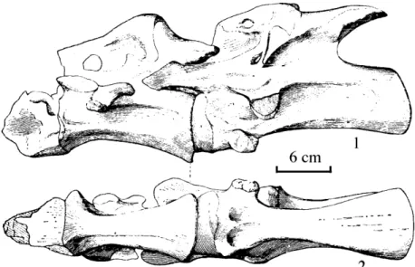
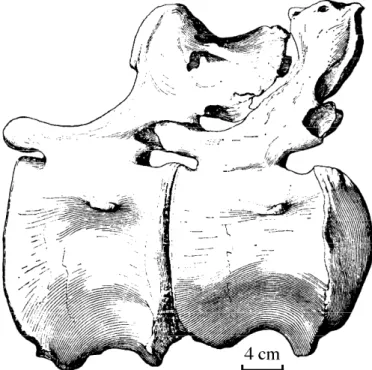
Sacrum (Table 1): The sacrum is a set of four relatively well preserved and tightly fused vertebrae. Only the most posterior neural spine is not fused with the other three. Centra
boundary lines are indistinct with only median depressions discernable. With the exception of the neural spine, the remaining neural arch features are not readily recognizable as the processes and sacral ribs are fused, and it is only possible to distinguish them ventrally. As with the cervicals and dorsals the entire sacrum has undergone compressional distortion but not as intensively as the aforementioned, with the four elements expanded anterolaterally and the
posterior two being been slightly contorted. The anterior end of S1 projects anteriorly while the posterior end of S4 projects posteriorly by 8 cm. Both anterior and posterior centra are bisected by a median longitudinal laminar ridge, the largest being 19.5 cm while its counterpart is 9.5 cm. At the anterior end the large hemisphere is on the right side, but posteriorly it is on the left side.
Sacral centra are fused into a single column which anteriorly is amphicoelus, in opposition to the procoelous caudal vertebrae. As such, the fused centra are exceptionally robust and lack pleurocoels. Intercentrum structure is only visible on the last centrum in ventral perspective as are the relatively distinct longitudinal laminae which connect the diapophyses and parapophyses. Ventrally, it is also evident that the lengths of the four vertebrae differ with S3 being the longest (22 cm), S2 is 18 cm, S4 is 14 cm and S1 is 11.5 cm. S3 possesses vestigial features of rib fusion, while the S4 centrum displays lateral depressions on each side, and a robust posterior margin with a small undulating lamina.
Sacral neural spines are weak and gracile, particularly the spine of S4, which is not fused to the other three and is situated approximately 10 cm distant. Its anterior side is convex, posterior side is concave, and it possesses a longitudinal median lamina. The remaining three fused neural spines possess four lateral longitudinal laminar ridges. The
Figure 8. Mamenchisaurus hochuanensis
caudal vertebrae.
1. Cd35, A. Right lateral view, B.Dorsal view, 2. Posterior view of Cd2.
anterior laminar ridge becomes the ventral contact for the prezygopophysis and extends dorsally directly to the anteromedial lamina of S1. On the right side this is conspicuous but on the left side is indistinct. This feature is not present on S2, although dorsally on S2 and 3, not only are laminar ridges well developed, but there exist distinct small flat dorsal and ventral processes which may represent vestiges for rib attachment. S4 postzygopophyses are indistinct. The posterodorsal end of the three fused neural spines is cap-shaped. At the base are two irregularly spaced small fossae and an anteroposteriorly rugose narrow
surface, the posterior of which is depressed and spoon shaped. S1 and S2 neural spines possess a fenestration between them.
Table 4. Caudal vertebrae measurements (mm).
Sequence Centrum length Post. height Post. breadth Total height Cd1(36) 120 335 200 800 Cd2(37) 150 350 150 710 Cd3(38) 140 335 150 700 Cd4(39) 145 320 150 665 Cd5(40) 150 310 135 630 Cd6(41) 160 295 135 600 Cd7(42) 150 285 150 570 Cd8(43) 160 265 140 495 Cd9(44) 160 255 165 455 Cd10(45) 150 240 170 455 Cd11(46) 150 220 155 450 Cd12(47) 160 200 160 400 Cd13(48) 160 185 165 380 Cd14(49) 160 180 170 370 Cd15(50) 160 165 165 360 Cd16(51) 160 160 135 355 Cd17(52) 160 150 140 330 Cd18(53) 170 145 130 305 Cd19(54) 170 135 130 275 Cd20(55) 175 130 120 175 Cd21(56) 170 130 120 245 Cd22(57) 165 120 110 225 Cd23(58) 165 115 110 210 Cd24(59) 165 100 105 210 Cd25(60) 150 100 95 175 Cd26(61) 150 95 95 175 Cd27(62) 150 95 90 160 Cd28(63) 150 90 90 145 Cd29(64) 150 85 85 150 Cd30(65) 140 75 80 135 Cd31(66) 145 75 80 120 Cd32(67) 130 70 75 120 Cd33(68) 130 55 65 100 Cd34(69) 110 55 60 95 Cd35(70) 115 55 60 90
Sacral ribs: None is complete although there are several distal ribs fused solidly to the dorsal ilia, and particularly on the left side, where proximal ends are solidly fused with the sacral centra. In addition there are several capitular pedicels. One relatively large (20 cm in length) piece may represent a proximal rib. It is relatively robust with several solid surficial transverse laminae. The end is missing prohibiting further description.
Caudal vertebrae (Plate V, Figs. 6,7,8,9,10, Text Fig. X) (Table 4): Thirty-five caudal vertebrae are preserved comprising complete centra, neural spines, and haemal arches. Posterior caudals are distorted although distortion and shifting is not evident anteriorly, with the exception of a slight amount at the anterior end in the opposite direction of the aforementioned vertebrae, or with the processes of the right side inclined
posteroventrally, but with the left side preserved normally. Anteriorly the vertebrae are higher than long but gradually diminish in height posteriorly. Caudal 12 is nearly equivalent in height and length and beginning with Cd13, length gradually increases relative to height. Most posteriorly, centra and neural spines become extremely low and flat.
The most anterior caudals are distinctly procoelous but this condition diminishes gradually posteriorly. Lengths of postzygopophyses on the anterior caudals are nearly equivalent to the centra lengths, but by Cd15 or 16 the postzygopophyses are barely visible and the procoelous condition has become weakened to the point of being nearly absent. From Cd17 posteriorly the vertebral condition approaches amphiplatyan and further posteriorly becomes very slightly platycoelus. However at Cd35 the procoelous condition reappears (although not in its typical state) with a hemispherical posterior end.
Transverse processes on the anterior caudals are relatively well developed and particularly on Cd1 with a length of 11 cm. They are relatively thin and flat dorsally and ventrally, and are anteroposteriorly broadened, but this condition gradually diminishes posteriorly and by Cd16 the transverse processes are completely lost. Cd1 is transitional, as there appears to be vestiges for rib fusion. The neural spine also resembles the sacral condition by being anteriorly projected and posteriorly concave and as such it could be regarded as a sacral vertebra
Caudal centra are relatively robust and lack distinct pleurocoels. Anterior caudal centra are laterally concave. Anterior and posterior margins display laminar ridges with the anterior being more well developed. The ventral centra are narrow and also concave. Pre-and postzygopophyses are extremely well developed Pre-and are in tight articulation.
Diapophyses or parapophyses are not well developed with only a single vestigial process (Table 4).
Whether or not Cd35 is the terminal vertebra is subject for discussion. It is the smallest within the vertebral sequence with the rate of size reduction extremely rapid. Cd31 is 14.5 cm in length while Cd35 diminishes to 11.5 cm and is distinctly procoelous with an apparently genuine hemispherical posterior terminus (refer to text fig. X,2) that is not the result of pathology or weathering conditions. Although Cd35 was the last vertebra to be excavated it is not necessarily the last element in the sequence, for it is possible that there
Figure 9. Mamenchisaurus
hochuanensis Cd7 1. Right lateral view,
was a cartilaginous terminal shaft, but one which could not be too long as the length of the individual nodes could not exceed the length of Cd35. The anterior ends of these elements were probably concave and in tight articulation with the terminal caudal. Additionally, Cd35’s neural spine and arch are extremely low, do not display thin and flat sides as do the other caudals, and are flattened dorsoventrally, particularly at their posterior end, which may reflect its relationship to additional flattened cartilaginous nodes. It should also be noted that the Hechuan specimen is distinct from the gracile and long terminal caudals on Diplodocus which do not gradually diminish in size and shorten, but in contrast gradually lengthen and become more slender with neural arches and spines nearly absent. On Mamenchisaurus these elements gradually shorten and thicken while becoming
dorsoventrally compressed. This tendency of rapid morphological change suggests that there may have been one more meter of cartilage articulated with Cd35 (Fig. 8,1).
Figure 10. Left lateral view of Mamenchisaurus hochuanensis caudals 12-13.
Caudal neural spines are still well developed, with the spine on Cd1 resembling the sacral condition. Cd2 neural spine resembles Cd1 but is not as distinctly concave. These spines gradually become more baton-shaped from Cd3-10 with an inflated terminal end and a thickly rounded posteromedial lamina. There is also a very slender rugose longitudinal ridge on each side of the posterior surface to facilitate caudal musculature. Beginning with Cd30 the dorsal spines become laterally compressed, anteroposteriorly extended, plate-shaped, and undistorted, as opposed to the anterior caudals. Height and breadth decrease rapidly posteriorly. The spines are widest posteromedially, some being 10 cm in breadth. Beginning with Cd20 the neural spine straightens to becomes basically anteroposteriorly aligned. On the posterior caudals the nerual spines are basically complete with only a small amount of dammage on #15, 19, 20, 24, 26, 27, 28, and 33). Additionally, some of these centra and spines are fused.
Haemal arches are relatively well preserved (arches 1-8 are complete) (Pl. V, Figs. 1,2,3,4,5) with their inception occurring between Cd1 and 2. Arch 1 is shorter than arch 2 and is laterally compressed with a slightly medially thickened lamina. It is rather long dorsoventrally rather long but extremely thin anteroposteriorly. Arch 4 is the longest at
33.5 cm, arch 1 is broadest at 16 cm, and arches 3 and 4 are the widest anteroposteriorly, but as opposed to arch 1 are the narrowest laterally. The first eight anterior haemal spines display a certain degree of posterior curvature with spine 2 as the most curved, which occurs not at its midpoint but more ventrally. From anterior perspective the proximal end of the arch appears to be tightly fused along a suture which is ventrally concave and on arch 1 is distinctly laterally butressed. Many of the haemal spines’ termini are bluntly rounded with the exception of spines 1 and 2, which are more acute, and spine 8 which is more inflated at its terminus. The haemal canal is a dorsally broad and ventrally narrow ellipse that is largest at the first haemal arch and which gradually attenuates posteriorly. Most noteworthy are the anteroposterior bifurcations of the haemal arches in lateral view at the midpoint of the caudal series, which initiates at Cd9 and is transitional in morphology to the following spines. The transition initiates as a small opening on the bluntly rounded distal end of Cd9, although this spine is broken. On spine 10 this opening is futher enlarged, and further posteriorly becomes quite prominent. At spine 14 the divergence angle of the bifurcated spine has not attained 90° but at spine 15 attains a right angle. By spine 17 the divergence angle approaches 180° and at spine 19 becomes a completely straight profile which continues to the terminal caudal. Distal ends are relatively sharp and gracile and the haemal canal becomes a dorsal broadened ventrally narrowed triangle. From a lateral perspective the anterior branch of the spine is shorter than the posterior branch with an anterior external angle smaller than the external angle of the posterior branch. Posterior to haemal arch 19, where the spines are completely straightened, the anterior and posterior branches are in contact due to the close proximity of the arches while the haemal arch openings become extremely close, providing protection for the haemal canal. This is particularly noticeable at the most posterior haemal arches where they have basically formed a canal that is tightly fused to the centra. Ventrally these arches appear gracile and long to resemble a transversely broadened “cross,” although the intersection is not always at the midpoint, it is more noticeable on Mamenchisaurus constructus, but is indistinct on the Hechuan specimen (Tables 5a,b).
Several caudals are described in more details as follows:
The centrum of Cd1 is the most procoelous, robust, and tallest in the caudal sequence, and is anteroposteriorly compressed with relatively well developed laminae between the centrum and neural arch. The rugosely textured neural spine is particularly well developed, with nine foramina and distinct vertical ridges on it. The
postzygopophyses are tightly articulated with the prezygopophyses of Cd2. The right half of the anterior sulcus is five cm deep on the right side due to compressional distortion. Diapophyses intersect lateral laminae.
Table 5a. Anterior haemal arch measurements (mm).
Sequence Length Horiz. breadth Vert. breadth 1* 300 160 55 2 310 125 75 3 330 110 75 4 335 115 80 5 330 110 75 6 285 105 70 7 235 105 75 8 225 100 70
Cd2 centrum is lower but longer than Cd1 with relatively small pleurocoels and undeveloped lateral laminae. Small foramina are only on the rugose neural arch. The centrum has undergone anteroposterolateral distortion which has created median laminae on the processes. Articular surfaces are smooth and glossy, the length of the arc on the left side of the median lamina is 19 cm, and the slightly medially concave diapophyses are not parallel but are ventrally oblique at a 70° angle. The medial sides of the postzygopophyses extend ventrally to intersect directly with the neural canal, which is a thinly tapered ellipse ventrally. The centrum is laterally flattened with a smooth ventral surface (Fig. 8, 2).
Cd3 and 4 are tightly articulated. Cd3 is typical in caudal morphology with relatively smooth and glossy lateral sides. The centrum of Cd4 is relatively well
developed, being 15 cm in length and with a neural arch that is comparable to Cd1 although distinctly more gracile.
Table 5b. Posterior haemal arch measurements (mm).
Sequence Length Horiz. breadth Vert. breadth 9*** 280 90 65 10** 11* 12* 100 13** 14* 15 140 90 16* 17 62 70 180 18 70 178 19 65 80 165 20 65 75 170 21 165 22 65 90 170 23 172 24 160 25* 26 48 77 150? 27 65 150? 28* 29 40 75 150 30* 31** 32* *Fragmentary ** Not Preserved
*** Initiates bifurcation transition phase
Cd7 is relatively well developed (Fig. 9) with a high rather smooth and glossy centrum, but is shorter than Cd6. Pre- and postzygopophyses are extremely well developed, the latter of which articulate tightly with Cd8. In cross-section, the ventrally directed diapophyses are anteroposteriorly elongated ellipses that still maintain small rectangular foramina at their center. The neural spine is rather high with a small foramen
on each side and differs from the other caudals with an apex that is not inflated but to the contrary is a gracile baton. The neural spine is distinctly anteriorly convex and posteriorly concave and has irregular lateral laminar ridges. Several of the ridges are separated by fossae which may reach 5 cm.
Cd12 and 13 are tightly articulated (Fig. 10). The centra are rather low and
elongated with a combined length of 320 cm. Diapophyses are not as conspicuous as those on the anterior caudals with their terminal ends inclined ventrally while gradually
attenuating laterally. Dorsoventrally they are thin and flat but are the same anteroposterior length as on the anterior caudals. Centra are distinctly medially concave, ventrally smooth, but laterally, and particularly dorsolaterally, are rather rugose in texture.
In summary, the Hechuan specimen has 70 vertebrae composed of 17 cervicals 9.8 m in length, 20 dorsals 3.26 m in length, four sacrals .7 m in length, and 35 preserved caudals that are six meters in length. It is estimated the skull was a half meter long and the cartilagineous posterior tail is estimated to be 1.6 m for a total body length of 21.86 m. This is the first sauropod discovered in China with such a length, and of particular interest is the length of the neck which is unprecedented by constituting one-half the total body length.
Appendicular skeleton:
Forelimbs and pectoral girdle:
Due to the affects of heavy weathering, there are only three fragments that may represent a portion of the scapula. These dark purple striated elements are thin and flat with a thicknesses of over one cm. One surface is relatively smooth while the opposing side is rather rugose. It is presumed the smooth portion was anteriorly directed due to its slight convexity. Restoration of the dorsoventral alignment is based principally upon the striations and it is regrettable that the largest piece does not exceed 15 cm, hence a
determination of which portion of the scapula they represent is difficult, but it is assumed they represent the midportion based upon their thicknesses.
The forelimbs are represented only by a humeral head, which is relatively robust, approximately 15 cm in thickness, and it closely resembles the Yongdeng County
specimen, and hence is estimated to be nearly equivalent in length. The posterior process of the articular head is well developed, while the articular surface itself is rugose. Ventrally there are several rounded laminae that lie perpendicular to the axis of the shaft. It is well preserved with glossy dermal bone, while gray-white endochondral bone is noticeable where it is broken.
Hindlimbs and pelvic girdle (Fig. 6):
These are basically complete and relatively well preserved, particularly the pelvic girdle. Two ilia are present, although the anterior portion of the left ilium is damaged; there are two relatively complete ischia but the right proximal end was not preserved; there is slight damage to the left pubic peduncle, and only two distal ends of the pubes are represented.
Ilium (Pl. VI, Fig. 1; Pl. VIII, Fig. 1): This is massive, robust, and concpicuously elongated anteriorly, being nearly twice the size as the Yongdeng specimen. A right angle is formed between the ventral margin and the anterior margin of the pubic peduncle. Posteriorly it is also very robust but does not project excessively posteriorly. A shallow depression lies along the median iliac plate. The margin of the posterior end (ischiac
process) descends abruptly. The pubic peduncle is robust and positioned centrally, or nearly traversing the sutures of the ilia, in which character it is more distinct than on any other sauropod. The anterior surface of the pubic peduncle is relatively rugose with a dorsoventral fossa. Half of the acetabulum is surrounded by the ilium due to the extreme development of the pubic peduncle. Dorsoventrally the peduncle is composed of three surfaces, the largest of which faces the acetabulum. The iliac surface is curved and rugose with visible basal processes and fossae. Shallow fossae on the iliac blade are directed medially in the standard condition. There is an extremely thick marginal lamina on the dorsal iliac blade which increases the pelvic robustness. Solid vestigial nodes for sacral rib attachment lie medially but are not in alignment and appear to represent paired series. The dorsal set is rather numerous and robust but the ventral set is poorly developed. Most interestingly are the presence of 2-3 cm thick projections along the dorsal margin of the left ilium which may be to strengthen the element. Both right and left ilia resemble each other in morphology (measurements provided in Table 6).
Pubis: (Plate VI, Fig. 3): Only the distal ends are preserved with the right one more complete than the left. The distal end of the right pubis is thickly expanded, being 32 cm in breadth and 16 cm thick, with a relatively flat medial surface, an extremely rugose lateral surface, and a rounded laminar ridge that lies most dorsolaterally . In vertical cross-section it is a bluntly rounded quadrangle. The left pubis is damaged prohibiting measurements, but it appears to have a more smooth surface than its opposing element.
Table 6. Hindlimb and sacral girdle measurements (mm).
Place of measurement
Length 900
Ilium Height Top-isc. proc. 500
Medial 130
Ischium Breadth Ventral 120
Pubis Ventral breadth 230
Femur Ventral breadth 415
Length 860
Right Dorsal breadth 335
Tibia Ventral breadth 280
Medial breadth 170
Length 880
Dorsal breadth 250
Fibula Ventral breadth 190
Medial breadth 110
Length 180
Astragalus Breadth 300
Height 150
Length 920
Ilium Top-pub. ped. 700
Height Top-isc. proc. 420
Length 930
Left Ischium Medial 110
Breadth Ventral 130
Length 190
Astragalus Breadth 310
Ischia (Pl. VI, Fig. 2; Pl. VIII, Fig. 2): These are relatively well preserved and smaller than the pubes. The left side is the most complete with slight damage only at the anterior side. It is “Y” shaped, medially thin and flat, and laterally projected to compose a robust medial lamina. Its midportion is extremely thin but thickens distally, curves laterally, and curves from its midportion dorsally. The proximal end composes the posteroventral margin of the acetabulum and is smooth both medial and laterally.
Compressional distortion has created several small angular fissures that run along the axis of the shaft, and hence the left and right ischia equal in shape for the left side is large with a bluntly rounded distal end while the right side’s distal end is bluntly angular. In cross section they differ with the right side being a compressed ellipse and the left side being a compressed triangle. Both rugose distal ends reflect massive musculature attachment. Although the distal end of the right ischium is inflated, it is not very prominent (Table 6).
The hindlimb is relatively well represented, however the left side is only represented by an astragalus.
Femur (Pl. VII, Fig. 3): Only the distal end is represented which is flat and smooth anteriorly as are both sides, but the posterior surface is rugose. Anteroposteriorly the element is rather thin and flat with an extremely deep trochlea between the two condyles, but anteriorly this depression is relatively shallow. From an anterior perspective, the tibial condyle is relatively large, as is the fibular condyle from a posterior perspective. A vertical groove runs dorsal to the condyles which becomes rugose at the articular contacts with the tibia and fibula. A posterior view of the tibial condyle reveals a posteriorly curved vertical lamina that penetrates the entire condyle. Based upon the morphology of the distal end, it is presumed that the femoral shaft distal to the fourth trochanter was relatively thin. Compared to the Yongdeng specimen this element is distinctly larger and consequently restored as longer, although its breadth is indeterminate.
Tibia (Pl. VII, Fig. 3): Completely preserved, the tibia is thin and flat, rather robust with smooth lateral sides, extremely broad proximally, and expanded distally. The dorsal margin is straight but distally the margin is concave at its midpoint to facilitate articulation with the astragalus. The thinnest portion of the shaft is ventral to the midpoint, the medial side is nearly straight, but the lateral side is curved. Compressional distortion has created several mud fulled longitudinal fissures upon the anterior and posterior surfaces which are largest anteriorly. The medial side is thin, narrow, and resembles a gently expanded lamina while laterally there lies a very fine laminar ridge with a slightly undulating margin. The proximal end broadens to the same width as the distal femur. A deep vertical fossa lies posterodorsally that is broad dorsally and narrow ventrally with a gentle lamina flanking each side. Posteroventrally there is an elliptical depression that lies very close to the medial margin. The distal articular surface is composed of two projections divided by a sulcus to facilitate the fibular hinge of the astragalus and articulation with the fibula.
Fibula (Pl. VII, Fig. 2): Completely represented and subjected to less distortion than the tibia this element is robust, thin, flat, and 20 cm longer than the tibia, which is a rare phenomenon among other species of sauropods. It is dorsally broad, thin, and flat while ventrally narrow, and especially thickly rounded with a hemispherical astragalar articular facet. The anterior side is concave medially to compose a vertically straight fossa, while the posterior side is laterally projected as a thick and gently rounded ridge. The entire element is curved with the medial margin rather embayed and with several undulations at its midpoint. As on the tibia, the fibula’s lateral margin is extremely thin while the medial margin is broader. Posterodorsally the entire shaft is a very slightly depressed dorsally broadened triangle which attenuates to becoming flat and smooth. The proximal articular surface for the femur is not entirely flat while the distal articular surface for astragalus is
extremely smooth on the medial side and posterior side for tight articulation with the fibular hinge of the astragalus. The entire element is posteriorly curved, which is another feature rarely observed within other sauropods.
Astragalus (Plate VII, Figs. 4 and 5; Pl. XIV, Fig. 3; Text Fig. 11): The lateral sides of this robust element are completely preserved. The left astragalus is 19 cm wide, or one cm wider than the right, but one cm shorter in addition to being slightly thicker. It is also not as robust and is rather rugose. From a ventral perspective its outline is a scalene triangle with the anterior margin as the longest side. The medial half is relatively smooth but the lateral half has many curved laminar ridges. The anterior surface is relatively flat and is not subdivided into three nodes as on the right astragalus, however, a slight anterior projection is visible in the center. The posterior surface is particularly rugose. A three cm deep rectangular fossa surrounded by several projections lies on the anteromedial side of the fibular keel which inclines toward the fibula. On the right astragalus there are two large well developed fossae, but on the left specimen these are not conspicuous.
The right astragalus in ventral
perspective is rectangular, flat, and has a few small fossae on its lateral side. Ventrally, a sulcus is visible at the posteromedial portion. Three small projections occur on the anterior margin which is rather rugose and displays an anterior projection that is concave on two sides at its midpoint, which causes the entire facet to be subdivided into three sections. The entire posterior side is rather flat, depressed at the center, and contains vertical cracks due to compressional distortion. Two large fossae, the medial being larger than the lateral, are
observable on the dorsal surface which lie on either side of the well developed slightly laterally positioned fibular keel. Within the larger fossa lies a series of semiradiating laminar ridges, and it is in juxtaposition to the tibia while the smaller fossa opposes the fibula. A vertical medial fibular keel such as this is not observed on other sauropods, and the fact that the two astragali are asymmetrical is a very strange and a unique character for this specimen.
Metatarsal IV: Preserved relatively completely, this robust rectangular element has expanded proximal and distal ends. The shaft is curved lateromedially, the rugose textured larger proximal end is laterally projected for articulation with the astragalus. The distal end is relatively smooth with a very slight medial depression suggesting rather tight articulation with the first phalanx on the dorsomedial side, while on the lateral side there is a gentle laterally projected lamina. The midpoint of the ventral side is relatively thin and ventral curvature is rather pronounced at this point where there is a relatively well developed depression. In summary, MtIV is short, broad, and dorsoventrally thin and flat to facilitate plantigrade locomotion.
Metatarsal V (Fig. 12.1): This is the smallest element in the metatarsal series, shorter than MtIV and thin and flat with an exceptionally broadened proximal end. It is umbrella-shaped with a raised proximal end and lowered distal end. Its length is 18 cm and
Figure 11. Mamenchisaurus
hochuanensis right astragalus. 1. Dorsal view, 2. Anterior view
width is 15 cm. The laterally flattened hemispherical proximal end is rugose. At its midpoint there is a slight lateral projection. The distal end is smooth with a straight lateral margin for tight articulation with the first phalanx. The proximal end is twice as large as the distal end and the midpoint of the shaft is extremely slender with a significant
depression on the medial side. Curvature is more pronounced than on MtIV and directed distomedial-proximalaterally. A gentle dorsal lamina runs along the axis of the shaft which is smooth dorsally but rugose ventrally.
Phalanges: Numerous of these elements are preserved including the ungual (claw) of digit I, phalanges 2 and 3 of digit II, phalanges 1,2,3 and 4 (claw) of digit III, phalanges 1,2, and 3 (claw) of digit IV, and the ungual (claw) of digit V. Phalanges are all relatively rugose with a wide range of morphological variation. The largest is Ph1 of digit III with its proximomedial side composed of a medial projection flanked by two flat depressions. The dorsal surface undulates while the lateral side displays a small foramen at its midpoint. Other characters resemble Ph1 of digit II and Ph1 of digit IV which are relatively large, thin, and flat. Remaining phalanges are comparatively small and represent simple flattened bony plates.
Ungual morphology varies with that of digit I the largest (Fig. 12.2, length 16 cm, height 14 cm), laterally flattened, crescentic, and with a distinct groove suggesting
cutaneous sheathing. A relatively smooth well developed laminar ridge runs along the midline. The apex of the claw is broken approximately one-sixth the distance from the terminus. Posteriorly, it is dorsally projected and ventrally concave with a smooth articular surface that represents tight articulation with distal MtI. In summary the first claw is particularly robust for facilitating locomotion of a cumbersome body on muddy or sandy substrate.
Digit III’s ungual is gracile, weak, and long. It is slightly thin and conical but terminates in a blunt apex. Both sides are well-grooved with the lateral side possessing two well developed shallow grooves, one being
anteroposteriorly directed and the other vertically directed such that posteriorly they intersect to form a right angle. Dorsally there is a gently rounded lamina while ventrally there is a curved rugose surface. The posterior end is extremely flat with a slight sulcus for articulation with its opposing phalanx.
The ungual of digit IV is extremely simple in morphology with a bluntly rounded apex. The ventral surface is inclined and rugose, a fossa lies dorsally, while there is no distinct boundary between the dorsal and lateral sides which gradually phase into each other. The medial surface undulates with an indistinct outline.
The ungual of digit V is particularly distinct by being extremely long with a
pentagonal dorsal surface which terminates in a thin and gracile flattened lamina. The medial side is less developed as is the anterolateral margin and lateral side which are extremely short. A small round foramen lies anterodorsally, while the proximal end is flat
Figure 12. Mamenchisaurus
hochuanensis right hindlimb elements. 1. Dorsal view of pes digit V. 2. Lateral view of pes digit I ungual.
and slightly projected for articulation with its opposing phalanx. The distal end is a gracile and thin flattened lamina. The ventral surface is saddle shaped with two rugose sides. It appears as though this element seldom made contact with the ground.
Phalanx 1 of digit II has an extremely odd morphology as the outline of the dorsal surface is trapezoidal and the posterior end is smaller than the anterior end. But the posterolateral side is 10 cm thick while the anterior end is 4 cm thick. The medial side is small, narrow, and rugose, while the lateral side is expanded with three vertically aligned depressions. The anterior articular surface is projected but smooth, although the dorsal surface is inclined and rugose with a shallow horizontal groove in the center.
Phalanx 2 of digit II is extremely thin, being, anteroposteriorly shortened to 3 cm, but it is extremely high laterally with an anterior end that is slightly concave. Its four sides are all rugose, the proximal end is concave, or saddle shaped, and smooth with a well developed articular surface reflecting tight articulation.
Phalanx 1 of digit III has been slightly compressionally distorted such that the dorsal and lateral sides are nearly the same surface. Both lateral and medial sides possess a large fossa for musculature attachment. The anterior margin has a projected ridge and the ventral surface is crescentic.
Phalanx 2 of digit III is anteroposteriorly shortened with a medial side larger than the lateral side. Dorsally it is very uneven with a shallow sulcus in the center. The ventral surface is narrow, and rugose. From the perspective of digital sequence this phalanx appears to have been enclosed within its anterior and posterior counterparts.
Phalanx 3 of digit III is smaller than the previous, uneven anteroposteriorly, and has a small foramen on its dorsal surface. The medial and lateral sides are equivalent shapes; the distal end is thin, narrow, and moderately rugose; and the proximal end is flat and smooth with a relatively well developed articular surface.
Phalanx 1 of digit IV is anteroposteriorly flattened with anterior and posterior ends that are thin, flat, rugose, and lack any foramina or projections. The medial side possesses a small and shallow groove that is bisected by a small basal projection. Ridges on the dorsal or lateral sides are also indistinct. Both proximal and distal articular surfaces are not well developed, which may indicate the presence of cartilage between phalanges and suggests that the digit was relatively weak or did not frequently make contact with the ground unless traversing soft substrate.
Phalanx 2 of digit IV is simple, weak, and gracile but differs from its preceeding counterpart by being anteroposteriorly larger. It is dorsoventrally thin and flat with a relatively smooth and flat rhomboid shaped dorsal surface. There are relatively few foramina, the anterior end is smaller than the posterior end, a slightly concave proximal articular surface is not well developed, and the distal end is also slightly concave. The ventral surface is rugose and the lateral surface possesses a shallow groove to facilitate musculature attachment.
In summary, the reduced digits IV and V suggest that Mamenchisuarus probably relied on digits I, II, and III for the majority of its support.
Table 7. Right pes measurements (mm).
Element Length Width Height
Digit I, ungual 160 75 140
Digit II, phalanx 1 60 85 90
Digit II, phalanx 2 40 75 55
Digit III, phalanx 1 70 110 95
Digit III, phalanx 2 40 85 60
Digit III, phalanx 3 25 60 40
Digit III, ungual 115 40 50
Metatarsal IV 200 150 75
Digit IV, phalanx I 60 95 70
Digit IV, ungual 40 72 35
Metatarsal V 180 150 75
Digit V, ungual 75 75 60
III Diagnosis and discussion
The Hechuan specimen is recognized as a member of the family (super family) Homalosauropididae rather than the Bothrosauropidae due to characters including its opisthocoelus cervical and dorsal vertebrae; the anterior and posterior extension of the sacrals; procoelous anterior caudal vertebrae; and the anterior and posterior bifurcation’s of the haemal spines on the mid to posterior caudals. Cranial and dental remains are not preserved but based upon the postcranial evidence, its phylogenetic position is recognized as follows:
Sauropoda
Bothrosauropodidae-Spoon shaped dentition with platycoelus caudals Homalosauropodidae-Pedicilate dentition with procoelous anterior caudals.
Titanosauridae Apatosauridae Diplodocidae Dicraeosauridae
Mamenchisauridae fam. nov. Mamenchisaurus
Mamenchisaurus hochuanensis sp. nov. M. constructus
The description provided above clearly demonstrates extremely autapomorphic characters on the Hechuan specimen which justify the erection of a new sauropod family. These include: the increased cervical count to 19 with each cervical unusually elongated and associated with cervical ribs over two meters in length; four fused sacral vertebrae (with fusion of the three anterior neural spines), the increased number of procoelous caudal vertebrae (16); the terminal vertebra being procoelous with a posterior projection; the bifurcation of medial and posterior caudal haemal spines; and well developed lateral processes on the caudals.
The locality of the Hechuan specimen is very close to that of the type specimen for the genus. In addition, characters shared with the type include the distinctively long neck to body size ratio; opisthocoelous cervical vertebrae, procoelous anterior caudal vertebrae; the anteroposterior bifurcation of a portion of the haemal spines; and the condition of the
pleurocoels on the dorsal vertebrae. It is important to note, however, that the Hechuan specimen is much more complete than the Type and greatly supplements C.C. Young’s 1958 description. The Type specimen from Yibin County was poorly preserved with many characters indistinct, such as the precise vertebral count within several sections of the torso, characters of the cervical vertebrae and ribs, the condition of sacral centra fusion, and the morphology of the terminal caudal vertebrae. But it is unclear whether the Hechuan and Yibin specimens are conspecific as the Type is smaller and the Hechuan specimen
possesses 19 cervicals with relatively long cervical ribs that have dagger-shaped rib heads. Furthermore, the condition of sacral neural spine fusion differs, there are 16 procoelous caudals, and the astragalus is relatively low. The type appears to have fewer cervical vertebrae and shorter cervical ribs with spoon-shaped rib heads. Sacral neural spines are unfused, there are fewer than 16 procoelous caudals, and the astragalus is higher. Hence the erection of Mamenchisaurus hochuanensis sp. nov. is warranted and an additional specimen recovered from Yongdeng, Gansu Province, that was described in 1958 as M. constructus is reassigned to this new species.
Other Chinese specimens that can be compared to include
Tienshanosaurus chitaiensis, Euhelopus zdanskyi, and Omeisaurus changshouensis, which are three distinct genera of bothrosauropods distinguished by the following characters:
Omeisaurus displays extremely well developed pseudospinous vertebral processes, possesses three sacral vertebrae, the first caudal vertebra articulates with a fan-shaped, thin, and flat caudal rib, and the proximal ends of the pubis and ischium are expanded, all of which clearly differ from M. hochuanensis.
Tienshanosaurus chitaiensis has relatively well developed pleurocoels, vertebrae are relatively short and particularly the caudal centra, haemal spines are unbifurcated, and generally the species is relatively small as the ilium is 57 cm compared to 92 cm for the ilium of M. hochuanensis. From the dorsal margin of the ilium to the pubic peduncle is 79 cm on the Hechuan specimen while on T. chitaiensis this measurement is 48 cm. The pubic peduncle on the former is rather central while on the latter it is anterior to the midline.
The vertebral count of Euhelopus is: cervicals-17, dorsals-15, and sacrals-3. The entire column is procoelous, robust, hemispherical in morphology, and the hindfeet are more plantigrade. These characters are sufficient to distinguish it from M. hochuanensis.
A comparison to the remaining known sauropods in China is not possible here due to their fragmentary nature, poor preservation, and their chronological disparity. In addition, it is doubtful whether some taxa are legitimate, including Chiayusaurus lacustris (Bohlin, 1953) from the Cretaceous of Gansu Province, and Mongolosaurus haplodon (Gilmore, 1933) from the Lower Cretaceous of Huru Ulan, Inner Mongolia.∗
Table 8 displays the principle characters among the type specimens for the various families within the superfamily (or family) Homalosauropodidae:
There are five genera in Table 8, with the exception of Mamenchisaurus, of which four represent families within the Homalosauropodidae. Here, a discussion of the derived taxon Euhelopus is warranted, which was erected in 1929 by the Swedish paleontologist C. Wiman, although he did not propose a precise phylogenetic position. More recently, several workers have placed this genus within the Bothrosauropodidae (Huene, 1932;
∗ Two more recent taxa of bothrosauropods were discovered in 1964 from the Tugulu Group of Wurhe, Xinjiang
Romer, 1956). But Piveteau (1955) assigned Euhelopus to the Titanosauridae, which is regarded here as inappropriate, for titanosaurs have a relatively short cervical series, the first caudal centrum is biconvex while remaining vertebrae are procoelous, anterior dorsal neural spines are relatively low, and they possess six sacral vertebrae. Euhelopus has an extremely long cervical series with relatively elongated vertebrae, three sacral vertebrae, undeveloped pleurocoels, and relatively high neural spines on the anterior dorsals. More importantly, the teeth of Euhelopus are spoon-shaped, which is characteristic for the Bothrosauropodidae. Consequently the taxonomy erected by Huene et al. is used, which places E. zdanskii as an independent family within the Bothrosauropidae.
Mamenchisaurus displays several characters that approach Euhelopus in the cervical and dorsal region, however, from cranial and dental evidence, the latter genus is excluded from the Homalosauropodidae. It would also be appropriate here to mention that some workers recognize the age of the sediments which produce Euhelopus to be Late Jurassic while others recognize it as Early Cretaceous. This text recognizes the former age based upon the taxon’s characters and its associated fauna.
With the exception of some questionable character conditions, the other four families within the Homalosauropodidae all share characters including procoelous anterior caudal vertebrae, relatively low and long gracile skulls with pedicilate toothed maxillae, and extremely small nostrils posteriorly positioned between the orbits.
A brief description of each of these families and a comparison to Mamenchisaurus follows :
Table 8 illustrates that the Apatosauridae possess 15 cervicals with well developed neural spines and 10 dorsals with weakly developed pleurocoels, six of which have bifid neural spines. Five amphicoelous sacrals are present but only three of these are fused. Anterior caudals are gently procoelous, cervical ribs are extremely short, and haemal arches are very weakly bifurcated. These characters all differ from
Mamenchisaurus hochuanensis excluding it from this family.
Dicraeosaurus is incompletely known and the sole representative for this East African family. It displays bifid dorsal spines; a count of 12 for both cervicals and dorsals which constitutes a relatively short neck; and dorsals that are not strongly opisthocoelous, relatively simply constructed, and do not have well developed pleurocoels. These
characters approach those for the Diplodocidae and distinguish it from Mamenchisaurus. Two other families in Table 8 share characters with Mamenchisaurus, the
Titanosauridae and Diplodocidae, although neither of these families are completely consistent with the Chinese form. Although the Titanosauridae share characters such as procoelous centra (with the exception of the first caudal which is biconvex), numerous other characters are inconsistent and hence the family is regarded as exclusive.
The Diplodocidae also share several characters including bifid dorsal neural spines, gently procoelous anterior caudals, bifurcated haemal spines and rather extended neural spines on the dorsals and caudals. However, distinguishing characters are predominant with the diplodocids possessing 15 cervicals, 10 dorsals and merely two fused sacrals. The cervical ribs are short, the tail is long, and the pubic peduncle is situated more anterior, which are clearly distinct from Mamenchisaurus.
Consequently, the erection of the new family Mammenchisauridae is justified. To date there are two species recognized for the family, M. constructus and M hochuanensis.
M. hochuanensis is currently the largest sauropod known from China and it is regretable that neither skull nor teeth are known for the family and hence the conclusions offered here may be subject to some revision. Regardless, the taxonomic status based upon the appendicular skeleton is considered reliable.
With the exception of Euhelopus the remaining Chinese sauropods, including Tienshansaurus and Omeisaurus are known solely from their post crania* which are all
assignable to the Homalosauropodidae and relate to Euhelopus. Finally, it is necessary to clarify that at numerous Chinese localities (including Guangyuan and Nanxiong) both spatulate and pedicilate sauropod teeth have been recovered indicating that both the Bothrosauropodidae and Homalosauropodidae were widely distributed in China and that the Chinese forms are probably associated with the euhelopids and mammenchisaurids.
4. Rediagnosis of the sauropod from Yongdeng, Gansu.
In the Haishiwan region of Yongdeng, Gansu Province, sauropods are relatively abundant. Between 1947 and 1956, staff members from IVPP made over four expeditions to the region and subsequently diagnosed the material as Mamenchisaurus constructus (Vertebrata PalAsiatica Vol. 2, No.1, 1958). This material was referenced during the study of M. hochuanensis, but in 1958 comparative material was relatively rare creating several discrepancies in osteological diagnoses, and hence a revision is hereby undertaken: The radius and ulna initialy attributed to the right side is now recognized to be on the left. Metacarpal and tarsal reconstruction has also been revised: the left forefoot is not preserved but the right foot preserves McII and V, the right hindfoot preserves Mt II and III, and the left hindfoot preserves MtII and V. Vertebral revisions are not extensive, but the second caudal is now recognized as CdI and CdXI is recognized as CdV. In addition, the ilium initially associated with the specimen is now recognized as being 1/3 too small such that the specimen probably represents a juvenile.
A comparison of the Yongdeng and Hechuan specimens indicates that they are more similar to each other than to the Yibin specimen. The former two, however, lack certain elements such that it is only possible to compare the caudal vertebrae, posterior limbs, and pelvic girdle.
The Hechuan specimen is slightly larger than the Yongdeng specimen and the Yongdeng specimen is much larger than the Yibin specimen. Hence, the closer similarity of former two.
The morphology of the ilia are very similar, with the pubic peduncle descending centrally. The lateral side of the iliac plate is concave and extends anteriorly such that the pubic peduncle forms a 90° angle with both the antero- and posteroventral margins. The two specimens differ as the Hechuan ilium is more robust while the Yongdeng specimen is smaller and more gracile. Ischia also display minor differences as the pubic peduncle is larger than the ischiac peduncle which thickens distally on the Yongdeng specimen, whereas there is no appreciable difference of peduncle size on the Hechuan specimen. Particularly noteworthy is that the Yongdeng ischia are also distinctly asymmetrical.
Femora are distinctly similar and are robust with a fibular condyle divided by a depression andwith the lateral side larger than the medial. On the Yongdeng specimen this depression is deeper. The fibulae are both proximally expanded and extremely thin and flat on both specimens, although the tibia differ with the Yongdeng specimen being relatively
* In 1966 several spatulate teeth and potential cranial fragments were collected from the Turpan Basin of
thickly rounded and elliptical in cross section but not broadened proximally while the Hechuan specimen is thin and flat with a noticeably broadened proximal end. The fibula is longer than the tibia on both specimens.
The proximal articular surface of the Yongdeng humerus resembles the Hechuan specimen by being relatively well developed and rugose, but the Hechuan specimen differs by possessing several transverse laminae.
The astragali are nearly identical in morphology. From a dorsal perspective, the Yongdeng specimen is also quadrilateral but it is slightly taller than the Hechuan specimen. Lateral metatarsals are relatively thin and flat although the Yongdeng specimen’s are slightly shorter with thicker proximal and distal ends, thinner shafts, and more rugose articular surfaces. The Yongdeng first claw is sharper, very laterally compressed, and has a gently rounded dorsal laminar ridge. Proximally it is slightly concave, ventrally it is crescentic, and on the medial side there is a strong groove that extends along the entire arc. Consequently, this specimen differs only in size, being 18 cm in length, 5.5 cm in breadth, and 10 cm high, while the equivalent measurements for the Hechuan specimen are 16, 7.5, and 14 cm. Remaining limb elements are similar with the Yongdeng specimen being smaller but morphologically similar.
Fourteen of the Yongdeng caudal vertebrae are extremely procoelous while this number is 16 on the Hechuan specimen. Parapophyses continue distally on the caudals until Cd18 while on the Hechuan specimen this number is 16. Posteriorly, vertebral centra shorten equivalently and caudal neural spines are anteriorly projected and posteriorly concave. The first haemal arch is slightly smaller than the second, and the tenth haemal spine is very slightly bifurcated. All of these features closely resemble the Hechuan specimen. The Yongdeng specimen preserves only 27 caudals but it is estimated from the measurements of Cd26 (length 15 cm and breadth 9 cm) and Cd27 (length 14 cm and breadth 8 cm) that the Yongdeng specimen caudal count is also probably over 30 and at most does not exceed 40, and that both would have similar tail lengths.
Ribs are extremely similar as being thin and flat with weathered glossy surface texture.
It is evident from the comparisons made above that these specimens are much more alike than distinct from each other and that both differ a bit more from the Yibin specimen, which is difficult to compare due to its poor state of preservation. Consequently, the Yongdeng specimen Mamenchisaurus constructus Young is synonymized with
M. hochuanensis Young and Chao with the Hechuan specimen now recognized as the type for the family and which is housed in the Chengdu Institute of Geology. The Yongdeng paratype is at the Institute of Vertebrate Paleontology and Paleoanthropology in Beijing.
Table 8: Comparison of six sauropod taxa.
M. hochuanensis
M.
constructus Titanosauridae Diplodocidae Apatosauridae
Dicraeosaur-idae Euhelopus Cervicals 19, extremely long. 33.5* ?May be very long Short 15, relatively long, 55* 15, relatively long, 54* 12,? 17, long 36* Dorsals 12, anterior 4 bifid --- ? 10, numerous bifid 10, anterior 6 bifid 12 15, at least 8 bifid Presacral opisthocoelous very weak pleurocoels opisthocoelous very weak pleurocoels weak pleurocoels strong pleurocoels weak pleurocoels weak pleurocoels more concave Sacrals 4, both ends convex, anterior 3 fused ? 6 3, ? 2 fused 5, amphicoelous, medial 3 fused ? 3 Caudals 35, anterior 16 procoelous Anterior 12 procoelous (2) Cd1 biconvex, others procoelous ? ant. caudals distinctly procoelous ? ---Cervical ribs Medials
extremely long Short and acute
--- Short Short ? Long
Haemal arches
Medials bifid
Medials
bifid ? Bifid Not bifid ? ?
Ilium Centrally positioned ? ? Not anteriorly extended Anteriorly extended ? ? Ischium Distally
rounded ? ? Inclined ? ? Inclined