Distant cis-regulatory elements in
human skeletal muscle differentiation
The MIT Faculty has made this article openly available.
Please share
how this access benefits you. Your story matters.
Citation
McCord, Rachel Patton, Vicky W. Zhou, Tiffany Yuh, and Martha L.
Bulyk. “Distant Cis-Regulatory Elements in Human Skeletal Muscle
Differentiation.” Genomics 98, no. 6 (December 2011): 401–411. ©
2011 Elsevier Inc.
As Published
http://dx.doi.org/10.1016/j.ygeno.2011.08.003
Publisher
Elsevier
Version
Final published version
Citable link
http://hdl.handle.net/1721.1/92312
Terms of Use
Article is made available in accordance with the publisher's
policy and may be subject to US copyright law. Please refer to the
publisher's site for terms of use.
Distant cis-regulatory elements in human skeletal muscle differentiation
Rachel Patton McCord
a,e,1, Vicky W. Zhou
a,d, Tiffany Yuh
a,f, Martha L. Bulyk
a,b,c,e,⁎
a
Division of Genetics, Department of Medicine, Brigham & Women's Hospital and Harvard Medical School, Boston, MA 02115, USA
b
Department of Pathology, Brigham & Women's Hospital and Harvard Medical School, Boston, MA 02115, USA
cHarvard-MIT Division of Health Sciences and Technology, Harvard Medical School, Boston, MA 02115, USA d
Biological and Biomedical Sciences Graduate Program, Harvard Medical School, Boston, MA 02115, USA
e
Harvard University Graduate Biophysics Program, Cambridge, MA 02138, USA
f
Department of Biology, Massachusetts Institute of Technology, Cambridge, MA 02139, USA
a b s t r a c t
a r t i c l e i n f o
Article history: Received 12 August 2011 Accepted 17 August 2011 Available online 23 August 2011 Keywords:
Cis-regulatory modules DNA looping interactions Transcriptional regulation Transcription factors DNA binding sites
Identifying gene regulatory elements and their target genes in human cells remains a significant challenge. Despite increasing evidence of physical interactions between distant regulatory elements and gene pro-moters in mammalian cells, many studies consider only promoter-proximal regulatory regions. We identify putative cis-regulatory modules (CRMs) in human skeletal muscle differentiation by combining myogenic TF binding data before and after differentiation with histone modification data in myoblasts. CRMs that are distant (N20 kb) from muscle gene promoters are common and are more likely than proximal promoter re-gions to show differentiation-specific changes in myogenic TF binding. We find that two of these distant CRMs, known to activate transcription in differentiating myoblasts, interact physically with gene promoters (PDLIM3 and ACTA1) during differentiation. Our results highlight the importance of considering distal CRMs in investigations of mammalian gene regulation and support the hypothesis that distant CRM-promoter loop-ing contacts are a general mechanism of gene regulation.
© 2011 Elsevier Inc. All rights reserved.
1. Introduction
The identification of genomic sequence regions that regulate genes in a condition-specific manner is essential to understanding how the same genome sequence can give rise to the diversity of cell types and functions observed in an organism. In an organism with a small ge-nome, such as yeast, the majority of gene regulation can be explained by transcription factor (TF) binding and chromatin modifications within approximately 600 bp to 1 kb of DNA sequence upstream of the regulat-ed gene[1,2]. In metazoans, numerous prior studies in a range of organ-isms from sea urchin to mammals have identified cis-regulatory modules (CRMs), consisting of clusters of TF binding sites, located next to (or with-in the with-introns of) the genes whose expression they regulate[3]. However, as compared to the yeast genome, metazoan genomes, in particular mammalian genomes, have a much higher proportion of noncoding se-quence, and recent research has highlighted the importance of more dis-tant CRMs in gene regulation within these genomes[4–6]. Much detailed work on distantly located transcriptional enhancers in Drosophila has
shown the importance of such distant regulatory elements and the ways that they can be directed to their target genes by insulator bound-aries and promoter targeting sequences[7]. In mammalian systems, the rules governing enhancer–promoter links are less evident, but certain amples of distant regulatory elements have been studied in detail. For ex-ample, the locus control region (LCR) of the murineβ-globin locus forms a GATA-1-dependent looping interaction with actively transcribed globin gene promoters located approximately 40–60 kb away in erythroid cells
[8,9]. Such results suggest that a complete understanding of gene regula-tion will require searching for CRMs distant from target genes and further studies of how these CRMs are directed to and regulate their target genes. The differentiation of human skeletal myoblasts into mature mus-clefibers requires the coordinated regulation of many genes and is es-sential for the development and maintenance of proper muscle function. This differentiation process can be easily induced and mon-itored in cell culture, making it a tractable model system for investi-gating the regulatory mechanisms underlying dynamic, tissue-specific gene expression in human. Prior studies have measured changes in gene expression and TF binding during differentiation in primary human skeletal muscle cells or in the similar C2C12 mouse skeletal muscle cell line[10–14]. These studies have shown that a master regulatory TF, MyoD (encoded by MYOD1), initiates the cas-cade of gene regulation that leads to fusion of undifferentiated myo-blast cells into multinucleated, elongated myotubes with developed contractile elements[10,15]. Other TFs in the myogenic regulatory factor (MRF) basic helix-loop-helix (bHLH) family – myogenin
Abbreviations: CRM, cis-regulatory module; 3C, Chromosome Conformation Cap-ture; TF, transcription factor.
⁎ Corresponding author at: Harvard Medical School New Research Building, Room 466D, 77 Avenue Louis Pasteur, Boston, MA 02115, USA. Fax: +1 617 525 4705.
E-mail address:[email protected](M.L. Bulyk).
1Current affiliation: Program in Gene Function and Expression, University of
Massachusetts Medical School, Worcester, MA 01605, USA.
0888-7543/$– see front matter © 2011 Elsevier Inc. All rights reserved.
doi:10.1016/j.ygeno.2011.08.003
Contents lists available atSciVerse ScienceDirect
Genomics
(MyoG, encoded by MYOG), Myf5 (MYF5), and MRF4 (MYF6)– some of which are activated directly by MyoD, work together with MyoD and other TFs such as Serum Response Factor (SRF) and the Myocyte Enhancer Factor 2 (Mef2) family to regulate the expression of genes involved in the continuation and completion of this differentiation process[10,15,16]. Using the sets of differentially expressed genes, conservation of sequence across species, and the DNA binding site motifs of myogenic TFs, CRMs responsible for coordinating changes in expression during myogenic differentiation have been predicted computationally[12,17,18]. However, unlike the β-globin locus in which some of the classic distant regulatory interactions have been characterized previously, little is known about the role of distant reg-ulatory modules in human skeletal muscle differentiation.
Previous efforts to characterize the transcriptional regulatory net-work in skeletal muscle differentiation have measured binding of myogenic TFs using chromatin immunoprecipitation followed by mi-croarray hybridization or sequencing (ChIP-chip or ChIP-Seq, respec-tively) in murine C2C12 cell lines[10,11,14] and have tested the regulatory function of some predicted TF-bound elements with re-porter assays[18]. While the results of these studies have revealed previously unknown regulatory connections between TFs and differ-entiation processes, they tended to focus on TF binding near pro-moters (approximately 1–4 kb upstream of transcriptional start sites (TSSs))[10,11,18]. In contrast, methods to predict CRMs involved in myogenic differentiation have identified candidate regulatory mod-ules by searching sequences up to 10 kb [17]or 50 kb [12]away from TSSs. A handful of the more distantly located predicted CRMs (approximately 20–30 kb upstream or downstream of TSSs) were found to activate expression in reporter assays specifically during myogenic differentiation and to be bound by myogenic TFs [12]. These results suggest that gene regulatory myogenic TF binding occurs at locations distant from the proximal promoter regions that have been the focus of most prior studies.
In this study, we identified genomic regions with a potential reg-ulatory role in myogenic differentiation by combining experimental measurements of myogenic TF binding before and after muscle differ-entiation with previously published locations of enhancer-associated chromatin modifications in myoblasts. We found such putative CRMs located both proximal to and distant from muscle genes, and ob-served that distant CRMs showed changes in TF binding during differ-entiation more often than did CRMs located in proximal promoter regions. Investigating the potential regulatory roles of such distant candidate CRMs, we found that a few are located near known micro-RNAs, which we propose might be involved in myogenic differentia-tion. To determine whether distant CRMs with no microRNA or protein-coding gene promoters nearby might form long-range loop-ing interactions with muscle gene promoters, we examined two dis-tant CRMs previously found to drive gene expression specifically during myogenic differentiation as case examples. We found that these CRMs form differentiation-specific physical interactions with their closest target genes. Our results provide further support that a complete understanding of transcriptional regulation in mammals will require consideration of CRMs located distant from their target genes in the genome sequence.
2. Results
2.1. A combination of myogenic transcription factor binding and enhancer-associated histone marks best distinguishes positive and negative control CRMs
To search for candidate cis-regulatory regions utilized in human skeletal muscle differentiation, we characterized the binding of the known myogenic TFs MyoG, MyoD, and SRF to regions surrounding muscle genes before and after differentiation. ChIP-chip analysis of TF binding in 100 kb regions surrounding each of 104 muscle genes
resulted in 1781 significant (Bonferroni-corrected; α=0.05), biologi-cally replicated 1-kb regions of DNA bound by either MyoG or SRF im-mediately before (0 h) differentiation and 3702 1-kb regions bound by either MyoG or SRF 48 h after differentiation.
Prior studies have shown that considering clusters of TF binding sites and certain chromatin modifications can aid in the identification of functional regulatory regions, narrowing the set of candidate CRMs from the large set of regions bound by single factors to a smaller, higher-confidence set[19–21]. Therefore, we investigated which com-binations of TF binding and chromatin features might be predictive of CRMs involved in myogenic differentiation. We considered the overlap of MyoG and SRF binding from our ChIP-chip data and the changes we measured in this binding from 0 h to 48 h of differentiation, as well as supplementary single-replicate ChIP-chip data that we collected for MyoD binding at 0 h and 48 h and for the enhancer associated protein p300 (EP300)[22]binding at 48 h after differentiation. In addition to our own experimental data, we considered previously published data on histone modifications in human myoblasts (corresponding approximately to the 0 h of differentiation timepoint), focusing on marks that have been shown to be associated with enhancers both near and far from gene TSSs (H3K4me1 and H3K27Ac)[23]. As an initial guide for future predictions of putative CRMs, combi-nations of the above features were evaluated for their ability to dis-tinguish between 9 positive control CRMs and 9 negative control genomic regions. These control regions were identified previously as either active or inactive, respectively, in regulating gene expres-sion during myogenic differentiation, as evidenced by their being occupied by MyoD, MyoG, and SRF by ChIP-qPCR and by their abil-ity to activate reporter gene expression during differentiation[12]
(Table 1).
We observed MyoG binding at 48 h after differentiation for all 9 pos-itive controls and only 2 of the negative controls, making this a strong indicator of CRMs active during myogenic differentiation. The binding of SRF, MyoD, and p300 at 48 h post-differentiation co-localized with MyoG binding at subsets of the known CRMs, but including the ChIP data for these factors did not help to further distinguish the positive control CRMs from the negative control regions (Table 1). The binding of MyoD or SRF alone at 48 h post-differentiation was less specific than MyoG binding for distinguishing these sets of regions: we ob-served SRF binding at 7 positive and 7 negative controls and MyoD binding at 7 positive and 3 negative controls.
Combining TF binding information with data on enhancer-associ-ated histone marks H3K4me1 and H3K27Ac in human myoblasts
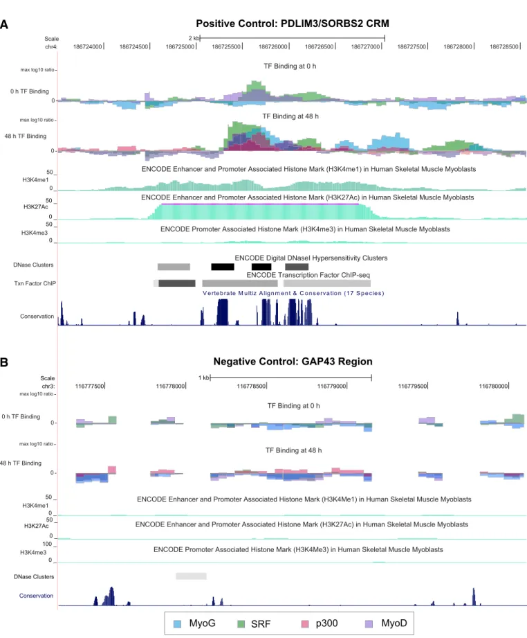
[23]further helped to distinguish the positive and negative controls. We found that the CRMs bound by MyoG at 48 h post-differentiation and that had either H3K4me1 or H3K27Ac modifications in myoblasts (i.e., immediately before differentiation) include all of the positive controls and none of the negative controls. MyoG binding at regions with enhancer-associated chromatin modifications not only identi-fied positive control CRMs located in proximal promoter regions, but also distinguished previously validated distant (greater than 20 kb away from TSS) CRMs from negative control genomic regions (Figs. 1A and B). Since this combination of enhancer marks and MyoG binding best discriminates the positive control CRMs from the negative controls, in subsequent analyses we considered all re-gions with these features as a set of putative muscle differentiation CRMs.
The promoter-associated histone mark H3K4me3[24]also provided a good distinction between the known CRMs and the negative controls, but the levels of this mark were only marginal for validated muscle differen-tiation enhancers more distant from promoters (Fig. 1A), in agreement with previous observations that H3K4me3 is generally not observed at enhancers located more than 2 kb away from TSS[23]. Therefore, we did not include this feature in subsequent analyses of CRMs since our an-alyses broadly encompassed both proximal and distant regulatory regions.
2.2. Comparison to computational prediction of CRMs
MyoG binding during differentiation and enhancer-associated his-tone marks in myoblasts not only distinguished positive control CRMs from negative control genomic regions, but also overlapped significantly with computational predictions of CRMs identified by searching for clus-ters of conserved MRF DNA binding site motif matches with the algo-rithm PhylCRM[12]. This MRF motif (the motif recognized by MyoG and MyoD) was more enriched among a set of genes upregulated during myogenic differentiation than any other combination of MRF, Mef2, SRF, or Tead motifs in a previous study[12]. Of the 1975 regions enriched for MyoG binding and enhancer histone marks, 1128 contain a CRM pre-dicted by PhylCRM (Supplementary Fig. S1). This significant overlap (pb1×10−14; Hypergeometric test) between computationally predicted
CRMs and experimentally identified putative CRMs further demonstrates that PhylCRM can be used successfully to predict likely CRMs and that the observed MyoG binding events are likely mediated by direct interactions with MyoG binding sites within the predicted CRMs. Neither correlation is perfect, however: 43% of regions with experimentally observed MyoG binding and chromatin modifications were not PhylCRM-predicted CRMs, and 67% of PhylCRM-predicted CRMs do not show these in vivo in-dicators of regulatory function. TF binding at regions not predicted by PhylCRM could be indirect association through other TFs, rather than by the myogenic factor binding motif. Likewise, PhylCRM predicted re-gions that are not bound could be CRMs in other conditions. Given the limitations of both experimental TF binding measurements and compu-tational predictions, it is important to combine such different sources of evidence to identify putative CRMs.
As a larger set of putative elements than the few positive and negative controls considered earlier, PhylCRM predictions allow us to further eval-uate the patterns of TF binding at putative regulatory regions. Interesting-ly, the binding of some TFs, such as SRF, that were not as specific as MyoG for positive control CRMs, do show significant overlap with PhylCRM-predicted CRMs. As measured by the sum of sensitivity and specificity, SRF binding at 48 h is nearly as effective as MyoG binding at 48 h in dis-criminating PhylCRM-predicted CRMs from genomic regions that did not contain PhylCRM hits (Supplementary Table S1). This suggests that SRF
binding is an informative feature of many muscle differentiation CRMs, even though it is not highly specific in our small set of validated positive and negative controls. The degree of overlap between computational pre-dictions and different combinations of TF binding and/or chromatin marks across different timepoints is shown in Supplementary Table S1. 2.3. Changes in TF binding during differentiation
Many of the genes whose predicted CRMs were included on our cus-tom-designed ChIP-chip microarray were chosen because they were upregulated over a timecourse of human skeletal muscle differentiation
[12]. To evaluate whether changes in TF binding might be a hallmark of cis-regulatory regions of muscle genes and might effect changes in their gene expression levels, we compared TF binding at 0 and +48 h relative to the induction of myoblast differentiation by serum removal. Changes in TF occupancy occurred at many genomic regions during differentia-tion; for example, 1033 of the 3007 regions bound by MyoG at 48 h were not bound by any of the examined myogenic TFs (MyoG, MyoD, or SRF) before differentiation (0 h). However, in most cases, including information about these changes in TF binding did not help to distin-guish the positive control CRMs from the negative control regions since the patterns of TF binding changes during differentiation vary across the known regulatory regions. For example, the ACTA1 promoter is bound by MyoG and SRF at both 0 and 48 h while the COX6A2 pro-moter is unbound before differentiation and then bound by MyoG, MyoD, and SRF at 48 h. The validated distant enhancer region between the PDLIM3 and SORBS2 genes is bound by MyoD and SRF at 0 h and then by an expanded set of factors (MyoG, MyoD, and SRF) with stron-ger peak intensities at 48 h (Fig. 1A). Only in the case of SRF binding did the ChIP timecourse data help to distinguish the positive control CRMs from negative control regions: of the 7 known CRMs bound by SRF at 48 h, 6 were also bound at 0 h, while in contrast only 1 of the 7 negative control regions bound at 48 h was also bound at 0 h.
In order to determine the value of any of the measured ChIP datasets in predicting the locations of TF binding events at 48 h during differenti-ation, we constructed a dependency network using the WinMine toolkit
[25]. A dependency network, like a Bayesian network,finds predictive
Table 1
‘X’ indicates binding of a TF to a region.
MyoG 0 h X X X H3K27Ac myoblasts X X X X X X X X X X H3K4me1 myoblasts X X X X X X X X X X X X SRF 0 h X X X X X X X MyoD 0 h X X X X X X X X X X X Region name ACTA1 promoter ACTA1 CRM PDLIM3 promoter PDLIM3/ SORBS2 CRM CAV3 TNNT2 COX6A2 HSPB3 CSRP3 GAP43 CPM MGLL BDKRB2 IGFBP4 S1PR2 KRT77 CLC HBZ Region coordinates (Hg18) chr1: 227635616−227637614 chr1: 227612394−227614392 chr4: 186693000−186695000 chr4: 186724927−186726925 chr3: 8749393−8751391 chr1: 199612744−199614742 chr16: 31346483−31348481 chr5: 53787965−53789963 chr11: 19179244−19181242 chr3: 116777470−116779468 chr12: 67659795−67661793 chr19: 44947497−44949495 chr12: 51369942−51371940 chr19: 10199535−10201533 chr14: 95762300−95764298 chr17: 35868970−35870968 chr3: 129031127−129033125 chr16: 148530−150528 MyoG 48 h X X X X X X X X X X X SRF 48 h X X X X X X X X X X X X X X MyoD 48 h X X X X X X X X X X p300 48 h X X X X X X X X X X X X Positive controls Negative controls
relationships between variables using conditional probability distribu-tions[26]. The derived network indicates which variables (in this case, TF binding events at a given genomic region) are well predicted by other variables. The dependency network for our data suggests that
MyoD binding at 0 h is more important for predicting the binding pat-terns of MyoG, SRF, MyoD, and p300 at 48 h after induction of differenti-ation than are either MyoG or SRF binding at 0 h or H3K4me1 marks in myoblasts (Supplementary Fig. S2). Thesefindings are consistent with
A
B
Fig. 1. ChIP-chip TF binding distinguishes positive and negative control regions. All data shown are log10ratios of IP to Input DNA signal and are normalized to the maximum ratio
for each TF dataset. A) Myogenic TFs MyoG, MyoD, and SRF are bound, and enhancer-associated histone marks are observed, at the location of a validated CRM driving expression during muscle differentiation. B) No significant peaks of TF binding are seen for a negative control genomic region (near the GAP43 gene).
prior literature on the pioneering role of MyoD in establishing cis-regula-tory sites in myogenic differentiation[10].
2.4. Locations of TF-bound, putative regulatory regions relative to genes Most regions previously known to have a role in myogenic differenti-ation (including most of the positive controls considered in the previous analyses) are located within proximal promoter regions of protein-coding genes (within 1–2 kb of TSS), but some of the putative regulatory
elements we identified are more distant from any protein-coding genes (N20 kb away from TSS;Fig. 1A). To further investigate the role of distant genomic regions in the regulation of myogenic differentiation, we ex-amined the distribution of distances between genes and putative regulatory elements enriched for myogenic TF binding and enhancer-associated histone modifications. There is an enrichment of these puta-tive regulatory regions close to gene TSS (within 1 kb) as compared with the locations of all possible 1-kb regions on our custom ChIP-chip array (pb1×10−14, Hypergeometric test; Fig. 2). Despite this
skew toward proximal promoter regions, about 80% of regions bound by MyoG at 48 h with H3K4me1 or H3K27Ac modifications fall outside of traditional promoters (1 kb from TSS) and, strikingly, 25% of such pu-tative CRMs are more than 20 kb away from any TSS. Interestingly, ge-nomic regions occupied by different TFs and with different binding dynamics during differentiation differ in their distances from gene TSS. Genomic regions that either lose or gain MyoG binding from 0 h to 48 h tend to be located further from TSSs than those that have con-stant MyoG binding at 0 and 48 h (p= 1.39 × 10− 4for constant binding versus gain of binding at 48 h; Kolmogorov–Smirnov (KS) test;Fig. 2A). Increased distance from gene promoters is also observed for regions that change in both MyoG and SRF binding between 0 and 48 h of differ-entiation as compared with those that are bound by both MyoG and SRF at both timepoints. In addition, regions bound by all 3 of these TFs (MyoG, MyoD, SRF) tend to lie closer to genes than regions with only one or two TFs bound (pb2.2×10−16for MyoG at 48 h vs. MyoG, SRF,
and MyoD at 48 h; KS test;Fig. 2B).
For 69% of the regions with enriched MyoG binding at 48 h and H3K4me1 or H3K27Ac marks in myoblasts, the closest gene is not known to be involved in myogenic differentiation and is not differen-tially expressed during this process. In fact, 90% of such putative CRMs are located outside of 1 kb muscle gene promoter regions and 56% are more than 20 kb away from a muscle gene TSS (Fig. 2C). Fur-thermore, for 30% of these MyoG-bound, H3K4me1 or H3K27Ac mod-ified regions, associating with the closest muscle gene along the linear chromosome requires skipping over intervening non-muscle genes.
We specifically included 100 genomic regions on our custom ChIP-chip array design that contained 287 high-scoring PhylCRM predictions of myogenic CRMs with clusters of evolutionarily conserved myogenic TF binding site matches, but were located far (N50 kb) from any annotat-ed muscle gene TSS. These distant prannotat-edictannotat-ed CRMs exhibitannotat-ed MyoG bind-ing and enhancer-associated histone marks at a somewhat lower frequency (26%) than PhylCRM-predicted CRMsb50 kb away from mus-cle gene TSS (35%). However, permutation testing showed that this level of overlap was greater than expected (p= 0.02) for randomly selected genomic regions represented on the array and much greater than expected (pb1×10−4) for genomic regions with the same median
dis-tance to the nearest TSS of any protein-coding gene (21.8 kb). The signif-icant overlap between MyoG binding events, H3K4me1 or H3K27Ac histone marks, and the PhylCRM computational CRM predictions indi-cates that these putative distant CRMs may indeed be biologically func-tional CRMs.
2.5. Potential regulation of microRNAs by CRMs distant from muscle protein-coding genes
To explore the possible functions of putative myogenic CRMs that are distant from muscle protein-coding genes, we searched for micro-RNA genes annotated in miRBase[27]that are located adjacent to these CRMs. We identified 14 different putative CRMs that are located within 25 kb of 10 different microRNA genes and that are closer to these microRNAs than to any known muscle protein-coding genes (Table 2). In 5 cases, the putative CRMs are closer to one of these microRNAs than to any protein-coding gene (Table 2). Some of these microRNAs that may be regulated by the myogenic TFs bound at these putative CRMs already have an annotated function in muscle differentiation (Table 2). For example, miR-133 and miR-1 (encoded
0 10 20 30 40 50 0.0 0.2 0.4 0.6 0.8 1.0
Distance from Peak to Nearest Gene
Distance to gene TSS (kb)
Cumulative Fraction of Peaks
MyoG 0h and 48h MyoG 48h only MyoG 0h only All Array Bins
0 10 20 30 40 50 0.0 0 .2 0.4 0 .6 0.8 1
.0 Distance from Peak to Nearest Gene
Distance to gene TSS (kb)
Cumulative Fraction of Peaks
MyoG 48h
MyoG 48h and Histone Marks MyoG and MyoD 48 h and Histone Marks MyoG, MyoD, SRF 48h and Histone Marks All Array Bins
0 20 40 60 80 100 0.0 0 .2 0.4 0 .6 0.8 1 .0
Distance from Peak to Nearest Muscle Gene
Distance to Muscle gene TSS (kb)
Cumulative Fraction of Peaks
MyoG 48h and Histone Marks All Array Bins
B
A
C
Fig. 2. Cumulative distributions of distances between putative CRMs and gene tran-scription start sites (TSS). A) Genomic regions bound by MyoG at both 0 and 48 h rel-ative to differentiation are located closer to gene promoters than those whose MyoG binding status changes between 0 h and 48 h. B) Regions bound by multiple myogenic TFs and with enhancer-associated“Histone Marks” (H3K4me1 and H3K27Ac) are lo-cated closer to promoter regions than regions with fewer bound factors C) Many puta-tive CRMs bound by MyoG and with enhancer-associated histone marks are located distant from the promoters of“muscle genes” that are differentially expressed during muscle differentiation or which have an annotated muscle function.“All Array Bins” in each case shows the cumulative distribution of distances to TSS for all 1-kb genomic regions covered on the ChIP-chip array.
by MIR133A1 and MIR1-2, respectively) influence skeletal muscle proliferation and differentiation, with miR-133 specifically downre-gulating the myogenic TF SRF, and miR-1 directly downredownre-gulating HDAC-4 (HDAC4) and thus indirectly modulating Mef2 gene expres-sion[28]. Additionally, miR-499 (encoded by MIR499) is differentially expressed during murine skeletal muscle differentiation[29]and is known to be involved in cardiac myocyte differentiation[30] and other processes in skeletal muscle[31]. The 7 remaining microRNAs (miR-9-2, miR-550a-2, miR-550b-2, miR-675, miR-3925, miR-4298, and miR-4322) we identified based on their proximity to putative myogenic CRMs do not yet have known regulatory functions in mus-cle differentiation and would be interesting to investigate further for their potential myogenic regulatory roles in future studies.
2.6. Differentiation-specific looping interactions between distant CRMs and muscle gene promoters
We used Chromosome Conformation Capture (3C)[32,33] experi-ments to investigate whether the putative myogenic CRMs that we found located distant from muscle genes form physical interactions with their adjacent, putative target genes during myogenic differentia-tion. We selected two CRMs that were bound by MyoG at 48 h after in-duction of differentiation, exhibited H3K4me1 and H3K27Ac
enhancer-associated histone marks, were found to drive expression of a reporter gene during differentiation[12], and were located at least 20 kb away from the TSS of the closest known protein-coding gene. The“ACTA1 CRM” (Fig. 3A) is located 23 kb downstream of the TSS of ACTA1, a gene which encodes the skeletal muscle alpha actin protein, an essential part of the contractile element in muscle. The “PDLIM3/SORBS2 CRM” (Fig. 3B) is located 32 kb upstream of PDLIM3, which encodes a cytoskel-etal organizing protein localized to the Z line of the sarcomere and which may also regulate the myogenic differentiation transcriptional network by affecting the nuclear localization of SRF[34]. This CRM is also located 240 kb downstream of SORBS2, another gene differentially expressed dur-ing myogenic differentiation.
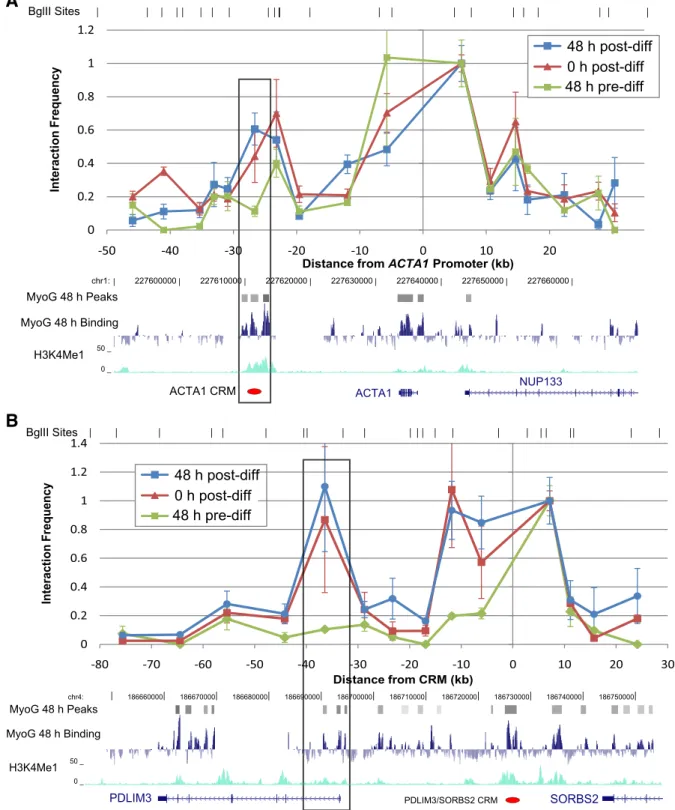
To assay the physical interactions between these two CRMs and their adjacent genes, we performed 3C experiments on human muscle cells 48 h before, immediately (“0 h”) before, and 48 h after induction of differentiation by serum removal. We identified “differentiation-specific interactions” by searching for any peaks of interaction in the 0 h or 48 h post-differentiation cells, outside of the neighboring +/−15 kb that may interact highly by random collisions with the fixed bait, that were higher than the 48 h pre-differentiation interaction level (seeMaterials and methods). The results of these experiments show evidence for a differentiation-specific interaction between the “ACTA1 CRM” and the proximal promoter region spanning from 1.6 kb
Table 2
All potential CRMs (bound by MyoG at 48 h and with enhancer-associated histone marks) that are within 25 kb of a miRNA gene and closer to a miRNA gene than to any muscle gene TSS. Some CRMs are closest to a miRNA gene while others are closer to some other protein-coding gene. Grey highlighting indicates miRNAs with a previously documented role in muscle differentiation.
miRNA closer than any gene
ChIP peak location (MyoG48h filtered by histone marks)
chr19: 10201001−10203000 chr20: 33035001−33036000 chr18: 17664001−17666000 chr20: 33047001−33048000 chr6: 36720001−36722000 chr7: 32733001−32736000 chr11: 1842001−1846000 chr11: 1831001−1832000 chr11: 1847001−1848000 chr11: 1848001−1849000 chr11: 1849001−1851000 chr5: 88014001−88018000 chr11: 1955001−1959000 chr11: 1852001−1862000 miRNA gene symbol MIR1-2 MIR133A1 MIR550A2 MIR550B2 MIR4298 MIR4298 MIR4322 MIR499 MIR499 MIR3925 MIR4298 MIR4298 MIR4298 MIR9-2 MIR675 MIR4298 Position relative to miRNA 5' end 1.95 kb upstream 5.26 kb upstream 4.62 kb upstream 4.71 kb downstream 89 bp upstream Closest gene NM_004230 (S1PR2) NM_020774 (MIB1) Position relative to gene TSS 948 bp downstream 89.5 kb downstream Closest muscle gene NM_015719 (COL5A3) NM_005257 (GATA6) Position rel. to muscle gene TSS 219.9 kb upstream 5.84 kb upstream NM_020884 (MYH7B) 28.6 kb downstream AB040945 (MYH7B) 9.1 kb downstream 338.4 kb upstream 7.66 kb downstream NM_020884 (MYH7B) 42.1 kb downstream AB040945 (MYH7B) 22.6 kb downstream 22.7 kb upstream NM_001220777 (CDKN1A) 31.2 kb upstream U03106 (CDKN1A) 33.5 kb upstream NR_003502 (ZNRF2P1) 414 bp downstream NM_198098 (AQP1) 1.81 Mb downstream 5.84 kb downstream NM_002339 (LSP1) 726 bp downstream NM_003282 (TNNI2) 14.7 kb downstream 6.65 kb upstream NM_001013253 (LSP1) 2.5 kb upstream NM_003282 (TNNI2) 27.2 kb downstream 10.2 kb upstream NM_001013253 (LSP1) 1.02 kb downstream NM_003282 (TNNI2) 30.7 kb downstream 11.2 kb upstream NM_001013254 (LSP1) 174 bp upstream NM_003282 (TNNI2) 31.7 kb downstream 12.7 kb upstream (LSP1) NM_001013255 1.3 kb downstream NM_003282 (TNNI2) 33.2 kb downstream 17.5 kb upstream NR_015436 (LINC00461) 376 bp downstream NM_ 001193349 (MEF2C) 139.5 kb downstream 17.6 kb downstream NR_024471 (MRPL23-AS1) 10.7 kb downstream NM_001042780 (TNNT3) 59.6 kb downstream 19.7 kb upstream NM_001013255 (LSP1) 8.3 kb downstream NM_003282 (TNNI2) 37.8 kb downstream
upstream to 1.3 kb downstream of the ACTA1 TSS (Fig. 3A). We con-firmed this looping interaction in cells at 48 h post-differentiation using two different restriction enzymes, BglII and AflII (Supplementary Fig. S3). We also found a differentiation-specific interaction between the“PDLIM3/SORBS2 CRM” and the region spanning from 3 kb upstream
to 4 kb downstream of the PDLIM3 TSS (Fig. 3B). This interaction was ob-served in primary myoblasts obtained from two different individuals (one male (Fig. 3B), and one female (Supplementary Fig. S4)).
The physical interactions between both of these previously identified CRMs[12]and their corresponding target gene promoters are absent
A
B
Fig. 3. 3C evidence that CRMs physically interact with muscle gene promoters 20–35 kb away during (0 h and 48 h) but not before (−48 h) differentiation. The y-axis shows the normalized ratio between the average quantity of the interaction-specific ligation PCR product vs. the BAC control. To allow relative comparisons between experiments, this ratio is normalized such that the interaction frequency between thefixed primer (located at 0) and its neighboring fragment is 1. Error bars=1 s.e.m. Restriction fragment boundaries are indicated above the graph. Note: not all restriction fragments are represented because some could not be assayed by good primers. A) A 3Cfixed primer at the ACTA1 promoter shows a significant interaction with a downstream CRM after, but not before, differentiation (pb0.05 by Student's t-test). B) A 3C fixed primer at the CRM upstream of PDLIM3 shows a specific interaction with the PDLIM3 promoter after, but not before, differentiation (pb0.05 by Student's t-test). In both cases, the interaction is first established as the cells begin to differentiate (0 h) and then increases during differentiation (48 h).
48 h before differentiation, but then become evident at 0 h, when the cells are confluent and elongating and differentiation is induced by serum removal. These CRM–promoter interaction peaks become more pronounced at 48 h after induction of differentiation (Fig. 3). These re-sults suggest that a differentiation-induced chromosome conformation is established at these gene loci as the cells are approaching confluence and may be initiating the differentiation process even before stimulation of differentiation by serum removal. These physical interaction data par-allel the previously published observation that the expression of the ACTA1 and PDLIM3 genes begins to increase from−48 h to 0 h[12]and our observations described above that predicted CRMs are often already bound by myogenic TFs at 0 h. Interestingly, the PDLIM3/SORBS2 CRM was observed to interact only with the PDLIM3 promoter region 23 kb downstream of the CRM but not with either of the two alternative pro-moters of SORBS2 located 240 kb and 385 kb upstream of the PDLIM3/ SORBS2 CRM (Supplementary Fig. S5). Thus, while the PDLIM3/SORBS2 CRM acts on its target promoter over a distance, the CRM selectively in-teracts with the closer promoter rather than the farther promoter. These observations suggest a model in which distant enhancers partici-pate in activating gene expression by looping to contact the promoters of the genes closest to them. Alternatively, the promoter selectivity could be due to promoter sequence elements that differ between genes to specify the appropriate interaction or cell-type specific insulator archi-tecture[35]. Interestingly, while the PDLIM3/SORBS2 CRM did not inter-act with the SORBS2 promoters, it did show evidence of differentiation-specific interaction with an intronic region of one of the SORBS2 isoforms where a peak of H3K4me1 enrichment is observed (Supplementary Fig. S4). This intronic region may be an additional regulatory region, and the observed contacts between these two noncoding regions raise the in-teresting possibility of combinatorial regulation of genes by multiple CRMs analogous to combinatorial TF input into transcriptional enhancers. 3. Discussion
In this study we found a variety of evidence that CRMs involved in myogenic differentiation can be located distant from the proximal promoter regions of muscle genes. When 50 kb of sequence upstream and downstream of muscle genes are considered, many genomic re-gions with the same TF binding patterns and histone modifications as those observed in known myogenic CRMs are located more than 20 kb away from muscle genes (56% of the putative set), and 90% of them fall outside of 1-kb proximal promoters.
We found that distant CRMs were more likely than proximal CRMs to show changes in MyoG and SRF binding after induction of human muscle differentiation. Thisfinding is in agreement with a prior study that characterized the genome-wide binding of MyoD during murine myogenic differentiation with ChIP-Seq[14]. In that study, Cao et al. noted that 90% of promoter proximal MyoD peaks were bound by MyoD both before and after differentiation and that most of the “differ-entially bound” regions tended to be located further from gene TSS. “Differentially bound” peaks of MyoD binding were found in that study to be more likely to regulate differentiation-specific gene expres-sion changes[14]. This suggests that the distant differentially bound MyoD peaks in that study and the distant putative CRMs we observed here to be differentially bound by MyoG and SRF are likely to be differ-entiation-specific regulatory regions. These results are also in agree-ment with the previous general observation that the chromatin state at promoters is often shared across cell types while distant putative reg-ulatory regions often have highly cell-type specific histone modifica-tions[36]. Beyond trends in binding dynamics with distance from gene TSS, we also observed that regions bound by three myogenic TFs tend to be located closer to promoters than regions bound by one or two TFs. This may relate to the phenomenon of TF binding“hotspots” (genomic regions where nearly every assayed TF is observed to be bound) observed infly and worm[37,38]. In this scenario, the regions bound within proximal promoters may be“hotspots” for TF binding,
while the distant regions are not hotspots but rather are bound selec-tively across timepoints and cell types.
The need to search for CRMs outside of proximal promoter regions increases the difficulty of a computational search by increasing the amount of DNA sequence to be examined. However, we previously found that the PhylCRM algorithm, which searches for clusters of con-served TF binding sites, was able to predict CRMs distant from muscle genes; these predicted CRMs were subsequently validated experi-mentally by ChIP-qPCR and by reporter assays[12]. Here, we found that two of these predicted and validated CRMs, the ACTA1 CRM and the PDLIM3/SORBS2 CRM, both located more than 20 kb from muscle genes, interact with adjacent gene promoters as assayed by 3C. Additionally, PhylCRM predicted a CRM 340 kb away from the nearest muscle gene TSS near a microRNA with a known role in mus-cle differentiation; we found this region to be bound by the myogenic TFs MyoG, MyoD, and SRF 48 h after the initiation of differentiation. Thus, computational predictions combined with experimental mea-surements of cell-type specific histone modifications and TF binding should provide a strong starting point for identifying active transcrip-tional enhancers both within and far from promoters in future studies of other cell types and processes. The DNA binding motif for the myo-genic factor Mef2, in combination with MyoG and SRF motifs, was found previously to help in identifying CRMs using PhylCRM[12]. The Mef2 antibodies available at the time of the ChIP-chip experiments presented here did not produce high-quality DNA binding profiles, but future experiments characterizing the binding of Mef2 during differen-tiation might provide further evidence for CRM locations.
After distant CRMs are identified, a remaining challenge is to de-termine which genes they regulate and the mechanisms by which they affect gene expression. The differentiation-specific interactions observed in this study by 3C for the CRMs located 20–35 kb away from ACTA1 and PDLIM3, combined with the myogenic TF binding ob-served with ChIP-chip and the upregulation of expression of these genes during differentiation, suggest a model in which these distal enhancers assist in activating gene expression by looping to contact the promoters of the closest genes. These are thefirst such distal in-teractions between a CRM and promoter that have been identified in mammalian muscle differentiation. Combined with the mounting evidence of CRM–promoter interactions in other cell types and sys-tems, these data suggest that long-distance physical interaction is a general mechanism of enhancer action rather than a mechanism spe-cific to a particular system. That these enhancer–promoter interac-tions begin to form at 0 h of differentiation suggests that a differentiation-specific set of looping interactions may be established before the resulting changes in gene expression and cell phenotype begin. This is consistent withfindings in other systems: in mouse ery-throid progenitors, theβ-globin LCR region exhibits interactions that are established before changes in gene expression occur[39], and in-teractions between enhancers and promoters implicated in hormone-responsive gene expression in mouse adenocarcinoma cells are al-ready present at lower levels before hormone treatment[40].
When identifying target genes for CRMs outside proximal pro-moter regions, some previous studies have used the simplifying as-sumption that CRMs are likely to interact with gene promoters lying within a domain bounded by CTCF sites, on the basis that CTCF often acts as an insulator across which boundary interactions are less likely to occur[14,41]. This assumption matches experimen-tal results in the case of the PDLIM3/SORBS2 CRM, which interacts with the PDLIM3 promoter in the same CTCF domain (as measured by a previous study in human skeletal muscle myoblasts[23]), but does not interact with the SORBS2 promoters that are separated from the CRM by several CTCF sites. However, the interaction we ob-served between the ACTA1 CRM and the ACTA1 promoter crosses a CTCF site, as does the interaction between the PDLIM3/SORBS2 CRM and the predicted CRM located 27 kb upstream of a SORBS2 pro-moter. Thus, our results suggest that the links between CRMs and
their target genes cannot always be predicted accurately by CTCF sites.
How enhancer–promoter interactions are established and why they are advantageous to myogenic differentiation are yet to be de-termined. It is possible that the myogenic TFs observed to bind to these interacting regions are also responsible for mediating these in-teractions. However, a full understanding of the interaction mechanism will require future work to search for sequence motifs recognized by other factors potentially involved in establishing chromosomal domains (CTCF, for example[39]), to experimentally purify factors associated with the interacting genomic regions[42], and to disrupt implicated DNA binding sites to test whether they are required for the physical interactions.
Although this study primarily focused on the regulatory links be-tween distant CRMs and target genes, we observed a differentiation-specific interaction between one distant CRM (the PDLIM3/SORBS2 CRM) and another region exhibiting the H3K4me1 enhancer-associated histone modification 265 kb away in the intron of SORBS2. This interac-tion raises the interesting possibility of combinatorial regulainterac-tion of genes by multiple CRMs, analogous to combinatorial TF input into tran-scriptional enhancers. Interestingly, while the PDLIM3/SORBS2 CRM in-teraction with the PDLIM3 promoter seems to be already present at the initiation of differentiation (0 h), the interaction with this other puta-tive CRM does not appear until 48 h into differentiation. This may indi-cate a distinct role of a CRM–CRM interaction in the timecourse of myogenic differentiation. Multiple distant (N50 kb away) enhancer re-gions have been previously shown to interact to select the appropriate promoter for expression of the major histocompatibility complex regu-lator CIITA[43]. Future work will be needed to determine the preva-lence of such interactions involving multiple CRMs and what role they might play in a process such as myogenic differentiation.
This study identifies putative CRMs for myogenic differentiation distant from muscle gene promoters and demonstrates that some of these CRMs physically interact with their target genes. These results highlight the importance of looking beyond the proximal promoter to understand the transcriptional regulation of genes involved in dif-ferentiation. These distant CRM–promoter interactions may relate to the global changes in genome organization observed as cells undergo differentiation[44]. In the future, combining computational enhancer predictions, experimental tests of distant CRMs, and global measure-ments of chromosome conformation[45]will help to further eluci-date the mechanisms of gene regulation during this differentiation process.
4. Materials and methods
4.1. Proliferation and differentiation of human myoblasts in cell culture Adult primary human skeletal myoblasts (Lonza) were grown in SkGM-2 medium (Lonza). Myogenic differentiation was stimulated by switching the culture medium to DMEM-F12 with 2% horse serum (Sigma) when the cells reached about 70% confluence. All time points referred to in this study are with respect to the time of switching to dif-ferentiation medium. The majority of the results presented here were obtained using adult male skeletal myoblasts; female skeletal myoblasts were used in one 3C experiment to confirm the reproducibility, and lack of gender-specificity, of the differentiation-specific physical interaction of the PDLIM3/SORBS2 CRM.
4.2. Design of the custom ChIP-chip array
We designed a custom ChIP-chip array using the Agilent 1x244K oligonucleotide array format (Agilent Technologies Inc., AMADID # 015037) to achieve 25-bp resolution tiling of 100 kb surrounding each of 104 selected human genes. The genes were chosen because they were either upregulated over the timecourse of myogenic
differentiation[12], or annotated by Gene Ontology[46]or other litera-ture as being a part of the muscle sarcomere. For each of these genes, the array covers −40 kb to +10 kb relative to transcription start and −10 kb to +40 kb relative to transcription stop. Additionally, the array sequence covers: 5 kb around each of 9 negative control regions previously found not to be bound by myogenic TFs[12]; 5 kb around each of 35 potentially regulatory noncoding SNPs (i.e., polymorphisms predicted to disrupt myogenic TF binding sites located within a compu-tationally predicted CRM); and 5 kb around each of 100 predicted CRMs which are distant (i.e., greater than 50 kb away from the TSS) from an-notated muscle-specific or differentially expressed genes[12,46]. 4.3. Chromatin immunoprecipitation with microarray hybridization (ChIP-chip)
ChIP experiments were performed using a modification of the ChIP protocol described previously[12]. Briefly, cells at various timepoints relative to differentiation were crosslinked with 1% formaldehyde for 20 min and then quenched by glycine addition. Cells were harvested by scraping and then lysed with detergents and mechanical stress. After cell lysis, crosslinked chromatin was sheared by sonication to an average length of ~600 bp, and then immunoprecipitated with relevant TF-specific antibodies (MyoD: sc-760; Myogenin: sc-576; SRF: sc-335; p300: sc-585; all from Santa Cruz Biotechnology, Inc.). A separate, mock‘no antibody’ (i.e., protein A beads without antibody) sample was processed as a negative control. The supernatant from the mock IP was saved as‘Input’ DNA. The purified DNA from each ChIP was am-plified and labeled with Cy5 (IPed DNA) or Cy3 (Input DNA) according to a published protocol[47]. Arrays were hybridized, washed, and scanned at 5-micron resolution according to recommendations from Agilent Technologies, Inc.
4.4. Computational analyses of ChIP-chip data
Agilent Feature Extraction software (version 9.1.3.1; Agilent Tech-nologies Inc., Santa Clara, CA) was used to calculate log10ratios of IP
DNA/Input DNA (Cy5/Cy3) for each microarray spot in each experi-ment and to remove spotsflagged as outliers because of debris on the slide or excessively high background. These log ratios were aver-aged across technical replicate experiments. ChIPOTle software[48]
was then used to identify peaks of statistically significant TF binding enrichment for the averaged technical replicate datasets (while keep-ing each biological replicate separate) uskeep-ing a slidkeep-ing window size (200 bp) that accounts for the density of tiling and the average length of the immunoprecipitated DNA fragments. A threshold on the calcu-lated p values for each peak was set so that the resulting FWER (α) was 0.05 by the Bonferroni correction. Thefinal set of significantly bound regions was then determined for each TF and each timepoint byfinding genomic regions with binding peak overlap between bio-logical replicates (peaks with overlapping genomic coordinates or centers separated by less than 800 bp were counted as overlapping) and without peaks in the negative control‘no antibody’ experiment.
Distances between significant ChIP-chip binding peaks and gene TSSs were calculated using RefSeq gene annotations, excluding micro-RNA genes. For the purpose of the analyses described in this study, we define “muscle genes” as genes that meet at least one of the following criteria: a) previously found to be differentially upregulated (FDR 5%) over the timecourse of human myogenic differentiation (clusters 0–5 in the Warner et al. publication)[12]; b) annotated with one or more of the following Gene Ontology (GO) terms: myofibril, sarcomere, con-tractilefiber, muscle cell differentiation, muscle contraction, or struc-tural constituent of muscle (GO database release 2011-01-06)[46]. The locations of microRNA genes were obtained from miRBase Release 16[27]. All genomic coordinates are from Hg18.
The WinMine toolkit[25]was used to create a dependency network describing the value of ChIP binding data for certain TFs at 0 or 48 h in
predicting other TF binding at 48 h. The model was learned on a 70% training set of the data using a kappa value of 0.01 and resulted in 63% probability of correctly predicting the values in the remaining (30%) test set of data.
4.5. Identifying predicted CRMs within genomic sequence using PhylCRM The PhylCRM algorithm was run with default parameters as de-scribed previously[12], searching for clusters of conserved MRF binding sites using the MYF motif (representing the specificities of MyoG and MyoD) from a prior study[49]. We chose to use MRF alone for the PhylCRM predictions because this motif was more enriched among a set of genes upregulated during myogenic differentiation than any other combination of MRF, Mef2, SRF, or Tead motifs in a previous study[12]. The phylogenetic tree used to evaluate binding site conser-vation in PhylCRM included human, chimp, macaque, mouse, rat, dog, cow, and opossum. From evaluation of the PhylCRM window scores for some previously experimentally tested CRMs, we set the threshold PhylCRM window score for calling a cluster of conserved MRF sites a predicted CRM at 1.5.
4.6. Chromosome Conformation Capture (3C)
3C experiments were performed following previously described pro-tocols[6]. Briefly, approximately 5×10⁷
human muscle cells were cross-linked in afinal concentration of 1% formaldehyde and harvested at each of two timepoints: 48 h before and 48 h after differentiation by serum deprivation. Chromatin extracted from these cells was digested with ei-ther an AflII or BglII restriction enzyme (New England Biolabs) and then incubated with T4 DNA ligase (New England Biolabs), such that regions of chromosome contact were ligated together. After reversing the formal-dehyde crosslinks, physical interactions between genomic regions were detected with PCR primers specific for each ligated interaction product. Primer pairs with melting temperatures of 56–60 °C, within 2 °C of each other, and of 35–50% GC content, were designed to uniquely amplify an approximately 100–300 bp region straddling the location of potential li-gation between two restriction fragments. PCR-amplified products were detected by quantitative real-time PCR (qPCR) on an iCycler (BioRad) with SybrGreen Supermix for iQ (BioRad) and standard protocols. The quantification by qPCR was validated by visualization on an agarose gel and quantification using QuantityOne gel image quantification software (BioRad). The qPCR quantification results were utilized for the final analysis.
To control for differences in primer efficiency, DNA fragments gener-ated from bacterial artificial chromosomes (BACs) spanning the genomic regions of interest were digested and randomly ligated (ACTA1: 1111E20, PDLIM3/SORBS2: CTD-2559A19, CTD-2194A4, and RP11-78H20). Quantified PCR products from each 3C reaction were then normalized by the quantified results from the same PCR amplifications performed on this BAC control library. For those regions for which an ANOVA test of interaction frequencies within a given genomic region (excluding fragments within 15 kb of thefixed primer, which are likely to show high levels of interactions from random collisions[50]) indicated the presence of a significant peak (pb0.05) within the data, we tested the observed peaks for statistically significant differences in interaction level between timepoints using a Student's t-test.
4.7. ChIP-chip data accession number
The raw and processed ChIP-chip microarray data are available in GEO series accession number GSE29865 (http://www.ncbi.nlm.nih. gov/geo/query/acc.cgi?acc=GSE29865).
Supplementary materials related to this article can be found online atdoi:10.1016/j.ygeno.2011.08.003.
Acknowledgments
The authors thank Job Dekker and Ye Zhan for providing training in the 3C technique, Jason Warner for assistance with array design and training in ChIP-chip experiments, Cherelle Walls for assistance with cell culture and ChIP-chip experiments, and Anthony Philippakis for assistance with PhylCRM and array design.
This work was supported by NIH/NHGRI grant # R21 HG005149 (M.L.B.). R.P.M. was supported in part by a National Science Founda-tion Graduate Research Fellowship.
References
[1] C. Zhu, K.J. Byers, R.P. McCord, Z. Shi, M.F. Berger, D.E. Newburger, K. Saulrieta, Z. Smith, M.V. Shah, M. Radhakrishnan, A.A. Philippakis, Y. Hu, F. De Masi, M. Pacek, A. Rolfs, T. Murthy, J. Labaer, M.L. Bulyk, High-resolution DNA-binding specificity analysis of yeast transcription factors, Genome Res. 19 (2009) 556–566. [2] G. Chua, M.D. Robinson, Q. Morris, T.R. Hughes, Transcriptional networks:
reverse-engineering gene regulation on a global scale, Curr. Opin. Microbiol. 7 (2004) 638–646.
[3] E.H. Davidson, Genomic Regulatory Systems: Development and Evolution, Academic Press, San Diego, 2001.
[4] T. Sexton, F. Bantignies, G. Cavalli, Genomic interactions: chromatin loops and gene meeting points in transcriptional regulation, Semin. Cell Dev. Biol. 20 (2009) 849–855.
[5] R.I. Kumaran, R. Thakar, D.L. Spector, Chromatin dynamics and gene positioning, Cell 132 (2008) 929–934.
[6] J. Dostie, T.A. Richmond, R.A. Arnaout, R.R. Selzer, W.L. Lee, T.A. Honan, E.D. Rubio, A. Krumm, J. Lamb, C. Nusbaum, R.D. Green, J. Dekker, Chromosome conformation capture carbon copy (5C): a massively parallel solution for mapping interactions between genomic elements, Genome Res. 16 (2006) 1299–1309.
[7] Q. Lin, L. Lin, J. Zhou, Chromatin insulator and the promoter targeting sequence modulate the timing of long-range enhancer–promoter interactions in the Drosophila embryo, Dev. Biol. 339 (2010) 329–337.
[8] B. Tolhuis, R.J. Palstra, E. Splinter, F. Grosveld, W. de Laat, Looping and interaction between hypersensitive sites in the active beta-globin locus, Mol. Cell 10 (2002) 1453–1465.
[9] C.R. Vakoc, D.L. Letting, N. Gheldof, T. Sawado, M.A. Bender, M. Groudine, M.J. Weiss, J. Dekker, G.A. Blobel, Proximity among distant regulatory elements at the beta-globin locus requires GATA-1 and FOG-1, Mol. Cell 17 (2005) 453–462. [10] Y. Cao, R.M. Kumar, B.H. Penn, C.A. Berkes, C. Kooperberg, L.A. Boyer, R.A. Young, S.J. Tapscott, Global and gene-specific analyses show distinct roles for Myod and Myog at a common set of promoters, EMBO J. 25 (2006) 502–511.
[11] A. Blais, M. Tsikitis, D. Acosta-Alvear, R. Sharan, Y. Kluger, B.D. Dynlacht, An initial blueprint for myogenic differentiation, Genes Dev. 19 (2005) 553–569. [12] J.B. Warner, A.A. Philippakis, S.A. Jaeger, F.S. He, J. Lin, M.L. Bulyk, Systematic
identification of mammalian regulatory motifs' target genes and functions, Nat. Methods 5 (2008) 347–353.
[13] D.A. Bergstrom, B.H. Penn, A. Strand, R.L. Perry, M.A. Rudnicki, S.J. Tapscott, Promoter-specific regulation of MyoD binding and signal transduction cooperate to pattern gene expression, Mol. Cell 9 (2002) 587–600.
[14] Y. Cao, Z. Yao, D. Sarkar, M. Lawrence, G.J. Sanchez, M.H. Parker, K.L. MacQuarrie, J. Davison, M.T. Morgan, W.L. Ruzzo, R.C. Gentleman, S.J. Tapscott, Genome-wide MyoD binding in skeletal muscle cells: a potential for broad cellular reprogramming, Dev. Cell 18 (2010) 662–674.
[15] C.A. Berkes, S.J. Tapscott, MyoD and the transcriptional control of myogenesis, Semin. Cell Dev. Biol. 16 (2005) 585–595.
[16] P.J. Gianakopoulos, V. Mehta, A. Voronova, Y. Cao, Z. Yao, J. Coutu, X. Wang, M.S. Waddington, S.J. Tapscott, I.S. Skerjanc, MyoD directly up-regulates premyogenic mesoderm factors during induction of skeletal myogenesis in stem cells, J. Biol. Chem. 286 (2011) 2517–2525.
[17] W. Thompson, M.J. Palumbo, W.W. Wasserman, J.S. Liu, C.E. Lawrence, Decoding human regulatory circuits, Genome Res. 14 (2004) 1967–1974.
[18] Q. Sun, G. Chen, J.W. Streb, X. Long, Y. Yang, C.J. Stoeckert Jr., J.M. Miano, Defining the mammalian CArGome, Genome Res. 16 (2006) 197–207.
[19] M.C. Frith, J.L. Spouge, U. Hansen, Z. Weng, Statistical significance of clusters of motifs represented by position specific scoring matrices in nucleotide sequences, Nucleic Acids Res. 30 (2002) 3214–3224.
[20] N.D. Heintzman, R.K. Stuart, G. Hon, Y. Fu, C.W. Ching, R.D. Hawkins, L.O. Barrera, S. Van Calcar, C. Qu, K.A. Ching, W. Wang, Z. Weng, R.D. Green, G.E. Crawford, B. Ren, Distinct and predictive chromatin signatures of transcriptional promoters and enhancers in the human genome, Nat. Genet. 39 (2007) 311–318.
[21] B.P. Berman, Y. Nibu, B.D. Pfeiffer, P. Tomancak, S.E. Celniker, M. Levine, G.M. Rubin, M.B. Eisen, Exploiting transcription factor binding site clustering to identify cis-regulatory modules involved in pattern formation in the Drosophila genome, Proc. Natl. Acad. Sci. U.S.A. 99 (2002) 757–762.
[22] A. Visel, M.J. Blow, Z. Li, T. Zhang, J.A. Akiyama, A. Holt, I. Plajzer-Frick, M. Shoukry, C. Wright, F. Chen, V. Afzal, B. Ren, E.M. Rubin, L.A. Pennacchio, ChIP-seq accurately predicts tissue-specific activity of enhancers, Nature 457 (2009) 854–858.
[23] J. Ernst, P. Kheradpour, T.S. Mikkelsen, N. Shoresh, L.D. Ward, C.B. Epstein, X. Zhang, L. Wang, R. Issner, M. Coyne, M. Ku, T. Durham, M. Kellis, B.E. Bernstein, Mapping and analysis of chromatin state dynamics in nine human cell types, Nature 473 (2011) 43–49.
[24] M.G. Guenther, S.S. Levine, L.A. Boyer, R. Jaenisch, R.A. Young, A chromatin landmark and transcription initiation at most promoters in human cells, Cell 130 (2007) 77–88. [25] D.M. Chickering, The WinMine Toolkit, in, Microsoft, , 2002.
[26] D. Heckerman, D.M. Chickering, C. Meek, R. Rounthwaite, C. Kadie, Dependency networks for inference, collaborativefiltering, and data visualization, J. Mach. Learn. Res. 1 (2000) 49–75.
[27] A. Kozomara, S. Griffiths-Jones, miRBase: integrating microRNA annotation and deep-sequencing data, Nucleic Acids Res. 39 (2011) D152–157.
[28] J.F. Chen, E.M. Mandel, J.M. Thomson, Q. Wu, T.E. Callis, S.M. Hammond, F.L. Conlon, D.Z. Wang, The role of microRNA-1 and microRNA-133 in skeletal muscle proliferation and differentiation, Nat. Genet. 38 (2006) 228–233.
[29] Y. Chen, J. Gelfond, L.M. McManus, P.K. Shireman, Temporal microRNA expression during in vitro myogenic progenitor cell proliferation and differentiation: regulation of proliferation by miR-682, Physiol. Genomics 43 (2010) 621–630.
[30] T. Hosoda, H. Zheng, M. Cabral-da-Silva, F. Sanada, N. Ide-Iwata, B. Ogorek, J. Ferreira-Martins, C. Arranto, D. D'Amario, F. Del Monte, K. Urbanek, D.A. D'Alessandro, R.E. Michler, P. Anversa, M. Rota, J. Kajstura, A. Leri, Human cardiac stem cell differentiation is regulated by a mircrine mechanism, Circulation 123 (2011) 1287–1296. [31] E. van Rooij, D. Quiat, B.A. Johnson, L.B. Sutherland, X. Qi, J.A. Richardson, R.J. Kelm
Jr., E.N. Olson, A family of microRNAs encoded by myosin genes governs myosin expression and muscle performance, Dev. Cell 17 (2009) 662–673.
[32] J. Dekker, K. Rippe, M. Dekker, N. Kleckner, Capturing chromosome conformation, Science 295 (2002) 1306–1311.
[33] H. Hagege, P. Klous, C. Braem, E. Splinter, J. Dekker, G. Cathala, W. de Laat, T. Forne, Quantitative analysis of chromosome conformation capture assays (3C-qPCR), Nat. Protoc. 2 (2007) 1722–1733.
[34] P. Pomies, M. Pashmforoush, C. Vegezzi, K.R. Chien, C. Auffray, M.C. Beckerle, The cytoskeleton-associated PDZ-LIM protein, ALP, acts on serum response factor activity to regulate muscle differentiation, Mol. Biol. Cell 18 (2007) 1723–1733. [35] A. Dean, In the loop: long range chromatin interactions and gene regulation, Brief.
Funct. Genomics 10 (2011) 3–10.
[36] N.D. Heintzman, G.C. Hon, R.D. Hawkins, P. Kheradpour, A. Stark, L.F. Harp, Z. Ye, L.K. Lee, R.K. Stuart, C.W. Ching, K.A. Ching, J.E. Antosiewicz-Bourget, H. Liu, X. Zhang, R.D. Green, V.V. Lobanenkov, R. Stewart, J.A. Thomson, G.E. Crawford, M. Kellis, B. Ren, Histone modifications at human enhancers reflect global cell-type-specific gene expression, Nature 459 (2009) 108–112.
[37] M.B. Gerstein, Z.J. Lu, E.L. Van Nostrand, C. Cheng, B.I. Arshinoff, T. Liu, K.Y. Yip, R. Robilotto, A. Rechtsteiner, K. Ikegami, P. Alves, A. Chateigner, M. Perry, M. Morris, R.K. Auerbach, X. Feng, J. Leng, A. Vielle, W. Niu, K. Rhrissorrakrai, A. Agarwal, R.P. Alexander, G. Barber, C.M. Brdlik, J. Brennan, J.J. Brouillet, A. Carr, M.S. Cheung, H. Clawson, S. Contrino, L.O. Dannenberg, A.F. Dernburg, A. Desai, L. Dick, A.C. Dose, J. Du, T. Egelhofer, S. Ercan, G. Euskirchen, B. Ewing, E.A. Feingold, R. Gassmann, P.J. Good, P. Green, F. Gullier, M. Gutwein, M.S. Guyer, L. Habegger, T. Han, J.G. Henik-off, S.R. Henz, A. Hinrichs, H. Holster, T. Hyman, A.L. Iniguez, J. Janette, M. Jensen, M. Kato, W.J. Kent, E. Kephart, V. Khivansara, E. Khurana, J.K. Kim, P. Kolasinska-Zwierz, E.C. Lai, I. Latorre, A. Leahey, S. Lewis, P. Lloyd, L. Lochovsky, R.F. Lowdon, Y. Lubling, R. Lyne, M. MacCoss, S.D. Mackowiak, M. Mangone, S. McKay, D. Mece-nas, G. Merrihew, D.M. Miller III, A. Muroyama, J.I. Murray, S.L. Ooi, H. Pham, T. Phippen, E.A. Preston, N. Rajewsky, G. Ratsch, H. Rosenbaum, J. Rozowsky, K.
Rutherford, P. Ruzanov, M. Sarov, R. Sasidharan, A. Sboner, P. Scheid, E. Segal, H. Shin, C. Shou, F.J. Slack, C. Slightam, R. Smith, W.C. Spencer, E.O. Stinson, S. Taing, T. Takasaki, D. Vafeados, K. Voronina, G. Wang, N.L. Washington, C.M. Whittle, B. Wu, K.K. Yan, G. Zeller, Z. Zha, M. Zhong, X. Zhou, J. Ahringer, S. Strome, K.C. Gunsalus, G. Micklem, X.S. Liu, V. Reinke, S.K. Kim, L.W. Hillier, S. Henikoff, F. Piano, M. Snyder, L. Stein, J.D. Lieb, R.H. Waterston, Integrative analysis of the Cae-norhabditis elegans genome by the modENCODE project, Science 330 (2010) 1775–1787.
[38] C. Moorman, L.V. Sun, J. Wang, E. de Wit, W. Talhout, L.D. Ward, F. Greil, X.J. Lu, K.P. White, H.J. Bussemaker, B. van Steensel, Hotspots of transcription factor colo-calization in the genome of Drosophila melanogaster, Proc. Natl. Acad. Sci. U.S.A. 103 (2006) 12027–12032.
[39] J.E. Phillips, V.G. Corces, CTCF: master weaver of the genome, Cell 137 (2009) 1194–1211.
[40] O. Hakim, M.H. Sung, T.C. Voss, E. Splinter, S. John, P.J. Sabo, R.E. Thurman, J.A. Sta-matoyannopoulos, W. de Laat, G.L. Hager, Diverse gene reprogramming events occur in the same spatial clusters of distal regulatory elements, Genome Res. 21 (2011) 697–706.
[41] N.D. Heintzman, B. Ren, Finding distal regulatory elements in the human genome, Curr. Opin. Genet. Dev. 19 (2009) 541–549.
[42] J. Dejardin, R.E. Kingston, Purification of proteins associated with specific genomic Loci, Cell 136 (2009) 175–186.
[43] Z. Ni, M. Abou El Hassan, Z. Xu, T. Yu, R. Bremner, The chromatin-remodeling zyme BRG1 coordinates CIITA induction through many interdependent distal en-hancers, Nat. Immunol. 9 (2008) 785–793.
[44] I. Rajapakse, M.D. Perlman, D. Scalzo, C. Kooperberg, M. Groudine, S.T. Kosak, The emergence of lineage-specific chromosomal topologies from coordinate gene regulation, Proc. Natl. Acad. Sci. U.S.A. 106 (2009) 6679–6684.
[45] E. Lieberman-Aiden, N.L. van Berkum, L. Williams, M. Imakaev, T. Ragoczy, A. Tell-ing, I. Amit, B.R. Lajoie, P.J. Sabo, M.O. Dorschner, R. Sandstrom, B. Bernstein, M.A. Bender, M. Groudine, A. Gnirke, J. Stamatoyannopoulos, L.A. Mirny, E.S. Lander, J. Dekker, Comprehensive mapping of long-range interactions reveals folding prin-ciples of the human genome, Science 326 (2009) 289–293.
[46] M.A. Harris, J. Clark, A. Ireland, J. Lomax, M. Ashburner, R. Foulger, K. Eilbeck, S. Lewis, B. Marshall, C. Mungall, J. Richter, G.M. Rubin, J.A. Blake, C. Bult, M. Dolan, H. Drabkin, J.T. Eppig, D.P. Hill, L. Ni, M. Ringwald, R. Balakrishnan, J.M. Cherry, K.R. Christie, M.C. Costanzo, S.S. Dwight, S. Engel, D.G. Fisk, J.E. Hirsch-man, E.L. Hong, R.S. Nash, A. SethuraHirsch-man, C.L. Theesfeld, D. Botstein, K. Dolinski, B. Feierbach, T. Berardini, S. Mundodi, S.Y. Rhee, R. Apweiler, D. Barrell, E. Camon, E. Dimmer, V. Lee, R. Chisholm, P. Gaudet, W. Kibbe, R. Kishore, E.M. Schwarz, P. Sternberg, M. Gwinn, L. Hannick, J. Wortman, M. Berriman, V. Wood, N. de la Cruz, P. Tonellato, P. Jaiswal, T. Seigfried, R. White, The Gene On-tology (GO) database and informatics resource, Nucleic Acids Res. 32 (2004) D258–261.
[47] T.I. Lee, S.E. Johnstone, R.A. Young, Chromatin immunoprecipitation and microarray-based analysis of protein location, Nat. Protoc. 1 (2006) 729–748. [48] M.J. Buck, A.B. Nobel, J.D. Lieb, ChIPOTle: a user-friendly tool for the analysis of
ChIP-chip data, Genome Biol. 6 (2005) R97.
[49] W.W. Wasserman, M. Palumbo, W. Thompson, J.W. Fickett, C.E. Lawrence, Human–mouse genome comparisons to locate regulatory sites, Nat. Genet. 26 (2000) 225–228.
[50] J. Dekker, The three‘C’ s of chromosome conformation capture: controls, controls, controls, Nat. Methods 3 (2006) 17–21.