HAL Id: inserm-00420400
https://www.hal.inserm.fr/inserm-00420400
Submitted on 26 Oct 2010
HAL is a multi-disciplinary open access
archive for the deposit and dissemination of
sci-entific research documents, whether they are
pub-lished or not. The documents may come from
teaching and research institutions in France or
abroad, or from public or private research centers.
L’archive ouverte pluridisciplinaire HAL, est
destinée au dépôt et à la diffusion de documents
scientifiques de niveau recherche, publiés ou non,
émanant des établissements d’enseignement et de
recherche français ou étrangers, des laboratoires
publics ou privés.
SWI/SNF deficiency results in aberrant chromatin
organization, mitotic failure, and diminished
proliferative capacity.
Ryan Bourgo, Hasan Siddiqui, Sejal Fox, David Solomon, Courtney Sansam,
Moshe Yaniv, Christian Muchardt, Daniel Metzger, Pierre Chambon, Charles
Roberts, et al.
To cite this version:
Ryan Bourgo, Hasan Siddiqui, Sejal Fox, David Solomon, Courtney Sansam, et al.. SWI/SNF
defi-ciency results in aberrant chromatin organization, mitotic failure, and diminished proliferative
capac-ity.: SWI/SNF Deficiency Causes Failed Mitosis. Molecular Biology of the Cell, American Society for
Cell Biology, 2009, 20 (14), pp.3192-9. �10.1091/mbc.E08-12-1224�. �inserm-00420400�
Vol. 20, 3192–3199, July 15, 2009
SWI/SNF Deficiency Results in Aberrant Chromatin
Organization, Mitotic Failure, and Diminished
Proliferative Capacity
Ryan J. Bourgo,* Hasan Siddiqui,
†Sejal Fox,
‡David Solomon,
§Courtney G. Sansam,
储Moshe Yaniv,
¶Christian Muchardt,
¶Daniel Metzger,
#Pierre Chambon,
#Charles W.M. Roberts,
储and Erik S. Knudsen*
*Department of Cancer Biology, Kimmel Cancer Center, Thomas Jefferson University, Philadelphia, PA 19107;
†
Human Cancer Genetics Program, Department of Molecular Virology, Immunology, and Medical Genetics,
Comprehensive Cancer Center, College of Medicine and Public Health, The Ohio State University, Columbus,
OH 43210;
‡Department of Cell and Cancer Biology, University of Cincinnati College of Medicine, Cincinnati,
OH 45267;
§Lombardi Comprehensive Cancer Center, Georgetown University School of Medicine,
Washington, DC, 20057;
¶Expression Ge´ne´tique et Maladies, Formation de Recherche en Evolution 2850 du
Centre National de la Recherche Scientifique, De´partement de Biologie du De´veloppement, Institut Pasteur,
75724 Paris, France;
#Institut de Ge´ne´tique et Biologie Mole´culaire et Cellulaire, Department of Functional
Genomics, F-67400 Illkirch, France; Institut National de la Sante´ et de la Recherche Me´dicale, U596, F-67400
Illkirch, France; Centre National de la Recherche Scientifique, Unite´ Mixte de Recherche 7104, F-67400 Illkirch,
France; Universite´ Louis Pasteur, F-67000 Strasbourg, France; Colle´ge de France, F-67400 Illkirch, France; and
储
Department of Pediatric Oncology, Dana-Farber Cancer Institute, Boston, MA 02115
Submitted December 19, 2008; Revised April 13, 2009; Accepted May 11, 2009 Monitoring Editor: J. Silvio Gutkind
Switch (SWI)/sucrose nonfermentable (SNF) is an evolutionarily conserved complex with ATPase function, capable of regulating nucleosome position to alter transcriptional programs within the cell. It is known that the SWI/SNF complex is responsible for regulation of many genes involved in cell cycle control and proliferation, and it has recently been implicated in cancer development. The ATPase action of SWI/SNF is conferred through either the brahma-related gene 1 (Brg1) or brahma (Brm) subunit of the complex, and it is of central importance to the modification of nucleosome position. In this study, the role of the Brg1 and Brm subunits were examined as they relate to chromatin structure and organization. Deletion of the Brg1 ATPase results in dissolution of pericentromeric heterochromatin domains and a redistribution of histone modifications associated with these structures. This effect was highly specific to Brg1 and is not reproduced by the loss of Brm or SNF5/BAF47/INI1. Brg1 deficiency is associated with the appearance of micronuclei and aberrant mitoses that are a by-product of dissociated chromatin structure. Thus, Brg1 plays a critical role in maintaining chromatin structural integrity.
INTRODUCTION
The switch/sucrose nonfermentable (SWI/SNF) complex uses the energy of ATP hydrolysis to modulate nucleosome position and density across promoters and thus plays an important role in regulating gene expression. The SWI/SNF ATPase function is necessary for its chromatin remodeling activities (Laurent et al., 1993), and this function is mani-fested through one of two mutually exclusive subunits, brahma-related gene 1 (Brg1) or brahma (Brm). Although
both Brg1 and Brm can function as the central ATPase in the SWI/SNF complex, each defines a discrete complex with unique biochemical activity. In addition, specific accessory factors interact with the ATPase subunits and play critical roles in chromatin remodeling activity. For example, SNF5/ BAF47/INI1 is present in both Brg1- and Brm-associated complexes and is required for maximal chromatin remodel-ing activity, both in vitro and in yeast. Surprisremodel-ingly, al-though Brg1 and Brm have⬎75% sequence homology, the impact of each individual subunit on chromatin biology and underlying cell biology is poorly understood (Phelan et al., 1999).
Due to its central function in chromatin remodeling, SWI/ SNF function is required in many facets of gene regulation. Correspondingly, it is estimated that approximately 5% of the yeast genome is transcriptionally regulated by the SWI/ SNF complex (Holstege et al., 1998; Sudarsanam et al., 2000). In mammalian cells, SWI/SNF plays a critical role in the activation of a diverse set of genes and has been shown to be recruited directly to transcriptional activation domains
(De-This article was published online ahead of print in MBC in Press (http://www.molbiolcell.org/cgi/doi/10.1091/mbc.E08 –12–1224) on May 20, 2009.
Address correspondence to: Ryan J. Bourgo (ryan.bourgo@jefferson. edu).
Abbreviations used: Brg1, brahma-related gene 1; Brm, brahma; RB, retinoblastoma; SNF, sucrose nonfermentable; SNF5, SNF5/BAF47/ INI1; SWI, switch.
roo and Archer, 2001; Hassan et al., 2001). In contrast, SWI/ SNF is also required for transcriptional repression. For ex-ample, transcriptional repression of cell cycle genes mediated by the retinoblastoma tumor suppressor is depen-dent on SWI/SNF, implicating the involvement of SWI/SNF in cellular proliferation (Gunawardena et al., 2004, 2007).
Paradoxically, disruption of SWI/SNF function has alter-natively been associated with tumorigenesis and lack of cellular/organismal viability. Specifically, loss or mutation of the SNF5/INI1 subunit is associated with rhabdoid tu-morigenesis, and it has been convincingly demonstrated that the SNF5/INI1 gene acts as a tumor suppressor (Biegel et al., 1999; Sevenet et al., 1999; Roberts et al., 2002). Correspond-ingly, diminished expression or loss of Brg1, Brm, Baf57, Baf180, and several other chromatin-modifying genes has been observed in cancer cell lines and tumor specimens (Gregory and Shiekhattar, 2004). However, these implica-tions in tumor development are seemingly at odds with important roles for SWI/SNF subunits in maintaining cellu-lar viability and proliferative capacity (Bultman et al., 2000). It is well known that chromosomal instability is a hall-mark of cancer. Interestingly, loss of chromatin regulatory proteins can have deleterious effects on chromatin structure that can compromise genome stability and fuel tumorigen-esis (David et al., 2003, 2006). Here, we used somatic cell culture models to define the impact of the SWI/SNF chro-matin-modifying complex on chromatin biology and cell cycle progression.
MATERIALS AND METHODS
Cell Culture and Adenoviral Transduction
Primary and 3T3 cell lines were cultured in DMEM supplemented with 10% heat-inactivated fetal bovine serum, 100 U/ml penicillin-streptomycin, and 2 mM l-glutamine at 37°C in 5% CO2. All cell lines were transduced with
adenoviruses encoding green fluorescent protein (GFP), GFP-Cre recombi-nase (Cre), or Cre for 120 h.
Immunoblotting
Cell lysates were resolved by SDS-polyacrylamide gel electrophoresis (PAGE) and transferred to Immobilon-P membrane (Millipore, Billerica, MA). Mem-branes were incubated with the following primary antibodies: Brg1 (sc-17796; Santa Cruz Biotechnology, Santa Cruz, CA), Brm (sc-17828; Santa Cruz Biotech-nology), vimentin (gift from Dr. Wallace Ip, University of Cincinnati, Cincinnati, OH), Cre (69050; Novagen, Madison, WI), p53 (CM5p; Novocastra, New Castle, United Kingdom), and lamin B (sc-6217; Santa Cruz Biotechnology).
Immunofluorescence and 5-Bromo-2ⴕ-deoxyuridine (BrdU) Incorporation
Cells were subjected to the indicated treatment, fixed with methanol:acetone (1:1) or 3.7% formaldehyde, and permeabilized with 0.3% Triton X-100. Cells were then stained for the indicated proteins. For BrdU, cells were subjected to the indicated treatment and incubated with BrdU reagent (GE Healthcare, Chalfont St. Giles, Buckinghamshire, United Kingdom) for 1 h before fixation in 3.7% formaldehyde. Cells were then permeabilized with 0.3% Triton X-100 in phosphate-buffered saline, and incubated with rat anti-BrdU. Cells were then stained with rhodamine anti-rat immunoglobulin G, and mounted on slides. In assays wherein abnormal heterochromatin was quantified, “abmal” was determined by disorganization or loss of clearly identifiable, nor-mally discrete and punctate (round) heterochromatin domains.
Flow Cytometry
BrdU reagent was added to cells for 1 h before harvest. Cells were then harvested by trypsinization, fixed with ethanol, and incubated with pro-pidium iodide and fluorescent anti-BrdU secondary antibody. Histograms represent 15,000 gated events. Histograms were analyzed using FlowJo soft-ware, version 8.7 (Tree Star, Ashland, OR).
Polymerase Chain Reaction (PCR)
Total genomic DNA was extracted using DNeasy blood and tissue kit (69504; QIAGEN, Valencia, CA). Indicated portions of DNA were amplified via polymerase chain reaction; amplified DNA was separated using a 1.5% aga-rose gel. For mouse genotyping (Brm), mouse tail DNA was extracted and
analyzed by PCR using a combination of three different primers: sense 5⬘-CCTGAGTCATTTGCTATAGCCTGTG-3⬘, antisense 5⬘-GGACTGCCAGCT-GCA-GAG-3⬘, and 5⬘-CATCGCCTTCTAT-CGCCTTC-3⬘. For mouse genotyp-ing (Brg1), mouse tail DNA was extracted and analyzed by usgenotyp-ing the following primers: sense 5⬘-GATCAGCTCATGCCCTAAGG-3⬘ and antisense 5⬘-CCTACAGTTCCATG-CAGCTGG-3⬘.
Retroviral Transduction, Plasmid Transfection, and Selection
Retroviral Transduction.Cells were transduced for 7 h with a retrovirus containing a p53 dominant-negative allele (p53DD). The transduced cells were selected for 14 d in 0.2 mg/ml G418 sulfate (Calbiochem, San Diego, CA).
Plasmid Transfection.Cells were transfected for 18 h using Lipofectin trans-fection reagent (Invitrogen, Carlsbad, CA) following manufacturer-provided protocol. After 48 h, cells were passaged into 2g/ml puromycin for 7 d. These cells were passaged again into 2g/ml puromycin for another 7 d before beginning experimental procedures.
RESULTS
Targeted Deletion of SWI/SNF ATPases in Cell Culture
The SWI/SNF complex is implicated in a wide range of highly cancer-relevant processes, such as transcriptional reg-ulation, cell cycle control, and DNA repair. Here, models of Brg1 and Brm knockout were used to directly define the impact of deficiency of these subunits on cellular function. To perform these studies, we utilized mouse adult fibro-blasts that were derived from Brg1f/f, Brm⫺/⫺, and
com-pound Brm⫺/⫺Brg1f/fmice. By employing Cre-recombinase
in vitro, any combination of core ATPase deficiency could be created (Supplemental Figure S1A). Cell cultures were trans-duced with adenoviruses encoding either GFP or GFP-Cre, and analyses of GFP-fluorescence revealed ⬎95% of cells were infected. Correspondingly, the Brg1loxallele was
effi-ciently recombined in those cultures subjected to infection with GFP-Cre encoding adenoviruses (Supplemental Figure S1B). Consistent with the genetic recombination, the expres-sion of Brg1 protein was significantly reduced at 120 h after infection (Figure 1A). As expected, the Brm knockout cell cultures exhibited the appropriate genomic organization of the targeted allele and failed to express Brm protein (Figure 1A). Thus, each combination of ATPase deficiency could be modeled in these cell cultures.
Loss of Brg1 Leads to Nuclear Malformations
Because SWI/SNF subunits have been shown to play im-portant roles in chromatin dynamics, nuclear structure and organization were examined in the context of ATPase defi-ciency. At 120 h after infection, cells were stained with Brg1 antibodies to unequivocally define Brg1-deficient cells, and chromatin was labeled with Hoechst 33258. Primary murine fibroblasts harbor highly discrete pericentromeric hetero-chromatin domains, which are readily identified through DNA staining. In cultures with intact ATPase function, these pronounced domains were clearly identifiable in ⬎90% of cells (Figure 1B). The deletion of Brg1 yielded a dramatic disruption of these chromatin domains, as evidenced by lack of defined borders and an overall dispersion of heterochro-matin (Figure 1B). Importantly, this effect occurred in the majority of cells lacking Brg1. In contrast, Brm deficiency lead to only a modest impact on the fidelity of such hetero-chromatin domains. To further explore the effect of Brg1 deletion on chromatin organization, nuclei were visualized by confocal microscopy. Optical sectioning revealed that heterochromatin domains have a discrete three-dimensional structure (Figure 1C). However, the deletion of Brg1 results in the loss of this structure and a corresponding
disorgani-SWI/SNF Deficiency Causes Failed Mitosis
zation of heterochromatin in the nucleoplasm (Figure 1C). Consistent with this assessment, the discrete heterochro-main doheterochro-mains are abnormally dispersed in quantitative monitoring of chromatin intensity (Figure 1D). To illustrate the specificity of this phenomenon to Brg1 loss, the acute deletion of SNF5 was achieved by Cre-recombinase expres-sion in SNF5f/fmouse embryonic fibroblasts. Strikingly,
de-letion of this SWI/SNF core-complex component yields no change in the structure or presence of heterochromatin do-mains (Figure 1E). Furthermore, these observed affects on heterochromatin are not an artifact of Cre-recombinase ex-pression, because intact chromatin domains are observed in other models that have been exposed to Cre-recombinase (Figure 1E). Thus, Brg1 specifically plays a critical role in the maintenance of heterochromatin domains.
Pericentromeric heterochromatin is characterized by spe-cific histone modifications. To delineate how Brg1 deficiency
Figure 1. Loss of core ATPases causes appearance of abnormal hetero-chromatin in primary fibroblasts. (A) Primary MAFs were transduced with adenovirus encoding either GFP or Cre-recombinase. At 120 h after infection, cells were harvested, and protein lysates were resolved by SDS-PAGE. The indicated proteins were detected by immunoblotting. (B) Cells were cultured on glass coverslips and transduced with adenovirus encod-ing either LacZ or Cre. At 120 h after infection, coverslips were fixed and stained with Hoechst dye and visualized by fluorescence microscopy. Percentage of cells with abnormal heterochromatin (HC) was counted. (C) Cells were prepared as described in B, stained with 4,6-diamidino-2-phenylindole (DAPI), and visualized by confocal microscopy. Serial sec-tions were imaged and stacked to create a three-dimensional image of chromatin structure. (D) Cells were visualized by confocal microscopy, and relative chromatin staining intensity was quantitatively graphed. Staining intensity represents chromatin condensation/organization, with red indicating the highest level of organization and blue indicating the least. (E) Cells were stained for SNF5, and with DAPI, they were visual-ized by confocal microscopy. Three-dimensional images of chromatin density were taken. The percentage of cells with abnormal heterochroma-tin was counted.
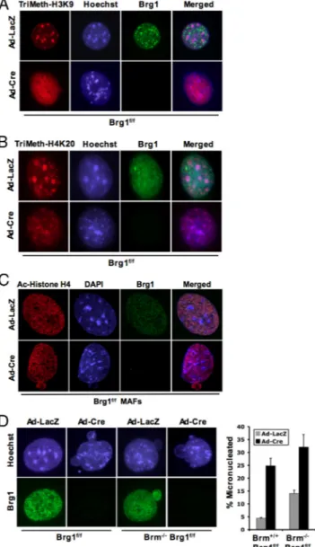
Figure 2. Brg1 is required to maintain specific histone methylation. Cells were cultured on glass coverslips and transduced with ade-novirus encoding either LacZ or Cre. (A–C) At 120 h after infection, coverslips were fixed and stained for the indicated modifications or proteins and visualized via immunofluorescence microscopy. (D) At 120 h after infection, coverslips were fixed and stained as indicated. The percentage of cells containing micronuclei was counted.
Figure 3. Brg1 deficiency causes micro-nuclei formation and reduces cellular pro-liferative capacity in vitro. Brg1f/f 3T3
MAFs were cultured and transduced with adenovirus encoding either LacZ or Cre for 120 h. (A) One hour before harvest, cells were pulse labeled with BrdU. Cov-erslips were fixed and stained for BrdU incorporation, visualized, and quantified. (B) Cells were seeded at equal density 7 d after infection. At indicated the times, plates were fixed and stained with 1% crystal violet. Five random fields per plate were counted, per time point—results are indicative of three separate experiments. (C) Coverslips were harvested at the in-dicated times, fixed, and stained for Brg1 expression. Cells were visualized by im-munofluorescence, and results are quantified at different days postinfection (DPI). (D) Coverslips were harvested 120 h after infection, fixed, SWI/SNF Deficiency Causes Failed Mitosis
influenced the distribution of these histone modifications, cells were costained for Brg1 (to define negative cells) and histone modifications. As shown in Figure 2A, histone H3-trimethyl lysine 9 modification is largely confined to peri-centromeric heterochromatin domains in cells harboring SWI/SNF ATPase activity. In contrast, there was a signifi-cant dispersion and redistribution of histone H3-trimethyl lysine 9 species with the deletion of Brg1. Similar results were observed with the histone H4-trimethyl lysine 20 mod-ification, which was altered in the absence of Brg1 (Figure 2B). Interestingly, this result was specific to Brg1 loss, be-cause no changes in histone modifications were observed in models of Brm and SNF5 loss (Supplemental Figure S2). Furthermore, this phenomenon is relatively specific to mod-ifications associated with heterochromatin, because acetyla-tion of histone H4 and histone H3 were not significantly altered with the deletion of Brg1 (Figure 2C; data not shown). Thus, loss of Brg1 impinges on both the structural fidelity of pericentromeric heterochromatin and modifica-tions that define these distinct chromatin structures.
Role of Brg1/Brm in Maintenance of Genomic Structure
Multiple studies have suggested that aberrations in chroma-tin organization can have significant effects on proliferation and genome stability. In the investigation of chromatin structure, it was apparent that deficiency of SWI/SNF sub-units was associated with a corresponding increase in cells harboring micronuclei (Figure 2D). Cells lacking Brm exhib-ited a modest yet significant increase in micronuclei forma-tion (Figure 2D), which is consistent with a previous report (Coisy-Quivy et al., 2006). However, the acute loss of Brg1 resulted in a striking increase in micronuclei. This level of micronuclei was subtly increased in the context of combined Brg1 and Brm deficiency (Figure 2D). Thus, the percentage of cells harboring micronuclei largely correlated with the aberrations in heterochromatin organization, wherein Brg1 deficiency was responsible for a more significant impact.
Although many attempts were made to establish long-term primary cultures lacking Brg1, these cells were ulti-mately selected against in culture. Because primary cells have a finite proliferative capacity, the Brg1f/f cells were
subjected to spontaneous immortalization by using a 3T3 protocol. Due to the unlimited proliferative capacity of this model, we could readily assess the effect of Brg1 loss on cell proliferation. Initially, BrdU incorporation was analyzed af-ter the deletion of Brg1 (Figure 3A). These analyses showed a significant reduction in BrdU incorporation with Brg1 loss. These effects were further confirmed by analyses of cellular proliferation, wherein the deletion of Brg1 initially sup-pressed proliferation (Figure 3B). However, direct analyses of Brg1 by immunostaining of these cultures showed the
emergence of Brg1-positive cells that, over time, began to represent the majority of the population (Figure 3C). This inhibition of proliferation was also associated with the for-mation of micronuclei in the immortalized compro-mised cultures. Nearly 60% of all 3T3 cells in these Brg1-compromised cultures harbored micronuclei (Figure 3D). These analyses imply that loss of Brg1 has a deleterious impact on proliferation.
Because aberrant nuclear structure could function to in-duce a DNA damage checkpoint, we evaluated the impact of disabling p53 function on proliferation in the absence of Brg1. Primary Brg1f/fmouse adult fibroblasts (MAFs) were
transduced with a retrovirus encoding p53DD. This results in aberrant stabilization of the endogenous p53 (Figure 3E) and immortalization of the cultures (data not shown), con-sistent with a previous report (Bosco et al., 2007). As with the 3T3 cultures, Brg1 deletion in this context resulted in a suppression of proliferation (Figure 3F). Furthermore, single cell analyses demonstrated that Brg1 deletion was selected against over extended culture (Figure 3G). These data sug-gest that the defects in chromatin organization experienced with loss of Brg1 result in the inhibition of proliferation, even with p53 function compromised.
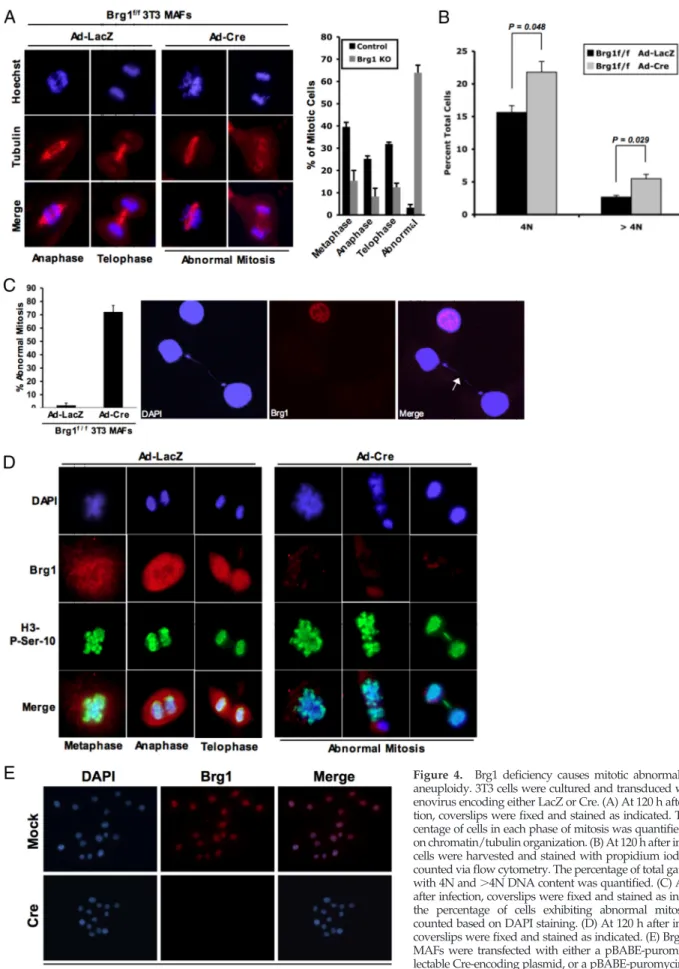
To determine the basis for the inhibition of proliferation, we initially analyzed the cell cycle distribution by flow cytometry. With acute deletion of Brg1 there was an increase in cells harboring 4N and⬎4N DNA content (Figure 4B). To explore whether Brg1 deletion induced alterations in mitotic entry or progression, the presence and stage of mitotic nuclei were evaluated. In immortalized cultures, mitotic cells were readily apparent with the absence of Brg1; however, these cells expressed hallmarks of aberrant mitosis that could be easily identified. This finding was even apparent in mixed cultures in which not all cells had lost Brg1 (Figure 4A). Importantly,⬎70% of cells harboring Brg1 deficiency exhib-ited some form of abnormal mitotic cell division (Figure 4C). Subsequently, we determined whether loss of Brg1 influ-enced the phosphorylation of Ser10 on histone H3, which is associated with mitotic entry. These analyses showed that loss of Brg1 did not preclude this histone modification (Fig-ure 4D). However, there was a significant selection against cells in anaphase or telophase. Those few Brg1-deficient cells in anaphase or telophase (Figure 4A) manifested mitotic bridges, lagging chromosomes, and evidence of mitotic ca-tastrophes (Figure 4D). Together, these studies indicate that although Brg1 is not required for mitotic entry, appropriate chromatin structure is requisite for proper and effective execution of mitosis. To rigorously identify whether cells lacking Brg1 could proliferate, a vector expressing Cre with a selectable marker was used. After selection, the majority of cells still expressed Brg1; however, rare Brg1-deficient cells were detected. Passaging these cells at low density enabled us to specifically interrogate whether single cells could pro-liferate into microcolonies. From ⬎150 colonies analyzed, only two demonstrated expansion in the absence of Brg1. Therefore, although there is a strong selection against Brg1 loss, few cells can ultimately proliferate under this subver-sive condition (Figure 4E and Supplemental Figure S3).
DISCUSSION
It is known that the SWI/SNF complex employs one of two different core ATPases, Brg1 or Brm, to remodel chromatin structure by repressing and facilitating transcription. In this study, the individual function of Brg1 and Brm on chromatin organization, nuclear structure, and mitotic division was determined.
Figure 3 (cont). and costained for DAPI and Brg1. The percentage of Brg1-positve or -negative cells containing micronuclei was quan-tified. (E–G) Brg1f/fprimary MAFs were cultured and transduced
with retrovirus encoding p53DD, resulting in immortalization of the cultures. (E) Cells were transduced with adenovirus encoding either LacZ or Cre. At 120 h after infection, cells were harvested and protein lysates were resolved by SDS-PAGE. Immunoblotting was performed for indicated proteins. (F) Cells were seeded at equal density 7 d after infection. At the indicated times, plates were fixed and stained with 1% crystal violet. Five random fields per plate were counted, per time point—results are indicative of three sepa-rate experiments. (G) Coverslips were harvested at the indicated times, fixed, and stained for Brg1 expression. Cells were visualized by immunofluorescence, and results are quantified at different DPI.
Figure 4. Brg1 deficiency causes mitotic abnormality and aneuploidy. 3T3 cells were cultured and transduced with ad-enovirus encoding either LacZ or Cre. (A) At 120 h after infec-tion, coverslips were fixed and stained as indicated. The per-centage of cells in each phase of mitosis was quantified based on chromatin/tubulin organization. (B) At 120 h after infection, cells were harvested and stained with propidium iodide and counted via flow cytometry. The percentage of total gated cells with 4N and⬎4N DNA content was quantified. (C) At 120 h after infection, coverslips were fixed and stained as indicated; the percentage of cells exhibiting abnormal mitosis was counted based on DAPI staining. (D) At 120 h after infection, coverslips were fixed and stained as indicated. (E) Brg1f/f3T3
MAFs were transfected with either a pBABE-puromycin se-lectable Cre-encoding plasmid, or a pBABE-puromycin select-able control plasmid. Cells were then passaged into media containing puromycin for 14 d. Cells (1⫻ 103) were plated
onto glass coverslips in a 10-cm dish and allowed to grow for 48 h, and then the cells were harvested and stained for Brg1 and DAPI.
SWI/SNF Deficiency Causes Failed Mitosis
The SWI/SNF complex is thought to use the Brg1 and Brm subunits interchangeably to mediate the ATPase function critical for chromatin remodeling (Roberts and Orkin, 2004). In terms of biochemical activity, there is significant func-tional redundancy between these core ATPases (Phelan et
al., 1999). Furthermore, several transcriptional processes can
be mediated via the activity of either ATPase. However, there are clear distinctions between Brg1 and Brm related to tissue-specific dependence and overall organismal surviv-ability (Muchardt and Yaniv, 2001; Kadam and Emerson, 2003). Our data show that deficiency of Brg1, but not Brm, leads to the dissolution of discrete pericentromeric hetero-chromatin domains. This finding suggests that the Brg1 ATPase either represents a larger fraction of the total AT-Pase protein in the cell or that it has distinct functions from Brm. However, this effect on chromatin is readily apparent via multiple approaches and results in the structural disper-sion of heterochromatin domains. These structures are known to be highly enriched in trimethylated histone H3 lysine 9 and histone H4 lysine 20. Correspondingly, Brg1 deletion results in a significant dissemination of these mod-ifications. Interestingly, our data reveal that loss of SNF5, another core subunit of the SWI/SNF complex, does not elicit dissolution of heterochromatin domains, nor does it affect the localization of trimethylation on histone H4 lysine 20 or histone H3 lysine 9 (Supplemental Figure S2). It is well established that these histone markers are not required for the structural maintenance of the heterochromatin domains, because deletion of Suv39H1/H2 and retinoblastoma (RB)-related family members results in the loss of trimethylation of histone H3 lysine 9 and histone H4 lysine 20 at hetero-chromatin domains, respectively, without compromising the integrity of the overall chromatin domain structure (Peters et
al., 2001; Gonzalo et al., 2005). Thus, the maintenance of
pericentromeric heterochromatin domains is hierarchical, with an underlying Brg1-dependent function that is critical for structural integrity.
The regulation of transcription and modulation of chro-matin structure are critically involved in cellular prolifera-tion, and aberrations associated with these processes are implicated in tumorigenesis. The effect of SWI/SNF ATPase deficiency on cellular proliferation remains the subject of controversy, as there are tumor cell lines which harbor dis-crete loss of both ATPases (Strobeck et al., 2002; Reisman et
al., 2003). These cell lines actively proliferate and are, in fact,
compromised for the appropriate response to growth inhib-itory signals as elicited through the RB pathway. Although such tumor cell models are important for interrogating path-ways, it is not possible to determine the cellular requirement for ATPases because other genetic events could obviate their necessity. Thus, the analyses of cultured cells from gene-targeted animals afford an opportunity to define their intrin-sic role in proliferation. Loss of Brm has minimal effect on proliferation, and primary and immortalized lines lacking Brm function can be readily propagated. However, Brg1 deletion resulted in a substantial reduction in cellular pro-liferation and BrdU incorporation and was selected against during culture. Moreover, this effect was observed in both primary cell culture and cultures specifically deficient in canonical p53 function. Thus, loss of Brg1 is not tolerated even in the context of rapidly proliferating immortalized populations. This finding is supportive of previous analyses in embryonal carcinoma cells (Sumi-Ichinose et al., 1997). Cell cycle analyses strongly suggest that the principle neg-ative impact of Brg1 deficiency on proliferation is manifest during mitotic progression. Our data support the notion that Brg1 deficiency can be overcome by virtue of additional
stochastic events; however, this process was highly sporadic even in the context of immortalized 3T3 populations and selection for Brg1 deletion. Thus, tumor cell lines and po-tentially other Brg1-deficient cell types, presumably use compensatory mechanisms to bypass the requirement that we observed.
Findings from multiple laboratories have suggested that deletion/loss of Brg1 may contribute to the genesis of cancer (Murphy et al., 1999; Lee et al., 2002); however, the underly-ing mechanism for this process is unclear. The data pre-sented here indicate that loss of Brg1 results in aberrant mitotic progression and provides evidence of genomic in-stability. These findings are supported by previous studies that show both the localization of SWI/SNF to mitotic chro-mosomes (Xue et al., 2000), and the requirement for related complexes for proper mitosis and chromosome maintenance (Baetz et al., 2004; Campsteijn et al., 2007). This phenomenon is also similar to that observed with the knockout of a critical centromeric protein, inner centromere protein, which dis-rupts chromatin structure and leads to genomic instability (Cutts et al., 1999). Importantly, normal mitotic progression is dependent upon proper centromeric function, and the loss of Brg1 seems to result in disassembly of these pericentro-meric heterochromatin domains. This suggests a mechanism for the observed mitotic abnormalities, because disruption of such domains compromises mitotic fidelity. It has been recently reported that tumors arising in Brg1⫹/⫺mice,
al-though not mimicking specific pathways, are best character-ized by genomic instability (Bultman et al., 2008). Impor-tantly, this study also concludes that tumor formation in Brg1⫹/⫺mice occurs due to haploinsufficiency rather than
loss of heterozygosity (Bultman et al., 2008), suggesting a lack of selection or proliferative advantage with the com-plete ablation of Brg1. Thus, in the context of Brg1 defi-ciency, resultant dispersion of pericentric heterochromatin domains and mitotic dysfunction could potentially repre-sent the underlying key etiological feature relevant to tu-morigenesis.
ACKNOWLEDGMENTS
We thank members of the Erik and Karen Knudsen’s laboratories for critical review of the manuscript and insightful discussion. We also thank Drs. Christian Muchardt and Moshe Yaniv for providing Brm nullizygous mice, Drs. Daniel Metzger and Pierre Chambon for providing Brg1f/fmice, and to
Dr. Charles Roberts for providing SNF5f/fmouse embryonic fibroblasts and
the pBABE-Puro and pBABE-Puro-Cre plasmids. Finally, we thank Dr. A. Kathleen McClendon for providing the p53DD retrovirus. E.S.K. is supported by National Cancer Institute grant CA 104213.
REFERENCES
Baetz, K. K., Krogan, N. J., Emili, A., Greenblatt, J., and Hieter, P. (2004). The ctf13–30/CTF13 genomic haploinsufficiency modifier screen identifies the yeast chromatin remodeling complex RSC, which is required for the estab-lishment of sister chromatid cohesion. Mol. Cell. Biol. 24, 1232–1244. Biegel, J. A., Zhou, J. Y., Rorke, L. B., Stenstrom, C., Wainwright, L. M., and Fogelgren, B. (1999). Germ-line and acquired mutations of INI1 in atypical teratoid and rhabdoid tumors. Cancer Res. 59, 74 –79.
Bosco, E. E., Wang, Y., Xu, H., Zilfou, J. T., Knudsen, K. E., Aronow, B. J., Lowe, S. W., and Knudsen, E. S. (2007). The retinoblastoma tumor suppressor modifies the therapeutic response of breast cancer. J. Clin. Invest. 117, 218 – 228.
Bultman, S., et al. (2000). A Brg1 null mutation in the mouse reveals functional differences among mammalian SWI/SNF complexes. Mol. Cell 6, 1287–1295. Bultman, S. J., Herschkowitz, J. I., Godfrey, V., Gebuhr, T. C., Yaniv, M., Perou, C. M., and Magnuson, T. (2008). Characterization of mammary tumors from Brg1 heterozygous mice. Oncogene 27, 460 – 468.
Campsteijn, C., Wijnands-Collin, A. M., and Logie, C. (2007). Reverse genetic analysis of the yeast RSC chromatin remodeler reveals a role for RSC3 and SNF5 homolog 1 in ploidy maintenance. PLoS Genet. 3, e92.
Coisy-Quivy, M., Disson, O., Roure, V., Muchardt, C., Blanchard, J. M., and Dantonel, J. C. (2006). Role for Brm in cell growth control. Cancer Res. 66, 5069 –5076.
Cutts, S. M., Fowler, K. J., Kile, B. T., Hii, L. L., O’Dowd, R. A., Hudson, D. F., Saffery, R., Kalitsis, P., Earle, E., and Choo, K. H. (1999). Defective chromo-some segregation, microtubule bundling and nuclear bridging in inner cen-tromere protein gene (Incenp)-disrupted mice. Hum. Mol. Genet. 8, 1145– 1155.
David, G., Dannenberg, J. H., Simpson, N., Finnerty, P. M., Miao, L., Turner, G. M., Ding, Z., Carrasco, R., and Depinho, R. A. (2006). Haploinsufficiency of the mSds3 chromatin regulator promotes chromosomal instability and cancer only upon complete neutralization of p53. Oncogene 25, 7354 –7360. David, G., Turner, G. M., Yao, Y., Protopopov, A., and DePinho, R. A. (2003). mSin3-associated protein, mSds3, is essential for pericentric heterochromatin formation and chromosome segregation in mammalian cells. Genes Dev. 17, 2396 –2405.
Deroo, B. J., and Archer, T. K. (2001). Glucocorticoid receptor-mediated chro-matin remodeling in vivo. Oncogene 20, 3039 –3046.
Gonzalo, S., et al. (2005). Role of the RB1 family in stabilizing histone meth-ylation at constitutive heterochromatin. Nat. Cell Biol. 7, 420 – 428. Gregory, R. I., and Shiekhattar, R. (2004). Chromatin modifiers and carcino-genesis. Trends Cell Biol. 14, 695–702.
Gunawardena, R. W., Fox, S. R., Siddiqui, H., and Knudsen, E. S. (2007). SWI/SNF activity is required for the repression of deoxyribonucleotide triphosphate metabolic enzymes via the recruitment of mSin3B. J. Biol. Chem.
282, 20116 –20123.
Gunawardena, R. W., Siddiqui, H., Solomon, D. A., Mayhew, C. N., Held, J., Angus, S. P., and Knudsen, E. S. (2004). Hierarchical requirement of SWI/SNF in retinoblastoma tumor suppressor-mediated repression of Plk1. J. Biol. Chem. 279, 29278 –29285.
Hassan, A. H., Neely, K. E., Vignali, M., Reese, J. C., and Workman, J. L. (2001). Promoter targeting of chromatin-modifying complexes. Front. Biosci.
6, D1054 –D1064.
Holstege, F. C., Jennings, E. G., Wyrick, J. J., Lee, T. I., Hengartner, C. J., Green, M. R., Golub, T. R., Lander, E. S., and Young, R. A. (1998). Dissecting the regulatory circuitry of a eukaryotic genome. Cell 95, 717–728.
Kadam, S., and Emerson, B. M. (2003). Transcriptional specificity of human SWI/SNF BRG1 and BRM chromatin remodeling complexes. Mol. Cell 11, 377–389.
Laurent, B. C., Treich, I., and Carlson, M. (1993). The yeast SNF2/SWI2 protein has DNA-stimulated ATPase activity required for transcriptional activation. Genes Dev. 7, 583–591.
Lee, D., Kim, J. W., Seo, T., Hwang, S. G., Choi, E. J., and Choe, J. (2002). SWI/SNF complex interacts with tumor suppressor p53 and is necessary for the activation of p53-mediated transcription. J. Biol. Chem. 277, 22330 –22337. Muchardt, C., and Yaniv, M. (2001). When the SWI/SNF complex remodels the cell cycle. Oncogene 20, 3067–3075.
Murphy, D. J., Hardy, S., and Engel, D. A. (1999). Human SWI-SNF compo-nent BRG1 represses transcription of the c-fos gene. Mol. Cell. Biol. 19, 2724 –2733.
Peters, A. H., et al. (2001). Loss of the Suv39h histone methyltransferases impairs mammalian heterochromatin and genome stability. Cell 107, 323–337. Phelan, M. L., Sif, S., Narlikar, G. J., and Kingston, R. E. (1999). Reconstitution of a core chromatin remodeling complex from SWI/SNF subunits. Mol. Cell
3, 247–253.
Reisman, D. N., Sciarrotta, J., Wang, W., Funkhouser, W. K., and Weissman, B. E. (2003). Loss of BRG1/BRM in human lung cancer cell lines and primary lung cancers: correlation with poor prognosis. Cancer Res. 63, 560 –566. Roberts, C. W., Leroux, M. M., Fleming, M. D., and Orkin, S. H. (2002). Highly penetrant, rapid tumorigenesis through conditional inversion of the tumor suppressor gene Snf5. Cancer Cell 2, 415– 425.
Roberts, C. W., and Orkin, S. H. (2004). The SWI/SNF complex– chromatin and cancer. Nat. Rev. Cancer 4, 133–142.
Sevenet, N., Sheridan, E., Amram, D., Schneider, P., Handgretinger, R., and Delattre, O. (1999). Constitutional mutations of the hSNF5/INI1 gene predis-pose to a variety of cancers. Am J. Hum. Genet. 65, 1342–1348.
Strobeck, M. W., Reisman, D. N., Gunawardena, R. W., Betz, B. L., Angus, S. P., Knudsen, K. E., Kowalik, T. F., Weissman, B. E., and Knudsen, E. S. (2002). Compensation of BRG-1 function by Brm: insight into the role of the core SWI-SNF subunits in retinoblastoma tumor suppressor signaling. J. Biol. Chem. 277, 4782– 4789.
Sudarsanam, P., Iyer, V. R., Brown, P. O., and Winston, F. (2000). Whole-genome expression analysis of snf/swi mutants of Saccharomyces cerevisiae. Proc. Natl. Acad Sci. USA 97, 3364 –3369.
Sumi-Ichinose, C., Ichinose, H., Metzger, D., and Chambon, P. (1997). SNF2beta-BRG1 is essential for the viability of F9 murine embryonal carci-noma cells. Mol. Cell. Biol. 17, 5976 –5986.
Xue, Y., Canman, J. C., Lee, C. S., Nie, Z., Yang, D., Moreno, G. T., Young, M. K., Salmon, E. D., and Wang, W. (2000). The human SWI/SNF-B chroma-tin-remodeling complex is related to yeast rsc and localizes at kinetochores of mitotic chromosomes. Proc. Natl. Acad. Sci. USA 97, 13015–13020.
SWI/SNF Deficiency Causes Failed Mitosis