HAL Id: hal-01923617
https://hal.archives-ouvertes.fr/hal-01923617
Submitted on 15 Nov 2018
HAL is a multi-disciplinary open access
archive for the deposit and dissemination of
sci-entific research documents, whether they are
pub-lished or not. The documents may come from
teaching and research institutions in France or
abroad, or from public or private research centers.
L’archive ouverte pluridisciplinaire HAL, est
destinée au dépôt et à la diffusion de documents
scientifiques de niveau recherche, publiés ou non,
émanant des établissements d’enseignement et de
recherche français ou étrangers, des laboratoires
publics ou privés.
Production of vineomycin A1 and chaetoglobosin A by
Streptomyces sp. PAL114
Adel Aouiche, Atika Meklat, Christian Bijani, Abdelghani Zitouni, Nasserdine
Sabaou, Florence Mathieu
To cite this version:
Adel Aouiche, Atika Meklat, Christian Bijani, Abdelghani Zitouni, Nasserdine Sabaou, et al..
Produc-tion of vineomycin A1 and chaetoglobosin A by Streptomyces sp. PAL114. Annals of Microbiology,
Springer, 2015, 65 (3), pp.1351-1359. �10.1007/s13213-014-0973-1�. �hal-01923617�
OATAO is an open access repository that collects the work of Toulouse
researchers and makes it freely available over the web where possible
Any correspondence concerning this service should be sent
to the repository administrator:
tech-oatao@listes-diff.inp-toulouse.fr
This is an author’s version published in: http://oatao.univ-toulouse.fr/20338
To cite this version:
Aouiche, Adel and Meklat, Atika and Bijani, Christian
and Zitouni, Abdelghani and
Sabaou, Nasserdine and Mathieu, Florence
Production of vineomycin A1 and
chaetoglobosin A by Streptomyces sp. PAL114. (2015) Annals of Microbiology, 65 (3).
1351-1359. ISSN 1590-4261
Official URL:
https://doi.org/10.1007/s13213-014-0973-1
Production
of vineomycin A1 and chaetoglobosin
A by
Streptomyces sp. PAL114
Adel Aouiche & Atika Meklat & Christian Bijani & Abdelghani Zitouni & Nasserdine Sabaou & Florence Mathieu
Abstract An actinobacteria strain PAL114, isolated from a Saharan soil in Algeria, produces bioactive compounds. Morphological and chemical studies indicated that this strain belongs to the genus Streptomyces. Analysis of the 16S rRNA gene sequence showed a similarity level of 99.8 % with S. griseoflavus LMG 19344T, the most closely related species. Two bioactive compounds, named P44 and P40, were extract-ed by dichloromethane from the cell-free supernatant broth and were purified by HPLC. Minimum inhibitory concentra-tions (MIC) of the compounds were determined against path-ogenic and toxigenic microorganisms, most of which are multiresistant to antibiotics. The P40 fraction showed a strong activity especially against Candida albicans, Bacillus subtilis, and Staphylococcus aureus and has lower MIC values than those of P44 against most microorganisms tested. Chemical structures of compounds were determined based on spectro-scopic and spectrometric analyses (UV-visible, mass,1H, and
13
C NMR spectra). The compounds P44 and P40 were iden-tified as vineomycin A1 and chaetoglobosin A, respectively. Vineomycin A1 is known to be produced by some Streptomyces species. However, chaetoglobosin A is known to be produced only by fungi belonging to the genera
Chaetomium, Penicillium, and Calonectria. This is the first time that chaetoglobosin A, known for its antimicrobial, anti-cancer, and cytotoxic effects, is reported in prokaryotes.
Keywords Streptomyces . Vineomycin A1 . Chaetoglobosin A . Chemical structure . Bioactive compounds
Introduction
Actinobacteria are Gram-positive bacteria with a percentage of guanine-cytosine higher than 55 %, and most of them produce mycelia. They are particularly interesting for their high capacity to produce secondary metabolites with diverse chemical structures (Watve et al.2001). Among these com-pounds, there are, for example, antivirals, antiparasitics, immunostimulants, and immunosuppressants. However, actinobacteria are especially known for the production of antibiotics (Takahashi and Omura2003; Solanki et al.2008). Indeed, over 45 % of bioactive molecules of microbial origin are produced by actinobacteria (Solecka et al.2012). Among these molecules, about 80 % are produced by species of the genus Streptomyces, the most common genus in the environ-ment (Demain2006; Jose et al.2011). The antibiotics secreted by this genus may have antibacterial or antifungal activities, or cytostatic and antitumor properties, such as adriamycin and anthramycin (Butler 2004). Many of these molecules have found an important therapeutic application (Jose and Jebakumar2013).
Our previous works showed the richness of Algeria Saharan soil with actinobacteria producers of bioactive com-pounds (Sabaou et al.1998). Therefore, many strains proved to be new species producing new bioactive molecules (Lamari et al.2002a,b; Zitouni et al.2004a,b,2005; Badji et al.2006,
2007; Merrouche et al.2010; Boubetra et al.2012).
A. Aouiche
:
A. Meklat:
A. Zitouni:
N. Sabaou (*)Laboratoire de Biologie des Systèmes Microbiens (LBSM), Ecole Normale Supérieure de Kouba, Alger, Algeria
e mail: sabaou@yahoo.fr C. Bijani
Laboratoire de Chimie de Coordination (LCC), CNRS, UPS, INPT, LCC, Université de Toulouse, 205, Route de Narbonne,
31077 Toulouse, France F. Mathieu (*)
Université de Toulouse, laboratoire de génie chimique UMR 5503 (CNRS/INPT/UPS), INP de Toulouse/ENSAT, 1 Avenue de l’Agrobiopôle, Castanet Tolosan cedex, France
A previous work performed on a Saharan actinobacteria strain named PAL114 revealed the production of two bioac-tive molecules identified as saquayamycins A and C, known as anticancer agents (Aouiche et al. 2014). In the present article, which is the continuation of a previously published work, the taxonomy of actinomycete strain and the produc-tion, purificaproduc-tion, structure elucidaproduc-tion, and bioactivity of two other bioactive molecules produced by the same strain are described.
Materials and methods
Identification of actinobacteria strain PAL114
The actinobacteria strain PAL114 was isolated from a Saharan soil collected from Ghardaïa province, south Algeria (Aouiche et al.2014).
The morphological and cultural characteristics were ob-served by naked-eye examination of 14 day-old cultures grown on various ISP (International Streptomyces Project) media: yeast extract–malt extract agar (ISP2), oatmeal agar (ISP3), inorganic salts–starch agar (ISP4), and glycerol–as-paragine agar (ISP5) (Shirling and Gottlieb1966), and also on Bennett medium (Waksman 1961). The micromorphology and sporulation were observed by light microscopy.
For chemotaxonomic analyses, biomass was obtained from a culture grown in shaken ISP2 medium (Shirling and Gottlieb
1966) and incubated at 30 °C for 4 days. Analysis of diaminopimelic acid isomers and whole-cell sugar pattern were carried out using the method of Becker et al. (1964) and Lechevalier and Lechevalier (1970). Phospholipids were analysed according to the procedures developed by Minnikin et al. (1977).
For the physiological study, 18 tests were used (Locci
1989), including the production of melanoid pigments on ISP6 and ISP7 media, the assimilation of nine carbohydrates as sole carbon source, the degradation of xanthine, the pro-duction of nitrate reductase, the sensitivities to sodium chlo-ride (7 % w/v), sodium azide (0.01 % w/v), phenol (0.1 % w/v), and penicillin (10 UI), and the growth at 45 °C.
For molecular analysis, DNA was extracted using the pro-cedure recommended by Liu et al. (2000). PCR amplification of the 16S rRNA gene sequence of strain PAL114 was per-formed using two primers: 27f (5′-AGAGTTTGATCCTGGC TCAG-3′) and 1492r (5′- GGTTACCTTGTTACGACTT-3′). The 16S rRNA gene sequence was amplified by PCR using an Invitrogen Kit and sequenced. The sequences obtained were compared with sequences present in the public sequence databases and with the EzTaxon-e server (Kim et al.2012).
Phylogenetic analyses were conducted using MEGA ver-sion 5.0 (Tamura et al.2011). The 16S rRNA gene sequence of strain PAL114 was aligned using the CLUSTAL W
program (Thompson et al. 1994) against corresponding nu-cleotide sequences of representatives of the Streptomyces genus retrieved from GenBank. Evolutionary distance matri-ces were generated as described by Jukes and Cantor (1969) and a phylogenetic tree was established using the neighbor-joining method of Saitou and Nei (1987). The topology of the tree was evaluated by bootstrap analysis (Felsenstein1985) using 1,000 resamplings.
Production and purification of antimicrobial compounds Production of bioactive compounds were conducted in ISP2 broth (malt extract 10 g, yeast extract 4 g, glucose 4 g, distilled water 1,000 mL, pH 7.2). A seed culture was prepared with the same medium and used to inoculate 80 Erlenmeyer flasks of 500 mL, each containing 100 mL of medium. The cultures were incubated on a rotary shaker (250 rpm) at 30 °C. The extraction of active compounds was performed on the fifth day, previously determined as being the day of optimum production. The ISP2 culture broth was centrifuged to elimi-nate cells. The cell-free supernatant was extracted with an equal volume of dichloromethane. The organic extract was concentrated to dryness by rotary evaporator under a vacuum at a temperature lower than 40 °C. The resulting dry extract was recuperated in 10 mL of methanol and bioassayed against Candida albicans M3, Aspergillus carbonarius M333, and Bacillus subtilis ATCC 6633 by paper disc diffusion method. Preparative chromatography with silica gel plates (Merck Art. 5735, Kiesselgel 60HF 254–366; 20×20 cm) was employed for the partial purification of bioactive product. TLC plates were developed in the solvent system ethyl ace-tate–methanol (100:15 v/v). The developed plates were air dried overnight to remove all traces of solvents. The separated compounds were visualized under UVat 254 nm (absorbance) and 365 nm (fluorescence), and the active spot was detected by bioautography (Betina1973). The retention factor (Rf) of the active spot was measured.
The final purification of bioactive compound was carried out by Waters reverse phase HPLC using an XBridge C18 (5 μm) column (200×10 mm, Waters) with a continuous linear gradient solvent system from 20 to 100 % meth-anol in water, a flow rate of 2 mL/min, and UV detec-tion at 220 and 254 nm. All peak fracdetec-tions were col-lected and tested against Candida albicans M3, Aspergillus carbonarius M333, and Bacillus subtilis ATCC 6633. The active fractions were re-injected into the HPLC system under the same conditions as previ-ously, until their final purification.
Identification of bioactive compounds
These analyses were made with the pure bioactive com-pounds. The UV spectra were determined with a
Shimadzu UV1605 spectrophotometer. The mass spectra were recorded on an LCQ ion-trap mass spectrometer (Finnigan MAT, San Jose, CA, USA) with nanospray ion electro-spray ionization (ESI) source (positive and negative ion modes). 1H and 13C NMR spectroscopy
were used for the characterization of two bioactive molecules. NMR sample was prepared by dissolving 3 mg of each compound in 600 μL of CD3OD. All
spectra were recorded on a Bruker Avance 500 spec-trometer equipped with a cryoprobe. All chemical shifts
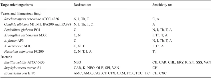
Table 1 Resistance pattern of target fungi and bacteria to antibiotics
Target microorganisms Resistant to: Sensitivity to: Yeasts and filamentous fungi
Saccharomyces cerevisiae ATCC 4226 N, I, Th, T C, A Candida albicans M1, M3, IPA200 and IPA988 N, I, Th, T, C A
Penicillium glabrum PG1 C N, I, Th, T, A Aspergillus carbonarius M333 C, N I, Th, T, A A. flavus AF3 C N, I, Th, T, A A. ochraceus AO1 C, N, T I, Th, A Fusarium culmorum FC200 C, N, T, I, A Th Bacteria
Bacillus subtilis ATCC 6633 NEO CH, CAR, CHL, ERY, K, SPI, SSS, VAN Staphylococcus aureus S1 CAR, K, NEO, OLE, SPI, VAN CH
Escherichia coli E195 AMC, AMX, CAZ, CF, CTX, CXM, FOX, TCC, TIC CH, CXC
A, amphotericin B; C, cycloheximide; I, itraconazole; N, nystatin; T, thioconazole; Th, terbinafine; AMX, amoxicillin; AMC, amoxicillin + clavulanic acid; CH, chloramphenicol; CAR, carbenicillin; CAZ, ceftazidime; CF, cefalotin; CTX, cefotaxime; CXC, cefotaxime + clavulanic acid; CXM, cefuroxime; ERY, erythromycin; FOX, cefoxitin; K, kanamycin; NEO, neomycin; OLE, oleandomycin; SPI, spiramycin; SSS, sulfamide; TCC, ticarcillin + clavulanic acid; TIC, ticarcillin; VAN, vancomycin
0.001
Streptomyces althioticusNRRL B 3981T (AY999791)
Streptomyces labedaeNBRC 15864T (AB184704)
Streptomyces erythrogriseusLMG 19406T(AJ781328)
Streptomyces griseoincarnatusLMG 19316T(AJ781321)
Streptomyces variabilisNBRC 12825T(AB184884)
Streptomyces griseorubensNBRC 12780T(AB184139)
Strain PAL114
Streptomyces griseoflavusLMG 19344T(AJ781322)
Streptomyces malachitofuscusNBRC 13059T(AB184282)
Streptomyces tendaeATCC 19812T(D63873)
Streptomyces flaveolusNBRC 3715T(AB184786)
Streptomyces paradoxusNBRC 14887T(AB184628)
Streptomyces azureusNBRC 12744T(AB184837)
Streptomyces viridochromogenesNBRC 3113T(AB184728)
Streptomyces ambofaciensATCC 23877T(M27245)
70 75 55 99 65 51 74
Fig. 1 Neighbor joining tree based on 16S rRNA gene sequences show ing the relation between strain PAL114 and type species of the genus Streptomyces. The numbers at the nodes indicate the levels of bootstrap
support (≥50 %) based on neighbour joining analyses of 1,000 resampled data sets. Bar, 0.001 nt substitution per nt position
for 1H and 13C are relative to TMS using 1H (residual) or 13C chemical shifts of the solvent as a secondary standard. The temperature was set at 298 K. All the 1H and 13C signals were assigned on the basis of chemical shifts, spin-spin coupling constants, splitting patterns and signal intensities, and by using 1H-1H COSY45,
1
H-1H TOCSY, 1H-13C HSQC, and 1H-13C HMBC ex-periments. Gradient-enhanced 1H COSY45 was realised including 36 scans for per increment. The mixing time for TOCSY was 60 ms and 96 scans per increment were accumulated. 1H-13C correlation spectra using a gradient-enhanced HSQC sequence (delay was optimised for 1JCH of 145 Hz) was obtained with 200 scans per
increment. Gradient-enhanced HMBC experiment was performed allowing 62.5 ms for long-range coupling evolution (340 scans were accumulated). Typically, 2,048 t2 data points were collected for 256 t1 increments.
Determination of minimum inhibitory concentrations Minimum inhibitory concentrations (MIC) of pure bio-active molecules were carried out using conventional agar dilution (Oki et al. 1990). Thirteen target micro-organisms, the majority of which are pathogenic or toxigenic to humans and multiresistant to antibiotics (Table 1), were used. Three bacteria (Bacillus subtilis AT C C 6 6 3 3 , S t a p h y l o c o c c u s a u re u s S 1 , a n d Escherichia coli E195), five filamentous fungi (Aspergillus carbonarius M333, A. flavus AF3, A. ochraceus AO1, Fusarium culmorum FC200, and Penicillium glabrum PG1), and five yeasts (Candida a l b i c a n s M 1 , M 3 , I PA 2 0 0 , a n d I PA 9 8 8 , a n d Saccharomyces cerevisiae ATCC 4226) were inoculated o n t o M ue l l e r H i n t o n m e d i u m f o r ba c t e r i a a n d Sabouraud medium for fungi, containing different con-centrations of active compounds (1, 2, 5, 10, 20, 30, 50, 75, and 100 μg/mL). After a growth period of 24– 48 h at 37 °C for bacteria and 48–72 h at 28 °C for fungi, the plates were examined for growth and the lowest bioactive compound concentration that inhibited the growth of each organism was determined. Mueller Hinton and Sabouraud media, without active compound and inoculated with target organisms, were used as control treatments.
Table 2 1H and 13C NMR data assignments of P44 compound in CD3OD at 298K. See Fig.2bfor numbering of hydrogen and carbon
atoms
1
H and13C number 1H chemical shift, ppm 13C chemical shift, ppm
1 1.33 14.04 2 4.61 70.21 3 197.28 4 7.04 144.19 5 6.08 126.13 6 5.34 95.13 7 1.25 16.10 8 4.24 66.33 9 3.71 76.40 10 1.51 1.69 24.40 11 1.97 23.93 12 5.25 92.10 13 1.39 24.90 14 81.95 15 1.99 2.32 42.61 16 79.80 17 79.50 18 205.28 19 2.73 2.93 50.18 20 6.90 116.65 21 6.41 145.40 22 139.03 23 138.50 24 182.30 25 137.68 26 130.97 27 182.30 28 157.40 29 114.00 30 7.89 132.84 31 7.62 118.63 32 1.38 17.39 33 3.56 75.18 34 3.15 86.45 35 3.82 71.13 36 1.41 2.49 39.28 37 4.92 70.98 38 1.23 15.72 39 4.37 67.34 40 3.76 76.28 41 1.99 2.11 23.93 42 1.51 1.69 24.40 43 5.01 98.92 44 1.33 14.04 45 4.61 70.21 46 197.34 47 7.02 144.09 Table 2 (continued) 1
H and13C number 1H chemical shift, ppm 13C chemical shift, ppm
48 6.09 126.18
Results and discussion Identification of strain PAL114
The strain PAL114 showed good growth on ISP2, ISP3, ISP4, ISP5, and Bennett media. The aerial and substrate mycelia were light to medium grey and light brown, respectively. A diffusible pigment with light brown color was produced on ISP2, ISP3, and Bennett media. The strain PAL114 formed a very well-developed aerial mycelium with long spiraled chains containing between 10 and 50 spores per chain. The spores were carried by sporophores and were oval and 1–1.5× 0 . 6–1 μm in size. The substrate mycelium was nonfragmented.
The chemotaxonomic study of strain PAL114 showed the presence of LL-diaminopimelic acid isomer and glycin in the cell wall. The whole-cell hydrolysates contained non-characteristic sugars (ribose, glucose, and galactose), typical
of cell wall type IC (Lechevalier and Lechevalier1970). The phospholipid profile contained phosphatidylethanolamine, corresponding to phospholipid type PII (Lechevalier et al.
1977). Based on the morphological and chemical characteris-tics, strain PAL114 was identified to the genus Streptomyces (Holt et al.1994).
The strain PAL114 used arabinose, fructose, inositol, man-nitol, melibiose, rhamnose, and xylose as carbon source, but not sucrose and raffinose. Xanthine was degraded and nitrate reduced. The strain grew at 45 °C in the presence of penicillin (10 UI), phenol (0.1 % w/v), sodium azide (0.01 % w/v), and NaCl (7 % w/v). The melanoid pigments were not produced on ISP6 and ISP7 media.
The alignment of the 16S rRNA gene sequence (1471 nucleotides) of strain PAL114 with those of Streptomyces reference species available in the GenBank database, can be seen in the neighbor-joining dendrogram (Fig.1). The simi-larity level was 99.8 % with S. griseoflavus LMG 19344T
Fig. 2 Structure of bioactive compound P44 (a) and HMBC and COSY correlations (b) Ann Microbiol
(Holt et al.1994), the most closely related species. The mor-phological and physiological properties of strain PAL114 are similar to those of the type strain of S. griseoflavus except for the tests of nitrate reduction and growth at 45 °C.
Purification of bioactive compounds
The dichloromethane extract was chromatographed by TLC and developed in an ethyl acetate–methanol system (100–15 v/v). After migration, we observed one active bioautographic compound, which showed a strong activity against Candida albicans M3 and Bacillus subtilis ATCC 6633. This com-pound (Rf=0.9), was selected and analyzed by HPLC. Two active fractions (P44 and P40) were purified with 80 % meth-anol in water. The fraction P40 was eluted at a retention time of 39.68 min and P44 at 44.08 min. A quantity of 5 mg was obtained for each molecule from 8 L of culture filtrate. Identification of bioactive compounds
The UV-VIS spectrum of the bioactive compound P44 showed maxima at 218, 318, and 438 nm.
The mass spectrum of the compound was obtained in negative mode. It yielded a pseudo-molecular ion [M - H] = 933. Thus, the molecular weight of antimicrobial compound is M=934.
The1H and13C chemical shifts of P44 compound are given in Table2 and structure in Fig. 2. The HSQC and HMBC spectra show 49 carbon signals for P44 molecule. It was possible to discern five ketone groups (δc182.30 to 205.28),
four hydroxyl groups (δc71.13 to 157.40), nine ether
func-tions (δc67.34 to 95.13), 13 sp2–hybridized carbons (δcfrom
114.00 to 144.09), and 13 sp3-hybridized carbons (δc14.04 to
42.61) for the P44 molecule. The hydrogens of the hydroxyl group are not observed due to rapid exchange with MeOD. The 2D1H-1H and1H-13C experiments, and especially the long range1H-13C couplings observed in the HMBC spectrum (see Fig. 2b), permitted us to established the connectivity between all the groups of the molecule.
The structure of the P44 molecule was determined by NMR and mass spectrometry to be vineomycin A1. It is an antibiotic belonging to the aquayamycin group, class of angucycline, family of anthracycline (Maskey et al.2003). It is known to be active against Gram-positive bacteria and sarcoma-180 solid tumors in mice (Imamura et al. 1982; Omura2011). Also, vineomycin A1 is a suitable treatment for hypertrophic scar tissue and keloid disease. It is known to be a potent inhibitor of prolyl 4-hydroxylase, an essential enzyme involved in the post translational modification of collagen causing several diseases such as fibrosis, arterio-sclerosis, and scleroderma (Cunliffe and Franklin 1986; Chen2007). Furthermore, cytostatic activity in vivo has been reported for vineomycins (Rohr and Thiericke 1992).
Vineomycins are known to be produced by actinobacteria, especially Streptomyces species such as S. matensis and S. albogriseolus (Ono et al.1974; Chen2007; Omura2011), but have never been reported in S. griseoflavus, to which our strain was closely related.
For the P40 compound, the UV-VIS spectrum showed maxima at 220 and 276 nm.
The mass spectrum of the compound was obtained in negative mode. It yielded a pseudo-molecular ion [M - H] = 527. Thus, the molecular weight of antimicrobial compound is M=528. The1H and13C chemical shifts of the P40 compound are given in Table3and structure in Fig.3. The HSQC and HMBC spectra show 32 carbon signals for P40. From these data, it was possible to discern two ketone groups
Table 3 1H and13C NMR data assignments of the P40 compound in CD3OD at 298 K. See Fig.3bfor numbering of hydrogen and carbon
atoms
1
H and13C number 1H chemical shift, ppm 13C chemical shift, ppm
1 6.95 124.2 2 108.0 3 127.5 4 136.6 5 7.26 111.3 6 7.49 118.1 7 7.00 118.4 8 7.03 120.7 9 2.84 32.1 10 3.91 57.7 11 2.89 46.4 12 63.1 13 174.3 14 1.73 36.3 15 58.0 16 2.78 62.1 17 2.05 48.9 18 1.08 12.1 19 1.30 18.4 20 6.04 128.3 21 5.13 132.7 22 2.00 2.29 41.3 23 2.47 31.8 24 5.53 139.2 25 132.3 26 5.00 81.5 27 200.5 28 6.13 131.7 29 7.27 134.4 30 197.3 31 1.00 19.8 32 1 33 9.5
(δc 197.3 and 200.5), one amide group (δc 174.3), one
hydroxyl group (δc 81.5), one epoxy group (δc 58.0 and
62.1), 14 sp2–hybridized carbons (δc from 108.0 to
136.6), and 14 sp3-hybridized carbons (δc 12.1 to
81.5). The hydrogens of the hydroxyl group are not observed due to rapid exchange with MeOD. The 2D
1
H-1H and 1H-13C experiments, and especially the long range 1H-13C couplings observed in the HMBC spec-trum (see Fig. 3b), permitted us to establish the con-nectivity between all the groups of the P40 molecule.
The structure of the P40 compound was determined by NMR and mass spectrometry to be chaetoglobosin A. This compound is known for its antibacterial, antifungal, phytotox-ic (June et al.1998; Larsen et al. 2005; Zhang et al.2013), anticancer, and cytotoxic effects towards mammal cells (Fisvad et al.2004; Fogle et al.2008), and nematicidal effects (Hu et al.2012). It has also been reported that this compound increases fibrinolytic activity in bovine animals (Shinohara et al. 2000). This compound is a cytochalasin derivative (Ohtsubo et al. 1978; June et al. 1998). Furthermore, it is considered a mycotoxin, and it is known to be produced only by fungi such as Chaetomium globosum (Hu et al. 2012), Penicillium discolor, P. expansum, P. marinum (Fisvad et al.
A
B Fig. 3 Structure of bioactive
compound P40 (a) and HMBC and COSY correlations (b)
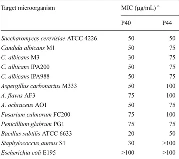
Table 4 Minimum inhibitory concentrations (MIC) of the bioactive compounds P44 and P40 secreted by the strain PAL114 against several fungi and bacteria
Target microorganism MIC (μg/mL)a
P40 P44
Saccharomyces cerevisiae ATCC 4226 50 50
Candida albicans M1 50 75 C. albicans M3 30 75 C. albicans IPA200 50 75 C. albicans IPA988 50 75 Aspergillus carbonarius M333 50 100 A. flavus AF3 75 100 A. ochraceus AO1 50 75 Fusarium culmorum FC200 75 100 Penicillium glabrum PG1 75 75 Bacillus subtilis ATCC 6633 20 50 Staphylococcus aureus S1 30 >100 Escherichia coli E195 >100 >100
a
2004), and Calonectria morganii (Von Wallbrunn et al.2001). It has never been described in prokaryotes. This is the first time that this compound is reported in the genus Streptomyces, belonging to the class of Actinobacteria. All experiments were made three times at different cultivation times to confirm fully the production of chaetoglobosin A by strain PAL114. Minimum inhibitory concentrations
Minimum inhibitory concentrations (MIC) of bioactive mol-ecules purified by HPLC are summarized in Table4.
For vineomycin A1 (P44), MIC values were between 50 and 75μg/mL for yeasts, and 75 and 100 μg/mL for filamen-tous fungi. For bacteria, Bacillus subtilis ATCC 6633 was the most sensitive (50 μg/mL). All other bacteria tested were resistant.
For chaetoglobosin A (P40), MIC values were between 30 and 75μg/mL for yeasts, 50 and 75 μg/mL for filamentous fungi, and 20 and 30μg/mL for Gram-positive bacteria. All Gram-negative bacteria tested were resistant. The most sensi-tive microorganisms were Bacillus subtilis ATCC 6633 (20 μg/mL), Staphylococcus aureus S1 (30 μg/mL), and Candida albicans M3 (30μg/mL).
In conclusion, the strain PAL114, closely related to S. griseoflavus, showed an antibacterial and antifungal activity against pathogenic and toxigenic microorganisms, most of which are resistant to several antibiotics. The bioactive com-pounds produced by the strain proved to be the vineomycin A1 and chaetoglobosin A.
Vineomycin A1 belongs to the same group, class, and family as saquayamycins (Aouiche et al.2014); it differs from these compounds only by the presence of a saccharide deriv-ative bonded to the carbon 40. Chaetoglobosin A is known to be produced only by a small number of fungal species. This is the first time that chaetoglobosin A, which, in addition to its antimicrobial activity, is also considered a mycotoxin in the scientific literature, is produced by prokaryotes such as Actinobacteria.
Moreover, strain PAL114 represents a sample which con-firms the potential of actinobacteria to produce a large variety of bioactive molecules.
References
Aouiche A, Bijani C, Zitouni A, Mathieu F, Sabaou N (2014) Antimicrobial activity of saquayamycins produced by Streptomyces sp. PAL114 isolated from a Saharan soil. J Mycol Med 24:17 23
Badji B, Zitouni A, Mathieu F, Lebrihi A, Sabaou N (2006) Antimicrobial compounds produced by Actinomadura sp. AC104 isolated from an Algerian Saharan soil. Can J Microbiol 52:373 382
Badji B, Mostefaoui A, Sabaou N, Lebrihi A, Mathieu F, Seguin E, Tillequin F (2007) Isolation and partial characterization of antimi crobial compounds from a new strain Nonomuraea sp. NM94. J Ind Microbiol 34:403 412
Becker B, Lechevalier MP, Gordon RE, Lechevalier HA (1964) Rapid differentiation between Nocardia and Streptomyces by paper chromatography of whole cell hydrolysates. Appl Microbiol 12:421 423
Betina V (1973) Bioautography in paper and thin layer chromatography and its scope in the antibiotic field. J Chromatogr 78:41 51 Boubetra D, Sabaou N, Zitouni A, Bijani C, Lebrihi A, Mathieu F (2012)
Taxonomy and chemical characterization of new antibiotics pro duced by Saccharothrix SA198 isolated from a Saharan soil. Microbiol Res 168:223 230
Butler MS (2004) The role of natural product chemistry in drug discov ery. J Nat Prod 67:2141 2215
Chen CL (2007) Methodologies for the synthesis of functionalized naph thols and progress toward the total synthesis of vineomycinone B2 methyl ester and actinophyllic acid. Ph.D. dissertation, University of Texas, Austin
Cunliffe CJ, Franklin TJ (1986) Inhibition of prolyl 4 hydroxylase by hydroxyanthraquinones. Biochem J 239:311 315
Demain AL (2006) From natural products discovery to commercializa tion: a success story. J Ind Microbiol Biotechnol 33:486 495 Felsenstein J (1985) Confidence limits on phylogenies: an approach using
the bootstrap. Evolution 39:783 791
Fisvad JC, Smedsgaard J, Larsen TO, Samson RA (2004) Mycotoxins, drugs and other extrolites produced by species in Penicillium sub genus Penicillium. Stud Mycol 49:201 241
Fogle MR, Douglas DR, Jumper CA, Straus DC (2008) Growth and mycotoxin production by Chaetomium globosum is favored in a neutral pH. Int J Mol Sci 9:2357 2365
Holt JG, Kreig NR, Sneath PHA, Staley JT, Williams ST (1994) Bergey’s manual of determinative bacteriology, 9th edn. Williams and Wilkins Co., Baltimore
Hu Y, Zhang W, Zhang P, Ruan W, Zhu X (2012) Nematicidal activity of chaetoglobosin A poduced by Chaetomium globosum NK102 against Meloidogyne incognita. J Agric Food Chem 61:41 46 Imamura N, Kakinuma K, Ikekawa N, Tanaka H, Omura S (1982)
Biosynthesis of vineomycins A1 and B2. J Antibiot 35:602 608 Jose PA, Jebakumar SRD (2013) Non streptomycete actinomycetes nour
ish the current microbial antibiotic drug discovery. Front Microbiol. doi:10.3389/fmicb.2013.00240
Jose PA, Santhi VS, Jebakumar SRD (2011) Phylogenetic affiliation, antimicrobial potential and PKS gene sequence analysis of moder ately halophilic Streptomyces sp. inhabiting an Indian saltpan. J Basic Microbiol 51:348 356
Jukes TH, Cantor CR (1969) Evolution of protein molecules. In: Munro HN (ed) Mammalian protein metabolism, vol 3. Academic, New York, pp 21 132
June N, Yeon SW, Paek NS, Kim TH, Kim YH, Kim CJ, Kim KW (1998) Isolation and structural determination of anti Helicobacter pylori compound from fungus 60686. San’oeb misaengmul haghoeji 26: 137 142
Kim OS, Cho YJ, Lee K, Yoon SH, Kim M, Na H, Park SC, Jeon YS, Lee JH et al (2012) Introducing EzTaxon e: a prokaryotic 16S rRNA Gene sequence database with phylotypes that represent uncultured species. Int J Syst Evol Microbiol 62:716 721
Lamari L, Zitouni A, Boudjella H, Badji B, Sabaou N, Lebrihi A, Lefebvre G, Seguin E, Tillequin F (2002a) New dithiolopyrrolone antibiotics from Saccharothrix sp. SA 233. I. Taxonomy, fermenta tion, isolation and biological activities. J Antibiot 55:696 701 Lamari L, Zitouni A, Dob T, Sabaou N, Lebrihi A, Germain P, Seguin E,
Tillequin F (2002b) New dithiolopyrrolone antibiotics from Saccharothrix sp. SA 233. II. Physicochemical properties and struc ture elucidation. J Antibiot 55:702 707
Larsen TO, Smedsgaard J, Nielsen KF, Hansen ME, Frisvad JC (2005) Phenotypic taxonomy and metabolite profiling in microbial drug discovery. Nat Prod Rep 22:672 693
Lechevalier MP, Lechevalier HA (1970) Chemical composition as a criterion in the classification of aerobic actinomycetes. Int J Syst Bacteriol 20:435 443
Lechevalier MP, De Bievre C, Lechevalier HA (1977) Chemotaxonomy of aerobic actinomycetes: phospholipid composition. Biochem Syst Ecol 5:249 260
Liu D, Coloe S, Baird R, Pedersen J (2000) Rapid mini preparation of fungal DNA for PCR. J Clin Microbiol 38:471
Locci R (1989) Streptomyces and related genera. In: Williams ST, Sharpe ME, Holt JG (eds) Bergey’s manual of systematic bacteriology, vol 4. Williams and Wilkins Co., Baltimore, pp 2451 2492
Maskey RP, Helmke E, Laatsch H (2003) Himalomycin A and B: isolation and structure elucidation of new fridamycin type antibiotics from a marine Streptomyces isolate. J Antibiot 56:942 949
Merrouche R, Bouras N, Coppel Y, Mathieu F, Monje MC, Sabaou N, Lebrihi A (2010) Dithiolopyrrolone antibiotic formation induced by adding valeric acid to the culture broth of Saccharothrix algeriensis. J Nat Prod 73:1164 1166
Minnikin DE, Patel PV, Alshamaony L, Goodfellow M (1977) Polar lipid composition in the classification of Nocardia and related bacteria. Int J Syst Bacteriol 27:104 117
Ohtsubo K, Saito M, Sekita S, Yoshihira K, Natori S (1978) Acute toxic effects of chaetoglobosin A, a new cytochalasan compound pro duced by Chaetomium globosum, on mice and rats. Jpn J Exp Med 48:105 110
Oki T, Tenmyo O, Tomatsu K, Kamei H (1990) Pradimicins A, B and C: new antifungal antibiotics. II. In vitro and in vivo biological activ ities. J Antibiot 43:763 770
Omura S (2011) Microbial metabolites: 45 years of wandering, wonder ing and discovering. Tetrahedron 67:6420 6459
Ono H, Harada S, Kishi T (1974) Maridomycin, a new macrolide antibi otic. VII. J Antibiot 27:442 448
Rohr J, Thiericke R (1992) Angucycline group antibiotics. Nat Prod Rep 9:103 137
Sabaou N, Boudjella H, Bennadji A, Mostefaoui A, Zitouni A, Lamari L, Bennadji H, Lefebvre G, Germain P (1998) Les sols des oasis du Sahara algérien, source d’actinomycètes rares producteurs d’antibiotiques. Sécheresse 9:147 153
Saitou N, Nei M (1987) The neighbor joining method: a new method for reconstructing phylogenetic trees. Mol Biol Evol 4:406 425 Shinohara C, Chikanishi T, Nakashima S, Hashimoto A, Hamanaka A,
Endo A, Hasumi K (2000) Enhancement of fibrinolytic activity of
vascular endothelial cells by chaetoglobosin A, crinipellin B, geodin and triticone B. J Antibiot 53:262 268
Shirling EB, Gottlieb D (1966) Methods for characterization of Streptomyces species. Int J Syst Bacteriol 13:313 340
Solanki R, Khanna M, Lal R (2008) Bioactive compounds from marine actinomycetes. Indian J Microbiol 48:410 431
Solecka J, Zajko J, Postek M, Rajnisz A (2012) Biologically active secondary metabolites from actinomycetes. Cent Eur J Biol 7:373 390
Takahashi Y, Omura S (2003) Isolation of new actinomycete strains for the screening of new bioactive compounds. J Gen Appl Microbiol 49:141 154
Tamura K, Peterson D, Peterson N, Stecher G, Nei M, Kumar S (2011) MEGA5: molecular evolutionary genetics analysis using maximum likelihood, evolutionary distance, and maximum parsimony methods. Mol Biol Evol 28:2731 2739
Thompson JD, Higgins DG, Gibson TJ (1994) CLUSTAL W: improving the sensitivity of progressive multiple sequence alignment through sequence weighing, position specific gap penalties and weight ma trix choice. Nucleic Acids Res 22:4673 4680
Von Wallbrunn C, Luftmann H, Bergander K, Meinhardt F (2001) Phytotoxic chaetoglobosins are produced by the plant pathogen Calonectria morganii (anamorph Cylindrocladium scoparium). J Gen Appl Microbiol 47:33 38
Waksman SA (1961) The Actinomycetes. Classification, identification and descriptions of genera and species, vol 2. Williams and Wilkins Co, Baltimore
Watve MG, Tickoo R, Jog MM, Bhole BD (2001) How many antibiotics are produced by the genus Streptomyces? Arch Microbiol 176:386 390
Zhang G, Zhang Y, Qin J, Qu X, Liu J, Li X, Pan H (2013) Antifungal metabolites produced by chaetomium globosum No.04, an endo phytic fungus isolated from Ginkgo biloba. Indian J Microbiol 53: 175 180
Zitouni A, Lamari L, Boudjella H, Badji B, Sabaou N, Gaouar A, Mathieu F, Lebrihi A, Labeda DP (2004a) Saccharothrix algeriensis sp. nov., isolated from Saharan soil. Int J Syst Evol Microbiol 54: 1377 1381
Zitouni A, Boudjella H, Mathieu F, Sabaou N, Lebrihi A (2004b) Mutactimycin PR, a new anthracycline antibiotic from Saccharothrix sp. SA 103. I. Taxonomy, fermentation, isolation and biological activities. J Antibiot 57:367 372
Zitouni A, Boudjella H, Lamari L, Badji B, Mathieu F, Lebrihi A, Sabaou N (2005) Nocardiopsis and Saccharothrix genera in Saharan soils in Algeria: isolation, biological activities and partial characterization of antibiotics. Res Microbiol 156:984 993