HAL Id: tel-00801614
https://tel.archives-ouvertes.fr/tel-00801614
Submitted on 17 Mar 2013
HAL is a multi-disciplinary open access archive for the deposit and dissemination of sci-entific research documents, whether they are pub-lished or not. The documents may come from teaching and research institutions in France or abroad, or from public or private research centers.
L’archive ouverte pluridisciplinaire HAL, est destinée au dépôt et à la diffusion de documents scientifiques de niveau recherche, publiés ou non, émanant des établissements d’enseignement et de recherche français ou étrangers, des laboratoires publics ou privés.
Characterizing interactions of AMP-activated protein
kinase & B-type creatine kinase
Anna Klaus (née Brückner)
To cite this version:
Anna Klaus (née Brückner). Interactomics of key kinases in energy metabolism : Characterizing interactions of AMP-activated protein kinase & B-type creatine kinase. Cellular Biology. Université de Grenoble, 2010. English. �tel-00801614�
2010
D
OCTORAT DE L
’U
NIVERSITE DE
G
RENOBLE
T
HESE
- Biologie Cellulaire -
Anna Brückner
Interactomics of key kinases
in energy metabolism
Characterizing interactions
of AMP-activated protein kinase & B-type creatine kinase
Jury
Pascual Sanz
Rapporteur
Theo Wallimann
Rapporteur
Daniel Auerbach
Examinateur
Hans Geiselmann
Examinateur
Xavier Leverve
Examinateur
Cécile Polge
Co-directeur
Je tiens à remercier :
Le Ministère de la Recherche ainsi que la Fondation de la Recherche Médicale qui m’ont accordé une bourse et ainsi permis de mener à bien ce projet de thèse.
Les membres du jury pour le temps ainsi que l’intérêt qu’ils portent à juger ce travail :
Pascual Sanz et Theo Wallimann pour avoir accepté de rapporter sur ces travaux. Je suis consciente de l’effort que cela implique et espère sincèrement que vous trouverez ce manuscrit intéressant.
Xavier Leverve, Daniel Auerbach et Hans Geisselmann pour l’intérêt qu’ils ont porté à cette étude en acceptant d’examiner ces travaux.
Mon directeur de thèse, Uwe Schlattner pour son accompagnement et son encadrement tout au long de ce travail, la confiance et la liberté qu’il m’a accordé tout en étant à l’écoute et d’un conseil précieux à toute question scientifique.
Ma co-directrice, Cécile Polge, pour m’avoir pris sous son aile au début de ma thèse et de m’avoir accompagné tout au long de celle-ci, même après son départ du Laboratoire. J’apprécie énormément non seulement nos discussions scientifiques mais également toute l’amitié que tu me portes et les bons moments que nous avons pu passer en dehors du Labo.
Toute l’équipe d’Uwe Schlattner. J’ai beaucoup apprécié de travailler avec vous ! Merci pour votre écoute, vos conseils et votre aide au quotidien.
Je remercie plus particulièrement Sacnicte, co-thésarde dans la lutte pour la compréhension de l’AMPK, d’avoir été à mes côtés du début à la fin de cette thèse. Tu m’as été d’un soutien particulier, surtout dans les moments de petites frustrations que le travail de recherche peut engendrer. Notre complicité au travail a contribué à bâtir une grande amitié qui, je l’espère profondément, persistera même si nos chemins de chercheuses se séparent. Je te souhaite énormément de courage pour la dernière ligne droite et beaucoup de bonheur et de chance pour tes projets à venir.
Merci également à Séverine, pour son énergie et sa sincérité et de m’avoir apporté un regard extérieur sur mes idées parfois bornées. Je te souhaite également beaucoup de courage pour cette fin de thèse – reste confiante et zen.
Merci à Sarah, la petite nouvelle, d’assurer la relève. Je te souhaite également du courage mais aussi beaucoup de chance pour les années de thèse à venir.
REMERCIEMENTS
Claire. J’ai adoré collaborer avec toi et je te remercie énormément pour le temps que tu as pris pour m’apprendre ton know-how sur l’isolement de mitochondries, synaptosomes et de vésicules synaptiques ainsi que sur l’oxygraphie. Je te remercie d’avoir élargi mon horizon scientifique et pour toutes ces théories qu’on a pu établir et discuter ensemble. Je te remercie également pour ton amitié et tous ces moments qu’on a pu passer ensemble au-delà du Labo.
Ich danke ganz herzlich der ganzen Truppe von Dualsystems für ihre Hilfe, ohne die ich nie so schnell und gut in meine Doktorarbeit hätte starten können. Merci für eure Gastfreundschaft während meines Aufenthalts bei euch, den ich in sehr guter Erinnerung behalte.
Je remercie tous les membres du LBFA. Merci de m’avoir accueilli chaleureusement et pour cette bonne ambiance qui faisait que je venais avec plaisir tous les jours au Labo.
Je remercie plus particulièrement toutes les « petites mains » du labo qui garantissent le bon fonctionnement au quotidien. Merci Mado pour la gestion de la vaisselle, des pointes etc., tu nous aides vraiment énormément ! Merci à Joëlle, d’avoir toujours répondu présent à mes soucis divers de pipette, commande, etc. Merci Stéphane pour la gestion de la radioactivité et la production d’AMPK qui m’a beaucoup aidé dans cette fin de thèse. Merci Cindy, pour les soins que tu portes aux animaux tous les jours. Merci Sarah de m’avoir toujours accueilli avec le sourire et pour ton aide administrative. Merci Gérard pour la gestion des commandes et ta lutte avec les fournisseurs. Merci Régis, notre géni du bricolage !
Merci aux anciens jeunes, Guillaume, Fred, Lolo, Alex, Rita pour les bons moments partagés au laboratoire ou au-delà et les nombreuses bières qu’on a pu boire tous ensemble.
Merci aux nouveaux jeunes, Zineb, Marcella, Evi, Marie, Nathalie et Shuji pour le „vent frais“ que vous avez apporté dans ma fin de thèse et pour les kilos que vous m’avez fait prendre avec vos délicieux gâteaux. Restez soudés et courageux!
Merci à tous ceux que je n’aurais pas nommé, chacun d’entre vous m’a apporté quelque chose durant ces quatre dernières années, scientifiquement et/ou humainement et je vous en suis très reconnaissante.
PARTI: STATE OF THE ART &AIM OF THIS WORK
AIM AND STRUCTURE OF THE THESIS 1
CHAPTER I. REGULATION OF CELLULAR AND WHOLE BODY ENERGY HOMEOSTASIS 3
1. Cellular energy metabolism 3
1.1. General information 3
1.2. Energy homeostasis 4
1.3. Brain energy metabolism 5
2. Creatine Kinase & other Phosphotransfer systems in energy homeostasis 6
2.1. Energy transfer systems 6
2.2. Tissue specific expression of Creatine Kinase isoenzymes 7
2.3. CK compartmentation and high energy phosphate channeling 7
3. A key-regulator of energy metabolism: AMP-activated protein kinase 9
3.1. Heterotrimer structure and expression 10
3.2. Regulation of AMPK 11
3.3. Downstream AMPK regulation 14
4. Cellular energy metabolism in pathologies 18
4.1. The metabolic syndrome 18
4.2. Energy metabolism in cancer 19
4.3. Neurodegenerative diseases 19
4.4. Brain creatine kinase-/creatine deficiency related diseases 20
4.5. AMPK deficiency related diseases 21
5. References 21
CHAPTER II. YEAST TWO-HYBRID, A POWERFUL TOOL FOR SYSTEMS BIOLOGY 31
1. Interactomics Take Center Stage in Systems Biology 31
1.1. A central role for protein interactions 31
1.2. Systems bioenergetics 32
1.3. Interactomics tools 33
2. Screening Technologies for Protein-Protein Interactions 33
2.1. Yeast two-hybrid 34
2.2. Affinity purification/mass spectrometry 34
2.3. Comparison of Y2H- and MS-based methods 35
3. Aiming at in vivo Interaction: The Yeast Two-Hybrid Approach 36
3.1. Historical perspectives: The principles of the approach 36
3.2. Choosing the right strategy: Available Y2H systems and their advantages 39
3.3. Dealing with doubt: Limitations of Y2H systems and methods for its validation 45
4. Further confirmation: Protein-Protein Interactions within Biological Systems 49
5. Conclusion 49
6. Acknowledgements 50
INDEX
PART II : ANALYSIS OF THE AMPK INTERACTION NETWORK
BY COMBINED INTERACTOMIC APPROACHES
AIM OF THIS PART 57
CHAPTER III. AMPK SUBSTRATES AND INTERACTION PARTNERS – AN OVERVIEW 59
1. Introduction 59
2. Methods 60
2.1. In silico search for AMPK substrates 60
2.2. In silico search for AMPK interacting proteins 60
2.3. Data analysis 61
3. Results 61
3.1. Experimental approaches to AMPK substrates and interacting proteins 61
3.2. Physiological role and subcellular localization of AMPK targets and interactors 62
3.3. A closer look on AMPK interacting proteins 64
4. Conclusion 65
4. References 66
CHAPTER IV. A CYTOSOLIC Y2H APPROACH REVEALS PHYSICAL ASSOCIATION BETWEEN AMPK AND INTRACELLULAR VESICLE TRANSPORT 69
1. Introduction 69
2. Results 70
2.1. Identification of novel interaction partners of AMPK in human brain 70
2.2. Analysis of candidate interaction with both truncated β-subunits
73
2.3. AMPK interaction with different VAMP family members
74
2.4. Co-immunoprecipitation of full length AMPK and VAMP2
75
2.5. VAMPs are no AMPK targets but might recruit AMPK to vesicles
75
2.6. Determination of the AMPK-VAMP interaction domain
79
2.7. Putative role of AMPK in neurotransmitter release
80
3. Discussion 82
4. References 86
CHAPTER V. A TWO-DIMENSIONAL SCREENING STRATEGY REVEALS NOVEL ISOFORM-SPECIFIC SUBSTRATES OF AMP-ACTIVATED PROTEIN KINASE 89
1. Introduction 89
2. Results 92
2.1. Setup of an in vitro target screen coupling biophysical interaction to a kinase assay 92
2.2. Identification of candidate targets of AMPK
94
2.3. FH and FABP1 preferentially interact with AMPK α2 and (partially) β2 subunits
94 2.4. FH and FABP1 are directly phosphorylated by AMPK221
95
2.5. FH and FABP1 are prefentially phosphorylated by α2-containing AMPK complex 96
3. Discussion 99
4. Acknowledgements 101
5. References 101
CHAPTER VI.HIGH AFFINITY INTERACTION OF AMP-ACTIVATED PROTEIN KINASE WITH
GLUTATHIONE TRANSFERASES MU AND PI LEADS TO THEIR
PHOSPHORYLATION AND PI-1 ENZYMATIC ACTIVATION 105
1. Introduction 105
2. Results 107
2.1. Schistosoma GST interacts directly with AMPK via its β-subunit N-terminal domain 107 2.2. Abundant mammalian GSTM1 and P1 show similar interaction with AMPK 109
2.3. GST/AMPK interaction is of high affinity 109
2.4. AMPK/GST interaction occurs in rat liver 110
2.5. AMPK activation reduces GST/AMPK interaction 111
2.6. GST/AMPK complex formation does not affect AMPK signaling 111
2.7. AMPK activation leads to phosphorylation of mammalian GST 112
2.8. GSTP1 phosphorylation increases catalytic efficiency 112
3. Discussion 114
4. Footnotes 118
5. References 118
Corrigendum 121
CHAPTER VII.DISCUSSION 123
1. AMPK is associated to intracellular trafficking 123
2. Alternative metabolic regulation 125
2.1. Pleiotropic functions of fumarate hydratase regulated by phosphorylation? 125
2.2. Putative regulation of FABP mediated FA transport 126
3. Regulation of cellular redox homeostasis and glutathionylation 127
4. Conclusion 128
INDEX
PART III : ANALYSIS OF BCK COMPARTMENTATION IN BRAIN
AIM OF THIS PART 133
CHAPTER VIII.BCK COMPARTMENTATION AND INTERACTOMICS 135
1. Cellular compartmentation 135
2. Energy availability and distribution in the brain 136
3. BCK interacting proteins 137
4. Further BCK subcellular localization 139
5. References 139
CHAPTER IX.BCK ASSOCIATION TO SUBCELLULAR STRUCTURES 143
1. Introduction 143
2. Results 144
2.1. Association of BCK to synaptic vesicles 144
2.2. Association of BCK to mitochondria 146
3. Discussion 149
4. References 151
CHAPTER X.ANALYSIS OF THE BCK INTERACTOME BY A CYTOSOLIC Y2H APPROACH 153
1. Introduction 153
1. Results 154
2.1. Identification of putative BCK interaction partners by a cytosolic Y2H approach 154
2.2. Evaluation of putative BCK interaction partners 155
2.3. BCK interacts with JWA only at the plasma membrane 156
2.4. BCK interacts with different VAMP family members 157
3. Discussion 158
4. References 161
CHAPTER XI.DISCUSSION:CELLULAR COMPARTMENTATION OF BCK 163
1. Synaptic vesicles 163
2. Mitochondria 165
3. Plasma membrane 167
PART IV : FINAL CONCLUSIONS & FUTURE PERSPECTIVES
FINAL CONCLUSIONS & FUTURE PERSPECTIVES (ENGLISH) 171
1. Analysis of the AMPK interactome 171
2. Analysis of BCK compartmentation 173
3. Putative interplay of AMPK and Creatine Kinase 174
4. References 176
CONCLUSIONS FINALES & PERSPECTIVES (FRANCAIS) 177
1. Analysis of the AMPK interactome 177
2. Analysis of BCK compartmentation 179
3. Putative interplay of AMPK and Creatine Kinase 181
4. References 183
PART V: EXPERIMENTAL PROCEDURES 1. Cloning 185
2. Protein expression and purification 187
3. Preparation of subcellular organelles from rat 189
4. Yeast two-hybrid procedures 191
5. Surface Plasmon Resonance (SPR) assays 193
6. Co-immunoprecipitation and co-pull down assays 195
7. AMPK phosphorylation assays 195
8. Measurement of enzymatic activities 198
9. Other methods 198
10. References 199
ANNEXES Annex 1: Abbreviations 201
Annex 2: AMPK substrates identified by individual studies or by phosphoscreening 203
Annex 3: Putative AMPK interacting proteins 205
Annex 4: Expression vectors based on Sfi1 cloning strategy 209
Annex 5: Summary of all constructs available for further investigations 213
PART
I
∼⋅∼
S
TATE OF THE ART
&
A
IM OF THIS WORK
A
IM OF THIST
HESISC
HAPTERI :
Regulation of cellular and whole body energy homeostasis
C
HAPTERII :
Part I Aim and structure of the Thesis
A
IM AND STRUCTURE OF THET
HESISA key property of complex biological systems is the presence of interaction networks crucial for all levels of cellular function, including the regulation of cellular energy. Two enzymes take center stage in the regulation of cellular and whole body energy metabolism to maintain energy homeostasis. Creatine kinase (CK) functions as an intracellular energy storage and transport system. AMP-activated protein kinase (AMPK) maintains cellular and whole body energy balance by regulating ATP supply and consumption at various levels. Both enzymes are supposed to require specific protein interactions for their cellular functions. Creatine kinase is partially localized in cellular microenvironments of high ATP turnover, providing a rapid energy transfer system based on creatine/phosphocreatine to use or restore local ATP pools. This requires direct interaction with ATPases or at least anchoring in their close vicinity. AMPK senses energy status and provides complex downstream signaling to specific key metabolic enzymes and genes. This requires substrate recognition and possibly also scaffolding and recruitment to specific pathways or subcellular sites to ensure specificity.
Thus, protein/protein interactions may be fundamental for the molecular mechanisms by which these two key kinases regulate energy homeostasis. The aim of this thesis was to apply state-of-the-art technology, in particular yeast two-hybrid (Y2H) systems, for a genome-wide, non-biased screening of cytosolic creatine kinase and AMPK protein interaction partners in the brain, and to perform initial characterization of candidates.
The thesis is structured in 5 different parts, most of them containing several chapters.
PART I – INTRODUCTION.
The current state of knowledge on creatine kinase and AMPK is presented in Chapter I, together with a short overview on cellular and whole body energy metabolism. Chapter II then summarizes the methodological basis of protein interactomics, in particular recent technological developments in the field of high-throughput interactomics research, with an emphasis on yeast two-hybrid (Y2H) screening.
PART II - AMPK
Chapter III summarizes all publically available data on interaction partners, but also on substrates of AMPK, a field that has experienced an explosive growth during the last few years. Chapter IV then describes application of a cytosolic, split protein Y2H approach to screen for novel protein interaction partners of AMPK in a human brain cDNA library, revealing several putative AMPK interactors and
associating the kinase, among others, to intracellular vesicle transport. Chapter V presents a second, new strategy combining a biophysical interaction approach (surface plasmon resonance) with an in
vitro phosphorylation assay, which allowed identification of targets with a preference for specific
AMPK isoforms. Finally, Chapter VI gives a detailed characterization of a new AMPK substrate, the glutathione S-transferase, revealing a link of AMPK to oxidative stress defense. All results on AMPK interaction partners are discussed together in Chapter VII.
PART III – BRAIN-TYPE CREATINE KINASE
Chapter VIII provides an introduction to creatine kinase compartmentation in the brain, and Chapter IX summarizes some preliminary experiments to characterize subcellular distribution of brain-type creatine kinase (BCK). A cytosolic, split protein Y2H screen applied to BCK and a human brain cDNA library is described in Chapter X. Results on BCK compartmentation and interaction partners are discussed in Chapter XI.
PART IV – FINAL SUMMARY, CONCLUSIONS AND OUTLOOK
This part summarizes the different experimental strategies, gives a synthetic discussion, provides main conclusions, and proposes some experimental strategies for future research.
PART V – EXPERIMENTAL PROCEDURES
All materials and methods used and applied in the experiments presented in this thesis are described here.
ANNEXES
Supplemental data, more detailed description of some materials (vectors, DNA constructs) and a CV are given here.
CHAPTER I Regulation of cellular and whole body energy homeostasis
-
C
HAPTERI
-
R
EGULATION OF CELLULAR AND WHOLE BODY ENERGY HOMEOSTASIS„A living organism continually increases its entropy [...] and thus tends to approach the dangerous state of maximum entropy, which is of death. It can only keep [...] alive, by continually drawing from its environment negative entropy. [...] To put it less paradoxically, the essential thing in metabolism is that the organism succeeds in freeing itself from all the entropy it cannot help producing while alive.“
Erwin Schrödinger – What is life?
1. C
ELLULAR ENERGY METABOLISM1.1. General information
Multicellular organisms are highly organized and mostly contain different organs with specific structure and function. Each organ is an ordered aggregate of communicating cells, and each cell contains a complex and dynamic network of macromolecules and chemical reactions. Considering this important role of organization, Erwin Schrödinger defined life as the struggle against disorder (defined by negative entropy change) (Schrödinger 1986). For this daily battle, each organism must take up, transform and distribute energy. Energy metabolism is therefore one of the fundamental prerequisites for life.
The cellular energy metabolism is mainly supported by the oxidation of carbohydrates (glucose) and lipids (fatty acids, FA) taken up with the food. Breakdown of glucose and FA results mainly in adenosine triphosphate (ATP) production, the universal cellular energy carrier. In cells, endergonic reactions are always coupled to exergonic reactions. Thus, metabolic processes use energy in form of “high energy” phosphate (β- and γ-phosphates in nucleoside triphosphates), converting ATP to ADP or AMP (Eq1 and 2). The latter are continuously reconverted into ATP during exergonic catabolic processes.
ATP + H2O → ADP + Pi ΔG˚ = −30.5 kJ/mol (−7.3 kcal/mol) (1)
ATP + H2O → AMP + PPi ΔG˚ = −45.6 kJ/mol (−10.9 kcal/mol) (2)
The initial step of cellular energy conversion consists in the transport of nutrients across the cell membrane, followed by their activation prior to entrance into the two major pathways of ATP
production, glycolysis and FA-β-oxidation. Glucose transport is regulated by a family of 13 carriers (GLUT), which differ in kinetic characteristics and are translocated from a cytosolic vesicular storage pool to the plasma membrane in energy requiring situations (Joost et al. 2001, Ishiki et al. 2005). One of these carriers, GLUT4, is directly linked to energy status as its translocation is regulated by insulin (Chang et al. 2004) and AMP-dependent protein kinase (AMPK) (Ishiki et al. 2005). Similar mechanisms are implicated in FA transport across the plasma membrane. The FA carrier FAT/CD36 is also stored in cytoplasmic vesicles and its recruitment to the plasma membrane also involves active AMPK (Koonen et al. 2005).
In the cell, glucose is then phosphorylated by hexokinase and subsequent enzymatic steps convert glucose-6-phosphate into pyruvate, yielding 2 ATP and 2 NADH molecules. NADH is an important coenzyme in living cells that functions as an electron donor in its reduced (NADH) or as an electron acceptor in its oxidized (NAD+) state. Under aerobic conditions, pyruvate is further metabolized in mitochondria. Oxidative decarboxylation leads to acetyl-CoA formation which than enters the tricarboxic acid (TCA) cycle where it is fully oxidized to carbon dioxide and water yielding ATP and redox equivalents in form of NADH and FADH2. The latter enter the mitochondrial electron transport
chain (or respiratory chain), which couples reactions between electron donors (such as NADH) and electron acceptors (such as O2) to the transfer of H+ ions across the mitochondrial inner membrane.
The created electrochemical (proton) gradient is then used by the mitochondrial ATP synthase complex to produce ATP, the last step in the process of oxidative phosphorylation (OxPhos, reviewed by (Leverve et al. 2007, Rigoulet et al. 2007)). Under anaerobic conditions (e.g. during extreme exercise) pyruvate is converted into lactate by reducing NADH to NAD+, again available for
glycolysis.
The stepwise breakdown of fatty acids in mitochondrial β-oxidation also generates acetyl-CoA, which is then further metabolized in mitochondria by the TCA cycle and OxPhos as described above to generate ATP.
1.2. Energy homeostasis
While every living organism has to strive for minimizing disorder, it also faces a continuously changing and fluctuating external environment, including food supply, temperature etc., resulting in different energetic requirements. However, the internal environment of an organism or a cell is regulated in a sophisticated way to maintain a rather stable and constant energy balance (Eq3) (Flier et al. 2000).
Energy intake = internal heat produced + external work + storage (3)
This energy homeostasis is ensured by adjustment of multiple dynamic equilibria and complex regulatory mechanisms. Indeed, intracellular metabolite and in particular ATP levels are maintained remarkably homeostatic despite changes in whole body energy expenditure (e.g. rest-work)
CHAPTER I Regulation of cellular and whole body energy homeostasis (Hochachka 2003). Disorders in energy homeostasis are related to diseases such as diabetes, obesity or cancer and can eventually lead to death.
1.3. Brain energy metabolism
During evolution, complex mechanisms have emerged to control whole body energy homeostasis of multicellular organisms. Paracrine, endocrine, neural and neuroendocrine signaling mechanisms work in concert to control energy metabolism and to coordinate behavioural aspects of energy balance, such as feeding and exercise. The central nervous system (CNS) is not only the main regulatory hub, but is itself a major energy consumer. The brain represents only 2 % of our body weight but receives 15 % of our cardiac output and even 20 % of total body oxygen and 25 % of total body glucose consumption (Ames 2000). Its energy is procured predominantly from carbohydrate (e.g. glucose) by an oxidative metabolism. Because of its tissue heterogeneity and rapidly changing energy demands, it remained difficult to monitor rates of energy generation and consumption of specific brain cells, and detailed organization of inter- and intracellular brain energy metabolism is still unclear. Generally it is assumed that the maintenance of the ionic concentration gradient via sodium pumps (Na+K+ATPases) constitutes the major energy requirement in brain, followed by Ca2+-handling (Table 1-1). Both
processes are of primary importance for neurotransmitter release and thus for brain function.
TABLE 1-1. Estimates of relative energy-demands in brain.
Energy-requiring processes Share (%)
Gated Na+ influx through plasma membrane 40-50%
Ca2+ influx from organelles and extracellular fluid 3-7%
Neurotransmitter processing 10-20%
Vegetative metabolism 5-15%
Intracellular signaling system 20-30%
Axonal and dendritic transport; other 20-30%
Principal energy-requiring processes with estimates of their relative energetic demands in brain. Energy requirements might however fluctuate between different cell types. (Table from (Ames 2000))
As mentioned above, one of the specificities of the CNS is its cellular heterogeneity. Brain cells differ in morphology, molecular composition or physiological activity. In the same way, energy metabolism seems to vary in the different cell types. There is for example evidence that glycolysis is separated from oxidative metabolism at some sites with lactate becoming an important substrate. Some glia are primarily glycolytic and the lactate they generate is transferred to adjacent neurons as substrate for their oxidative metabolism in what has been characterized as the astrocyte-neuron lactate shuttle (Bittar et al. 1996).
Beside this intercellular organization of energy metabolism, its intracellular compartmentation has also to be considered. Mechanisms to facilitate energy transfer within cells include the close spatial organization of ATP generating enzymes (Knull 1978, Campanella et al. 2005) and the juxtaposition of sites of ATP generation, like mitochondria, with sites of consumption (Wong-Riley 1989). In cases
where generation and consumption are separated, a transfer system for “energy-rich” phosphate moieties (∼P) like creatine kinase (see below) maintains a rapid transfer between both sites.
2. C
REATINEK
INASE&
OTHERP
HOSPHOTRANSFER SYSTEMS IN ENERGY HOMEOSTASIS2.1. Energy transfer systems
It has been well accepted that many biochemical processes are spatially confined within cells, leading to a locally increased demand for ATP and release of ADP. Some cell types as muscles and neurons can drastically increase their metabolic activity during short time periods resulting in highly fluctuating energy demands. ATP production by OxPhos or glycolysis does not obligatorily concur temporally and spatially with cellular energy requirements. To avoid such disequilibria, metabolic enzymes and mitochondria are able to relocate to sites of increased energy consumption. It has i.e. been reported that glycolytic enzymes are able to form functional complexes and to bind to Na+K+ATPases (Knull 1978, Campanella et al. 2005). In skeletal muscle and cardiac cells, in which an
oxidative metabolism predominates, mitochondria are highly organized and form a regular arrangement with elements of the cytoskeleton (Kuznetsov 2007). In addition a relocation of mitochondria along microtubules and actin filaments has been observed (Hollenbeck et al. 2005, Boldogh et al. 2007). Beside the ability to couple primary ATP production sites physically to ATP consumption sites, cells also possess further energy transfer or buffering systems.
These phosphotransfer systems include three families of enzymes: the nucleoside diphosphate kinases (NDPK), the adenylate kinase (AK) and the phosphagen kinases, with creatine kinases (CK) as the most prominent sub-family.
NDPKs control intracellular nucleotide homeostasis, catalyzing the reversible exchange of the γ−phosphate between tri- and diphosphonucleosides: N1TP + N2DP ↔ N1DP + N2TP. Cellular
concentrations of the different nucleotides suggest that reaction in vivo mainly proceeds in the direction of using ATP, e.g. formed by oxidative phosphorylation, to generate the other triphosphonucleosides necessary for specific reactions. Through the action of NDPK, high intracellular ATP concentration is used for buffering local NTP pools (Schlattner et al. 2009).
AK controls homeostasis among adenine nucleotides. The reaction: 2ADP ↔ ATP + AMP is also able to increase the energy gain per ATP molecule by using its β-phosphate (in ADP) as energy source. This reaction is also the major source of AMP by converting any drop in ATP into a rise in AMP concentration (Zeleznikar et al. 1995).
CK is the major enzyme in higher eukaryotes managing high and fluctuating energy demands and energy homeostasis (Wallimann et al. 1992). CK isoenzymes catalyze the reversible phosphate transfer from ATP to creatine (Cr) yielding phosphocreatine (PCr) and ADP: ATP+Cr ↔ ADP + PCr and will be described in more detail below.
CHAPTER I Regulation of cellular and whole body energy homeostasis
2.2. Tissue specific expression of creatine kinase isoenzymes
Five different oligomeric isoforms of creatine kinase are expressed in mammals, encoded by four different genes (reviewed by (Wallimann et al. 1992)). Their tissue and compartment specificity is one of the important properties for their function in cellular energy metabolism. Most mammalian tissues express a combination of two CK isoenzymes, one cytosolic dimeric isoform, either muscle-type MMCK or brain-type BBCK, and one mitochondrial mostly octameric isoform, either sarcomeric sMtCK or ubiquitous uMtCK. The combination of MMCK and sMtCK is expressed in differentiated cardiac and skeletal muscle, whereas the combination of BBCK and uMtCK is more widely distributed and was detected in brain (in neuronal and retina photoreceptor cells, hair bundle of the inner ear and astrocytes), smooth muscle, kidney, endothelial cells, spermatozoa and skin. CK expression levels are very low or absent in liver. A fifth dimeric isoenzyme (MBCK), formed by one momomer of each, MMCK and BBCK, is only expressed in adult cardiac muscle and transiently during muscle differentiation.
2.3. CK compartmentation and high energy phosphate channeling
The different CK isoenzymes are not entirely soluble, but located at specific sites within their cellular compartments. MtCKs are located in the innermembrane space of mitochondria and are functionally coupled to the voltage dependent anion channel (VDAC) of the mitochondrial outer membrane and to the adenine nucleotide transporter (ANT) of the mitochondrial inner membrane. ATP generated by OxPhos is transported by ANT and, thanks to the functional coupling to MtCK, directly transphosphorylated into PCr and ADP. The latter can directly reenter into the matrix via ANT and activate OxPhos. PCr can exit and Cr enters the mitochondria via VDAC. Altogether, this leads to high-energy phosphate channeling from the mitochondrial matrix to the cytosol (Fig. 1-1, bottom part).
In the cytosol, BBCK or MMCK assume local ATP regeneration from PCr at cellular ATP consuming sites (Fig. 1-1, upper part). In fact, a fraction of cytosolic CK was found permanently bound or transiently associated to the plasma membrane (Wallimann et al. 1985) and to subcellular structures (i.e. to the myofibrillar M-band (Turner et al. 1973) or I-band (Kraft et al. 2000)) or to compartments, such as the sarcoplasmic reticulum (SR) (Rossi et al. 1990) or the Golgi apparatus (Burklen et al. 2007). Bound CK is supposed to provide required ATP and to be functionally coupled to membrane ATPases as the Na+K+ pump (Guerrero et al. 1997) at the plasma membrane or the Ca2+ pump at the SR (Rossi et al. 1990). The remaining soluble part of cytosolic CK would be involved in buffering the global ATP/ADP ratio (Fig. 1-1, central part).
These observations led to the model of the CK-PCr circuit detailed in Fig. 1-1. In particular in some large eukaryotic cells, diffusion-based adenylate transfer may become limiting, and fluctuating metabolic situations may lead to transient accumulation of ATP at the generating and ADP, Pi and H+
at the consuming sites, provoking fatal inhibitory effects e.g. on ATPases. To overcome such diffusional limitations and local accumulation of adenylates, the high cellular concentrations of Cr and
PCr (up to 30 mM) and the higher diffusibility of the smaller PCr can be exploited to constitute a temporal PCr/CK shuttle (Fig. 1-1) (Wallimann et al. 2007).
It has also been proposed that a near-equilibrium network of CK enzymes transmits high-energy phosphoryl fluxes by rapidly equilibrating reactions. In this way the incoming ligand at one end may trigger propagation of a flux wave through the network without moving along the pathway. Beside its ability to work nearly without a concentration gradient, this type of energy transmission may be much faster (Dzeja et al. 2003). However, the complexity of the intracellular environment seems to be too high and CK enzyme concentrations too low to support this hypothesis for long distance shuttling. In contrast, in the vicinity of ATPases for example, such a model may apply in form of the so-called “functional coupling”.
FIGURE 1-1. The CK-phosphocreatine circuit. Models for a circuit in brain (A) or muscle (B).
LOWER PART: In both tissues, mitochondrial MtCK (uMtCK in non-muscle tissues and sMtCK
in muscle) is specifically located in the mitochondrial inner membrane space where it is functionally coupled to adenine nucleotide translocator (ANT) of the mitochondrial inner membrane and to the voltage dependent anion channel (VDAC) of the mitochondrial outer membrane. MtCK accepts mitochondrially generated ATP and phosphorylates it into PCr, which will than leave the mitochondria through VDAC whereas generated ADP enters the mitochondrial matrix to be rephosphorylated into ATP. CENTRAL PART: The cytosolic CK isoenzymes (MMCK in muscle and BBCK in non-muscle tissues) form an efficient ATP transfer system through the transmission of high-energy phosphoryl fluxes by rapidly equilibrating near-equilibrium reactions, buffering the global cellular ATP pool. UPPER PART: Cytosolic CK isoenzymes associated in the vicinity of ATP-consuming reactions maintain local ATP pools (e.g. in (A) the Na+/K+ pump regulating neuronal K+ fluxes across the plasma membrane, or in (B) the
Ca2+ pump SERCA at the sarcoplasmic/endoplasmic reticulum membrane of muscle). This shuttle system allows maintaining constant, elevated ATP concentrations in all cellular compartments.
CHAPTER I Regulation of cellular and whole body energy homeostasis
3. A
KEY-
REGULATOR OF ENERGY METABOLISM:
AMP-
ACTIVATED PROTEIN KINASEThe PCr/CK shuttle constitutes the first safeguard to prevent sudden drops of ATP concentration. However, once the PCr pool is exhausted, the ATP concentration also declines, accompanied by a rise in ADP. Through the action of AK, some ATP can be generated from ADP, but this will generate at the same time a sharp increase in AMP concentration. This is the signal for AMP-activated protein kinase (AMPK) which, as indicated by its name, is sensitive to intracellular AMP levels, and is at the front line to sense any ATP decrease translated into a rise in AMP levels (Fig. 1-2).
FIGURE 1-2. Regulation of cellular energy state by an interplay of CK, AK and AMPK. ATP is consumed by ATPases (2) and the energy is converted, among other purposes, for muscle contraction, ion pumping, and protein synthesis. The cellular ATP/ADP ratio is kept high by the action of two energy-related kinases: creatine kinase (CK) (1), and adenylate kinase (AdK) (3). The ADP generated upon hydrolysis by ATPases is recharged into ATP by the action of CK, which draws its energy from a phosphocreatine (PCr) pool, and by AdK, which uses two ADP molecules to regenerate one ATP and to generate one AMP molecule. AMP serves as a “second messenger” for cellular energy stress and stimulates AMPK (4). Phosphorylation by either of the two upstream kinases, i.e., LKB1 or CaMKK, at Thr172 in the α-subunit causes its activation, with AMP inhibiting dephosphorylation. Activated and fully AMP-stimulated AMPK then upregulates catabolic pathways for ATP production and suppresses anabolic pathways that would consume ATP. Figure modified after (Neumann et al. 2007).
AMPK is an evolutionary conserved and ubiquitously expressed serine/threonine kinase. In response to diverse external (e.g. hormones, nutrients) and internal (e.g. AMP) stimuli signaling an impaired cellular energy state in diverse physiological and pathological situations, AMPK is activated to inhibit ATP-consuming (anabolic) processes (e.g. fatty acid and glycogen synthesis) and to stimulate ATP-generating (catabolic) processes (e.g. fatty acid oxidation, glucose uptake, glycolysis). Beyond the role of AMPK in the regulation of cellular energy homeostasis, the enzyme seems also to be a nutrient sensor and to play a role in appetite regulation and in energy-dependent regulation of cell shape and cancer signalling (Hardie 2007, Steinberg et al. 2009).
3.1. Heterotrimer structure and expression
AMPK is a heterotrimer composed of one catalytic subunit α and two regulatory subunits β and γ. Each subunit includes different isoforms encoded by different genes (α1, α2, β1, β2, γ1, γ2, γ3). The AMPK α1 and α2 subunits are similar, including a conserved N-terminal catalytic domain and divergent C-terminal domains bearing an autoinhibitory sequence (AIS) and a β subunit interaction domain. The AMPK β subunits differ in the first 65 amino acids (aa) but are otherwise highly conserved, bearing a central glycogen binding domain (GBD) and a C-terminal scaffolding domain for binding both α and γ subunits. The AMPK γ subunits differ in length but share a conserved C-terminal domain including four tandem repeat sequences, the cystathionine β-synthetase (CBS) domains. The CBS motifs are functionally organized in two pairs, called Bateman domains, known to bind AMP or ATP (Adams et al. 2004, Scott et al. 2004). The γ2 and γ3 isoforms contain N-terminal extensions which are subject to truncation by RNA splicing and whose function is currently unknown. The large number of subunit isoforms lead to at least 12 possible combinations within a given AMPK heterotrimer.
AMPK subunits show differential tissue-specific expression and activation. In rat, AMPK α1 is equally expressed in heart, liver, kidney, brain, spleen, lung and skeletal muscle whereas AMPK α2 is highly abundant in skeletal muscle, but less abundant in heart, liver, brain and kidney, and detectable in lung (Stapleton et al. 1996). The β1 subunit shows a similar widespread expression profile as α1, whereas β2 is highly expressed in skeletal muscle and heart and to lower extent in brain, placenta, liver and pancreas (Thornton et al. 1998). The γ1 isoform is ubiquitously expressed, whereas γ3 expression seems to be limited to skeletal muscle. The γ2 isoform is also widely expressed but most abundant in heart followed by brain, placenta and skeletal muscle (Cheung et al. 2000). The expression profile of the different subunits let to the conclusion that AMPK α1β1γ1 is probably the predominant complex in most cell types. Interestingly the skeletal muscle is the only tissue that expresses all subunit isoforms including γ3, and it has been proposed that the predominant heterotrimer in this tissue is α2β2γ3 (Mahlapuu et al. 2004). The specific role of the different heterotrimer combinations is not yet clear. However it appears that the different AMPK isoform combinations may vary in some properties. In rat pancreatic β-cells, α2 subunit displayed an equal distribution between the nucleus and the cytosol whereas α1 subunit was much more abundant in the cytosol (Salt et al. 1998a). AMPK complexes purified from rat liver containing α2 rather than α1 isoform showed a greater dependence on AMP in vitro (Salt et al. 1998a). Finally, in vitro phosphorylation assays of variant peptides suggest a slight difference in AMPK α1 or α2 target recognition (Woods et al. 1996).
CHAPTER I Regulation of cellular and whole body energy homeostasis
3.2. Regulation of AMPK
3.2.1. Molecular regulation
3.2.1.1. Regulation by phosphorylation
As the main regulatory mechanism indispensable for AMPK activation, phosphorylation at Thr172 located in the activation loop of the AMPK α kinase domain was described (Hawley et al. 1996). Three mammalian AMPK upstream kinases have since been identified. The major upstream kinase in most cell types was shown to be the Ser/Thr kinase LKB1 in complex with its two accessory subunits STRADα/β and MO25α/β (Hawley et al. 2003, Woods et al. 2003a). LKB1 has originally been identified as a tumor suppressor whose inactivating mutations lead to the Peutz-Jeghers syndrome, an inherited susceptibility to different human cancers (reviewed by (Alessi et al. 2006)). LKB1 also functions upstream of 12 other AMPK-related kinases and it appears to be constitutively active (Lizcano et al. 2004). In cells with low or no expression of LKB1, the Ca2+/calmodulin-dependent
protein kinase kinase β (CamKKβ) mediates AMPK activation in a Ca2+-dependent manner (Hawley
et al. 2005, Hurley et al. 2005, Woods et al. 2005). CamKK is highly expressed in brain, and K+
depolarization followed by an increase of intracellular Ca2+ leads to a threefold activation of AMPK, independent of any change in the cellular AMP/ATP ratio (Hawley et al. 2005). CamKK mediated AMPK activation might thus anticipate increasing energy turnover which usually accompanies Ca2+
release in this tissue. Recently, the transforming growth factor-β activated kinase-1 (TAK1) was identified as an Snf1 activator in yeast and an AMPK upstream kinase in vitro (Momcilovic et al. 2006). In addition it was shown that TAK1-deficient MEF cells displayed reduced oligomycin, AICAR or metformin mediated AMPK activation (Xie et al. 2006).
While the α subunit Thr172 is the major AMPK activation site phosphorylated by the presented upstream kinases, both α and β subunits have multiple phosphorylation sites, many of them targeted by autophosphorylation, the functional roles of which are poorly understood (Mitchelhill et al. 1997, Woods et al. 2003b). It was shown that the activation of the forskolin/isobutylmethylxanthinine-mediated cAMP pathway led to increased α1 Ser485 (α2 Ser491) phosphorylation, necessary for reduced Thr172 phosphorylation (Hurley et al. 2006). Similar results were obtained in response to insulin where PKB mediated α1 Ser485 (α2 Ser491) phosphorylation inhibited ischemia-induced AMPK activation (Horman et al. 2006).
3.2.1.2. Allosteric regulation
Besides direct covalent activation of the α-subunit Thr172 via upstream kinases by phosphorylation, AMPK is activated by allosteric binding of AMP to the γ-subunit as mentioned above. Two different mechanisms for the allosteric activation by AMP have been proposed. First, AMP binding to the γ-subunit seems to inhibit α Thr172 dephosphorylation by the protein phosphatase 2Cα (PP2Cα) and others (Suter et al. 2006, Sanders et al. 2007b) via a conformational change (Riek et
al. 2008). A second hypothesis is that an internal autoinhibitory sequence present in the first CBS domain of the γ subunit and which resembles the sequence of an AMPK target sites might sequester the catalytic α subunit (Scott et al. 2007). However this latter hypothesis was not supported by in vitro phosphorylation assays, in which AMP did not affect α Thr172 phosphorylation (Suter et al. 2006, Sanders et al. 2007b). Nevertheless, since the major AMPK kinase (LKB1) is constitutively active, the allosteric effect of AMP or other mechanisms like regulated dephosphorylation of Thr172seem to play a major role in regulating AMPK activity.
The β subunit has also been implicated in AMPK activity regulation. Abolishing β myristoylation by a β G2A mutation caused a fourfold activity increase, and truncation of the first 63 amino acids even a fivefold increase in activity (Warden et al. 2001, Hudson et al. 2003). On the other hand glycogen as well as other synthetic branched oligosaccharides were found to inhibit AMPK activity by binding to the β GBD domain (McBride et al. 2009).
3.2.2. Cellular regulation
AMPK is activated in response to any metabolic situation that increases AMP/ATP ratios such as glucose deprivation (Salt et al. 1998b), muscle contraction (Winder et al. 1996), hypoxia (Evans et al. 2005a), oxidative stress (Choi et al. 2001) or treatment with metabolic poisons. However hyperosomotic stress (Fryer et al. 2002), several endocrine signals (e.g. leptin, adiponectin) or synthetic compounds (e.g. A769662, A23187) activate AMPK independent of cellular AMP levels (Hawley et al. 2010) either directly or via the upstream kinase or phosphatase pathways.
AMPK has emerged as a whole body energy sensor as its activity is modulated by cytokines and other hormones that regulate whole-body energy balance (Kahn et al. 2005). A summary of known AMPK endocrine regulators is given in Table 1-2. Regulation of AMPK by these factors partially depends on the tissue. While in peripheral tissues leptin activates and ghrelin inhibits AMPK in the regulation of FA oxidation and glucose uptake, their effects in hypothalamus are different, since they inhibit (leptin) or stimulate (ghrelin) AMPK-controlled food intake (for reviews see (Kahn et al. 2005, Steinberg et al. 2009)).
Some natural plant metabolites and synthetic compounds are also able to activate AMPK (reviewed in (Hwang et al. 2009, Steinberg et al. 2009)). Most of these activators function through an increase in intracellular AMP levels, mainly by targeting mitochondrial ATP generation. The synthetic anti-diabetic drug metformin activates AMPK mainly in liver through increase in intracellular AMP levels as a consequence of the inhibition of complex I of the mitochondrial respiratory chain (El-Mir et al. 2000, Hawley et al. 2010). Resveratrol, a polyphenol component of red wine, stimulates AMPK in liver, skeletal muscle, neurons and cancer probably also by an increase in AMP due to mitochondrial ATPase inhibition. (Steinberg et al. 2009).
Different other chemical compounds are currently used to study the AMPK pathway. These compounds include inhibitors of ATP synthesizing pathways (e.g. oligomycin, dinitorphenol, 2-deoxyglucose) and 5-aminoimidazole-4-carboxyamide riboside (AICAR) that is converted into the
CHAPTER I Regulation of cellular and whole body energy homeostasis AMP mimick ZMP (Corton et al. 1995). Recently, a specific AMPK activator has been developed by Abbott Laboratories. A-769662 directly binds to the GBD of the β-subunit and has become a widely used activator in AMPK pathway research due to its few off-target effects (Sanders et al. 2007a). A summary of currently frequently used AMPK activators is presented in Table 1-3.
TABLE 1-2. AMPK regulation by different endocrine cytokines/hormones.
Compound regulation AMPK Mechanism Tissue References
leptin + indirect AMP increase Muscle (Minokoshi et al. 2002)
leptin - melanocortin receptor signalling ? hypothalamus (Minokoshi et al. 2004)
interleukin-6 + ? Muscle (Al-Khalili et al. 2006, Carey et al. 2006)
tumor necrosis factor α
(TNFα) -
increased PP2C
expression Muscle (Steinberg et al. 2006b)
resistin - ? liver, muscle, adipose tissue (Satoh et al. 2004)
ghrelin + G protein coupled
receptor signalling (CamKK activation)?
hypothalamus, heart (Nakazato et al. 2001, Kola et al. 2005)
ghrelin - liver (Kola et al. 2005)
adiponectin + Adiponectin receptor 1 signalling? muscle, adipose tissue, hypothalamus (Yamauchi et al. 2002, Wu et al. 2003)
estrogen + ? muscle (D'Eon et al. 2005)
dihydrotestosterone - ? adipocytes (McInnes et al. 2006)
TABLE 1-3. Pharmacological AMPK activators.
Compound AMPK activation mechanismus Usage References
FA α-lipoic acid ? natural compound (Kim et al. 2004, Lee et al. 2005)
Resveratrol AMP increase (γ-subunit) natural component of red wine (Steinberg et al. 2009)
Beberine ? Chinese herbal medicine (Steinberg et al. 2009)
Biguanides
(metformin/phenfromin) AMP increase (γ-subunit) Anti-diabetic drug (Fryer et al. 2002, Hawley et al. 2010) Thiazolidinediones AMP increase (γ-subunit) Anti-diabetic drug (Fryer et al. 2002, Hawley et al. 2010)
Phenobarbital AMP increase (γ-subunit) Anesthetic (Rencurel et al. 2005)
AICAR ZMP (AMP mimick) (γ-subunit) Research (Sullivan et al. 1994)
A-769662 Direct AMPK binding (β-subunit) Research (Cool et al. 2006)
A-23187 Ca2+ increase & CamKK activation
(α-subunit phosphorylation) Research (Hawley et al. 2005)
Oligomycin AMP increase (γ-subunit) Research (Pan et al. 2002)
Dinitrophenol AMP increase (γ-subunit) Research (Hayashi et al. 2000)
Sorbitol Osmotic stress Sugar substitute (Mao et al. 2004)
3.3. Downstream AMPK regulation
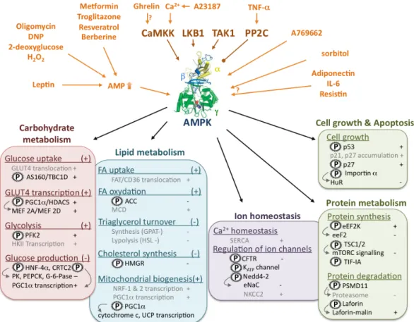
As already mentioned, AMPK activation stimulates ATP producing catabolic pathways and inhibits ATP consuming anabolic pathways, both by rapid effects via direct phosphorylation of metabolic enzymes and by long-term effects via regulation of transcription. The principal processes upstream and downstream of AMPK activation are summarized in Figures 1-3 and 1-4.
FIGURE 1-3. AMPK activating pathways and downstream targets. UPPER PART: Upstream signalling. AMPK is activated allosterically by pathways increasing cellular AMP/ATP ratios (top left) and covalently by phosphorylation (top center) via upstream kinases, or inactivated by dephosphorylation via protein phosphatases like PP1 or others. Other activators (A769662) act directly on AMPK, independent of AMP, or have unknown mechanisms (top right). LOWER
PART: Downstream signalling. Phosphorylation by AMPK is indicated by (P), activation by (+)
and inhibition by (-), arrows link primary phosphorylations to further downstream events, indirect AMPK effects are shown in grey. Activated AMPK stimulates e.g. glucose and FA uptake, glycolysis and FA oxidation, as well as transcription of mitochondrial proteins and mitochondrial proliferation in general. It inhibits energy requiring gluconeogenesis in liver, and FA/cholesterol biosynthesis. Other energy requiring processes such as cell growth, protein synthesis and degradation and regulation of intracellular ion concentrations are also regulated by AMPK.
Abbreviations: AMPK kinases/phosphatases - LKB1, Liver kinase B1; CamKK, Ca2+/calmodulin
dependent kinase kinase; TAK1, transforming growth factor-β activated kinase 1; PP2C, protein phosphatase 2C AMPK substrates - ACC, acetyl-CoA carboxylase; AS160, AKT substrate of 160 kDa; CFTR, cystic fibrosis transmembrane conductance regulator Cl- channel; CREB, CRE
binding protein; EF2K, elongation factor 2 kinase; eeF2, endothelial elongation factor; eNaC, epithelial Na+ channel; G-6-Pase, glucose 6-phosphatase; GLUT, glucose transporter; GPAT, glycerol-3-phosphate acyl transferase; HDAC, histone deacetylase; HMGR, 3-hydroxy-3-methylglutaryl coenzyme A reductase; HNF-4α, hepatic nuclear factor 4α; HSL, hormone-sensitive lipase; MCD, malonyl-CoA decarboxylase; MEF, myocyte enhancer factor; mTORC, mammalian target of rapamycin complex; NKCC, Na+-K+-2Cl- cotransporter; NRF, Nuclear respiratory factor; PEPCK, phosphoenol-pyruvate kinase; PGC1α, PPARγ co-activator-1α; PFK2, 6-phosphofructo-2-kinase; PK, pyruvate kinase; SERCA, sarcoplasmic/endoplasmic reticulum
CHAPTER I Regulation of cellular and whole body energy homeostasis calcium ATPase; TSC, Tuberous Sclerosis Complex; UCP, uncoupling protein. AMPK structure adapted from (Townley et al. 2007).
FIGURE 1-4. Cellular AMPK downstream signaling. Schematic representation of the AMPK downstream signaling processes mentioned in Fig. 1-3 and their subcellular localization.
3.3.1. Regulation of carbohydrate, lipid and whole body metabolism
Carbohydrates and lipids are the principal sources of energy for mammalian organisms. Supply, storage and processing of both, glucose and FA is crucial not only for cellular but also for whole body energy metabolism. An integrated metabolic control system is required to fine-tune the distribution between carbohydrate and lipid metabolism in response to a large number of physiological stimuli (e.g. exercise, fasting, feeding). AMPK controls both metabolic pathways by exerting a tight acute as well as transcriptional control.
3.3.1.1. Regulation of carbohydrate metabolism
Cellular glucose uptake is mainly regulated by the glucose transporter (GLUT) family. These transporters are stored in intracellular vesicles and translocated to the plasma membrane mainly in response to insulin (reviewed in (Ishiki et al. 2005)). Activation of AMPK leads to increased GLUT4 translocation in muscle, cardiomyocytes and adipocytes (Kurth-Kraczek et al. 1999, Yamaguchi et al. 2005, Webster et al. 2010) and to increased GLUT3 translocation in neurons (Weisova et al. 2009). Recently, AMPK was shown to phosphorylate the AKT substrate AS160, regulating its binding to 14-3-3 and thus promoting vesicle translocation regulated by the monomeric G protein Rab (Treebak et al. 2006, Sakamoto et al. 2008). However, the exact molecular mechanism of AMPK-mediated GLUT translocation remains unclear. In addition to GLUT4 translocation, AMPK is also involved in GLUT4 expression by phosphorylation of both peroxisome proliferator-activated receptor gamma co-activator-1 alpha (PGC-co-activator-1α) and histone deacetylase (HDAC) 5, which promote myocyte-enhancer factor (MEF)-dependent GLUT4 transcription (Steinberg et al. 2009) (Fig. 1-4).
Glycolysis is increased by activated AMPK either in short-term by 6-phosphofructosekinase-2 (PFK2) phosphorylation (Marsin et al. 2000) or in long-term by stimulation of hexokinase II (HKII) transcription (Stoppani et al. 2002).
In liver, AMPK negatively regulates the transcription of gluconeogenic enzymes L-type pyruvate kinase (L-PK), phosphoenol-pyruvate carboxylase (PEPCK) and glucose-6-phosphatase (G-6-Pase) by phosphorylation of transcription factor HNF-4α and transcriptional activator CREB (Steinberg et al. 2009).
3.3.1.2. Regulation of lipid metabolism
Besides glucose, FA are the main substrates for cellular energy metabolism. As for glucose, the first step consists in FA uptake, which is regulated, similar to glucose uptake, by transmembrane transporters like FAT/CD36. CD36 is stored in intracellular vesicles similar to GLUT4, and AMPK activation by AICAR or oligomycin leads also to CD36 translocation (van Oort et al. 2009). However, the molecular mechanism remains unknown.
The longest known AMPK target is acetyl-CoA carboxylase (ACC). ACC catalyzes the carboxylation of acetyl-CoA to malonyl-CoA. Malonyl-CoA is a precursor in FA synthesis and acts as an allosterical inhibitor of mitochondrial carnitine palmitoyltransferase 1 (CPT1) involved in FA
CHAPTER I Regulation of cellular and whole body energy homeostasis import, thus preventing FA β-oxidation. AMPK mediated phosphorylation inhibits ACC activity and therefore suppresses FA synthesis, while increasing at the same time mitochondrial FA β–oxidation (Munday 2002) (Fig. 1-4).
AMPK is suggested to inhibit both, triacylglycerol (TG) synthesis and degradation. One of the first steps of TG synthesis is regulated by glycerol-3-phosphate acyl transferase (GPAT) whose activity was reduced by AMPK in vitro and in AICAR-treated hepatocytes (Muoio et al. 1999). AMPK also phosphorylates hormone-sensitive lipase (HSL), preventing its activation by PKA and thus TG degradation (Watt et al. 2006).
Finally, AMPK phosphorylates and inhibits 3-hydroxy-3-methylglutaryl-CoA reductase (HMGR) preventing energy consuming cholesterol synthesis (Clarke et al. 1990).
3.3.1.3. Regulation of whole body energy metabolism
The idea that AMPK acts not only as a cellular energy sensor, but regulates the whole body energy status, has emerged from the findings that hypothalamic AMPK is regulated by food intake (Minokoshi et al. 2004) and that this regulation is mediated by the anorexigenic hormone leptin and the orexigenic hormone ghrelin (Andersson et al. 2004). Indeed, injection of adenovirus encoding a constitutive active AMPK mutant into the hypothalamus increased body weight and food uptake while a dominant negative mutant had the opposite effect (Minokoshi et al. 2004). However, the molecular mechanism by which AMPK regulates these processes remains unclear.
3.3.2. Regulation of protein metabolism, cell growth and other processes
Beside the regulation of major pathways in primary energy metabolism, AMPK seems to regulate other energy consuming processes, including protein synthesis, cell proliferation and cellular ion homeostasis.
First evidences for AMPK-mediated protein synthesis and cell growth came from the identification of tuberous sclerosis 2 (TSC2) as AMPK substrate (Inoki et al. 2003). TSC2 is a GTPase-activating protein which inactivates the small G-protein Rheb and thus exerts a critical negative regulation of downstream mammalian target of rapamycin complex 1 (mTORC1) (Yang et al. 2007). TSC2 is inhibited by AMPK, thus preventing mTORC1 downstream signalling involved in ribosome biogenesis, peptide translation and cell growth (Kwiatkowski et al. 2005). In addition, AMPK also inhibits mTORC1 assembly by phosphorylation of the TORC1 subunit raptor (Gwinn et al. 2008). In addition to protein synthesis, the mTORC1 pathway was shown to implicate AMPK also in autophagy (Meijer et al. 2007).
AMPK also phosphorylates and activates the eukaryotic elongation factor 2 kinase (eEF2K), which in turn inhibits eEF2 involved in peptide elongation (Browne et al. 2004). In addition, AMPK phosphorylates the RNA polymerase I-associated transcription factor TIF-IA, thus downregulating ribosomal RNA synthesis (Hoppe et al. 2009). AMPK activation also reduces the cytoplasmic level of the RNA-binding protein HuR, a mRNA stabilizing protein involved in regulation of cyclin A, cyclin
B1 and p21 expression (Wang et al. 2002). This might be due to increased AMPK-mediated importin α phosphorylation which stimulates HuR nuclear import (Wang et al. 2004). AMPK also phosphorylates the tumor suppressor p53 and the cyclin-dependent kinase inhibitor p27kip1, both contributing to cell cycle arrest (Jones et al. 2005, Liang et al. 2007).
AMPK seems not only to inhibit protein synthesis and cell growth, but also proteasome-dependent degradation (Viana et al. 2008). Indeed, AICAR treatment of MEF cells resulted in a 20% decrease of proteasome-dependent protein degradation, whereas no effect was observed in α1-/- ,α2-/- knock out MEF (Viana et al. 2008). AMPK was not involved in lysosomal-dependent protein degradation (Viana et al. 2008). AMPK phosphorylates and interacts with the 26s proteasome subunit PSMD11, indicating that AMPK might regulate the proteasome via direct phosphorylation of PSMD11 (Moreno et al. 2009). In addition, AMPK was shown to interact with and to phosphorylate laforin resulting in laforin-malin complex formation (Solaz-Fuster et al. 2008). The laforin-laforin-malin complex promotes protein-ubiquitination and degradation e.g. of type 1 protein phosphatase (PP1) which positively regulates glycogen accumulation (Solaz-Fuster et al. 2008).
In addition to protein metabolism and cell cycle progression, the maintenance of ion gradients across cell membranes and intracellular ion homeostasis are further very energy-demanding processes. Thus it is little surprising that AMPK might also regulate these processes. Indeed, different ion transporters seem to be regulated by AMPK. These include cystic fibrosis transmembrane conductance regulator Cl- channel (CFTR) (King et al. 2009), ATP-sensitive potassium (K
ATP) channel (Chang et
al. 2009) and epithelial Na+ channel (eNaC) (Carattino et al. 2005) which are all inhibited by AMPK activation, whereas Na+-K+-2Cl- cotransporter (NKCC2) (Fraser et al. 2007) seems to be activated.
Recently AMPK activity has also been reported to stimulate the sarcoplasmic/endoplasmic reticulum calcium ATPase by preventing its oxidation (Dong et al. 2010a, Dong et al. 2010b).
4. C
ELLULAR ENERGY METABOLISM IN PATHOLOGIES4.1. The metabolic syndrome
Impaired whole body energy balance is the major reason of poor human health worldwide. On the one hand, in 2006, malnutrition was responsible for almost 60% of total mortality worldwide. On the other hand, the industrialized world faces more and more problems related to overweight and obesity. In 2005, 66 % of the US American population and 38% of the French population were considered to have overweight (OECD 2005). The World Health Organization estimates that 1 billion adults worldwide have overweight and that 30 % of them are even clinically obese (body mass index > 30).
Obesity is often associated with a number of other health problems (type 2 diabetes, hypertension blood fat disorder, fatty liver and cardiovascular disease) that are summarized as the metabolic syndrome. Indeed, obesity raises the risk for developing type 2 diabetes by a factor of 30-40. Type 2 diabetes is characterized by insulin resistance and high blood glucose indicating impaired cellular glucose uptake. AMPK can bypass and replace insulin-signaling in many respects. It is recognized to
CHAPTER I Regulation of cellular and whole body energy homeostasis be a major energy metabolism-regulating enzyme whose activity has been implicated in both lipid and glucose metabolism, processes which are impaired in obesity and diabetes (Viollet et al. 2007, Hardie 2008, Viollet et al. 2009). Actually, AMPK activation with AICAR increases glucose uptake and FA oxidation in obese diabetic rodents (Bergeron et al. 2001) and humans (Koistinen et al. 2003, Steinberg et al. 2004, Steinberg et al. 2006a) suggesting a therapeutic potential of AMPK activators for bypassing skeletal muscle insulin resistance. Indeed, metformin, the major anti-diabetic drug that inhibits complex I of the mitochondrial respiratory chain, also activates AMPK (Musi et al. 2002, Zou et al. 2004). Chronic AICAR treatment was also shown to reduce blood pressure in obese Zucker rats by a still unknown mechanism (Buhl et al. 2002).
4.2. Energy metabolism in cancer
Carcinogenesis is caused by mutations of the genetic material in normal cells, which disturbs the normal balance between proliferation and cell death. This process is accompanied by changes in energy requirements due to an increase in energy costly processes like biomass production and genome replication. Ninety years ago Otto Warburg observed that tumor cells change their glucose metabolism from OxPhos to aerobic glycolysis (Warburg effect) (reviewed in (DeBerardinis 2008)). Indeed, even if the latter provides ATP less efficiently than OxPhos, it generates at the same time metabolic intermediates important for cell growth and proliferation. It is thus not surprising that tumor suppressors and proto-oncogenes often affect metabolism. Mutations in these genes can promote a metabolic phenotype more supportive for accelerated cell growth and proliferation.
The AMPK signaling network contains a number of such tumor suppressor genes including its upstream kinase LKB1 and its downstream targets p53 and TSC1/2. Rather rare LKB1 mutations lead to increased incidence of epithelial cancers and hamartomas, and its inactivation is estimated to occur in 30-50 % of lung adenocarcinomas, 20% of squamous cell carcinomas and 10% of lung large cell carcinomas (Ji et al. 2007). The role of LKB1 in the activation of AMPK suggests that pharmacological AMPK activation might be associated with reduced cancer incidence and progression. Indeed, AICAR-induced AMPK activation in breast cancer cell lines blocks their proliferation and reduced tumor growth in nude mice (Swinnen et al. 2005). In addition, diabetic patients treated with metformin show a 30% reduction in deaths from cancers versus those treated with either sulfonylurea or insulin (Evans et al. 2005b).
4.3. Neurodegenerative diseases
A common feature of neurodegenerative disorders such as Huntington’s disease (HD), amyotrophic lateral sclerosis (ALS), Parkinson’s disease (PD) and Alzheimer disease (AD) are mutations in nuclear or mitochondrial DNA leading to impaired mitochondrial function and energy metabolism disturbance (Beal 2000). In AD, the cerebral glucose metabolism is decreased, accompanied by impaired OxPhos. In addition, AD is characterized by accumulation of pathological protein aggregates like extracellular