Case report
Large thoracic duct cyst ± a case report and review of the literature
Alexandros Karajiannis
a, Thorsten Krueger
b,*, Eduard Stauffer
c, Hans-Beat Ris
baDepartment of Thoracic and Cardiovascular Surgery, University Hospital of Bern, Bern, Switzerland bDepartment of Surgery, University Hospital of Lausanne, Lausanne, Switzerland
cInstitute of Pathology, University Hospital of Bern, Bern, Switzerland
Received 7 June 1999; received in revised form 7 March 2000; accepted 29 March 2000
Abstract
Large thoracic duct cysts are rare and standard lateral thoracotomy is usually used for resection. In the reported case the combination of an antero-lateral thoracotomy with a partial longitudinal median sternotomy (hemiclamshell approach) allowed an excellent visualization and dissection of a large thoracic duct cyst expanding in the anterior cervico-thoracic junction, and was associated with an uncomplicated recovery. q 2000 Elsevier Science B.V. All rights reserved.
Keywords: Thoracic duct cyst; Trap door incision; Case report; Clamshell; Mediastinum
1. Case report
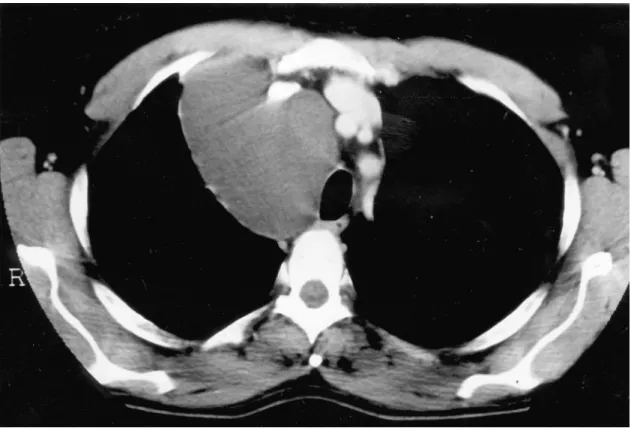
A 49-year-old man was referred to our institution with a large cystic tumour localized in the right upper anterior mediastinum. Bronchoscopy showed a slight compression of the middle and distal part of the trachea. The tumour was discovered 8 years before admission and ®ne needle aspira-tion revealed milky-yellowish ¯uid leading to the assump-tion of a thoracic duct cyst. No further treatment was done at that time. A CT-scan of the chest at admission revealed a huge cystic mass in the right superior anterior mediastinum in close relationship to the superior vena cava, the ascending aorta, the oesophagus, the trachea, which was deviated and impressed, and the right phrenic and recurrent nerves (Fig. 1). The maximum diameter was 12 cm. Physical examina-tion of the asymptomatic patient yielded no symptoms such as stridor, dyspnea or vein distension. Lung function testing and laboratory control revealed normal values.
It was decided to resect the lesion due to its unknown aetiology. Owing to its localization in the anterolateral superior mediastinum, a hemiclamshell thoracotomy was chosen and the cystic tumour was dissected without dif®-culty. The cyst originated from the thoracic duct and was situated in close relationship to the right subclavian vessels, the brachiocephalic trunk, the superior vena cava, the middle and distal trachea and the oesophagus, the ascending aorta and the azygos vein, with displacement of the right
upper lobe of the lung. The cystic tumour was dissected from these structures and removed after ligating and divid-ing the thoracic duct as well as several efferent and afferent lymphatic vessels. The phrenic and recurrent nerves were visualized and preserved.
The postoperative recovery was uneventful. Pain was controlled by continuous peridural analgesia for 7 days. The patient was discharged from the hospital on the 10th postoperative day with low dose oral analgesics. The 6-month follow-up showed an asymptomatic patient with a regular performance status and normal pulmonary function. The intraoperative suspicion of a thoracic duct cyst was con®rmed by histological examination. The cyst was unilo-cular and contained yellowish cloudy serous ¯uid. The weight was 395 g with a size of 8 £ 10 £ 12 cm. Microsco-pically, the cyst's wall contained of an inner single layer of ¯at endothelial cells, smooth muscle cells and islets of lympho-reticular tissue (Fig 2).
2. Discussion
A cyst of the thoracic duct is an uncommon disease. A review of 42 surgically treated congenital mediastinal cysts showed only one thoracic duct cyst [1]. The ®rst reported case was found during an autopsy examination by Carbone in 1892 and only 27 cases of thoracic duct cyst have been reported since Emerson [2] described the ®rst case diag-nosed during the patient's lifetime. The cyst in our patient was found in the superior anterior mediastinum, in accor-dance with approximately one of three previous reports.
European Journal of Cardio-thoracic Surgery 17 (2000) 754±756
1010-7940/00/$ - see front matter q 2000 Elsevier Science B.V. All rights reserved. PII: S1010-7940(00)00447-4
www.elsevier.com/locate/ejcts
* Corresponding author. Tel.: 141-21-314-2408; fax: 141-21-314-2358. E-mail address: [email protected] (T. Krueger).
A. Karajiannis et al. / European Journal of Cardio-thoracic Surgery 17 (2000) 754±756 755
Fig. 1. Computed tomography of the chest. Large cystic lesion localized in the superior anterior mediastinum.
Fig. 2. Histology (hematoxylin/eosin staining). Cyst wall containing of an inner single layer of ¯at endothelial cells, smooth muscle cells and islets of lympho-reticular tissue.
In contrast to our patient, symptoms were reported in other reports such as dyspnea on effort, thoracic or back discomfort and dysphagia, often aggravated by intake of food. Life-threatening complications, such as acute respira-tory insuf®ciency, have also been reported [3].
Computed tomography rose the suspicion of a cystic lesion, but failed to clear its dignity. The differential diag-nosis included pericardial or pleural mesothelial cysts, tera-tomatous cysts, bronchial or oesophageal cysts, thymic cysts and neurenteric and lymphangiomatous cysts [1]. Only three of the published cases of mediastinal thoracic duct cysts were diagnosed prior to surgery [4±6]. Lymphangiography and direct injection of contrast into the cyst with visualiza-tion of a communicavisualiza-tion between the cyst and the thoracic duct have been reported. The diagnosis in our patient was based on the relation between the cyst and the thoracic duct as assessed during the operation and on the microscopic ®ndings displaying the cyst's wall with an inner single layer of ¯at endothelial cells, smooth muscle cells and islets of lympho-reticular tissue [7].
Surgical resection of the cyst is usually recommended. Only one investigator suggested an expectative approach in asymptomatic patients with con®rmed diagnosis [6]. Surgical resection was performed via a lateral thoracotomy in 15 of 20 reviewed cases [8]. The most common compli-cation seen after surgery was chylothorax [7,9], requiring re-operation. As an alternative to standard thoracotomy the hemiclamshell approach may be used to access large mediastinal tumours situated in the superior part of the mediastinum, since the adjacent structures, especially the recurrent nerve may be better visualized by use of this approach than a lateral thoracotomy. The hemiclamshell approach offers an excellent visualization of the entire mediastinum and the entire cervico-thoracic junction with little morbidity regarding shoulder girdle function, chest wall complaints or pulmonary function as compared to stan-dard thoracotomy [10]. Video-assisted thoracoscopic surgery (VATS) may offer an alternative to resect benign mediastinal tumours [11]. However, it may jeopardize
important structures such as the recurrent or phrenic nerves in situations with large expanding cysts situated at the cervico-thoracic junction. Moreover, the risk of rupture of the cyst during dissection with subsequent spillage of its content within the chest cavity is increased by use of VATS as compared by an open approach.
The frequency of complaints, the risk of potential life-threatening complications and the need to establish a diag-nosis justify the resection of those cysts even in asympto-matic patients. The surgical access has to be chosen according to the size and localization of the lesion in order to achieve complete resection with save control of adjacent structures and efferent and afferent lymphatic vessels.
References
[1] Ochsner JL, Ochsner SF. Congenital cysts of the mediastinum: 20-year experience with 42 cases. Ann Surg 1966;163:909±920. [2] Emerson GL. Supradiaphragmatic thoracic-duct cyst. An unusual
mediastinal tumor. N Engl J Med 1950;242:575±578.
[3] Fromang DR, Seltzer MB, Tobias J. Mediastinal compression and acute respiratory insuf®ciency. Chest 1975;67:725±727.
[4] Hori S, Harada K, Morimoto S, Uchida H, Okumura K. Lymphangio-graphic demonstration of thoracic duct cyst. Chest 1980;78:652±654. [5] Tsuchiya R, Sugiura Y, Ogata T, Suemasu K. Thoracic duct cyst of
the mediastinum. J Thorac Cardiovasc Surg 1980;79:856±859. [6] Morettin LB, Timothy EA. Thoracic duct cyst: diagnosis with needle
aspiration. Radiology 1986;161:437±438.
[7] Mori M, Kidogawa H, Isoshima K. Thoracic duct cyst in the medias-tinum. Thorax 1992;47:325±326.
[8] Okabe K, Miura K, Konishi H, Hara K, Nobuyoshi S. Thoracic duct cyst of the mediastinum: case report. Scand J Thorac Cardiovasc Surg 1993;27:175±177.
[9] Cervantes-Perez P, Fuentes-Maldonado R. Thoracic duct cyst of the mediastinum: case report. Chest 1976;70:411.
[10] Lardinois D, Sippel M, Gugger M, Dusmet M, Ris HB. Morbidity and validity of the hemiclamshell approach for thoracic surgery. Eur J Cardio-thorac Surg 1999;16(2):194±199.
[11] Demmy TL, Krasna MJ, Detterbeck FC, Kline GG, Kohman LJ, DeCamp Jr MM, Wain JC. Multicenter VATS experience with mediastinal tumors. Ann Thorac Surg 1998;66:187±192.
A. Karajiannis et al. / European Journal of Cardio-thoracic Surgery 17 (2000) 754±756 756