No improvement of survival with reduced- versus
high-intensity conditioning for allogeneic stem cell
transplants in Ewing tumor patients
U. Thiel
1, A. Wawer
1, P. Wolf
2, M. Badoglio
3, A. Santucci
4, T. Klingebiel
5, O. Basu
5, A. Borkhardt
6,
H.-J. Laws
6, Y. Kodera
7,8, A. Yoshimi
8, C. Peters
9, R. Ladenstein
9, A. Pession
10, A. Prete
10,
E.-C. Urban
11, W. Schwinger
11, P. Bordigoni
12, A. Salmon
12, M. A. Diaz
13, B. Afanasyev
14, I. Lisukov
14,
E. Morozova
14, A. Toren
15, B. Bielorai
15, J. Korsakas
16, F. Fagioli
17, D. Caselli
18, G. Ehninger
19,
B. Gruhn
20, U. Dirksen
21, F. Abdel-Rahman
22, M. Aglietta
23, E. Mastrodicasa
4, M. Torrent
24,
P. Corradini
25, F. Demeocq
26, G. Dini
27, P. Dreger
28, M. Eyrich
29, J. Gozdzik
30, F. Guilhot
31, E. Holler
32,
E. Koscielniak
33, C. Messina
34, D. Nachbaur
35, R. Sabbatini
36, E. Oldani
37, H. Ottinger
38, H. Ozsahin
39,
R. Schots
40, S. Siena
41, J. Stein
42, S. Sufliarska
43, A. Unal
44, M. Ussowicz
45, P. Schneider
46,
W. Woessmann
47, H. Ju¨rgens
21, M. Bregni
48& S. Burdach
1* on behalf of the Solid Tumor Working
Party (STWP) and the Pediatric Disease Working Party (PDWP) of the European Group for Blood and
Marrow Transplantation (EBMT), the Asia Pacific Blood and Marrow Transplantation (APBMT), the
Pediatric Registry for Stem Cell Transplantations (PRST) and the MetaEICESS Study Group
1
Department of Pediatrics and Wilhelm Sander Sarcoma Unit MRI, Pediatric Oncology Center, Technische Universita¨t Mu¨nchen;2
Institute for Medical Statistics and Epidemiology, Klinikum rechts der Isar der Technischen Universita¨t Mu¨nchen, Munich, Germany;3
EBMT Data & Study Office, Hopital Saint-Antoine, Assistance Publique des Hoˆpitaux de Paris and UPMC Univ Paris 06, Paris, France;4
Section of Pediatric Hematology & Oncology, University of Perugia, Perugia, Italy;5
Children’s Hospital III, Department of Pediatrics, Johann Wolfgang Goethe University, Frankfurt;6
Department of Pediatric Oncology, Hematology and Clinical Immunology, Heinrich Heine University, Du¨sseldorf, Germany;7
Department of Promotion for Blood and Marrow Transplantation, Aichi Medical University, Aichi;8
APBMT Data Center, Nagoya University School of Medicine, Nagoya, Japan;9
Department of Pediatrics, St. Anna Kinderspital, Vienna, Austria;10
Department of Scienze Pediatriche Mediche e Chirurgiche, Ospedale S Orsola Malpighi, Bologna, Italy;11
Department of Pediatrics, Medical University of Graz, Graz, Austria;12
Service de Transplantation Medullaire, CHU de Nancy Brabois, Vandoeuvre-les-Nancy, France;13
Department of Pediatrics, Division of Pediatric Hematology-Oncology and Hematopoietic Stem Cell Transplantation and Cell Therapy Unit, Hospital Infantil Universitario Nin˜o Jesus, Madrid, Spain;14St. Petersburg State Medical Pavlov University, Ratsa Gorbacheva Memorial Children‘s Institute, Department of Hematology and Transplantology, St. Petersburg, Russia;15Pediatric Hemato-Oncology Unit, Sheba Medical Center (affiliated to the Sackler Faculty of Medicine), Tel Hashomer, Israel; 16
Department of Hematology, Oncology and Transfusion Medicine Center, Vilnius University Hospital Santariskiu Clinics, Vilnius, Lithuania;17Stem Cell Transplantation and Cellular Therapy Unit, Pediatric Onco-Hematology Division, ‘‘Regina Margherita’’ Children’s Hospital, Turin;18
Department of Oncoematologia Pediatrica, Azienda Ospedaliero-Universitaria Meyer, Florence, Italy;19
Department of Internal Medicine I, University Hospital Carl Gustav Carus, Dresden;20
Department of Pediatrics, University of Jena, Jena; 21
Department of Pediatric Hematology and Oncology, University Children’s Hospital, Mu¨nster, Germany;22
The Bone Marrow and Stem Cell Transplantation Program, King Hussein Cancer Center, Amman, Jordan;23
Department of Istituto per la Ricerca e la Cura del Cancro, Turin, Italy;24
Hospital de la Santa Creu i Sant Pau, Department of Pediatrics, Barcelona, Spain;25
Department of Hematology - Bone Marrow Transplantation Unit, Istituto Nazionale dei Tumori, University of Milano, Milan, Italy;26 Centre Hospitalier et Universitaire de Clermont-Ferrand, Service de Pe´diatrie B et Unite´ Bioclinique de The´rapie Cellulaire, Clermont-Ferrand, France;27
Department of UO Ematologia ed Oncologia Pediatrica, Istituto G Gaslini, Genova, Italy;28
Department of Internal Medicine V, University of Heidelberg, Heidelberg;29
Children’s Hospital, Department of Paediatric Stem Cell Transplantation, University of Wu¨rzburg, Wu¨rzburg, Germany;30
Transplantation Centre, University Children’s Hospital, Cracow, Poland;31
Department of Hematology, University Hospital, Poitiers, France;32
Department of Hematology and Oncology, University of Regensburg, Regensburg;33
Department of Pediatrics 5 (Oncology, Hematology, Immunology), Olga Hospital, Klinikum Stuttgart, Stuttgart, Germany;34
Hemo/Oncology, Department of Pediatrics, Hospital-University of Padova, Padova, Italy;35
University Hospital of Innsbruck, Internal Medicine V, Department of Hematology and Oncology, Innsbruck, Austria;36
Department of Oncology, Haematology, and Respiratory Diseases, Policlinico di Modena, Modena;37
Department of U.S.C. Ematologia, Ospedali Riuniti, Bergamo, Italy;38
Department of Bone Marrow Transplantation, University Hospital of Essen, Essen, Germany;39
Paediatric Oncology Unit, University of Geneva Children’s Hospital, Geneva, Switzerland;40
Division of Clinical Hematology and BMT Unit, University Hospital Brussels, Brussels, Belgium;41
Department of S. C. Divisione Oncologia Falck and S. C. Divisione Anatomia Patologica, Ospedale Niguarda Ca’ Granda, Milan, Italy;42Bone marrow Transplant Unit, Department of Pediatric Hematology-Oncology, Schneider Children’s Medical Center of Israel, Petach Tikva, Israel;43Bone Marrow Transplantation Unit, Department of Pediatrics, Comenius University Medical School, Bratislava, Slovak Republic;44Institutions Erciyes Medical School, Department of Hematology and Oncology, Kapadokya BMT Center, Kayseri, Turkey;45Department of Pediatric Hematology, Oncology and Bone Marrow Transplantation, Wroclaw Medical University, Wroclaw, Poland;46
Department of Pediatric Hematology and Oncology, Hoˆpital Charles Nicolle, Rouen, France;47
Department of Pediatric Hematology and Oncology, University Hospital, Giessen, Germany;48
Unit of Medical Oncology, Ospedale San Giuseppe, Milan, Italy
Received 25 October 2010; accepted 3 November 2010
Background:
Outcomes of Ewing tumor (ET) patients treated with allogeneic stem celloriginal
article
*Correspondence to: Prof. S. Burdach, Department of Pediatrics, Wilhelm Sander Sarcoma Unit and Roman Herzog Comprehensive Cancer Center, MRI, Technische Universita¨t Mu¨nchen, 81664 Mu¨nchen, Germany. Tel: +49-89-3068-2260 or -2261; Fax: +49-89-3068-3954; E-mail: [email protected]
transplantation (allo-SCT) were compared regarding the use of reduced-intensity conditioning (RIC) and high-intensity conditioning (HIC) regimens as well as human leukocyte antigen (HLA)-matched and HLA-mismatched grafts.
Patients and methods:
We retrospectively analyzed data of 87 ET patients from the European Group for Blood and Marrow Transplantation, Pediatric Registry for Stem Cell Transplantations, Asia Pacific Blood and MarrowTransplantation and MetaEICESS registries treated with allo-SCT. Fifty patients received RIC (group A) and 37 patients received HIC (group B). Twenty-four patients received HLA-mismatched grafts and 63 received HLA-matched grafts.
Results:
Median overall survival was 7.9 months [61.24, 95% confidence interval (CI) 5.44–10.31] for group A and 4.4 months (61.06, 95% CI 2.29–6.43) for group B patients (P= 1.3). Death of complications (DOC) occurred in 4 of 50 (0.08) and death of disease (DOD) in 33 of 50 (0.66) group A and in 16 of 37 (0.43) and 17 of 37 (0.46) group B patients, respectively. DOC incidence was decreased (P< 0.01) and DOD/relapse increased (P < 0.01) in group A compared with group B. HLA mismatch was not generally associated with graft-versus-Ewing tumor effect (GvETE).Conclusions:
There was no improvement of survival with RIC compared with HIC due to increased DOD/relapse incidence after RIC despite less DOC incidence. This implicates general absence of a clinically relevant GvETE with current protocols.Key words:
advanced-stage Ewing tumor, allogeneic stem cell transplantation, graft versus tumor effect, haploidentical stem cell transplantation, reduced/high-intensity conditioning chemotherapyintroduction
Ewing tumors (ET) constitute a cancer entity of highly
malignant, small, round blue cell tumors with features of
neuroectodermal or endothelial differentiation [1–5]. They are
defined by the expression of a chimeric transcript, commonly
deriving from the reciprocal translocation t(11;22)(q24;q12)
that results in formation of an EWS/ETS fusion gene, whose
detection permits a specific molecular genetic diagnosis [6, 7].
The incidence is 3.3 per million in the Western hemisphere with
a peak incidence at the age of 15 years [8]. The most frequent
localization of disease onset is bone tissue, preferentially long
bones and pelvis. Rarely, soft tissue may harbor the primary
tumor site [2]. Metastatic potential is high; approximately 75%
of patients present with local disease at the time of diagnosis,
whereas the remaining 25% initially present with metastatic
disease. Radical surgery alone results in metastatic relapse in the
majority of cases, suggesting occult metastases at diagnosis even
in apparently localized disease. Despite the impressive
improvement of treatment in the last decades using multimodal
approaches, 5-year overall survival (OS) of ET patients with
localized disease remains at 70%, dropping down to <15% in
patients with multifocal primary disease or with early relapse in
most studies. Use of involved compartment irradiation and
systemic high-dose therapy, both with stem cell rescue, may
increase long-term cure rates. However, additional
improvements of survival are warranted [9–14].
The existence of a clinically relevant graft-versus-tumor effect
in patients with ET after allogeneic stem cell transplantation
(allo-SCT) has been a matter of debate [15–17]. Evidence for
a graft-versus-Ewing tumor effect (GvETE) could solely be
deduced from case reports [18–21]. Allo-SCT following
high-intensity conditioning (HIC) or even reduced-high-intensity
conditioning (RIC) regimens has thus remained a merely
experimental therapy option in advanced-stage Ewing tumor
(AET) patients and a consensus about eligibility criteria is
lacking. In order to clarify this issue, we carried out
a retrospective European Group for Blood and Marrow
Transplantation (EBMT), Pediatric Registry for Stem Cell
Transplantations (PRST), Asia Pacific Blood and Marrow
Transplantation (APBMT) and MetaEICESS registries based
analysis of 87 ET patients who had either received RIC or HIC
before either human leukocyte antigen (HLA)-mismatched or
HLA-matched allo-SCT.
patients and methods
study design and data provenience
We collected and evaluated data of 87 patients who were diagnosed with ET from 1984 to 2010 from the EBMT (n = 69), APBMT (n = 5), PRST (n = 18) and MetaEICESS (n = 4) registries. Some patients were listed in more than one registry. Inclusion criterion was diagnosis of Ewing sarcoma family of tumors. Diagnosis was based on histopathological examination and in recently diagnosed patients confirmed by
molecular genetic detection of ET-specific translocations. In the following sections, patient numbers are followed by specification of respective proportions given in brackets when appropriate, except when data were unavailable.
definitions
Engraftment was defined as an absolute neutrophil count ‡0.5 · 109/l after allo-SCT. In case, patients died within £100 days after allo-SCT or when information was unavailable, chronic graft versus host disease (cGvHD) was considered as not assessable. Death of complications (DOC) constituted any kind of treatment-related death occurring after allo-SCT in the absence of disease evidence, including engraftment failure. In contrast, the definition of death of disease (DOD) comprised any death directly related to either disease progression or relapse. Progressive disease (PD) was defined as ‡50% progression of tumor volume; stable disease (SD) included <50% progression, partial remission (PR) as ‡50% reduction and complete remission (CR) as absence of detectable disease. Residual disease (RD) included both PD and PR. Relapse-free survival (RFS) was defined as the time period from the last allo-SCT until the occurrence of any local or metastatic ET evidence in patients who had reached CR after treatment. Early relapse was defined as relapse occurrence £24 months after diagnosis as opposed to the definition of late relapse (‡24 months after diagnosis). Multifocal disease was defined as three or more involved bone sites and/or bone marrow (BM) involvement at diagnosis. Secondary malignancy was defined as any occurrence of post-allo-SCT malignancies other than ET.
HLA mismatch was defined as one or more allele mismatch in HLA class 1 and/or HLA class 2.
patients
The study population consisted of 49 (0.56) male and 38 (0.44) female patients. Median age at allo-SCT was 17 years (range 3–49 years). Depending on the used conditioning regimen before allo-SCT, all patients were assigned either to group A (RIC) or to group B (HIC). Group A comprised 50 of 87 (0.57) and group B 37 of 87 (0.43) patients. In total, 63 of 87 (0.72) patients received grafts from either HLA-matched related or HLA-matched unrelated donors, whereas 24 of 87 (0.28) patients received either haploidentical or otherwise HLA-mismatched grafts. Eligibility for allo-SCT was decided upon the following criteria: local and/or metastatic relapse (n = 27), multifocal primary with/without RD (n = 46) and autograft mobilization failure (n = 1). After induction and conditioning treatment, 42 of 87 (0.48) patients were transplanted in CR, 29 (0.33) in PR and 14 (0.16) in PD. Graft source was only BM in 33 (0.38) patients, only peripheral blood (PB) in 48 (0.55), BM and PB in 2 (0.02) patients and only cord blood in 1 (0.01) patient. Thirty-four (0.39) patients had received autologous grafts, 1 (0.01) patient had received allogeneic graft and 45 (0.52) patients had not received any other graft before allo-SCT. In group A, 4 (0.08) patients were transplanted before the year 2000 and 46 (0.92) in 2000 or later, whereas in group B, 29 (0.78) patients were transplanted <2000 and 8 (0.22) ‡2000. The database contained some previously published patient data [18, 20]. Age and stage distribution did not differ significantly between both groups. Precise patient data of both groups are given in Table 1. All patients or their guardians signed informed consent before therapy. Treatment application relied upon institutional review board approvals according to the precepts established by the Helsinki Conference Declaration.
conditioning regimens and graft versus host disease
prophylaxis
RIC regimens were mainly based on the use of fludarabine (60–210 mg/m2; n = 47), alone or in combination with either/or the following drugs: melphalan (MEL; 70–150 mg/m2; n = 15), busulfan (BU; 3–12 mg/kg; n = 13), thiotepa (TT; 5–10 mg/kg; n = 26), cyclophosphamide (CYC; 30–120 mg/kg; n = 14), treosulfan (TREO; 36 or 42 mg/m2; n = 2), topotecan (6 mg/m2; n = 3), carmustine (320 mg/m2; n = 1) or etoposide (ETO; dosage unavailable; n = 1) or other nonspecified drugs in nonmyeloablative doses (n = 1) with or without the application of 2–4 Gy total body irradiation (TBI; n = 5). Furthermore, in some RIC patients, treatment comprised the application of antithymocyte globuline (ATG; n = 13), muromonab-CD3 (OKT3; n = 8) and/or alemtuzumab (n = 1). HIC regimens were mainly based on the use of TBI (10–14 Gy; n = 23) in combination with either/or the following drugs: MEL (120–210 mg/m2; n =
25), BU (8–16 mg/kg; n = 9), ETO (800–2100 mg/m2; n = 18), carboplatin
(800–1500 mg/m2; n = 9), CYC (45–150 mg/kg; n = 7), TT (600 or 3300 mg/m2; n = 3), TREO (42 mg/m2; n = 2), vincristine (2 or 2.5 mg/m2; n = 2)
or other nonspecified drugs in myeloablative doses (n = 1). At least one group B patient received additional ATG. For assessment of conditioning regimens, only the effect of the latest allo-SCT was analyzed even when some patients had received auto- or allografts before. Graft versus host disease (GvHD) prophylaxis included methotrexate, mycophenolat-mofetil, cyclosporine A and/or prednisolone. Individual regimens are provided in the supplemental Tables S1 and S2 (available at Annals of Oncology online).
statistical analysis
End points were assessed upon the date of last patient contact. Final database update was conducted in June 2010. Statistical analyses were carried out using R 2.11.0 (The R Foundation for Statistical Computing,
Vienna, Austria) and the PASW Statistics v18.0 (SPSS Inc, Chicago, IL) software. Time values for DOC and relapse/DOD estimates were assessed starting on the date of the last allo-SCT until last follow-up (FU) and for OS, until FU and/or until the occurring event was death independent of the cause. In multivariate analyses, considered variables were patient age, sex, disease status and conditioning regimen for allo-SCT. Hazard ratios, standard errors and confidence intervals (CI) are given when appropriate.
For calculation of OS probabilities, the Kaplan–Meier estimate was used. OS curves were compared using the two-tailed log-rank test. Associations of patient characteristics and conditioning regimens with OS were evaluated in multivariate analyses using Cox proportional hazards.
Cumulative incidence curves were applied to estimate the occurrence of relapse/DOD and DOC, with DOC being a competing event for relapse/ DOD and vice versa. For comparison of relapse/DOD incidence between HLA-matched and HLA-mismatched patients, DOC was the competing event. When DOD/relapse and DOC were competing events, uni- and multivariate risk analyses were conducted using the cmprsk and the crr-addson packages designed for R, as proposed by Scrucca et al. [22, 23]. In uni- and multivariate analyses, probabilities of relapse/DOD or DOC for potential explanatory variables were compared using Gray’s test or Wald test, respectively. A P value <0.05 was considered statistically significant.
results
engraftment and GvHD
Seventy-nine (0.91) patients engrafted successfully, whereas 7
(0.08) patients failed to engraft and for 1 patient, engraftment
data were not available. Overall, acute graft versus host disease
(aGvHD) was reported in 39 (0.45) patients and absent in 44
(0.51). Overall, cGvHD occurred in 12 (0.14) patients, was
absent in 31 (0.36) patients and was not assessable due to either
death/last FU before day 100 after allo-SCT in 37 (0.42)
patients or to otherwise unavailable information in 7 (0.08)
patients. Within group A, 15 (0.30) patients presented with
limited and 10 (0.20) with extensive aGvHD, whereas 24 (0.48)
patients did not develop aGvHD. Four (0.08) group A patients
developed limited, 3 (0.06) developed extensive and 17 (0.34)
did not have cGvHD. Status information remained unavailable
in 26 (0.52) group A patients due to early death or last FU
before day 100 after allo-SCT. Within group B, eight (0.22)
patients developed limited and six (0.16) developed extensive
aGvHD. In 20 (0.54) group B patients, aGvHD was not
detectable. Limited cGvHD was present in three (0.08) and
extensive cGvHD in two (0.05) further patients. Status
information remained unavailable in 18 (0.49) group B patients
due to early death or last FU before day 100 after allo-SCT. An
overview is given in Table 2.
overall survival
The 5-year OS estimate for groups A and B was 0.15 (60.07)
and 0.10 (60.05), respectively. OS time did not differ
significantly between both groups (Figure 1; log rank P =
0.133). At the time of data censure, 37 of 50 (0.74) group A and
33 of 37 (0.89) group B patients had died due to disease or due
to complications. Thirteen (0.26) group A and four (0.11)
group B patients were alive in CR except for one group A
patient who had SD. Median OS was 7.9 months (61.24; 95%
CI 5.44–10.31) for group A and 4.4 months (61.06; 95% CI
Table 1. Patient characteristics
Parameter Group A (RIC, n = 50) Group B (HIC, n = 37) Number Fraction Number Fraction Median age at diagnosis 15 15 Range 4–49 2–40 £15 years 30 0.60 21 0.57 >15 years 20 0.40 16 0.43 Median age at allo-SCT 17 16 Range 7–49 3–41 £15 years 19 0.38 20 0.54 >15 years 31 0.62 19 0.51 Gender Male 28 21 Female 22 16 Period of diagnosis <2000 11 0.22 28 0.76 ‡2000 39 0.78 9 0.24 Period of allo-SCT <2000 4 0.08 25 0.68 ‡2000 46 0.92 12 0.32
Risk profile leading to allo-SCT
Early relapsea 7 0.14 5 0.14 Late relapsea 11 0.22 4 0.11 Multifocal primary and/or residual disease 29 0.58 17 0.46 Autograft mobilization failure 0 0.00 1 0.03 Not available 3 0.06 10 0.27 Primary site
Local bone only 15 0.30 8 0.22 Local bone and
pulmonal or/and lymph node metastases
8 0.16 2 0.05
Multifocal 21 0.42 17 0.46
Local soft tissue only
1 0.02 0 0.00
Not available 5 0.10 10 0.27 Local treatment modality
Surgery only 12 0.24 5 0.14 Irradiation only 11 0.22 8 0.22 Surgery + irradiation 19 0.38 15 0.41 None 3 0.06 0 0.00 Not available 5 0.10 9 0.24 Previous graft No previous graft 14 0.28 31 0.84 Allogeneic graft once 1 0.02 0 0.00 Autologous graft once 24 0.48 1 0.03 Autologous graft twice or more 7 0.14 2 0.05 Not available 4 0.08 3 0.08 Table 2. Results
Parameter Group A (RIC, n = 50) Group B (HIC, n = 37) Number Fraction Number Fraction Engraftment Success 47 0.94 32 0.86 Failure 3 0.06 4 0.11 Not available 0 0.00 1 0.03 aGvHD None 24 0.48 20 0.54 Limited 15 0.30 8 0.22 Extensive 10 0.20 6 0.16 Not available 1 0.02 3 0.08 cGvHD None 17 0.34 14 0.38 Limited 4 0.08 3 0.08 Extensive 3 0.06 2 0.05 NA due to death or last FU £ day 100 22 0.44 15 0.41 Not available 4 0.08 3 0.08 Outcome DOC 4 0.08 16 0.43 DOD 33 0.66 17 0.46 Alive at last FU 13 0.26 4 0.11 Median OS (months after allo-SCT)
Median 7.9 4.4
Range 0–67 0–213
RIC, reduced-intensity conditioning; HIC, high-intensity conditioning; aGvHD, acute graft versus host disease; cGvHD, chronic graft versus host disease, FU, follow-up; DOC, death of complications; DOD, death of disease; OS, overall survival; allo-SCT, allogeneic stem cell transplantation. Table 1. (Continued)
Parameter Group A (RIC, n = 50) Group B (HIC, n = 37) Number Fraction Number Fraction Donor HLA match
Matched related 27 0.54 32 0.86 Matched unrelated 3 0.06 1 0.03 Mismatchedb 20 0.40 4 0.11 Graft source BM only 8 0.16 25 0.68 PB only 39 0.78 9 0.24 BM + PB 2 0.04 0 0.00 CB only 1 0.02 0 0.00 Not available 0 0.00 3 0.08 Pretransplant status CR 22 0.44 20 0.54 PR 17 0.34 12 0.32 PD 10 0.20 4 0.11 Not available 1 0.02 1 0.03
aIncluding multifocal relapse in some patients.
bOne or more allele mismatch in HLA class 1 and/or HLA class 2.
RIC, reduced-intensity conditioning; HIC, high-intensity conditioning; allo-SCT, allogeneic stem cell transplantation; HLA, human leukocyte antigen; BM, bone marrow; PB, peripheral blood; CB, cord blood; CR, complete remission; PR, partial remission; PD, progressive disease.
2.29–6.43) for group B patients, respectively. An overview is
provided in Table 2.
relapse and DOD
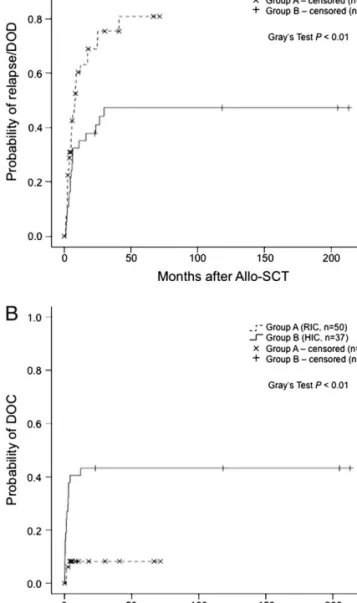
The 5-year estimate for relapse/DOD was 0.81 (60.08) for
group A patients and 0.47 (60.09) for group B patients.
Overall, 33 of 50 (0.66) group A patients and 17of 37 (0.46)
group B patients had a relapse and/or died of their disease
(Table 2). Overall, risk of relapse/DOD was significantly higher
within group A as compared with group B (Figure 2A, Gray’s
test P = 0.009).
death of complications
The 5-year and overall DOC estimate was 0.08 (60.04) for
group A and 0.43 (60.08) for group B patients. Main reasons
causing DOC were occurrences of infection, hemorrhage,
veno-occlusive disease and GvHD. Overall, risk of DOC was
significantly lower within group A as compared with group B
(Figure 2B, Gray’s test P = 0.0002).
HLA-mismatched versus HLA-matched grafts
In this setting, time periods from the date of the last allo-SCT
until the occurrence of relapse/DOD were compared between
those patients who had received HLA-mismatched with those
who had received HLA-matched grafts. DOC was considered as
a competing event for relapse/DOD. Twenty-four of 87 (0.28)
patients had received HLA-mismatched and 63 of 87 (0.72)
patients had received HLA-matched grafts. The 3-year estimate
for relapse/DOD was 0.62 (60.13) for patients with
mismatched grafts and 0.64 (60.06) for patients with
HLA-matched grafts. The 3-year estimate for DOC was 0.22 (60.09)
for patients with HLA-mismatched grafts and 0.24 (60.05) for
patients with HLA-matched grafts. Neither group had an OS or
RFS advantage (Figure 3A, Gray’s test P = 0.89; Figure 3B,
Gray’s test P = 0.95).
multivariate analysis
Upon multivariate analyses, none of the considered variables
age, sex, conditioning regimen and disease stage at allo-SCT
had an influence on OS outcome. Age just marginally failed to
reach statistical significance as an influential factor for OS
(Wald test P = 0.05). Employment of RIC was associated with
an increased relapse/DOD versus DOC rate (Wald test P =
0.021) compared with HIC regimens (Table 3).
Figure 2. Probabilities and group comparisons of time periods from the date of allogeneic stem cell transplantation (allo-SCT) until (A) relapse/ death of disease (DOD) and (B) death of complications (DOC) between patients treated with reduced-intensity conditioning (RIC; group A, n = 50) versus high-intensity conditioning (HIC; group B, n = 37) regimen in a competitive risk setting between DOD and DOC. Patients alive at last follow-up were censored. P values < 0.05 (Gray’s test) indicate significant difference.
Figure 1. Overall survival probabilities from the date of allogeneic stem cell transplantation (allo-SCT) for patients treated with reduced-intensity conditioning (RIC; group A, n = 50) versus high-intensity conditioning (HIC; group B, n = 37) regimen; patients alive at last follow-up were censored. The differences are not significant (log rank, P > 0.5).
discussion
Allo-SCT has been applied as a therapeutic option for patients
with AET [18]. Koscielniak et al. as well as Lucas et al. reported
on AET patients who had experienced tumor regression
following allo-SCT [20, 21]. However, the GvETE may be
associated with toxicity of GvHD. In addition to natural killer
cell-mediated toxicity, the broad T-cell allo-response poses
a significant problem in clinical transplantation across HLA
incompatibilities [24].
The use of myeloablative conditioning before allo-SCT has
led to improved disease response but implicated pronounced
treatment-related toxicity [14, 25–27], which in turn has led to
a shift toward the implementation of less toxic RIC regimens in
the last decade. This shift was expected to diminish incidence of
DOC and thus to enhance the conditions for a GvETE to
engage. We retrospectively compared clinical outcome of
allo-SCT in ET patients who had either received RIC (group A) or
HIC (group B) before allo-SCT in regard to age at
transplantation, toxicity, RFS and OS. Median age, disease stage
and indications for allo-SCT were comparable. Both groups
contained HLA-related and nonrelated matched and
mismatched donor/recipient combinations, including
haploidentical transplants. We hypothesized a clinically
relevant GvETE in ET patients treated with allo-SCT and
assessed its possible presence by comparing the relapse/DOD
rates between both groups. In group A (RIC), DOC rate (0.08)
was reduced and DOD rate (0.66) was increased compared with
group B (HIC, DOC 0.43 and DOD 0.46). Compared with the
application of HIC, RIC-mediated protection from DOC was
significantly higher in uni- and multivariate analyses (P < 0.05).
However, the lower DOC rate in the RIC-treated patients did
not translate into improved survival but was associated with
a higher relapse/DOD rate in group A as compared with group
B and did not improve OS. Sex, disease status and age at
allo-SCT were neither associated with improved time until relapse/
DOD nor with OS in either setting.
Despite higher incidence of relapse/DOD due to lower
incidence of DOC in patients treated with RIC, comparison of
RFS and OS between both groups demonstrated that neither
Figure 3. Comparison of survival rates in a competitive risk setting between relapse/death of disease (DOD) and death of complications (DOC) from the date of allogeneic stem cell transplantation (allo-SCT) until relapse/DOD (A) or until DOC (B) of patients receiving human leukocyte antigen (HLA)-mismatched or HLA-matched grafts. Patients alive at last follow-up were censored. Neither graft type provides a significant survival advantage (Gray’s test, P > 0.5).
Table 3. Multivariate analysis
HR SE 95% CI P valuea OS Recipient age 1.03 0.01 1.00–1.05 0.050 Sex 0.640 Male Referent Female 0.88 0.27 0.52–1.50 Conditioning 0.068 HIC Referent RIC 0.60 0.28 0.35–1.04 Disease stage at allo-SCT 0.110 RD Referent CR 0.67 0.25 0.41–1.10 Relapse/DOD Recipient age 0.99 0.02 0.95–1.03 0.500 Sex 0.580 Male Referent Female 0.85 0.30 0.48–1.51 Conditioning 0.021 HIC Referent RIC 2.10 0.31 1.11–3.75 Disease stage at allo-SCT 0.250 RD Referent CR 0.71 0.30 0.40–1.27 aWald test.
HR, hazard ratio; SE, standard error; CI, confidence interval; OS, overall survival; RIC, reduced-intensity conditioning; HIC, high-intensity conditioning; RD, residual disease; CR, complete remission; allo-SCT, allogeneic stem cell transplantation; DOD, death of disease.
type of conditioning was associated with significantly better
survival rates. This finding indirectly implicates the lack of
a clinically relevant GvETE in current settings. A DOC rate of
0.43 in patients receiving HIC confirms the results of our study
conducted 10 years ago, which revealed a DOC rate of 0.40 in
AET patients treated with matched allogeneic sibling grafts after
HIC [18].
Some patients survived long term after allo-SCT and within
this group some had received haploidentical grafts. We
expected that the application of mismatched grafts would lead
to protection via GvETE and therefore compared relapse/DOD
and DOC outcomes of patients receiving HLA-matched versus
HLA-mismatched grafts, but we could not detect any difference
regarding RFS or OS. In anticipation of a supposed GvETE, the
application of haploidentical transplantations in AET patients
constitutes an approach that has been increasingly
implemented notably during the last decade. In our study,
among those patients who were alive at the time point of data
censure, some had received haploidentical grafts just recently
and were alive, so the long-term effect of this particular
allo-SCT setting cannot yet be determined. Additionally, we did not
address the role of donor lymphocyte infusions (DLI).
Therefore, the question whether allo-SCT in haploidentical
graft settings may yield a clinically relevant GvETE with or
without the use of DLI will remain subject to ongoing
prospective studies. As GvETE may depend on ET-specific as
well as non-ET-specific antigen presentation, it will also be
important to identify further biological eligibility criteria for
haploidentical allo-SCT, i.e. certain HLA mismatch
constellations when choosing the donor.
For data interpretation, it also has to be considered that
group A (RIC) patients were mainly treated during the last 10
years in contrast to group B (HIC) patients who were mostly
treated before this period. This might have an impact on DOC
comparability between both groups, e.g. due to improvement
in supportive therapy, especially for GvHD prophylaxis and
treatment. Furthermore, initial and relapse systemic and local
treatment differed between patients depending on center and
time period and many group A patients had received prior
grafts before allo-SCT. On the other hand, decreased relapse
rates after HIC versus RIC provide further evidence for
antitumor efficacy of high-dose therapy in ET.
Our results emphasize the need for additional therapeutic
strategies for AET that yield antitumor effectiveness with
minimal toxic side-effects. The identification of ET-specific
target antigens [1] may give way to a personalized immune
therapy using ET antigen-specific allo-restricted T cells in
addition to the application of allo-SCT, rendering future
application of DLI more specific and thus more efficacious and
less toxic as seen in the treatment of other cancer entities [24,
28].
In our present study, we aimed to match patient groups as
closely as possible, however, as the effect of allo-SCT demands
large ET patient numbers, the effect of heterogeneity, i.e.
regarding initial diagnostic and local/systemic therapeutic
approaches [29–31] within our study population could not
entirely be taken into account for assessment of comparability.
This is why the conclusions drawn from the present data should
be handled carefully. However, despite the limitations
associated with all retrospective studies, this is, to the best of
our knowledge, the first study to provide a systematic
assessment of outcomes of a large number of ET patients
receiving allo-SCT in different therapeutic settings within
Europe and beyond. A prospective international approach is
warranted to further clarify the issue of allo-SCT in AET.
acknowledgements
The authors wish to thank all patients and their families as well
as all data managers, physicians and nurses for their
contribution to this study.
funding
The study was supported by unrestricted grants to S.B. from the
Wilhelm Sander-Stiftung (2006.109.1), Else Kro¨ner–Fresenius–
Stiftung (P31/08//A123/07), BMBF (TranSaRNet
FK:01GM0870), AmGen and Chugai, as well as to S.B. and G.R.
from the Deutsche Kinderkrebsstiftung (DKS 2010.07). H. J.
and U.D. were supported by the Deutsche Krebshilfe (50-2551
Ju¨3; 50-2551-Ju¨4), the Federal Ministry of Education and
Research Germany, BMBF (TranSaRNet 01GM0869) and the
Deutsches Zentrum fu¨r Luft- und Raumfahrt e.V. E.K. was
supported by a grant from Deutsche Krebshilfe (DKS 50-2635).
The PRST was kindly supported by the Deutsche
Knochenmarkspenderdatei (DKMS).
disclosure
The authors declare no conflict of interest.
references
1. Staege MS, Hutter C, Neumann I et al. DNA microarrays reveal relationship of Ewing family tumors to both endothelial and fetal neural crest-derived cells and define novel targets. Cancer Res 2004; 64: 8213–8221.
2. Schmidt D, Harms D, Burdach S. Malignant peripheral neuroectodermal tumours of childhood and adolescence. Virchows Arch A Pathol Anat Histopathol 1985; 406: 351–365.
3. Richter GH, Plehm S, Fasan A et al. EZH2 is a mediator of EWS/FLI1 driven tumor growth and metastasis blocking endothelial and neuro-ectodermal differentiation. Proc Natl Acad Sci U S A 2009; 106: 5324–5329.
4. Burdach S, Plehm S, Unland R et al. Epigenetic maintenance of stemness and malignancy in peripheral neuroectodermal tumors by EZH2. Cell Cycle 2009; 8: 1991–1996.
5. Schmidt D, Herrmann C, Jurgens H, Harms D. Malignant peripheral neuroectodermal tumor and its necessary distinction from Ewing’s sarcoma. A report from the Kiel Pediatric Tumor Registry. Cancer 1991; 68: 2251–2259. 6. Delattre O, Zucman J, Plougastel B et al. Gene fusion with an ETS DNA-binding
domain caused by chromosome translocation in human tumours. Nature 1992; 359: 162–165.
7. Dockhorn-Dworniczak B, Schafer KL, Dantcheva R et al. [Molecular genetic detection of t(11;22)(q24;12) translocation in Ewing sarcoma and malignant peripheral neuroectodermal tumors]. Pathologe 1994; 15: 103–112. 8. Cotterill SJ, Ahrens S, Paulussen M et al. Prognostic factors in Ewing’s tumor of
bone: analysis of 975 patients from the European Intergroup Cooperative Ewing’s Sarcoma Study Group. J Clin Oncol 2000; 18: 3108–3114.
9. Paulussen M, Braun-Munzinger G, Burdach S et al. [Results of treatment of primary exclusively pulmonary metastatic Ewing sarcoma. A retrospective analysis of 41 patients]. Klin Padiatr 1993; 205: 210–216.
10. Burdach S. Treatment of advanced Ewing tumors by combined radiochemotherapy and engineered cellular transplants. Pediatr Transplant 2004; 8 (Suppl 5): 67–82.
11. Paulussen M, Ahrens S, Burdach S et al. Primary metastatic (stage IV) Ewing tumor: survival analysis of 171 patients from the EICESS studies. European Intergroup Cooperative Ewing Sarcoma Studies. Ann Oncol 1998; 9: 275–281. 12. Meyers PA, Krailo MD, Ladanyi M et al. High-dose melphalan, etoposide, total-body irradiation, and autologous stem-cell reconstitution as consolidation therapy for high-risk Ewing’s sarcoma does not improve prognosis. J Clin Oncol 2001; 19: 2812–2820.
13. Burdach S, Thiel U, Schoniger M et al. Total body MRI-governed involved compartment irradiation combined with high-dose chemotherapy and stem cell rescue improves long-term survival in Ewing tumor patients with multiple primary bone metastases. Bone Marrow Transplant 2010; 45: 483–489.
14. Burdach S, Meyer-Bahlburg A, Laws HJ et al. High-dose therapy for patients with primary multifocal and early relapsed Ewing’s tumors: results of two consecutive regimens assessing the role of total-body irradiation. J Clin Oncol 2003; 21: 3072–3078.
15. Childs R, Chernoff A, Contentin N et al. Regression of metastatic renal-cell carcinoma after nonmyeloablative allogeneic peripheral-blood stem-cell transplantation. N Engl J Med 2000; 343: 750–758.
16. Kolb HJ, Schattenberg A, Goldman JM et al. Graft-versus-leukemia effect of donor lymphocyte transfusions in marrow grafted patients. Blood 1995; 86: 2041–2050.
17. Bregni M, Bernardi M, Ciceri F, Peccatori J. Allogeneic stem cell transplantation for the treatment of advanced solid tumors. Springer Semin Immunopathol 2004; 26: 95–108.
18. Burdach S, van Kaick B, Laws HJ et al. Allogeneic and autologous stem-cell transplantation in advanced Ewing tumors. An update after long-term follow-up from two centers of the European Intergroup study EICESS. Stem-Cell Transplant Programs at Dusseldorf University Medical Center, Germany and St. Anna Kinderspital, Vienna, Austria. Ann Oncol 2000; 11: 1451–1462.
19. Lang P, Pfeiffer M, Muller I et al. Haploidentical stem cell transplantation in patients with pediatric solid tumors: preliminary results of a pilot study and analysis of graft versus tumor effects. Klin Padiatr 2006; 218: 321–326. 20. Koscielniak E, Gross-Wieltsch U, Treuner J et al. Graft-versus-Ewing sarcoma
effect and long-term remission induced by haploidentical stem-cell
transplantation in a patient with relapse of metastatic disease. J Clin Oncol 2005; 23: 242–244.
21. Lucas KG, Schwartz C, Kaplan J. Allogeneic stem cell transplantation in a patient with relapsed Ewing sarcoma. Pediatr Blood Cancer 2008; 51: 142–144. 22. Scrucca L, Santucci A, Aversa F. Competing risk analysis using R: an easy guide
for clinicians. Bone Marrow Transplant 2007; 40: 381–387.
23. Scrucca L, Santucci A, Aversa F. Regression modeling of competing risk using R: an in depth guide for clinicians. Bone Marrow Transplant 2010; 45: 1388–1395. 24. Kolb HJ. Graft-versus-leukemia effects of transplantation and donor
lymphocytes. Blood 2008; 112: 4371–4383.
25. Burdach S, Peters C, Paulussen M et al. Improved relapse free survival in patients with poor prognosis Ewing’s sarcoma after consolidation with hyperfractionated total body irradiation and fractionated high dose melphalan followed by high dose etoposide and hematopoietic rescue. Bone Marrow Transplant 1991; 7 (Suppl 2): 95.
26. Frohlich B, Ahrens S, Burdach S et al. [High-dosage chemotherapy in primary metastasized and relapsed Ewing’s sarcoma. (EI)CESS]. Klin Padiatr 1999; 211: 284–290.
27. Kushner BH, Meyers PA. How effective is dose-intensive/myeloablative therapy against Ewing’s sarcoma/primitive neuroectodermal tumor metastatic to bone or bone marrow? The Memorial Sloan-Kettering experience and a literature review. J Clin Oncol 2001; 19: 870–880.
28. Kyriakou C, Canals C, Finke J et al. Allogeneic stem cell transplantation is able to induce long-term remissions in angioimmunoblastic T-cell lymphoma: a retrospective study from the lymphoma working party of the European group for blood and marrow transplantation. J Clin Oncol 2009; 27: 3951–3958. 29. Franzius C, Daldrup-Link HE, Wagner-Bohn A et al. FDG-PET for detection of
recurrences from malignant primary bone tumors: comparison with conventional imaging. Ann Oncol 2002; 13: 157–160.
30. Haeusler J, Ranft A, Boelling T et al. The value of local treatment in patients with primary, disseminated, multifocal Ewing sarcoma (PDMES). Cancer 2010; 116: 443–450.
31. Paulussen M, Craft AW, Lewis I et al. Results of the EICESS-92 Study: two randomized trials of Ewing’s sarcoma treatment–cyclophosphamide compared with ifosfamide in standard-risk patients and assessment of benefit of etoposide added to standard treatment in high-risk patients. J Clin Oncol 2008; 26: 4385–4393.