Clinical Perspectives
Coronary stenosis vasoconstriction: impact on myocardial
ischaemia
Introduction
Coronary vasomotor tone has been recognized to
play a crucial role in determinating the ischaemic
threshold and the occurrence of spontaneous as well
as exercise-induced myocardial ischaemia. The
tra-ditional concept of the 'rigid tube' has been changed
during the past 15 years and the coronary arteries
are considered today to represent a 'dynamic tube'.
Changes in coronary artery dimensions are caused
through the contraction and relaxation of the smooth
musculature within the vessel wall. Vasoactive
sub-stances released from the endothelium play a crucial
role in the regulation of vessel size and coronary
vasomotor tone. The endothelium is the largest and
most active paracrine organ in the body, producing
potent vasoactive, procoagulant, fibrinolytic, and
anticoagulant substances. Thus, a diseased coronary
endothelium may have a dramatic effect on the
func-tion of the vessels and may cause or contribute to the
occurrence of myocardial ischaemia under high
de-mand situations, such as physical exercise or mental
stress. An important variable in this regulation is
coronary blood flow.
whereas a-adrenergic blockade has been shown to
reduce attacks of variant angina and to abolish
coronary vasoconstriction. However, an increase in
/?-adrenergic tone has been associated with coronary
artery vasodilation'
21.
Coronary vasomotion is an important
deter-minant of myocardial perfusion, not only in normals
but also in patients with angina pectoris'
3'.
Distur-bances in vasomotion are closely linked to the
development of atherosclerosis and play an integral
part in the pathophysiology of myocardial
ischae-mia'
41. In patients with angina pectoris, there is a
circadian variation in ischaemic events, being most
frequent in the morning hours'
51. This is due to the
enhanced release of epinephrine and
norepine-phrine'
61, which results in an increase in heart rate and
blood pressure as well as enhanced platelet
aggrega-bility
[7]. Accordingly, vascular resistance has been
shown to be increased and ischaemic threshold to
be reduced in the morning hours'
81. This enhanced
release of catecholamines leads to coronary
vaso-constriction and transient reductions in coronary
blood flow with a spontaneous decrease in anginal
threshold, a condition referred to as 'mixed angina'
191.
Pathophysiologic determinants of
coronary vasomotion
Regional coronary blood flow is dependent on
several factors such as perfusion pressure, metabolic
demands, coronary resistance and vasomotor tone
1'
1.
Coronary vasomotor tone is the result of
vaso-constricting and vasodilating stimuli originating from
the autonomic nervous system, hormones and vessel
(wall-derived) vasoactive substances. Several studies
have shown the presence of both a- and /?-adrenergic
receptors in animal and human coronary arteries. An
increase in a-adrenergic tone has been associated with
the occurrence of coronary artery vasoconstriction,
Correspondence: Otto M. Hess, MD, Professor of Cardiology,
Department of Internal Medicine, Inselspital, CH-3010 Bern, Switzerland.
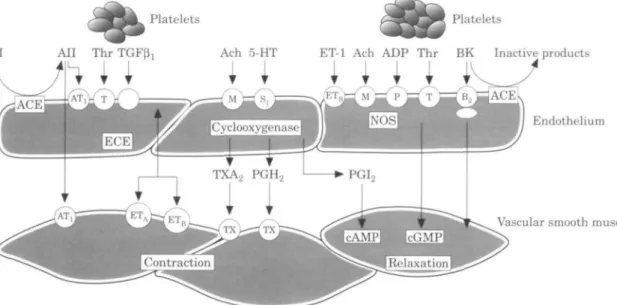
Role of the endothelium
The endothelium modulates coronary vasomotion by
the uptake, storage, and metabolism of numerous
substances'
10"
121, such as serotonin, norepinephrine,
bradykinin, acetylcholine etc. (Fig. 1).
Paracrine-released substances (autacoids) such as the
endothelium-derived relaxing factor (EDRF),
prosta-cyclin (PG1
2), platelet activating factor, and
thelin are synthesized and released from the
endo-thelium for vascular control and haemodynamic
homeostasis. Nitric oxide has been recognized to
be EDRF, although some of the pharmacological
actions of EDRF cannot be explained by nitric oxide
solely. Nitric oxide is continuously synthesized and
released, both without stimulation (basal nitric oxide
release) and with stimulation (stimulated release)
elicited by local and circulating agonists (such as
Platelets Platelets
AI All Thr TGF0, Ach o-HT ET-1 Ach ADP Thr BK Inactive products
i I i I i
/ETu
Endothelium
Vascular smooth muscle
Figure 1 Determinants of coronary vasomotor tone. In an intact endothelium, various stimulatory
compounds elicit NO-mediated dilation or vasoconstriction. ACE=angiotensin converting enzyme;
Ach=acetylcholine; ADP=adenosine diphosphate; Bk = bradykinin; cAMP/cGMP=cyclic adenosine/
guanidine monophosphate; ECE=endothelin converting enzyme; EDHF=endothelial derived hyperpolarizing
factor; ET-l=endothelin-l; 5HT=5-hydroxytryptamine (serotonin); L-arg=L-arginine; NO = nitric oxide;
PGH
2=prostaglandin H
2; PGl
2=prostacyclin 1
2; TGF/?=tranforming growth factor /?,; Thr=thrombin;
TXA
2=thrombozane A
2. Circles represent receptors (AT = angiotensinergic; B = bradykininergic;
M = muscarinergic; P = purinergic; T=thrombin receptor). (Reproduced with permission from Liischer TF and
Noll G. The endothelium in coronary vascular control. In: Heart disease — Update 3. Philadelphia: W. B.
Saunders Company, 1995: 2).
bradykinin, serotonin, norepinephrine) or, most
importantly by viscous drug-induced shear stress
from the perfusing blood (resulting in flow-dependent
dilation). The pulsatile stretching of the endothelium
adds to this mechanically stimulated nitric oxide
release'
13!. Furthermore, all vasoactive compounds
and drugs modulate endothelial nitric oxide synthesis
and release in either a receptor-dependent or in a
flow-dependent (=shear stress-mediated) manner'
14'.
Under physiological conditions, the continuous
dila-tor action of endothelial autacoids compensates for
the continuous constrictor effect of norepinephrine
released from the nerve endings'
15'. In patients with
impaired endothelial function flow-dependent, nitric
oxide-mediated dilator responses are diminished'
16'
and at the same time the myogenic constrictor
responses are enhanced resulting in an enhanced
autoregulatory response'
171.
Previous studies on coronary vasomotor tone
in humans used conventional coronary angiography
for measuring changes in response to various
phar-macological stimuli'
18"
20'. Recently, Schachinger and
Zeiher'
4' combined angiographic measurements and
intracoronary ultrasound for calculation of
vaso-motor tone. They used the ratio of a change in
circumference divided by the total vasomotor range
as a measure of vasomotor tone. However, pressure
as an important determinant of vascular tone was
neglected in this model'
41.
Vasomotion in coronary artery disease
Normal vessels
A number of pharmacological agents such as
sero-tonin, norepinephrine, vasopressin, papaverine, but
mainly acetylcholine have been used to study the
vasomotor response of coronary arteries'
21"
24'. In
clinical studies, intracoronary acetylcholine has been
shown to constrict coronary arteries in the presence
of atherosclerotic lesions, whereas it dilates normal
coronary arteries'
25'. Constriction during static but
dilation during dynamic exercise has been reported
previously'
3'. A heterogeneous response of
angio-graphically smooth vessels has been described.
Undetected atherosclerosis in angiographically
nor-mal vessels of patients with coronary artery disease
may account for insufficient vasodilator response due
to endothelial dysfunction. Progressive impairment of
endothelial function has been reported with different
stages of coronary atherosclerosis in patients with
angiographically smooth coronary arteries'
16'. Thus,
a functional disorder of the 'normal' coronary
175
150
125 -9 Q 100
-Rest 2 min Ex max. Ex Nitro si
Figure 2 Normal vessels (open squares) show
exercise-induced dilation which is further enhanced after sublingual
nitroglycerin (Nitro si). In contrast, stenoses show
exercise-induced vasoconstriction, which is reversed after
sublingual nitroglycerin. delta-Ex, percent change of
cross-sectional area.
arteries may account for the abnormal response of
various physiological and pharmacological stimuli in
patients with coronary artery disease.
Stenosis
The observation that stenotic arteries can change
its diameter depends on the fact that approximately
70% of all stenotic lesions are eccentric and have
a normal musculo-elastic wall segment within the
stenosis'
3'
26'
271. Intracoronary acetylcholine induces
vasoconstriction of stenotic coronary arteries. In
con-trast to the normal coronary arteries, constriction of
stenotic vessels has been reported during static'
281and
dynamic exercise'
3'
29'
301(Fig. 2). The mechanism of
coronary stenosis narrowing during exercise or after
intracoronary acetylcholine is not clear but might be
explained by either:
(1) endothelial dysfunction with insufficient
produc-tion of EDRF
(2) an increase in circulating catecholamines during
exercise'
31"
33'
(3) platelet aggregation with release of the
vaso-constricting compounds serotonin and
thrombo-xane A
2(4) a passive collapse of the free vessel wall when
coronary flow velocity increases during exercise
(Bernoulli mechanism)'
34' and/or
(5) a reduction of coronary blood flow increase
dur-ing exercise due to tachycardia with a decrease in
diastolic perfusion time'
301'
Cardiovascular risk factors and
coronary vasomotion
Hyper cholesterolaemia
There has been increasing interest in the literature
on the functional impact of hypercholesterolaemia
on coronary vascular function ' ' . Impairment of
endothelium-dependent vasodilation in
angiographi-cally normal coronary arteries has been reported in
the presence of hypercholesterolaemia'
16'
37'
381. This
has been attributed to a dysfunction of the
endo-thelium via an attenuation of EDRF release by
oxidised low-density lipoproteins (LDL)'
39'
401and/or
stimulation of endothelin-mRNA as well as the
release of endothelin'
4'
1. Lerman and co-workers'
42'
showed that hypercholesterolaemia elevates plasma
endothelin concentration and enhances coronary
artery tissue endothelin immunoreactivity, which is
thought to be responsible for abnormal endothelial
function. These changes play an important role in the
development of early atherosclerotic lesions, which
is characterized by functional alterations of the
endothelial cell before morphological changes are
detectable. Hypercholesterolaemia impairs
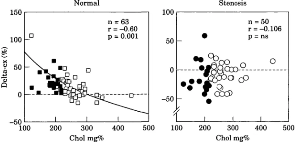
exercise-induced coronary vasodilation in angiographically
normal coronary arteries: as total serum cholesterol
increases exercise-induced dilation is diminished. In
humans, an inverse correlation between total or LDL
cholesterol and exercise-induced vasodilation in
angiographically normal coronary arteries has been
reported by Seiler et a/.'
43' (Fig. 3). An increase in
serum cholesterol above 200 mg. 100ml"
1reduces
exercise-induced vasodilation to 12%, whereas in the
presence of a normal serum cholesterol vasodilation
amounts to 20%. When serum cholesterol is increased
above 250 mg . 100 ml" ' coronary vasomotion is
abolished (+4% vasodilation during exercise, P=ns).
However, no influence of serum cholesterol was
observed in stenotic vessels. Similar observations
have been made in children with familiar
hyper-lipidaemia'
441and in porcine coronary arteries with
diet-induced hypercholesterolaemia'
45'.
The effect of hypercholesterolaemia on human
vascular function has been evaluated by Creager et al.
in the forearm of patients with normal and those
with elevated serum cholesterol. The forearm vessels
were chosen for investigation since they almost
never show atherosclerotic changes'
46'. There was
a diminished flow increase to methacholine in
150 100 50 0 D
• ^
Normal•
'-MD D 1 1 n r P = 63 = -0.60 = 0.001 D 1 Stenosis 100 50 -100 200 300 400 Choi mg% 500 50 -100 4-r
»
i n r P ocP
= 50 = -0.106 = ns O 1 0 200 300 400 Choi m g % 500Figure 3 An inverse correlation is found between total serum cholesterol and
exercise-induced vasomotion of angiographically normal but not of stenotic
cor-onary artery segments. Choi=total serum cholesterol; delta-Ex=percent change of
cross-sectional luminal area. Solid symbols=normal serum cholesterol; open
symbols=elevated serum cholesterol.
hypercholesterolaemic patients compared to normal
subjects. They concluded that hypercholesterolaemia
in the absence of atherosclerosis is associated with
abnormal function of the vascular endothelium.
Thus, abnormal coronary vasomotion in patients
with hypercholesterolaemia
143'
471may not necessarily
be associated with overt atherosclerosis. Thus,
two-different mechanisms of hypercholesterolaemia in the
pathophysiology of coronary artery disease must be
postulated: (1) a direct (toxic?) effect of oxidized
LDL-cholesterol on the endothelium (functional
dis-order) and (2) a chronic effect inducing structural
changes of the vessel wall.
Hypertension
High blood pressure has a direct effect on the arteries
which is characterized by structural changes of the
vessel wall such as media hypertrophy, increase in
endothelial cell volume, microvascular rarefication,
and augmentation of the extracellular matrix'
48"
511.
These changes may lead to impaired
endothelium-dependent relaxation'
52'
531. In hypertensive patients
with angiographically documented coronary artery
disease, Frielingsdorf et al. found a markedly blunted
vasodilatory response of non-stenotic vessels
com-pared with normotensive control subjects'
541(Fig. 4).
Furthermore, reduced coronary vasodilation of
nor-mal coronary arteries was found in response to
exer-cise, whereas endothelium-independent vasodilation
to nitroglycerin was maintained in hypertensive
patients. This suggests a preserved function of the
vascular smooth musculature but a primary defect of
the 'normal' epicardial coronary arteries in
hyperten-30 20 10 -10 -20 -30 Normal Stenosis p < 0.025 p < 0.025
-Figure 4 Luminal area change during exercise of normal
and stenotic coronary arteries in hypertensive patients
(•) and normotensive control subjects (D) with coronary
artery disease. Values are mean ± SEM.
sive patients with coronary artery disease. Alterations
of endothelial function are probably a consequence
rather than a cause of high blood pressure, and hence
the degree of endothelial dysfunction and its
mechan-ism change with increasing severity and duration of
hypertension. In animal models with antihypertensive
treatment, reductions of blood pressure are able to
reverse endothelial dysfunction'
55'
561, although the
exact mechanism is not fully understood.
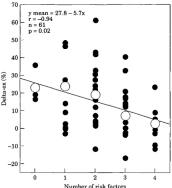
60 50 40 lta-e x (% ) to c o o o p 10 0 - 1 0 - 2 0 y r n P -( -mean = = -0.94 = 61 = 0.02 • 1 27.8-5.7x
a
•
t '
i j
i »t
i 0 1 2 3 4Number of risk factors
Figure 5 There is a significant, inverse correlation
be-tween the number of cardiovascular risk factors (horizon-tal axis) and mean exercise-induced percent change in coronary luminal area (delta-ex, %; vertical axis). O=mean delta-ex (%).
Other coronary risk factors
Although hypercholesterolaemia and hypertension
have been identified as major risk factors for
endothelial dysfunction, several additional coronary
risk factors are known to be responsible for
impaired coronary vasomotion. Long-term cigarette
smoking has been found to be associated with
impaired endothelium-dependent coronary
vaso-dilation regardless of the presence or absence of
coronary atherosclerotic lesions'
571. A significant
inverse correlation has been reported between the
mean exercise-induced vasomotor response and the
number of coronary risk factors'
43', such as
hyper-cholesterolaemia, hypertension, family history,
obes-ity and smoking, indicating that the accumulation of
different risk factors show an additive (adverse) effect
(Fig. 5)
cells'
581. An accelerated form of this process can be
induced by a denuding, deep endothelial injury as
it often occurs during percutaneous transluminal
coronary angioplasty, in patients undergoing
cor-onary bypass grafting, or by an immune injury such
as in patients undergoing heart transplantation'
591.
A direct relationship between left ventricular
muscle mass and coronary dimensions has been
reported in experimental animals and clinical
studies'
60"
661. As a regulatory mechanism for this
increase in vessel dimensions, blood flow velocity has
been postulated. An increase in mean flow velocity
has been associated with a change in shear stress,
which has been shown to be a mediator for the release
of EDRF, i.e. nitric oxide. In low and moderate
grades of left ventricular hypertrophy, an appropriate
increase in coronary artery size was reported, whereas
in severe left ventricular hypertrophy an
inappro-priate growth of the coronary arteries with a
reduc-tion in cross-secreduc-tional vessel area per 100 g muscle
mass was described in primary'
671as well as in
secondary'
67'
681left ventricular hypertrophy. In severe
hypertrophy the coronary arteries seem to be too
small for the increase in left ventricular muscle mass
as a result of inadequate growth of the coronary
arteries. Furthermore, impaired vasodilator capacity
of the epicardial coronary arteries in patients with
secondary left ventricular hypertrophy has been
reported previously'
691. This has been attributed to
an increase in resting flow. Although the size of the
coronary arteries is not a limiting factor for
myo-cardial perfusion, functional adaptation via the
release of EDRF seems to be inadequate in severe
hypertrophy. Insufficient growth of the coronary
arteries has been observed in severe left ventricular
hypertrophy'
701and, thus, may explain the occurrence
of myocardial ischaemia under high demand
situ-ations such as exercise. The exact control mechanism
of the coronary growth is not completely understood,
but may involve several factors, such as structural
(vascular remodelling) and functional (endothelial
dysfunction) changes'
671.
P. KAUFMANN
L. MANDINOV
O. M. HESS
Inselspital,
Bern, Switzerland
Vascular remodelling
The basis for the development of atherosclerosis is
damage to the arterial endothelium with
accumu-lation of lipids, adhesion of monocytes, and platelet
aggregation. Release of various growth factors leads
to the migration and proliferation of smooth muscle
References
[1] Mohrman DE, Feigl EO. Competition between sympathetic vasoconstriction and metabolic vasodilation in the canine coronary circulation. Circ Res 1978; 42: 79-86.
[2] Vatner SF, Hintze TH, Macho P. Regulation of large cor-onary arteries by beta-adrenergic mechanisms in the conscious dog. Circ Res 1982; 51: 56-66.
[3] Hess OM, Bortone A, Eid K et al. Coronary vasomotor tone during static and dynamic exercise. Eur Heart J 1989; 10 (Suppl F): 105-10.
[4] Schaechinger V, Zeiher AM. Quantitative assessment of coronary vasoreactivity in humans in vivo. Importance of baseline vasomotor tone in atherosclerosis. Circulation 1995; 92: 2087-94.
[5] Rocco MB, Barry J, Campbell S et al. Circadian variation of transient myocardial ischemia in patients with coronary artery disease. Circulation 1987; 75: 395-400.
[6] Linsell CR, Lightman SL, Mullen PE, Brown MJ, Causon RC. Circadian rhythms of epinephrine and norepinephrine in man. J Clin Endocrinol Metab 1985; 60: 1210-15.
[7] Tofler GH, Brezinski D, Schafer Al etal. Concurrent morning increase in platelet aggregability and the risk of myocardial infarction and sudden cardiac death. N Engl J Med 1987; 316: 1514-18.
[8] Quyyumi AA, Panza JA, Diodati JG, Lakatos E, Epstein SE. Circadian variation in ischemic threshold. A mechanism un-derlying the circadian variation in ischemic events. Circulation 1992; 86: 22-8.
[9] Maseri A, Chierchia A, Kaski JC. Mixed angina pectoris. Am J Cardiol 1985; 56: 3OE-33E.
[10] Furchgott RF. Role of endothelium in responses of vascular smooth muscle. Circ Res 1983; 53: 557-73.
[11] Furchgott RF, Zawadzki JV. The obligatory role of endo-thelial cells in the relaxation of arterial smooth muscle by acetylcholine. Nature 1980; 288: 373-6.
[12] Vanhoutte PM, Rubanyi GM, Miller VM, Houston DS. Modulation of vascular smooth muscle contraction by the endothelium. Annu Rev Physiol 1986; 48: 307-20.
[13] Pohl U, Holtz J, Busse R, Bassenge E. Crucial role of endothelium in the vasodilator response to increased flow in vivo. Hypertension 1986; 8: 37-44.
[14] Hecker M, Mulsch A, Bassenge E, Busse R. Vasoconstriction and increased flow: two principal mechanisms of shear stress-dependent endothelial autacoid release. Am J Physiol 1993; 265: 828-33.
[15] Bassenge E. Coronary vasomotor responses: role of endothe-lium and nitrovasodilators. Cardiovasc Drugs Ther 1994; 8: 601-10.
[16] Zeiher AM, Drexler H, Wollschlager H, Just H. Modulation of coronary vasomotor tone in humans. Progressive endo-thelial dysfunction with different stages of coronary athero-sclerosis. Circulation 1991; 83: 391-401.
[17] Pohl U, Lamontagne D, Bassenge E, Busse R. Attenuation of coronary autoregulation in the isolated rabbit heart by endothelium derived nitric oxide. Cardiovasc Res 1994; 28: 414-19.
[18] Doriot PA, Guggenheim N, Dorsaz PA, Rutishauser W. Morphometric versus densitometric assessment of coronary vasomotor tone — an overview. Eur Heart J 1989; 10 (Suppl F): 49-53.
[19] Lichtlen PR, RafBenbeul W, Jost S, Berger C. Coronary vasomotor tone in large epicardial coronary arteries with special emphasis on beta-adrenergic vasomotion, effects of beta-blockade. Basic Res Cardiol 1990; 1: 335-346.
[20] Hoshio A, Miyakoda H, Fukuki M, Yamasaki J, Kotake H, Mashiba H. Significance of coronary artery tone assessed by coronary responses to ergonovine and nitrate. Jpn Circ J 1991: 55: 33-40.
[21] Furchgott RF. The role of endothelium in the responses of vascular smooth muscle to drugs. Annu Rev Pharmacol Toxicol 1984; 24: 175-97.
[22] Cohen RA, Shepherd JT. Vanhoutte PM. Inhibitory role of the endothelium in the response of isolated coronary arteries to platelets. Science 1983; 221: 273-4.
[23] Cocks TM, Angus JA. Endothelium-dependent relaxation of coronary arteries by noradrenaline and serotonin. Nature
1983; 305: 627-30.
[24] Harrison DG. From isolated vessels to the catheterization laboratory. Studies of endothelial function in the coronary circulation of humans. Circulation 1989; 80: 703-6.
[25] Werns SW, Walton JA, Hsia HH, Nabel EG, Sanz ML, Pitt B. Evidence of endothelial dysfunction in angiographically normal coronary arteries of patients with coronary artery disease. Circulation 1989; 79: 287-91.
[26] Freudenberg H, Lichtlen PR. [The normal wall segment in coronary stenoses — a postmortal study (author's transl)]. Z Kardiol 1981; 70: 863-9.
[27] Saner HE, Gobel FL, Salomonowitz E, Erlien DA, Edwards JE. The disease-free wall in coronary atherosclerosis: its relation to degree of obstruction. J Am Coll Cardiol 1985; 6: 1096-9.
[28] Brown BG, Lee AB, Bolson EL, Dodge HT. Reflex constric-tion of significant coronary stenosis as a mechanism contrib-uting to ischemic left ventricular dysfunction during isometric exercise. Circulation 1984; 70: 18-24.
[29] Kaufmann P, Vassalli G, Utzinger U, Hess OM. Coronary vasomotion during dynamic exercise: influence of intravenous and intracoronary nicardipine. J Am Coll Cardiol 1995; 26: 624-31.
[30] Gage JE, Hess OM, Murakami T, Ritter M, Grimm J, Krayenbuehl HP. Vasoconstriction of stenotic coronary arteries during dynamic exercise in patients with classic angina pectoris: reversibility by nitroglycerin. Circulation 1986; 73: 865-76. [31] Bortone AS, Hess OM, Eberli FR et al. Abnormal coronary
vasomotion during exercise in patients with normal coronary arteries and reduced coronary flow reserve. Circulation 1989; 79: 516-27.
[32] Gaglione A, Hess OM, Corin WJ, Ritter M, Grimm J, Krayenbuehl HP. Is there coronary vasoconstriction after intracoronary beta-adrenergic blockade in patients with coronary artery disease. J Am Coll Cardiol 1987; 10: 299-310. [33] Gordon JB, Ganz P, Nabel EG et al. Atherosclerosis influ-ences the vasomotor response of epicardial coronary arteries to exercise. J Clin Invest 1989; 83: 1946-52.
[34] Brown BG, Bolson EL, Dodge HT. Dynamic mechanisms in human coronary stenosis. Circulation 1984; 70: 917-22. [35] Habib JB, Bossaller C, Wells S, Williams C, Morrisett JD,
Henry PD. Preservation of endothelium-dependent vascular relaxation in cholesterol-fed rabbit by treatment with the calcium blocker PN 200110. Circ Res 1986; 58: 305-9. [36] Verbeuren TJ, Jordaens FH, Zonnekeyn LL, Van HC, Coene
MC, Herman AG. Effect of hypercholesterolemia on vascular reactivity in the rabbit. I. Endothelium-dependent and endothelium-independent contractions and relaxations in isolated rabbit arteries of control and hypercholesterolemic rabbits. Circ Res 1986; 58: 552-64.
[37] Vita JA, Treasure CB, Nabel EG et al. Coronary vasomotor response to acetylcholine relates to risk factors for coronary artery disease. Circulation 1990; 81: 491-7.
[38] Harrison DG, Armstrong ML, Freiman PC, Heistad DD. Restoration of endothelium-dependent relaxation by dietary treatment of atherosclerosis. J Clin Invest 1987; 80: 1808-11. [39] Andrews HE, Bruckdorfer KR, Dunn RC, Jacobs M. Low-density lipoproteins inhibit endothelium-dependent relaxation in rabbit aorta. Nature 1987; 327: 237-9.
[40] Tanner FC, Noll G, Boulanger CM, Luscher TF. Oxidized low density lipoproteins inhibit relaxations of porcine cor-onary arteries. Role of scavenger receptor and endothelium-derived nitric oxide. Circulation 1991; 83: 2012-20.
[41] Boulanger CM, Tanner FC, Bea ML, Hahn AW, Werner A, Luscher TF. Oxidized low density lipoproteins induce mRNA expression and release of endothelin from human and porcine endothelium. Circ Res 1992; 70: 1191-7.
[42] Lerman A, Webster MWI, Chesebro JH et al. Circulating and tissue endothelin immunoreactivity in hypercholesterolemic pigs. Circulation 1993; 88: 2923-8.
[43] Seiler C, Hess OM, Buechi M, Suter TM, Krayenbuehl HP. Influence of serum cholesterol and other coronary risk factors on vasomotion of angiographically normal coronary arteries. Circulation 1993; 88: 2139^8.
[44] Sorensen KE, Celermajer DS, Georgakopoulos D, Hatcher G, Betteridge DJ, Deanfield JE. Impairment of endothelium-dependent dilation is an early event in children with familial
hypercholesterolemia and is related to the lipoprotein(a) level. J Clin Invest 1994; 93: 50-5.
[45] Shimokawa H, Vanhoutte PM. Impaired endothelium-dependent relaxation to aggregating platelets and related vasoactive substances in porcine coronary arteries in hyper-cholesterolemia and atherosclerosis. Circ Res 1989; 64:
900-14.
[46] Creager MA, Gallagher SJ, Girerd XJ, Coleman SM, Dzau VJ, Cooke JP. L-arginine improves endothelium-dependent vasodilation in hypercholesterolemic humans. J Clin Invest
1992; 90: 1248-53.
[47] Kaufmann P, Vassalli G, Seiler C, Hess OM. Abnormal coronary vasomotion in hypercholesterolemia: reversibility by calcium antagonists. Circulation 1994; (Abstr Suppl) 90: 606.
[48] Greene AS, Tonellato PJ, Lui J, Lombard JH, Cowley AWJ. Microvascular rerefication and tissue vascular resistance in hypertension. Am J Physiol 1989; 256: HI26-31.
[49] Haudenschild CC, Prescott MF, Chobanian AV. Effects of hypertension and its reversal on aortic intimal lesions of the rat. Hypertension 1980; 2: 33-44.
[50] Schwartz SM, Campbell GR, Campbell JH. Replication of smooth muscle cells in vascular disease. Circ Res 1986; 58: 427-44.
[51] Tomanek RJ, Palmer PJ, Pieffer GW, Schrieber K, Eastham CL, Marcus ML. Morphometry of canine coronary arteries, arterioles, and capillaries during hypertension and left ventricular hypertrophy. Circ Res 1986; 58: 38^6.
[52] Brush JE, Faxon DP, Salmon S, Jacobs AK, Ryan TJ. Abnormal endothelial-dependent coronary vasomotion in hy-pertensive patients. J Am Coll Cardiol 1992; 19: 809-15. [53] Treasure CB, Manoukian SV, Klein JL et al. Epicardial
coronary artery responses to acetylcholine are impaired in hypertensive patients. Circ Res 1992; 71: 776-81.
[54] Frielingsdorf J, Seiler C, Kaufmann P, Vassalli G, Suter T, Hess OM. Normalization of Abnormal Coronary Vasomotion by Calcium Antagonists in Patients With Hypertension. Circulation 1996; 93: 1380-7.
[55] Liischer TF, Vanhoutte PM, Raij L. Antihypertensive treat-ment normalizes decreased endothelium-dependent relax-ations in rats with salt- induced hypertension. Hypertension
1987, 6: 938-43.
[56] Dohi Y, Criscione L, Pfeiffer K, Liischer TR. Angiotensin blockade or calcium antagonists improve endothelial dysfunc-tion in hypertension: studies in perfused mesenteric resistance arteries. J Cardiovasc Pharmacol 1994; 24: 372-9.
[57] Zeiher AM, Schschinger V, Minners J. Long-term cigarette smoking impairs endothelium-dependent coronary arterial vasodilator function. Circulation 1995; 92: 1094-100. [58] Ross R. The pathogenesis of atherosclerosis — an update. N
Engl J Med 1986; 314: 488-500.
[59] Ip JH, Fuster V. Badminon L, Badminon J, Taubman MB, Chesebro JH. Syndromes of accelerated atherosclerosis: role of vascular injury and smooth muscle cell proliferation. J Am Coll Cardiol 1990; 15: 1667-87.
[60] O'Keefe JJ, Owen RM, Bove AA. Influence of left ventricular mass on coronary artery cross-sectional area. Am J Cardiol
1987; 59: 1395-7.
[61] Rembert JC, Kleinman LH, Fedor JM, Wechsler AS, Greenfield JJ. Myocardial blood flow distribution in concen-tric left venconcen-tricular hypertrophy. J Clin Invest 1978; 62: 379-86.
[62] Roberts CS, Roberts WC. Cross-sectional area of the proxi-mal portions of the three major epicardial coronary arteries in 98 necropsy patients with different coronary events. Relation-ship to heart weight, age and sex. Circulation 1980; 62: 953-9. [63] MacAlpin RN, Abbasi AS, Grollman JJ, Eber L. Human
coronary artery size during life. A cinearteriographic study. Radiology 1973; 108: 567-76.
[64] Wicker P, Tarazi RC. Coronary blood flow in left ventricular hypertrophy: a review of experimental data. Eur Heart J 1982; 3 (Suppl A): 111-18.
[65] Eberli FR, Ritter M, Schwitter J et al. Coronary reserve in patients with aortic valve disease before and after successful aortic valve replacement. Eur Heart J 1991; 12: 127-38. [66] Koiwa Y, Bahn RC, Ritman EL. Regional myocardial volume
perfused by the coronary artery branch: estimation in vivo. Circulation 1986; 74: 157-63.
[67] Kaufmann P, Vassalli G, Lupi-Wagner S, Jenni R, Hess OM. Coronary artery dimensions in primary and secondary left ventricular hypertrophy. J Am Coll Cardiol 1996; 28: 745-50 [68] Villari B, Hess OM, Meier C et al. Regression of coronary artery dimensions after successful aortic valve replacement. Circulation 1992; 85: 972-8.
[69] Vassalli G, Kaufmann P, Villari B et al. Reduced epicardial coronary vasodilator capacity in patients with left ventricular hypertrophy. Circulation 1995; 91: 2916-23.
[70] Villari B, Hess OM, Moccetti D, Vassalli G, Krayenbuehl HP. Effect of progression of left ventricular hypertrophy on cor-onary artery dimensions in aortic valve disease. J Am Coll Cardiol 1992; 20: 1073-9.