Hypoxia-induced vascular endothelial growth
factor expression causes vascular leakage in the
brain
Heike J. Schoch,
1Silvia Fischer
2and Hugo H. Marti
1,31Department of Molecular Cell Biology, Max Planck
Institute for Physiological and Clinical Research,
2Department of Anesthesiology and Intensive Care,
Kerckhoff Clinic, Bad Nauheim, Germany and3Institute of
Physiology, University of ZuÈrich, ZuÈrich, Switzerland
Correspondence to: PD Dr med Hugo H. Marti, University of ZuÈrich, Institute of Physiology, Winterthurerstrasse 190, CH-8057 ZuÈrich, Switzerland
E-mail: [email protected]
Summary
Formation of cerebral oedema caused by vascular leak-age is a major problem in various injuries of the CNS, such as stroke, head injury and high-altitude illness. A common feature of all these disorders is the fact that they are associated with tissue hypoxia. Hypoxia has therefore been suggested to be an important pathogenic factor for the induction of vascular leakage in the brain. Vascular endothelial growth factor (VEGF) is known as the major inducer of angiogenesis. Originally, however, it was described as a vascular permeability factor. As VEGF gene expression was shown to be up-regulated by hypoxia, increased VEGF expression may link hypoxia and vascular leakage in the CNS in vivo. To delineate the role of VEGF in vascular leakage in the brain, we studied the effect of hypoxia on VEGF
expression and vascular permeability in the brains of mice in vivo. Hypoxic exposure led to a signi®cant increase in the levels of VEGF mRNA and protein in mouse brain that correlated with the severity of the hypoxic stimulus. Measurement of vascular permeabil-ity using the ¯uorescent marker sodium ¯uorescein revealed a two-fold increase in ¯uorescence intensity in hypoxic brains, indicative of signi®cant vascular leak-age. Inhibition of VEGF activity by a neutralizing anti-body completely blocked the hypoxia-induced increase in vascular permeability. In conclusion, our data show that VEGF is responsible for hypoxia-induced augmen-tation in vascular leakage following tissue hypoxia. Our ®ndings might provide the basis for new therapeutic concepts for the treatment of cerebral oedema.
Keywords: vascular permeability; oedema; VEGF; HACE; ischaemia
Abbreviations: BBB = blood±brain barrier; HACE = high-altitude cerebral oedema; HIF = hypoxia-inducible factor; r.f.u. = relative ¯uorescence unit; VEGF = vascular endothelial growth factor
Introduction
Oedema formation is a major life-threatening complication of various injuries of the CNS, such as head injury (Murakami et al., 1999; Pilitsis and Rengachary, 2001), tumour growth (Papadopoulos et al., 2001) and cerebral ischaemia (Lipton, 1999; Rosenberg, 1999). It also occurs during high-altitude illness (Hackett and Roach, 2001). Acute mountain sickness and high-altitude cerebral oedema (HACE) refer to cerebral abnormalities of high-altitude illness and are syndromes that occur in unacclimatized persons on ascent to high altitude (Hackett, 1999). Acute mountain sickness is characterized by the presence of headache, nausea, insomnia, dizziness and fatigue (Hackett and Roach, 2001) and is probably due to the formation of mild cerebral oedema (Hackett, 1999). HACE, clinically de®ned as the onset of ataxia and altered
con-sciousness, has been considered to be the end-stage of acute mountain sickness, eventually leading to death caused by brain herniation (Hackett and Roach, 2001). While the clinical aspects of HACE are well established, its pathophy-siology remains elusive. Recent evidence suggests that HACE is associated with osmotic cell swelling, vasogenic oedema and biochemical alteration of the blood±brain barrier (BBB) (Severinghaus, 1995; Hackett, 1999; Hackett and Roach, 2001). Vasogenic brain oedema is de®ned as the translocation of proteins and ¯uid from the vascular space across the BBB (Hackett, 1999). The underlying molecular and pathogenic mechanisms are poorly understood. Several years ago, Severinghaus and Xu hypothesized that the vascular endothelial growth factor (VEGF) might be respon-ã Guarantors of Brain 2002
sible for the vascular leakage and oedema formation in the brain that occurs during high-altitude exposure (Severinghaus, 1995; Xu and Severinghaus, 1998). VEGF is the most prominent member of an increasing family of angiogenic growth factors and plays a key role in new vessel growth. VEGF is an endothelial mitogen and exists as several isoforms derived from a single gene by alternative splicing (Robinson and Singer, 2001). It is ubiquitously expressed, binds to two endothelial tyrosine receptors, VEGF receptor 1 (VEGFR-1, Flt-1) and VEGFR-2 (Flk-1/KDR), and is also a ligand for the semaphorin receptors neuropilin 1 and neuropilin 2 (Robinson and Singer, 2001).
There is good evidence to support the hypothesis of Severinghaus and Xu. First, all the above-mentioned brain pathologies involving oedema formation are associated with tissue hypoxia. For example, hypoxic regions have been identi®ed around brain tumours (Shweiki et al., 1992; Evans et al., 1997; Damert et al., 1997; Talks et al., 2000). Also, during cerebral ischaemia, a region around the core of the infarctÐthe so-called penumbraÐsuffers from hypoxia (Evans et al., 1997; Read et al., 1998; Marti et al., 2000). Finally, at high altitude, reduced ambient pO2levels result in
systemic tissue hypoxia that also affects the brain. Thus, tissue hypoxia could play an important role in the pathogen-esis of vascular leakage leading to brain oedema formation, e.g. by activating speci®c genes. Secondly, VEGF is among the best-known of the genes that are induced by hypoxia (Shweiki et al., 1992; Marti and Risau, 1998). VEGF gene expression is known to be activated under hypoxic conditions at the level of transcription, increased stability of the mRNA and preferential translation (Ikeda et al., 1995; Levy et al., 1995; Stein et al., 1998). Transcriptional activation is achieved by the transcription factors hypoxia-inducible factor 1 (HIF-1) and HIF-2 (Forsythe et al., 1996; Ema et al., 1997). HIF-1 binds to a hypoxia response element (HRE) in the 5¢-¯anking region of the VEGF gene, thereby enhancing transcription of the gene (Forsythe et al., 1996). Increased VEGF levels in hypoxic tissues are thought to induce an angiogenic reaction that enables increased delivery of nutrients and oxygen to the hypoxic cells through newly formed vessels (Shweiki et al., 1992; Marti and Risau, 1999). Tissue hypoxia and angiogenesis are thus interdependent, and have indeed been observed to coincide during cerebral ischaemia (Marti et al., 2000), tumour growth (Damert et al., 1997) and chronic hypoxic situations (Harik et al., 1995; LaManna and Harik, 1997). Thirdly, VEGF is expressed in the CNS in astrocytes and, after hypoxic exposure, in neurones (Marti and Risau, 1998; Ogunshola et al., 2000). Finally, VEGF was originally isolated as a factor leading to increased vascular permeability in tumours and was named vascular permeability factor (Senger et al., 1983). Thus, VEGF might be a signi®cant contributor to hypoxia-induced vascular leakage and oedema formation in the brain (Xu and Severinghaus, 1998). If so, inhibition of VEGF action could prevent brain oedema formation and thus be useful as a therapeutic strategy to treat patients with HACE
and other diseases associated with oedema formation. We therefore analysed vascular leakage and expression of VEGF during systemic normobaric hypoxia in the brains of mice in vivo and inhibited VEGF action by systemic application of a neutralizing anti-VEGF antibody.
Material and methods
Animals
All experiments were performed according to protocols approved by the RegierungspraÈsidium Darmstadt. Adult C57/ BL6 mice were exposed to normobaric hypoxia at 12±6% oxygen (corresponding to an altitude of 4000±9500 m) for 24 h or were kept at room air pressure. Hypoxia was achieved by substituting nitrogen for oxygen using a Digamix 5SA 18/ 3a pump (H. WoÈsthoff, Bochum, Germany) after gradual adaptation over a period of 1 h (Marti and Risau, 1998). Mice had free access to food and water. Following hypoxic exposure, the animals were killed by decapitation and the dissected brains were frozen in liquid nitrogen or embedded for cryosectioning.
Determination of brain water content and
vascular permeability
To quantify brain water content in brains from normoxic and hypoxic animals, the wet weights of the brains were determined immediately after removal. The samples were dried at 110°C for 24 h and reweighed to give the dry weight. The difference between the wet and dry weights was taken as the in vivo water content.
To quantify vascular permeability of brain vessels, 200 ml of sodium ¯uorescein (Sigma, Taufkirchen, Germany) at a concentration of 6 mg/ml in PBS (phosphate-buffered saline) was injected through the tail vein after 24 h of hypoxic or normoxic exposure. Sodium ¯uorescein (MW 376.3) is a ¯uorescent tracer that does not cross an intact BBB (Williams et al., 1984). Thirty minutes later, mice were anaesthetized and then perfused with PBS (20 ml) through the left heart ventricle to remove the ¯uorescent tracer from the vascular bed. Subsequently, both hemispheres of the brain were removed and frozen in liquid nitrogen. To assess ¯uores-cence, brain hemispheres were homogenized in 0.5 M borate buffer (pH 10) and centrifuged (3000 r.p.m.) for 15 min at 4°C, and the supernatant was added to 1.2 ml of ethanol to precipitate proteins. Probes were again centrifuged (13 000 r.p.m.) for 20 min at 4°C and the ¯uorescence of the supernatant was measured at 485 nm at an excitation wavelength of 330 nm using a Lambda Fluoro 320 Fluoroscope (MWG Biotech, Ebersberg, Germany) (Baba et al., 1988). Results are presented as relative ¯uorescence units (r.f.u.) per mg of brain tissue. In a second set of experiments, a neutralizing goat anti-mouse VEGF antibody or a corresponding control goat IgG antibody (R&D Systems, Minneapolis, MN, USA) was injected intraperitoneally at
100 mg (dissolved in 100 ml PBS) immediately prior to the 24 h hypoxic or normoxic exposure.
RNA extraction, Northern blot analysis and in
situ hybridization
Total RNA was extracted using peqGOLD RNA pure (Peqlab Biotechnologie, Weingarten, Germany) essentially as de-scribed (Wenger et al., 1996). Northern blot analysis was performed as described (Marti et al., 1996; Wenger et al., 1996). Hybridization patterns with RNA for the ribosomal protein L28, which remains unaffected by hypoxia (Wenger et al., 1995), were used to correct for loading differences of total RNA on each lane. To quantify VEGF levels, the relative intensities for the hybridization signal (VEGF/L28) were calculated and are presented as percentages of the normoxic control. VEGF in situ hybridization was performed exactly as described (Breier et al., 1992; Marti and Risau, 1998).
Mouse VEGF immunoassay
Cortical brain tissue lysates from normoxic and hypoxic animals were prepared as described (Marti et al., 2000). Mouse VEGF was quanti®ed using a commercially available immunoassay kit (Quantikine M; R&D Systems) as described (Marti et al., 2000).
Presentation of data and statistics
Steady-state mRNA levels for VEGF and L28 were recorded and quanti®ed using a Bio-Imaging Analyzer (BAS-2500; Fuji, Raytest GmbH, Straubenhardt, Germany). VEGF signals were then normalized to the signal obtained with the L28 cDNA probe to correct for loading and blotting differences. For hypoxia-inducible expression of VEGF, mRNA and
protein levels were expressed as percentages of the corres-ponding normoxic control. For the statistical analysis of the data, means and standard deviations of n experiments were determined and Student's t-test was performed.
Results
Normobaric hypoxia induces vascular leakage
in the brain
To mimic a stay at high altitude, mice were exposed to various fractions of inspiratory oxygen in a normobaric hypoxic chamber for a period of 24 h. To explore whether exposure to severe hypoxia led to the occurrence of HACE, we ®rst examined changes in brain water content as a sign of oedema formation in this organ. Brain water content increased slightly, but not signi®cantly, from 77.99 6 0.31% (n = 11) in normoxic controls to 78.92 6 1.96 (n = 12) after hypoxic exposure. The small increase observed might have been due to considerable water loss resulting from severe hyperventilation, as animals that were exposed for 24 h to hypoxia lost more than 10% of their body weight, whereas the normoxic control mice kept their weight constant.
To ®nd out directly whether vascular leakage had occurred as a result of hypoxic exposure, we quanti®ed vascular permeability by a direct method. Sodium ¯uorescein was injected through the tail vein and allowed to circulate. Sodium ¯uorescein is a ¯uorescence tracer that can be measured easily in homogenates of brain hemispheres to exactly quantify dye extravasation and thus vascular permeability.
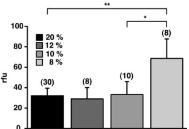
Relative ¯uorescence intensity in the brain of mice exposed to 8% oxygen increased more than two-fold (P < 0.0001) compared with normoxic controls, indicative of increased vascular leakage of the dye (Fig. 1). Fluorescence in control brain hemispheres was 31.38 6 4.59 r.f.u./mg brain tissue (n = 30) and increased to 68.70 6 18.97 r.f.u./mg (n = 8) at 8% oxygen. To address the question whether there is a threshold level above which vascular permeability is unaffected, we exposed mice in addition to 12 and 10% oxygen. Under both conditions, dye extravasation was not signi®cantly different from that in normoxic control animals (12% oxygen, 29.04 6 11.12 r.f.u./mg, n = 8; 10% oxygen, 33.38 6 12.58 r.f.u./mg, n = 10). Thus, vascular leakage of ¯uorescent dye during severe hypoxic exposure suggests that during exposure to 8% oxygen a signi®cant increase in vascular permeability of cerebral microvessels occurred. Furthermore, there was a sharp and signi®cant increase in vascular permeability when animals were exposed to 8% oxygen compared with exposure to 10% (P < 0.001)
Increased VEGF gene expression in the brain is
dependent on severity of hypoxia
An important question concerns the mechanisms that may link tissue hypoxia and increased vascular permeability. A promising candidate is the angiogenic growth factor VEGF. Fig. 1 Two-fold increase in vascular permeability after exposure
to 8% oxygen. Sodium ¯uorescein injected intravenously in controls or hypoxic mice was quanti®ed following homogenization of brain hemispheres. Results are expressed as relative
¯uorescence units (r.f.u.). Values are mean and standard deviation. **P < 0.0001; *P < 0.001; n = 8±30 as indicated.
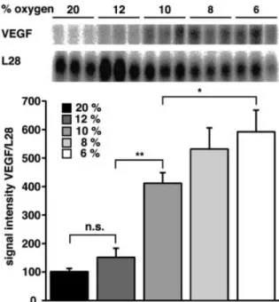
Expression of VEGF mRNA and protein levels in normoxic and hypoxic brains was therefore analysed using Northern blotting, in situ hybridization and ELISA (enzyme-linked immunosorbent assay) techniques. VEGF mRNA was detect-able by Northern blot analysis in all brain samples tested (Fig. 2, upper panel). In normoxic brains, though, mRNA levels were very low, but increased slightly after exposure to 12% oxygen. A signi®cant increase was seen at 10% oxygen, and mRNA levels rose further at 8 and 6% oxygen (Fig. 2, lower panel). VEGF mRNA levels did not differ signi®cantly between 6 and 8% oxygen.
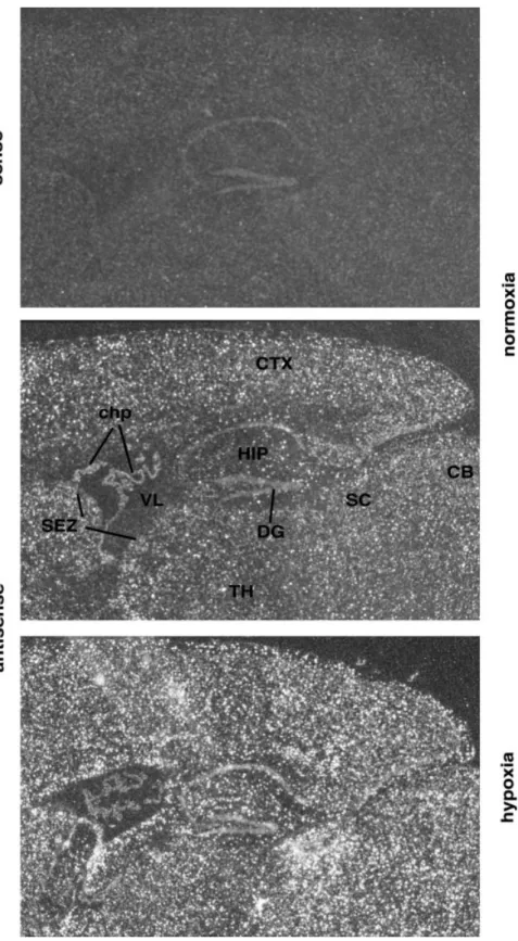
To analyse the regional distribution of VEGF expression under normoxic and hypoxic conditions, in situ hybridization experiments were performed. In normoxic controls, VEGF mRNA expression was very low and was homogeneous throughout the brain (Fig. 3). Stronger expression was detected only in the epithelial cells of the choroid plexus, as described previously (Marti and Risau, 1998). After hypoxic exposure, levels of VEGF mRNA were clearly increased all over the cortex, in the hippocampus, cerebellum and the subventricular zone, whereas expression was dimin-ished in the choroid plexus. Thus, with the exception of epithelial cells of the choroid plexus, exposure to hypoxic hypoxia led to general activation of VEGF gene expression in the whole brain. Taken together, our results demonstrate a
strong, continuous, oxygen-regulated increase in VEGF gene expression throughout the brain.
To con®rm these mRNA data at the protein level, we isolated total cortical proteins and quanti®ed the amount of VEGF protein present by ELISA. The basal normoxic level of VEGF was 2.4 6 0.9 ng/g total protein, which increased to 9.6 6 1.8 and 11.6 6 5.0 ng/g after exposure to 12 and 10% oxygen, respectively. A signi®-cant further increase was found at 8% (19.5 6 2.3 ng/g total protein) and 6% (21.9 6 3.6 ng/g total protein) oxygen (P < 0.05) (Fig. 4). Again, the VEGF level did not differ signi®cantly between 6 and 8% oxygen. Thus, both RNA and protein data con®rm a continuous increase in VEGF production in the brain when systemic oxygen availability is decreased progressively. These results implicate VEGF in the generation of vascular leakage and brain oedema during episodes of severe tissue hypoxia.
Blocking VEGF action prevents vascular
permeability
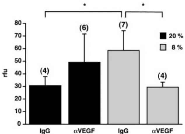
To further test the hypothesis that VEGF confers hypoxia-related leakage of cerebral blood vessels and may thus be responsible for the occurrence of HACE, neutralizing anti-bodies directed against mouse VEGF were applied immedi-ately before hypoxic exposure by injecting 100 mg of a neutralizing goat anti-mouse VEGF antibody intraperitone-ally. An isotypic goat IgG antibody was used as a speci®c negative control. As expected from the ®rst round of experiments, vascular permeability increased after hypoxic exposure to 8% oxygen, i.e. the r.f.u. level rose from 30.69 6 7.16 (n = 4) in normoxic controls to 58.60 6 15.63 per mg brain tissue (n = 7) in hypoxic animals (Fig. 5). In sharp contrast, the hypoxic increase in vascular permeability in mice treated with VEGF antibody was completely prevented (29.59 6 3.92 r.f.u./mg, n = 4), demonstrating the causative relationship between hypoxic VEGF induction and hypoxia-induced vascular leakage in the brain.
Discussion
This study demonstrates that inhibition of VEGF action is able to prevent hypoxia-induced vascular leakage in the brain. Our data support the hypothesis that exposure to systemic hypoxia activates VEGF gene transcription in the CNS, leading to increased VEGF protein levels, which in turn increase vascular permeability in brain microvessels. Furthermore, our results demonstrate the existence of a threshold level at ~10% oxygen at which VEGF expression starts to increase but no change in vascular permeability is yet observed. Our ®ndings provide the basis for the treatment of oedema formation in the CNS that is associated with brain injury, stroke and HACE.
Fig. 2 Increased expression of VEGF mRNA in mouse brain after hypoxic stimulation. Total RNA was extracted from brains of normal mice and mice exposed to 6±12% oxygen for 24 h. (Upper panel) Northern blots of total RNA sequentially hybridized with a
32P-labelled probe for murine VEGF and the ribosomal protein
L28. (Lower panel) Mean and standard deviation (n = 3) of VEGF mRNA pixel densities as quanti®ed with a Phosphoimager and corrected for L28. Normoxic control was set to 100%. **P < 0.001; *P < 0.05; n.s., not signi®cant.
Fig. 3 VEGF mRNA expression detected by in situ hybridization on sagittal sections of mouse brain. Following exposure to 8% oxygen (lower panel), VEGF mRNA is upregulated homogeneously all over the brain compared with normoxic controls (middle panel). (Upper panel) Hybridization with a VEGF sense probe. CB = cerebellum; chp = choroid plexus; CTX = cerebral cortex; DG = dentate gyrus; HIP = hippocampal region; SC = superior colliculus; SEZ = subependymal zone; TH = thalamus; VL = lateral ventricle.
Inverse correlation between VEGF expression
and inspired oxygen concentration
We noted that VEGF expression started to increase at ~10% oxygen, but only after exposure to 8% oxygen were signi®cant changes in vascular permeability in the brain microvessels observed, suggestive of the existence of de®ned threshold levels for VEGF expression and permeability changes. Activation of VEGF expression during hypoxic exposure has been investigated in a number of studies, although these investigated mostly the effects of exposure time, not variation in oxygen concentration (Patt et al., 1998; Xu and Severinghaus, 1998; Kuo et al., 1999). Increased VEGF gene expression is thus found consistently when inspired oxygen levels fall below 10%, which corresponds to the amount of oxygen present at ~5300 m altitude. In our mouse model, vascular leakage was detected only after 24 h of exposure to 8% oxygen, corresponding to 7100 m altitude. The occurrence of HACE in humans is increasingly common and severe at altitudes higher than 4000 m, while the incidence is clearly reduced at 3000 m (Severinghaus. 1995). Thus, also in humans there is a clear relation between the occurrence of HACE symptoms and the degree of hypoxia. The difference in the degree of hypoxia required to elicit vascular leakage and oedema formation in humans and mice might be explained by the fact that in humans it is mostly clinical symptoms that are considered (Hackett and Roach, 2001), whereas in our mouse model we sought direct proof of brain vascular leakage. It might also be due to species differences, since it has been shown that small animals have a higher capillary density in the muscles compared with larger species (Schmidt-Nielsen and Pennycuik, 1961). This may also hold true for the brain, resulting in a shorter diffusion distance from the capillary to the metabolizing cell, thus making mice more resistant to hypoxia than man.
VEGF gene expression is transcriptionally regulated by HIF-1 (Forsythe et al., 1996). We have previously demon-strated in vitro that HIF-1 activation is inversely related to oxygen concentration, with a half-maximal activation of HIF
at 1.5±2% oxygen (Jiang et al., 1996). Although we did not measure pO2within the brain parenchyma of our mice, data
from the literature suggest that normal tissue pO2levels in the
brain are in the range of 20 mm Hg (2.5% oxygen) (Whalen et al., 1970). During hypoxic exposure, levels certainly drop further, to a range where even small changes in pO2lead to
strong activation of HIF-1. Indeed, it was shown very recently that HIF-1 protein accumulated gradually in the brain within 2±6 h when mice were exposed to decreasing amounts of oxygen ranging from 21 to 6% (Stroka et al., 2001). Thus, activation of HIF-1 precedes upregulation of VEGF expres-sion, which will then lead to increased permeability, given a strong enough hypoxic stimulus.
A clinically important question which arises from our ®ndings that different hypoxic threshold levels exist for the activation of VEGF gene expression and the induction of vascular leakage is whether VEGF levels can be used as a marker to identify patients at risk of developing HACE. As it is not feasible to analyse brain VEGF levels in humans, one might look at plasma VEGF levels. In the plasma of our hypoxic mice (n = 3), we found that VEGF levels were elevated less than two-fold and non-signi®cantly at both 12 and 8% oxygen (3.9 6 0.63 and 3.7 6 0.57 ng VEGF/ml, respectively) compared with normoxic controls (2.4 6 0.27 ng VEGF/ml). This result is in good agreement with a study in humans demonstrating no increase in plasma VEGF at 4200 m and no correlation of VEGF with oxygen saturation or symptoms of acute mountain sickness (Maloney et al., 2000). Furthermore, in a recent study investigating plasma VEGF levels in humans at high altitude (4559 m), no correlation between VEGF and the occurrence of acute mountain sickness symptoms was found either, although VEGF in the plasma was signi®cantly increased after hypoxic exposure (Walter et al., 2001). Thus, as VEGF is a classical paracrine factor, released by one cell and acting on an adjacent Fig. 4 Increased VEGF protein levels in the hypoxic brain. The
amount of VEGF protein present in the brain was measured using an ELISA (enzyme-linked immunosorbent assay) speci®c for murine VEGF. Values are mean and standard deviation (n = 3). **P < 0.01; *P < 0.05. Note the inverse correlation between VEGF protein level and oxygen concentration.
Fig. 5 Blocking VEGF action prevents hypoxia-induced vascular leakage in the brain. A neutralizing anti-VEGF antibody (aVEGF) or an isotypic goat antibody (IgG) was injected intraperitoneally before exposure to 20% (black columns) or 8% oxygen (grey columns) for 24 h. Vascular permeability was assessed as described in Figure 2. Values are mean and standard deviation. *P < 0.01; n = 4±7 as indicated.
endothelial cell, systemic VEGF levels do not appear to be useful for the direct identi®cation of patients at risk of HACE formation.
Anti-VEGF treatment blocks vascular leakage
VEGF has been considered as a causative agent in HACE formation, although no direct data were presented (Xu and Severinghaus, 1998). Our results now show that there is a direct causative relation between inhibition of VEGF action and the prevention of hypoxia-induced vascular leakage. Further indirect evidence is coming from a number of studies demonstrating a correlation between increased VEGF expres-sion and oedema formation in brain tumours (Machein and Plate, 2000) and brain injury (Nag et al., 1997; Papavassiliou et al., 1997) as well as in cerebral ischaemia (Zhang et al., 2000). Inhibition of VEGF action has been considered to be useful in a variety of brain pathologies. An anti-VEGF strategy is currently being used in various clinical trials to treat tumour patients with the aim of inhibiting tumour-induced angiogenesis, thereby depriving the tumour cells of nutrients and oxygen (Carmeliet and Jain, 2000). Anti-VEGF treatment may have, in addition, a bene®cial effect in these patients by reducing tumour-induced oedema formation. Furthermore, during stroke, oedema formation occurring around infarcted brain tissue was signi®cantly reduced by the fusion protein mFlt(1-3)±IgG, which sequesters VEGF (van Bruggen et al., 1999). Recent data demonstrated that Src family kinases are involved in the VEGF-mediated augmen-tation of vascular permeability (Eliceiri et al., 1999). Blockade of Src activity in mice provided cerebral protection following stroke due to reduced brain oedema formation (Paul et al., 2001). Our results are in line with these reports and now suggest that blocking VEGF action may be a more speci®c therapeutic approach to the treatment of HACE. This notion is further sustained by the recent ®nding that dexamethasone, which is widely used at high altitude as well as in the clinic to reduce cerebral oedema, is capable of inhibiting hypoxia-induced VEGF gene expression (Heiss et al., 1996; Fischer et al., 2001).
Putative mechanisms involved in
VEGF-mediated vascular permeability
It has been shown both in vitro (Fischer et al., 1999) and in vivo (Mayhan, 1999) that the VEGF-mediated increase in permeability of the BBB involves nitric oxide- and cGMP (cyclic guanosine monophosphate)-dependent pathways. It remains unclear, however, where these molecules exert their action. It has been hypothesized that VEGF may induce the opening of interendothelial tight junctions or change the endothelial cell phenotype, or even alter pinocytic transport through the endothelial cell (Mayhan, 2001). On the one hand, topical administration or intradermal injection of VEGF can transform continuous endothelium into fenestrated
endothelium and thereby increase vascular permeability (Roberts and Palade, 1995). On the other hand, BBB permeability may be controlled by phosphorylation of tight-junction proteins (Papadopoulos et al., 2001). For example, occludin and zonula occludens 1 (ZO-1), which are both important constituents of the tight junction, are rapidly phosphorylated by VEGF treatment (Antonetti et al., 1999). Furthermore, VEGF reduced occludin expression and dis-rupted both ZO-1 and occludin organization at the tight junction (Wang et al., 2001). In an in vitro model of the BBB, we have recently shown that hypoxia-induced hyperperme-ability involves changes in the expression of ZO-1 and that these changes are mediated by VEGF (Fischer et al., 2002). Taken together, these results suggest that alteration of tight-junction protein expression and phosphorylation may regulate vascular permeability.
Future research, including the generation and analysis of mice with inducible overexpression of VEGF under the control of brain-speci®c promoters, will help us to understand fully the mechanisms whereby VEGF induces permeability and to unravel the different effects of VEGF in the CNS involving angiogenesis, permeability induction and neuro-protection. In summary, we have shown that hypoxia-induced VEGF expression in the brain is responsible for vascular leakage. These ®ndings provide the background for the development of therapeutic strategies to treat HACE based on the inhibition of VEGF action.
Acknowledgements
This work was supported by a grant from the Hartmann MuÈller-Stiftung, the Stiftung fuÈr wissenschaftliche Forschung an der UniversitaÈt ZuÈrich and the Kerckhoff Klinik Bad Nauheim. We wish to thank M. Euler for technical assistance and Lara Ogunshola and Roland H. Wenger for critically reading the manuscript.
References
Antonetti DA, Barber AJ, Hollinger LA, Wolpert EB, Gardner TW. Vascular endothelial growth factor induces rapid phosphorylation of tight junction proteins occludin and zonula occludens 1. A potential mechanism for vascular permeability in diabetic retinopathy and tumors. J Biol Chem 1999; 274: 23463±7.
Baba M, Oishi R, Saeki K. Enhancement of blood±brain barrier permeability to sodium ¯uorescein by stimulation of mu opioid receptors in mice. Naunyn Schmiedebergs Arch Pharmacol 1988; 337: 423±8.
Breier G, Albrecht U, Sterrer S, Risau W. Expression of vascular endothelial growth factor during embryonic angiogenesis and endothelial cell differentiation. Development 1992; 114: 521±32. Carmeliet P, Jain RK. Angiogenesis in cancer and other diseases. [Review]. Nature 2000; 407: 249±57.
Damert A, Machein M, Breier G, Fujita MQ, Hanahan D, Risau W, et al. Up-regulation of vascular endothelial growth factor expression
in a rat glioma is conferred by two distinct hypoxia-driven mechanisms. Cancer Res 1997; 57: 3860±4.
Eliceiri BP, Paul R, Schwartzberg PL, Hood JD, Leng J, Cheresh DA. Selective requirement for Src kinases during VEGF-induced angiogenesis and vascular permeability. Mol Cell 1999; 4: 915±24. Ema M, Taya S, Yokotani N, Sogawa K, Matsuda Y, Fujii-Kuriyama Y. A novel bHLH-PAS factor with close sequence similarity to hypoxia-inducible factor 1a regulates the VEGF expression and is potentially involved in lung and vascular development. Proc Natl Acad Sci USA 1997; 94: 4273±8. Evans SM, Bergeron M, Ferriero DM, Sharp FR, Hermeking H, Kitsis RN, et al. Imaging hypoxia in diseased tissues. Adv Exp Med Biol 1997; 428: 595±603.
Fischer S, Clauss M, Wiesnet M, Renz D, Schaper W, Karliczek GF. Hypoxia induces permeability in brain microvessel endothelial cells via VEGF and NO. Am J Physiol 1999; 276: C812±20. Fischer S, Renz D, Schaper W, Karliczek GF. In vitro effects of dexamethasone on hypoxia-induced hyperpermeability and expression of vascular endothelial growth factor. Eur J Pharmacol 2001; 411: 231±43.
Fischer S, Wobben M, Marti HH, Renz D, Schaper W. Hypoxia-induced hyperpermeability in brain microvessel endothelial cells involves VEGF-mediated changes in the expression of zonula occludens 1. Microvasc Res 2002; 63: 70±80.
Forsythe JA, Jiang B-H, Iyer NV, Agani F, Leung SW, Koos RD, et al. Activation of vascular endothelial growth factor gene transcription by hypoxia-inducible factor 1. Mol Cell Biol 1996; 16: 4604±13.
Hackett PH. High altitude cerebral edema and acute mountain sickness: a pathophysiology update. [Review]. Adv Exp Med Biol 1999; 474: 23±45.
Hackett PH, Roach RC. High altitude illness. [Review]. New Engl J Med 2001; 345: 107±14.
Harik SI, Hritz MA, LaManna JC. Hypoxia-induced brain angiogenesis in the adult rat. J Physiol 1995; 485: 525±30. Heiss JD, Papavassiliou E, Merrill MJ, Nieman L, Knightly JJ, Walbridge S, et al. Mechanism of dexamethasone suppression of brain tumor-associated vascular permeability in rats: involvement of the glucocorticoid receptor and vascular permeability factor. J Clin Invest 1996; 98: 1400±8.
Ikeda E, Achen MG, Breier G, Risau W. Hypoxia-induced transcriptional activation and increased mRNA stability of vascular endothelial growth factor in C6 glioma cells. J Biol Chem 1995; 270: 19761±6.
Jiang BH, Semenza GL, Bauer C, Marti HH. Hypoxia-inducible factor 1 levels vary exponentially over a physiologically relevant range of O2tension. Am J Physiol 1996; 271: C1172±80. Kuo NT, Benhayon D, Przybylski RJ, Martin RJ, LaManna JC. Prolonged hypoxia increases vascular endothelial growth factor mRNA and protein in adult mouse brain. J Appl Physiol 1999; 86: 260±4.
LaManna JC, Harik SI. Brain metabolic and vascular adaptations to hypoxia in the rat. [Review]. Adv Exp Med Biol 1997; 428: 163±7.
Levy AP, Levy NS, Wegner S, Goldberg MA. Transcriptional regulation of the rat vascular endothelial growth factor gene by hypoxia. J Biol Chem 1995; 270: 13333±40.
Lipton P. Ischemic cell death in brain neurons. [Review]. Physiol Rev 1999; 79: 1431±568.
Machein MR, Plate KH. VEGF in brain tumors. [Review]. J Neurooncol 2000; 50: 109±20.
Maloney J, Wang D, Duncan T, Voelkel N, Ruoss S. Plasma vascular endothelial growth factor in acute mountain sickness. Chest 2000; 118: 47±52.
Marti HH, Risau W. Systemic hypoxia changes the organ-speci®c distribution of vascular endothelial growth factor and its receptors. Proc Natl Acad Sci USA 1998; 95: 15809±14.
Marti HH, Risau W. Angiogenesis in ischemic disease. [Review]. Thromb Haemost 1999; 82 Suppl 1: 44±52.
Marti HH, Wenger RH, Rivas LA, Straumann U, Digicaylioglu M, Henn V, et al. Erythropoietin gene expression in human, monkey and murine brain. Eur J Neurosci 1996; 8: 666±76.
Marti HJ, Bernaudin M, Bellail A, Schoch H, Euler M, Petit E, et al. Hypoxia-induced vascular endothelial growth factor expression precedes neovascularization after cerebral ischemia. Am J Pathol 2000; 156: 965±76.
Mayhan WG. VEGF increases permeability of the blood±brain barrier via a nitric oxide synthase/cGMP-dependent pathway. Am J Physiol 1999; 276: C1148±53.
Mayhan WG. Regulation of blood±brain barrier permeability. Microcirculation 2001; 8: 89±104.
Murakami K, Kondo T, Yang G, Chen SF, Morita-Fujimura Y, Chan PH. Cold injury in mice: a model to study mechanisms of brain edema and neuronal apoptosis. [Review]. Prog Neurobiol 1999; 57: 289±99.
Nag S, Takahashi JL, Kilty DW. Role of vascular endothelial growth factor in blood±brain barrier breakdown and angiogenesis in brain trauma. J Neuropathol Exp Neurol 1997; 56: 912±21. Ogunshola OO, Stewart WB, Mihalcik V, Solli T, Madri JA, Ment LR. Neuronal VEGF expression correlates with angiogenesis in postnatal developing rat brain. Brain Res Dev Brain Res 2000; 119: 139±53.
Papadopoulos MC, Saadoun S, Davies DC, Bell BA. Emerging molecular mechanisms of brain tumour oedema. [Review]. Br J Neurosurg 2001; 15: 101±8.
Papavassiliou E, Gogate N, Proescholdt M, Heiss JD, Walbridge S, Edwards NA, et al. Vascular endothelial growth factor (vascular permeability factor) expression in injured rat brain. J Neurosci Res 1997; 49: 451±60.
Patt S, Danner S, TheÂallier-Janko AÂ, Breier G, Hottenrott G, Plate KH, et al. Upregulation of vascular endothelial growth factor in severe chronic brain hypoxia of the rat. Neurosci Lett 1998; 252: 199±202.
Paul R, Zhang ZG, Eliceiri BP, Jiang Q, Boccia AD, Zhang RL, et al. Src de®ciency or blockade of Src activity in mice provides cerebral protection following stroke. Nat Med 2001; 7: 222±7.
Pilitsis JG, Rengachary SS. Complications of head injury. [Review]. Neurol Res 2001; 23: 227±36.
Read SJ, Hirano T, Abbott DF, Sachinidis JI, Tochon-Danguy HJ, Chan JG, et al. Identifying hypoxic tissue after acute ischemic stroke using PET and18F-¯uoromisonidazole. Neurology 1998; 51:
1617±21.
Roberts WG, Palade GE. Increased microvascular permeability and endothelial fenestration induced by vascular endothelial growth factor. J Cell Sci 1995; 108: 2369±79.
Robinson CJ, Stringer SE. The splice variants of vascular endothelial growth factor (VEGF) and their receptors. [Review]. J Cell Sci 2001; 114: 853±65.
Rosenberg GA. Ischemic brain edema. [Review]. Prog Cardiovasc Dis 1999; 42: 209±16.
Schmidt-Nielsen K, Pennycuik P. Capillary density in mammals in relation to body size and oxygen consumption. Am J Physiol 1961; 200: 746±50.
Senger DR, Galli SJ, Dvorak AM, Perruzzi CA, Harvey VS, Dvorak HF. Tumor cells secrete a vascular permeability factor that promotes accumulation of ascites ¯uid. Science 1983; 219: 983±5. Severinghaus JW. Hypothetical roles of angiogenesis, osmotic swelling, and ischemia in high-altitude cerebral edema. [Review]. J Appl Physiol 1995; 79: 375±9.
Shweiki D, Itin A, Soffer D, Keshet E. Vascular endothelial growth factor induced by hypoxia may mediate hypoxia-initiated angiogenesis. Nature 1992; 359: 843±5.
Stein I, Itin A, Einat P, Skaliter R, Grossman Z, Keshet E. Translation of vascular endothelial growth factor mRNA by internal ribosome entry: implications for translation under hypoxia. Mol Cell Biol 1998; 18: 3112±9.
Stroka DM, Burkhardt T, Desbaillets I, Wenger RH, Neil DA, Bauer C, et al. HIF-1 is expressed in normoxic tissue and displays an organ-speci®c regulation under systemic hypoxia. FASEB J 2001; 15: 2445±53.
Talks KL, Turley H, Gatter KC, Maxwell PH, Pugh CW, Ratcliffe PJ, et al. The expression and distribution of the hypoxia-inducible
factors HIF-1a and HIF-2a in normal human tissues, cancers, and tumor-associated macrophages. Am J Pathol 2000; 157: 411±21. van Bruggen N, Thibodeaux H, Palmer JT, Lee WP, Fu L, Cairns B, et al. VEGF antagonism reduces edema formation and tissue damage after ischemia/reperfusion injury in the mouse brain. J Clin Invest 1999; 104: 1613±20.
Walter R, Maggiorini M, Scherrer U, Contesse J, Reinhart WH. Effects of high-altitude exposure on vascular endothelial growth factor levels in man. Eur J Appl Physiol 2001; 85: 113±7. Wang W, Dentler WL, Borchardt RT. VEGF increases BMEC monolayer permeability by affecting occludin expression and tight junction assembly. Am J Physiol 2001; 280: H434±40.
Wenger RH, Rolfs A, Marti HH, Bauer C, Gassmann M. Hypoxia, a novel inducer of acute phase gene expression in a human hepatoma cell line. J Biol Chem 1995; 270: 27865±70.
Wenger RH, Marti HH, Schuerer-Maly CC, Kvietikova I, Bauer C, Gassmann M, et al. Hypoxic induction of gene expression in chronic granulomatous disease-derived B-cell lines: oxygen sensing is independent of the cytochrome b558-containing nicotinamide adenine dinucleotide phosphate oxidase. Blood 1996; 87: 756±61. Whalen WJ, Gan®eld R, Nair P. Effects of breathing O2or O2+ CO2and of the injection of neurohumors on the pO2of cat cerebral cortex. Stroke 1970; 1: 194±200.
Williams WM, Hoss W, Formaniak M, Michaelson SM. Effect of 2450 MHz microwave energy on the blood±brain barrier to hydrophilic molecules. A. Effect on the permeability to sodium ¯uorescein. Brain Res 1984; 319: 165±70.
Xu F, Severinghaus JW. Rat brain VEGF expression in alveolar hypoxia: possible role in high-altitude cerebral edema. J Appl Physiol 1998; 85: 53±7.
Zhang ZG, Zhang L, Jiang Q, Zhang R, Davies K, Powers C, et al. VEGF enhances angiogenesis and promotes blood±brain barrier leakage in the ischemic brain. J Clin Invest 2000; 106: 829±38. Received March 5, 2002. Revised June 1, 2002.