International Immunology, Vol. 7, No. 3, pp. 369-379
CD40 signalling in ileal Peyer's patch B
cells: implications for T cell-dependent
antigen selection
Philip Griebel and Giorgio Ferrari
Basel Institute for Immunology, 487 Grenzacherstrasse, Postfach, CH-4005 Basel, Switzerland Key words: cAMP, CD40 ligand, cell death, cognate T-B interactions, sheep, T cells
Abstract
The ileal Peyer's patch (PP) plays a central role in B cell development in young sheep and it is hypothesized that this B cell development occurs independent of extrinsic antigen and T cells. Therefore, it was of interest to examine ileal PP follicular (iPf) B cell responses to CD40 ligand, a molecule integral to T cell-dependent B cell development. A variable level of CD40 expression was detected on a subpopulation of iPfB cells and J558L cells, expressing a membrane form of mouse CD40 ligand (mCD40L), interacted specifically with the CD40 molecule on iPfB cells. In response to mCD40L the non-S phase iPfB cells were rescued from apoptotic cell death and there was a marked proliferative response but viable cell number remained relatively constant. The mCD40L also induced decreased cytoplasmic cAMP levels, blocked anti-lg-induced iPfB cell death and induced functional IL-2 receptor expression on a subpopulation of iPfB cells. Many of the mCD40L-induced responses of iPfB cells were similar to those reported for germinal centre and immature B cells, and indicated that a cognate T cell-B cell interaction could influence iPfB cell proliferation and differentiation. Finally, that mCD40L induced iPfB cell activation and differentiation was evident as increased expression of CD5, the BAQ44A molecule, the CACT65A molecule and the expansion of surface lgG1 + B cells. These mCD40L-induced phenotypic changes were also observed on subpopulations of freshly isolated iPfB cells and jejunal PP follicular B cells. However, few iPfB cells had a phenotype similar to that observed in co-culture with mCD40L and this suggested that T cell-dependent B cell development may play a minor role in ileal PP B cell development. The possible significance of CD40 signalling is discussed in terms of the selection of iPfB cells during development.
Introduction
The interaction of the CD40 molecule, expressed on B cells (1), and the CD40 ligand (CD40L), expressed primarily on activated CD4 T cells (2), plays a central role in regulating T cell-dependent B cell growth and differentiation. This has been clearly demonstrated with in vitro systems where CD40 signalling induced B cell proliferation, cytokine respons-iveness, isotype switching, and differentiation into plasma-blasts and plasma cells (reviewed in 3). Furthermore, in vivo studies have shown that blocking the interaction of CD40 with CD40L inhibits primary and secondary immune responses to T cell-dependent antigens (4), and identification of a defective CD40L molecule in humans with X-linked hyper-IgM syndrome confirmed that signalling was essential for germinal centre formation and isotype switching (5). CD40 signalling may play a role in both extrafollicular and germinal centre B cell
development. In particular, CD40 may provide an essential co-signal with surface (s)lg for the rescue of centrocytes from cell death (6-8). Associated with this rescue from cell death is a reduction in cytoplasmic cAMP levels (9) and induction of the Bcl-2 protein (10). Thus, CD40 signalling results in numerous changes in B cell physiology that influence both B cell growth and differentiation during a response to a T cell-dependent antigen.
The ileal Peyer's Patch (PP) of sheep plays a central role in B cell development. In young sheep, - 7 0 % of all B cells are located within lymphoid follicles of the ileal PP (11) and emigrant B cells make a major contribution to the B cell populations in all lymphoid tissues (12,13). The ileal PP follicular (iPf) B cells express membrane IgM and Ig light chain (14) but are phenatypically and functionally immature
Correspondence to: P. Griebel
(15-17). Furthermore, the ileal PP is a site of Ig repertoire diversification by somatic hypermutation (18), but iPfB cell development is distinct from the T cell-dependent and antigen-dependent development of germinal centre B cells (4,5). In contrast, iPfB cell proliferation appears to be antigen independent (19), the lymphoid follicles involute in mature animals (20), and there is a paucity of T cells in ileal PP lymphoid follicles (15).
The paucity of T cells in the ileal PP, relative to jejunal PPs, and the initiation of iPfB cell proliferation prior to birth have been interpreted as evidence that iPfB cell development occurs independently of antigen and T cells (20). However, clustering of somatic mutations to the CDR regions of Ig X light chain V genes (18), the marked increase in iPfB cell emigration following birth (12) and the premature involution of ileal PP follicles in sterile gut-loops (19) could equally be interpreted as evidence that antigen, and perhaps T cells, do play a role in both iPfB cell proliferation and the selection of emigrant B cells. A T cell-dependent antigen selection of emigrant iPfB cells would further imply that the TCR repertoire 'shaped' the Ig repertoire of circulating B cells. As one approach to examining the possibility that T cell-dependent antigens may be important for iPfB cell development we studied their responses to CD40L, a molecule integral to T cell-B cell interactions. We found that iPfB cells expressed functional CD40 and that CD40 signalling influenced both iPfB cell growth and differentiation. Finally, CD40L altered the phenotype of cultured iPfB cells and we then analysed the phenotype of freshly isolated iPfB cells for evidence of similar phenotypic changes.
Methods
Reagents
Recombinant human (rh)IL-2 was purchased from British Biotechnology (Oxford, UK); cholera toxin and propidium iodide (PI) were purchased from Calbiochem-Behring (La Jolla, CA); 3-isobutyl-1-methyl-xanthine (IBMX), phorbol myr-istate acetate (PMA), calcium ionophore A23187, mitomycin C and the PKH26 Red Fluorescent Cell Linker Kit were purchased from Sigma (Buchs, Switzerland). F(ab')2 rabbit anti-sheep IgM was purchased from Cappel Laboratories (West Chester, PA). The 5-bromo-2'-deoxy-uridine (BrdU) and mouse mAb specific for BrdU were purchased from Boehringer-Mannheim (Mannheim, Germany). The isotype-specific biotinylated and FITC- or phycoerythrin (PE)-conjug-ated goat anti-mouse Ig antibodies were purchased from Southern Biotechnology (Birmingham, AL). The following mouse mAbs were used: Plg45A (sheep IgM), Blg312D3 (Sheep IgA), Blg715A (sheep lgG1), BAQ44A(B cell differenti-ation molecule), CACT65A (B cell activdifferenti-ation molecule) and CACT116A (sheep IL-2 receptor; CD25); and were purchased from VMRD (Pullman, WA); and ST1a (CD5) and 17D-13 (CD4) were produced at the Basel Institute for Immunology, Basel, Switzerland. Iscove's modified Dulbecco's modified Eagle's medium (IMDM) and fetal bovine serum (FBS) were purchased from Gibco/BRL (Basel, Switzerland).
Cell isolation and culture
All experiments were conducted with tissues collected from 6-10 week old, Swiss White Alpine lambs or 144 day gestation
feti (Versuchsbetrieb Sennweid, Olsburg, Switzerland) of either sex. Lymphoid follicles were released from ileum and jejunal PPs by separating the mucosa from the muscularis externa with a scalpel blade. The intact lymphoid follicles were separated by washing gently with a swirling motion in a Petri dish and then aspirating the medium. The washings were repeated until extrafollicular cells were depleted and follicles were then disrupted by repeated pipetting before filtering the cells through a 20 urn nylon mesh (Small Parts, Miami Lakes, FL). The washed jejunal PP follicles were incubated at 37°C for 10 min in 0.02% EDTA (Flow, Irvine, UK) and then disrupted by pipetting before filtering cells. These procedures produced single cell suspensions with >97% viability. All procedures were conducted with ice-cold PBS or IMDM and cells were maintained on ice prior to culture. Blood mononuclear cells (BMCs) were isolated by centrifugation over a 60% Percoll solution (Pharmacia, Uppsala, Sweden). Cells were cultured in serum-replaced IMDM, supplemented as previously described (21), and 2% FBS was added. Cultures for viable cell number, flow cytometry and cAMP assays were performed in 6-well culture dishes (Nunc, Roskilde, Denmark) with 10 x 106 iPfB cells/well cultured in a final volume of 5 ml medium. During long-term co-culture, iPfB cells were transferred every 2-4 days to fresh medium and re-stimulated with CD40L/J558 cells, with or without rhlL-2 (10 ng/ml). Proliferation assays were performed in 96-well plates with 2 x 10⁵ iPfB cells/well and 1 (xCi [3H]thymidine (Amersham, Buckinghamshire, UK) added 4 h prior to harvesting cells. The [3H]thymidine incorporation was determined using standard methods with a Betaplate 96-well Harvester and Betamax liquid scintillation counter (LKB Wallac, Turku, Finland). Data presented are the mean ± SD of values from five replicate cultures. Viable cell number was determined from counts of total cell number (Coulter Counter ZM; Coulter Electronic, Hialeah, FL) multiplied by the percent cells in R1 (see Fig. 4b). Differences in viable cell number and BrdU incorporation were compared using a two-way, one-tailed Student's t-test (22).
Soluble hCD40-Hn, soluble mCD40L-CD8cc and mCD40LJ J558L cells
The production of soluble recombinant fusion proteins of human CD40 with human Ign [hCD40-Hn; (2)] and murine (m)CD40L-CD8a (23) was as described. mCD40L was cloned into a plasmid construct to give membrane expression and permit L-histidinol selection of transfected J558L cells (pro-duced by P. Lane, Basel Institute for Immunology, Basel, Switzerland). A transfected J558L clone, expressing high levels of CD40L, was selected (CD40L/J558). The CD40L/ J558 cells were prepared for co-culture by incubating with 50 ng/ml mitomycin C for 1 h at 37°C and y-irradiating with 6000 rad. This treatment did not reduce membrane expression of mCD40L, as detected by hCD40-Hn binding, but inhibited all CD40L/J558 proliferation ([3H]thymidine incorporation) and cells remained intact for 4-5 days.
PKH labelling
The manufacturer's protocol (Sigma) was followed for PKH26 labelling of cell suspensions with a 3 min incubation. Viability of labelled cells was confirmed by flow cytometric analysis of
FSC and co-culture of labelled and unlabelled iPfB cells confirmed there was no difference in cell survival. However, during a 48 h co-culture there was low level PKH26 transfer to unlabelled cells that was apparent as a shift in mean fluorescence intensity (FL3) from 4.55 (control) to 6.88 (control cells co-cultured with PKH26-labelled cells).
Flow cytometry
Phenotypic analyses of cultured cells were restricted to viable cells (Pl~ FALShi) and mAb binding for single and dual-labelling was detected with FITC- and PE-conjugated isotype specific goat anti-mouse Ig antibodies. The mCD4OL-CD8oc protein was detected with PE-conjugated rat monoclonal anti-mouse CD8oc (Caltag, San Francisco, CA) and hCD40-Hu. was detected with PE-conjugated goat anti-human IgM (Southern Biotechnology). Cytophilic Ig was removed prior to analyses of slg on freshly isolated cells by incubating cells at 37°C for 30 min in PBS. This procedure was repeated after pelleting cells and resuspending in fresh PBS. All analyses were performed with a FACScan, using Lysis II program, and cell sorting (R1; see Fig. 3b) was performed with a FACStar Plus (Becton Dickinson, Mountain View, CA).
cAMP assay
iPfB cells were stimulated as described in Results and 2 x 10⁶ viable cells were sorted (R1; see Fig. 3b) for duplicate assays. cAMP was allowed to accumulate during a 1 h incubation with IBMX (5 x 1CT4 M) before cells were pelleted, solubilized in 500 u.l absolute ethanol and insoluble material separated by centrifugation. The supernatant of each sample was desic-cated in a rotary vacuum drier (Speed-Vac; Savant Instru-ments, Farmingdale, NY) and then reconstituted in 100 |il of cAMP buffer. cAMP was quantitated with a competitive binding radioimmunoassay (NEN-Dupon, Regensdorf, Switzerland). BrdU incorporation and detection
Lambs were injected intravenously with 20 mg BrdU/kg 30 min prior to tissue collection. Cytospots were prepared with freshly isolated ileal or jejunal PP follicular cells or FACStar sorted cultured iPfB cells (R1; see Fig. 4b). Incorporated BrdU was detected as described previously (17) with the modification that BrdU-specific mAb was detected with biotinylated goat anti-mouse lgG1 and developed with the Vectastain Elite ABC Kit (Vector, Burlingame, CA) using 3,3-diaminobenzidine (Fluka, Buchs, Switzerland) as the sub-strate.
Results
iPfB cells express a functional CD40 molecule
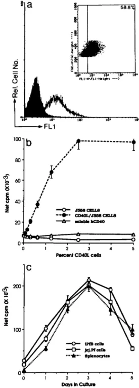
The binding of soluble mCD40L-CD8a fusion protein indi-cated that a subpopulation (56.2 ± 6.4%; n = 6 lambs) of iPfB cells expressed a detectable level of the CD40 molecule (Fig. 1a). A variable level of CD40 expression was apparent from the histogram width and an analysis of forward light scatter (FSC; relative cell size) and mCD40L-CD8a labelling intensity revealed an association between cell size and CD40 expression: high level CD40 expression was restricted to large (FSChi) B cells while a subpopulation of small (FSC'°) B cells did not express a detectable level of CD40 (Fig. 1a, inset). This suggested that the avidity of the soluble
mCD40L-iP i* 100 80 40-
20-A
J5MCSLL8 CDML/JSM CELLS viable hdMO 2 3 4 ParcantCDttlcaOs 200-8I
2 3 Days In Cuttur*Fig. 1. Expression of functional CD40 molecules on a subpopulation of iPfB cells, (a) Flow cytometric analysis of soluble mCD40L-CD8a binding by iPfB cells. A variable level of mCD40L-CD8a binding was apparent from the range of fluorescence intensity (open histogram) when compared with control (solid histogram). The dot scatter plot (insert) revealed -60% iPfB cells bound a detectable level of mCD40L-CD8a. The highest level of binding was on large cells (FSChi) and many small cells were negative or labelled at a low level,
(b) Co-culture with mCD40L-transfected cells (CD40L/J558) induced a dose-dependent proliferative response not observed with non-transfected cells (J558) or CD40L/J558 cells pre-incubated with soluble hCD40-Hn. (c) Proliferative responses induced by CD40L/ J558 cells were similar in amplitude and kinetics for B cells from a variety of lymphoid tissues. Data presented are mean ± SD of values from four replicate cultures.
45
20Fig. 2. A comparison of soluble mCD40L-CD8a binding by BMCs, freshly isolated iPfB cells and cultured iPfB cells, (a) A distinct subpopulation of BMCs bound soluble mCD40L-CD8a. (b) A low but detectable level of mCD4OL-CD8oc binding was associated with most FALShi iPfB cells and a subpopulation of FSC10 iPfB cells, (c) A high
level of mCD40L-CD8o binding was observed for all viable iPfB cells (Pl~) cultured for 72 h in IMDM + 2% FBS. (d) A high level of mCD40L-CD8a binding was observed for all iPfB cells following a 72 h co-culture with mCD401_/J558 cells. The percent cells binding a detectable level of soluble mCD40L-CD8a is indicated in the upper right corner and background fluorescence was determined using cells reacted with PE-conjugated rat anti-mouse CD8a. The x-axis presents the log mean fluorescence intensity of mCD40L-CD8a binding as detected with PE-rat anti-mouse CD8a (CD40) and the y-axis presents relative cell size as forward light scatter (FSC).
CD8a fusion protein may limit the detection of CD40 expressed on small iPfB cells. However, soluble mCD40L-CD8a fusion protein clearly labelled a distinct population of
Control Anti-lg CD40L/U558 CD40UJ558 + Anti-lg
o
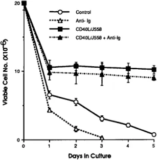
2 3 Days In CultureFig. 3. mCD40L inhibited iPfB cell death. Co-culture for 24 h with 5% CD40L/J558 cells (CD40L/J558) rescued 50-60% of iPfB cells but viable cell number then remained relatively constant during a 5 day culture period. Addition of soluble rabbit anti-sheep IgM (anti-lg; 40 ng/ml) to cultures resulted in the death of all iPfB cells within 72 h. This cell death exceeded that observed in the absence of anti-lg (control). Addition of soluble anti-lg had no effect on viable iPfB cell number during co-culture with CD40LAJ558 cells (CD40L/J558 + anti-lg). Data presented are mean ± SD of values from three replicate experiments.
blood mononuclear cells (BMC) (Fig. 2a) and the percent slgM+ cells (40.1 ± 4.8%; n = 5) approximated the percent cells labelled by soluble mCD40L-CD8a fusion protein (41.5 ± 5.9%; n = 5). Dual-labelling for slgM and CD40 expression indicated that all slgM+ BMC expressed CD40 but a small subpopulation of slgM" cells also bound detectable levels of soluble mCD40L-CD8a fusion protein (data not shown). In contrast, -95% of all iPfB cells are slgM+ but only 60% of these cells bound a detectable level of soluble mCD40L-CD8a fusion protein (Fig. 2b). Furthermore, despite the rela-tively larger size (greater FSC) of iPfB cells, when compared with BMC, there was a relatively lower level of mCD40L-CD8a fusion protein binding on the iPfB cells. Finally, all viable iPfB cells (PI"), surviving 72 h in culture in either the absence (Fig. 2c) or presence of CD40L/J558 cells (Fig. 2d), bound a high level of soluble mCD40L-CD8a fusion protein. Notable in these 72 h cultures was the absence of viable, FALS10 iPfB cells which were observed in cell suspensions prepared from ileal PP lymphoid follicles (Fig. 2b).
The specificity of the interaction between mCD40L-CD8a and the sheep CD40 molecule was determined by co-culturing iPfB cells with CD40L/J558 cells. A marked proliferative response was observed when iPfB cells were co-cultured CD40L/J558 cells (Fig. 1b). In contrast, neither non-trans-fected J558L cells nor CD40L7J558 cells, pre-incubated for 60 min with soluble hCD40-Hn to block the CD40 molecule, induced an increased iPfB cell proliferative response (Fig. 1b). The CD40LAJ558-induced proliferative responses were also dose-dependent. Increased iPfB cell proliferative responses were apparent in 48 h co-cultures with as few as
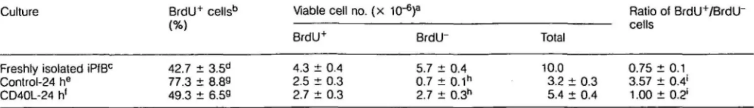
Table 1. mCD40L rescues non-S-phase iPfB cells from cell death
Culture
Freshly isolated iPfBc
Control-24 he CD40L-24 h1 BrdU+ 42.7 ± 77.3 ± 49.3 ± cells'3 3.5d 8.89 6.59 Viable BrdU4 4.3 ± 2.5 ± 2.7 ± cell no. ( x 1C 0.4 0.3 0.3 ,-6) a BrdU" 5.7 ± 0.7 ± 2.7 ± 0.4 0.1h 0.3h Total 1O.0 3.2 ± 5.4 ± 0.3 0.4 Ratio cells 0.75 3.57 1.00 of BrdU+/BrdU" ±0.1 ±0.4* ± 0.21 aEach culture initiated with 10 x 106 viable iPfB cells. Percent viable cells determined by PI exclusion and FSC (Fig. 4b; R1).
bBrdU incorporation was detected by immunoperoxidase stain and determined from counts of 500 cells per cytospot of sorted viable cells
(Fig. 4b;R1).
cLamb injected with 20 mg BrdU/kg body weight 30 min prior to iPfB cell isolation. dData presented are mean ± SD of values from seven experiments.
eiPfB cells cultured in IMDM/P + 2% FBS.
•iPfB cells co-cultured with 5% CD40L/J558 cells.
9"iSignificant differences ( P < 0.01) between control and CD40L cultures.
0.15% mCD40L/J558 cells and maximal proliferative responses were observed with 2.5% mCD40L/J558 cells. Finally, B cells from a variety of lymphoid tissues displayed a proliferative response of similar amplitude and kinetics when co-cultured with CD40L/J558 cells (Fig. 1c). Also apparent in co-cultures of iPfB cells and mCD40L/J558 was a clustering of B cells on the surface of mCD40LVJ558 cells and the size of these cell clusters increased during co-culture (data not shown).
mCD40L rescues non-S-phase iPfB cells from cell death mCD40L not only induced a proliferative response (Fig. 1b) but in 24 h co-cultures with mCD40L/J558 cells there was a marked increase in viable iPfB cell number when compared to 'control' cultures (Fig. 3). However, from day 2 to 8 of co-culture viable iPfB cell number remained relatively constant. This raised questions regarding which iPfB cells responded to mCD40L and how proliferative responses increased despite a constant viable cell number. To determine if CD40 signalling altered the fate of both resting and proliferating iPfB cells the S-phase iPfB cells were labelled with BrdU during a 30 min in vivo exposure. Approximately 40% iPfB cells had incorporated BrdU and in 24 h 'control' cultures the S-phase cells displayed a relatively greater survival than non-S-phase cells (Table 1). Only 12% of non-S-phase cell were viable after 24 h in culture while 58% of S-phase iPfB cells were viable after the same period. However, co-culture with CD40L7 J558 cells increased 4-fold the survival of non-S-phase cells (BrdU") but did not significantly change the survival of S-phase cells (BrdU+) (Table 1). To further characterize the mCD40L rescue of iPfB cells from cell death the iPfB cells were labelled with the lipophilic fluorochrome PKH26 prior to culture (Fig. 4a). This permitted an analysis of iPfB cell survival and proliferation during a longer co-culture period with CD40L7 J558 cells. PKH26 inserts into cell membranes and with each division distributes equally to progeny cells. A uniformly high level of PKH26 labelling was achieved (Fig. 4a; histogram ii) and mean PKH26 labelling intensity of viable iPfB cells declined ~4-fold during a 4 day culture period (Fig. 4a; histogram iii). This confirmed that most viable iPfB cells had undergone approximately two cell divisions. However, the
FL3
Fig. 4. mCD40L rescued iPfB cells from cell death but did not alter the rate of proliferation, (a) iPfB cells labelled with PKH26 (histogram ii; MFI = 3571) displayed high intensity fluorescence when compared with unlabelled cells (histogram i). Following 4 days in culture PKH26 labelling declined -4-fold for viable iPfB cells both in the presence or absence of CD40L/J558 cells [histogram iii; MFI = 325 (control) and 349 (CD40L/J558 cells)], (b) Viable iPfB cells (R1) were defined as a population that excluded PI (FL3 low) and displayed FSC greater than Pl+ cells, (c) In control cultures there was cell death with each
cell division which was apparent in the continuous contour plot for PKH26 fluorescence (FL3) of the FALS'° cells located beneath the population of FSChi viable cells (10,000 cells from R1). The contour
plot intensity for FALSto cells-also indicated cell death was greatest
prior to the first division (FL3 > 103). (d) Co-culture with CD40L/J558
cells eliminated much of the PKH26 contour plot for dead cells (FALSto) except for a small population that never divided (FALS10; FL3
increased width of histogram iii, when compared to histogram ii, indicated considerable variation in cell division within the viable iPfB cell population. For these analyses viable iPfB cells (Fig. 4b; R1) were identified as PI" (2.5 ng/ml PI; FL3'°) and FSChi. Dead iPfB cells were identified as Pl+ (FL3hi) and FSC'°. However, Pl~ cells (FL3'°) that were FSC'° were excluded from R1 after Giemsa stained cytospots of FACStar sorted cells revealed this population had nuclear condensa-tion and fragmentacondensa-tion typical of apoptotic cells (data not shown). Furthermore, during a continual lOminflowcytometric analysis there was a progressive increase in the level of PI incorporation in this population of FSC10 cells (data not shown). Thus, subsequent analyses of iPfB cell survival in culture, with or without CD40L/J558 cells, was determined by collecting 10,000 events in R1 (viable cells) and comparing this to the total number of PKH26 labelled events collected (Fig. 4c and d). When the contour plots were compared for control cultures (Fig. 4c) and co-cultures with CD40L/J558 cells (Fig. 4d) there was a noticeable reduction in the frequency of PKH26-labelled (FL3) dead cells (FSC'°) following co-culture with CD40L/J558 cells. Control cultures displayed extensive cell death prior to the first cell division (FL3 >103) and cell death with each cell division was evident in the continuous FL3 contour plot for FSC'° cells (Fig. 4c). In contrast, iPfB cell death during co-culture with CD40L/J558 appeared to be much lower and restricted primarily to cells that had not divided (FL3 >103; Fig. 4d). This was consistent with data from Table 1 that indicated 35% of non-S-phase iPfB cells died during the first 24 h of co-culture with CD40L/J558 cells. However, with no apparent cell death (Fig. 4d) and an average doubling time of 48 h (Fig. 4a) the viable iPfB cell number should have increased significantly after 4-5 days in co-culture with mCD40L (Fig. 2). These observations indicate that iPfB cell death continued in co-cultures with CD40L/J558 cells but iPfB cells may have died by necrosis which would result in cell disintegration and no PKH26-labeled cell frag-ments would be detected.
mCD40L inhibits anti-lg induced iPfB cell death
Antigen recognition by the Ig receptor should be part of T cell-dependent B cell development (3) but slg on iPfB cells transduces a signal that induces cell death (16). This was apparent in cultures with soluble rabbit anti-sheep Ig where few iPfB cells survived after 72 h and the decline in viable cell number exceeded that of control cultures (Fig. 3). Thus, we determined if mCD40L could alter iPfB cell death induced by slgM signalling. The mCD40L completely inhibited anti-lg-induced iPfB cell death [40 ng/ml F(ab')2 rabbit anti-sheep IgM] with viable cell number similar for iPfB cells co-cultured with CD40L/J558 cells in the presence or absence of anti-lg (Fig. 3). It has also been observed that CD40L induced decreased cAMP levels in human germinal centre B cells and this second messenger pathway may play a role in determining the survival of these cells (9). Therefore, it was of interest to examine the effects of mCD40L on cAMP levels in iPfB cells and to extend previous observations by examining cAMP levels during CD40 and slgM co-signaling. Freshly isolated iPfB cells had elevated cytoplasmic cAMP levels (Fig. 5; control-0 h) relative to blood lymphocytes (1.5 ± 0.6
Culture Treatment CD40L • Antl-Ig - 24 h CD40L • Antl-Ig - 4 b C D 4 0 L - 4 b Antl-lg 3 4 b Antl-lg - 4 b C o n t r o l - 2 4 b Control - 4 b C o n t r o l - O b 0 10 20 Cytoplasmic cAMP- pmol/ml
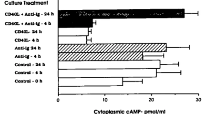
Fig. 5. mCD40L reduced the level of cytoplasmic cAMP in iPfB cells
but co-signalling by slg induced elevated cAMP levels. Addition of soluble anti-lg to cultured iPfB cells (Anti-lg-4 h; anti-lg-24 h) did not significantly change cAMP levels. In contrast, co-culture with CD40L/ J558 cells markedly reduced iPfB cell cytoplasmic cAMP levels within 4 h (CD40L-4 h) and cAMP levels remained low after a 24 h culture (CD40L-24 h). Addition of soluble anti-lg during iPfB cell co-culture with CD40L/J558 cells did not alter cytoplasmic cAMP levels after 4 h (CD40L + anti-lg-4 h) but cAMP levels increased after 24 h (CD40L + anti-lg-24h). Data presented are the mean ± SD of values from three experiments and assays were performed in duplicate with 1X10⁶ viable iPfB cells (R1; Fig. 2b).
pmol/ml) and maintained an elevated cAMP level during culture (Fig. 5; control-4 h; control-24 h). The addition of soluble anti-lg did not appear to alter cAMP levels in viable iPfB cells (Fig. 5) but a 1 h incubation with cholera toxin (1X10~9 M) increased the cytoplasmic cAMP level 5-fold (153.8 ± 16.1 pmol/ml for 106 viable cells) and a 1 h incubation with phorbol ester (PMA; 200 nM) plus calcium ionophore A23187 (200 nM) induced a 3-fold decrease (5.7 ± 0.3 pmol/ ml for 106 viable cells). These changes in cytoplasmic cAMP levels persisted for at least 24 h. The mCD40L also induced a 4-fold decrease in cAMP levels within 4 h that persisted for at least 24 h (Fig. 5). However, when iPfB cells were co-cultured with CD40L/J558 cells and anti-sIgM (40 ng/ml) then, after a 24 h incubation, there was a marked elevation of cAMP levels (Fig. 5). Thus, iPfB cell survival during CD40 and slg co-signalling was not dependent on a mCD40L-induced inhibition of cAMP levels.
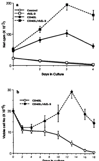
mCD40L induces IL-2 expression and iPfB cell differentiation Antigen-dependent T cell-B cell interactions include the pro-duction of a number of T cell cytokines and an increasing number of cytokines have been shown to influence B cell growth and differentiation in conjunction with CD40 signalling (reviewed in 3). Thus, we investigated the possibility that mCD40L altered iPfB cell cytokine responsiveness. Flow cytometric analyses, following iPfB cell co-culture for 48 h with CD40L/J558 cells, revealed a 10-fold increase in the percent cells expressing detectable levels of IL-2R and the percent cells expressing IL-2R increased further in the presence of rhlL-2 (10 ng/ml; Table 2). Greater than 50% of iPfB cells expressed detectable levels of IL-2R after 8-10 days of co-culture with CD40L/J558 cells and rhlL-2. That the IL-2R was functional was evident in the rhlL-2-induced
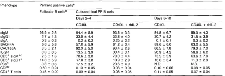
Table 2. iPfB cell phenotypic changes during co-culture with CD40L/J558 cells and rhlL-2 Phenotype slgM slgGI slgA BAQ44A CACT65A IL-2R CD5+ slgM+ c CD5+ slgG1 + PCsd CD5hl CD4+ T cells
Percent positive cellsa
Follicular B cellsb 96.5 ± 2.8 2.7 ± 1.3 0.3 d 6.6 d 3.5 d 0.8 d 2.5 d 14.8 d 0.8 d t 0.3 t 5.8 : 2.1 i 0.5 t 1.8 t 5.9 t 0.6 0.50 ± 0.24 0.45 ± 0.20 Cultured ileal Days 2-4 CD40L 94.4 ± 5.9 33.0 ± 4.4 0.2 ± 0.2 57.0 ± 5.9 92.0 ± 5.0 13.5 ± 3.0 78.6 ± 3.0 17.0 ± 3.0 17.5 ± 3.2 0.10 ± 0.05 0.09 ± 0.04 PP B cells CD40L + rhlL-2 93.8 d 33.8 d 0.25 d 61.2 d 93.4 d 30.4 d 78.0 d 10.9 d 23.6 d t 3.3 t 4.0 t 0.2 t 3.4 t 2.8 t 3.1 t 4.4 t 2.9 t 4.9 0.08 ± 0.04 0.08 ± 0.05 Days 8-10 CD40L 84.8 ± 6.7 30.7 ± 4.2 0.1 : 89.6 : 86.5 : 20.9 : 73.5 : 16.0 : t0.1 t 6.0 t 7.8 t 4.2 t 5.9 t 3.4 N.D. 0.10 ± 0.06 0.11 ± 0 . 0 5 CD40L + rhlL-2 89.0 ± 4.3 31.5 ± 3.9 0 . 3 : 6 3 . 0 : 79.0 d 58.6 d 63.6 d 11.3 d t 0.2 t 5.5 t 7.0 t 6.2 t 7.9 i 2.8 N.D. 0.08 ± 0.05 0.07 ± 0.04
aData presented are mean ± SD of values from six experiments. bCell suspension prepared from isolated lymphoid follicles of the ileal PP. cValues presented are the percent slgM+ or s l g G I+ cells that co-expressed CD5.
dPCs were quantitated from counts of 1000 cells in cytospots of sorted viable cells (Fig. 4b; R1). Isotype specific plasma cells (IgM, lgG1,
IgA) were detected by immunoperoxidase staining but >95% of plasma cells were consistently lgM+.
proliferative response induced by mCD40L (Fig. 6a) and the increased iPfB cell viable number when rhlL-2 was added to iPfB cell co-cultures with CD40L/J558 cells (Fig. 6b). A 6-6 day lag in the rhlL-2-induced increase in iPfB cell viable number may be consistent with the expansion of a subpopul-ation of IL-2R expressing cells (Table 2).
Phenotypic analyses revealed other changes that occurred when iPfB cells and CD40L/J558 cells were co-cultured. Within 48 h the CACT65A activation molecule (25) was expressed on most iPfB cells but the percent iPfB cells with detectable expression declined after 8-10 days (Table 2) and was detected on <20% of cells following 16-18 days of co-culture with CD40L/J558 cells and rhlL-2 (data not shown). Also, within 48-96 h of co-culture the percent s l g G I+ B cells increased 10-fold and this apparent isotype-switching was not influenced by the addition of rhlL-2 (Table 2). To determine if IgGi isotype switching or the expansion of an isotype switched population of iPfB cells occurred we examined the responses of naive iPfB cells isolated from three feti (day 144 of a 150 day gestation period) and co-cultured with CD40L/ J558 ± rhlL-2. The fetal iPfB cells and iPfB cells from lambs displayed similar proliferative responses and changes in viable cell number during co-culture with CD40L/J558 cells ± rhlL-2 (data not shown). However, after fetal iPfB cells were co-cultured with CD40L/J558 cells and rhlL-2 for 7-10 days there were only 3.8 ± 1.2% (n = 3) s l g G I+ cells and 0.1 ± 0.1% (n = 3) slgA+ cells. Thus, the interaction with CD40L and IL-2 induced very little isotype switching in naive iPfB cells and the large increase in s l g G I+ cells, observed for lambs, probably results from the expansion of isotype-switched B cells. Furthermore, few slgA+ iPfB cells were observed during the co-culture of fetal or lamb iPfB cells with CD40L/J558 cells ± rhlL-2 (Table 2). The mouse mAbs specific for sheep IgA and lgG1 are the same isotype (IgGi).
Thus the absence of detectable IgA provided a control for the specificity of lgG1 detection and also indicated that ileal PP follicles were not a site for the generation of IgA committed B cells. The large increase in plasma cells following 2-4 days of culture indicated that CD40 signalling may induce B cell differentiation but this was largely restricted to lgM+ plasma cells. Finally, co-culture with CD40L/J558 cells consistently induced low level CD5 expression on - 8 0 % of slgM+ and 10-20% of s l g G I+ iPfB cells and the BAQ44A differentiation molecule was expressed on 50-60% of iPfB cells (Table 2). In the absence of rhlL-2, the BAQ44A molecule was expressed on most iPfB cells after 8-10 days in co-culture with CD40L7 558 cells. Less than 0.5% T cells (CD5hi; CD4+) were present in iPfB cell suspensions and the number of T cells declined to the limit of detection during the co-culture period (Table 2). Furthermore, 1-2% of the cells in iPfB cell suspensions were macrophages and stromal cells but after three passages during co-culture with CD40L/J558 cells the plastic-adherent macrophages were no longer evident (data not shown). Analysis of CD5, BAO44A and Ig isotype expression on ileal and jejunal PP follicular B cells
The phenotypic analyses of iPfB cells co-cultured with CD40L7 J558 cells indicated that mCD40L altered the expression of BAQ44A, CD5 and slgGI (Table 2). Therefore, it was of interest to analyse the expression of these antigens on lymphocytes freshly isolated from lymphoid follicles of the ileal PP for possible evidence of T cell-dependent B cell develoment. The phenotype of lymphocytes isolated from lymphoid follicles of the jejunal PP was also analysed since this gut-associated tissue appears to function as a typical antigen-dependent secondary lymphoid tissue (20). That the jejunal PP functions to generate many antigen-specific B cells was evident in the high frequency of CD4+ T cells together
Table 3. Analysis of CD5, BAQ44A and Ig expression by cells isolated from lymphoid follicles of the ileal and jejunal PPs
2 3 Days In Culture
6 8 10 12 Days in culture
16 18
Fig. 6. mCD40L induced IL-2 responsiveness apparent as increased iPfB cell proliferation and viable cell number following addition of rhlL-2. (a) iPfB cell proliferation declined during culture (control) and addition of rhlL-2 (rhlL-2) did not significantly change the proliferative response. Co-culture with 5% CD40L/J558 cells (CD40L) significantly increased the iPfB cell proliferative response and induced responsiveness to rhlL-2 (10 ng/ml; CD40L/rhlL-2). The iPfB cell responsiveness to rhlL-2 was maximal following a 72 h co-culture with CD40L/J558 cells, (b) During long-term co-cultures with CD40LJ J558 cells (CD40L) viable iPfB cell number remained relatively constant for 7-10 days and then declined. In contrast, addition of 10 ng/ml rhlL-2 (CD40L/rhlL-2) resulted in a significantly increased viable iPfB cell number between days 10 and 15 of co-culture. Data presented are mean ± SD of values from three experiments. iPfB cells were transferred to fresh co-cultures at each data point.
with l g G i+ and lgA+ B cells (Table 3). On the basis of BAQ44A, CD5 and slgG1 expression a similar estimate was obtained that the jejunal PP had a 10-fold greater level of T cell-dependent B cell development than did the ileal PP (Table 3). The paucity of lgA+ B cells and isotype-switched plasma cells (PCs) in ileal PP follicles made it difficult to compare the frequency of these population between the two PPs but the frequency of lgM+ PCs, relative to slgM+ B cell number, was similar for both ileal and jejunal PP follicles (Table 3). Finally, the large SD for numbers of PCs and lgG1 isotype-switched
Phenotype slgM slgG1 slgA BAQ44A CD5+ slgM+ b C D 5+s l g G 1+ c lgM+ PCsb lgG+ PCs lgA+ PCs CD4+ T cells Percent positive Ileal PP follicular cells 96.5 ± 2.8 2.7 ± 1.3 0.3 ± 0.3 6.6 ± 5.8 2.5 ± 1.8 14.8 ± 5.9 0.8 ± 0.6 0.1 ± 0.3 <0.01 0.45 ± 0.20 cells3 Jejunal PP follicular cells 39.6 ± 7.4 26.5 ± 8.4 16.3 ± 7.8 79.0 ± 6.9 28.9 ± 8.6 56.0 ± 7.0 0.4 ± 0.3 2.8 ± 0.6 2.1 ± 0.5 13.8 ± 5.3 Comparative frequency jejunal PP/ ileal PP 0.4 9.8 54.3 12.0 11.6 3.8 0.5 28.0 NDd 30.6
aData presented are mean ± SD of values from the analyses of
cell suspensions prepared from isolated lymphoid follicles of the jejunal and ileal PPs of six lambs (6-10 weeks old).
bPCs were quantitated from counts of 1000 cells in cytospots and
isotype specific plasma cells (IgM, lgG1, IgA) were detected by immunoperoxidase staining.
cValues presented are the percent slgM+ or slgG1+ cells that
co-expressed CD5.
dND, not determined.
B cells in the ileal PP follicles reflected the broad variation among lambs, as might be expected if variable antigenic experiences altered iPfB cell differentiation.
Discussion
The present investigation determined that a subpopulation of ileal PP B cells expressed CD40 and it was functional. This was evident in the binding of soluble mCD40L (Figs 1a and 2) and the induction of a B cell proliferative response that was specific for mCD40L expressed on transfected J558L cells (Fig. 1b). Interspecies conservation of CD40 and CD40L functional domains was apparent when recombinant mouse and human CD40L displayed equivalent mitogenic activity with human and mouse B cells (26) and now in the mitogenic activity of mCD40L with sheep B cells (Fig. 1). Furthermore, a conservation of the role of CD40 in B cell development was apparent in the marked similarity between many mCD40L-induced iPfB cell responses and those reported for mouse and human B cells (reviewed in 3). These similarities included rescue from cell death (Figs 3 and 4), blocking anti-lg-induced cell death (Fig. 3), a reduction in cytoplasmic cAMP (Fig. 5), the induction of functional IL-2R (Fig. 6) and supporting the development of slgG1+ B cells (Table 2). All these responses could contribute to either B cell growth or differentiation and provide indirect evidence that T cell-dependent interactions may play a role in iPfB cell development. However, experi-ments must now be designed to directly determine if either iPfB cell proliferation or differentiation is influenced by T cell-dependent antigens.
The relatively low level of CD40 expression on a subpopul-ation of iPfB cells (Fig. 2) contrasts with the report that all B cells express CD40, with a lower level of CD40 on resting B
cells and increased CD40 expression following B cell activa-tion (1). The analyses of sheep B cells in blood (Fig. 2a) indicated that these resting B cells actually express a higher level of CD40 than the much larger, rapidly dividing iPfB cells (Fig. 2b). The apparent absence of CD40 on some FSC'° iPfB cells (Fig. 2b) suggested that CD40 expression may be down-regulated on B cells destined to undergo cell death. This conclusion is supported by the observations that FSC'° iPfB cells are destined to die (Fig. 4b) and the CD4CT, FSC'° iPfB cells disappear during culture (Fig. 2c and d). The apparent absence of CD40 expression, or CD40L binding, on a subpop-ulation of FSC'° iPfB cells cannot be explained simply by smaller cell size since this population has a FSC similar to blood lymphocytes (Fig. 2). Furthermore, that CD40L rescued -50% of non-S-phase iPfB cells from cell death is consistent with the expression of a functional CD40 molecule on a subpopulation of FSC'° iPfB cells (Table 1). Thus, there may be a brief period during which resting iPfB cells either interact with CD40L on T cells or become committed to cell death. Proliferating iPfB cells (BrdU+) are located primarily in the outer follicle and non-S-phase (BrdlT) iPfB cells are located in the central follicle, upper follicle, and dome region [P. Griebel, unpublished observation; (27)]. CD4+ T cells are distributed in the same regions as non-S-phase iPfB cells (15) suggesting it may be primarily resting iPfB cells that would interact with CD4+ T cells. If this is true, then CD40 signalling may play a primary role in iPfB cell differentiation rather than iPfB cell proliferation. This conclusion would be consistent with observations that B cell emigration from the ileal PP increased markedly in lambs after birth (12), a time when antigen exposure occurs, and that B cell numbers were decreased in the blood of young lambs that had been thymectomized and were deficient in CD4+ T cells (28).
A further suggestion that mCD40L may play a role in iPfB cell differentiation came from the observation that mCD40L inhibited anti-lg-induced iPfB cell death (Fig. 3). CD40 signal-ling can also rescue B lymphoma cell lines from anti-lg-induced cell death (7,8) but this is the first time that a normal, immature B cell population has been shown to exhibit such a response. CD40 signalling can also rescue human germinal centre B cells from programmed cell death (6) and associated with this response is a reduced level of cytoplasmic cAMP (9). This led to the suggestion that the cAMP-dependent second messenger pathway may influence germinal centre B cell survival and the mCD40L-induced decrease in iPfB cell cAMP levels (Fig. 5) may be consistent with this hypothesis. However, CD40 and slgM co-signalling resulted in an elevated level of cAMP (Fig. 5) indicating that CD40 signalling does not simply block slgM signalling. It may be that co-signalling by these molecules resulted in a different state of iPfB cell activation or differentiation. The state of B cell differentiation can determine the level of cAMP induced by CD40L since mature B cells respond with elevated cAMP levels (9). Thus, it may be that co-signalling by slgM and CD40 induces the differentiation of immature B cells.
In a number of culture systems, CD40 signalling was mitogenic for a variety of B cell populations (reviewed in 3). The CD40L/J558 cells were also mitogenic for iPfB cells (Fig. 1b) but this proliferation was of limited duration despite re-stimulation with fresh CD40L/J558 cells every 3-4 days (Fig.
6b). Other co-signals may be necessary to sustain iPfB cell proliferation since mCD40L rescued non-S-phase iPfB cells from cell death but viable cell number remained relatively constant (Fig. 3). Several cytokines have been identified that, together with CD40 signalling, support B cell proliferation (reviewed in 3). In particular, IL-4 has been shown to support long-term growth of human B cells co-stimulated through CD40 (29). Human IL-4 has no cross-species activity with sheep iPfB cells (21) but rhlL-2 was mitogenic following CD40 signalling (Fig.6a). CD40L induction of 2R (CD25) and IL-2-responsiveness have also been reported for human tonsillar B cells (30,31) but only a subpopulation of iPfB cells expressed IL-2R following CD40 signalling (Table 2) and the expansion of this subpopulation required exogenous IL-2 (Fig. 6b). Thus, the limited proliferative response of iPfB cells to rhlL-2 and mCD40L co-stimulation may be consistent with previous observations that indicated IL-2 played a role in iPfB cell differentiation (14). Other unidentified cytokines or cell-cell interactions may be more important in regulating the growth of the functionally unique iPfB cell population (17,21). The B cell proliferation in ileal PP follicles is well established prior to birth (27) but it remains to be determined if T cell-B cell interactions may amplify post-natal iPfB cell proliferation.
CD40 signalling resulted in a number of phenotypic changes in the iPfB cell population. Expression of the CACT65A molecule was previously observed following iPfB cell activation by phorbol ester and calcium ionophore (25), and increased expression of the BAQ44A molecule was associated with iPfB cell differentiation (14,15). However, this is the first time that a receptor-ligand interaction has been identified that can induce the expression of these two molec-ules on iPfB cells. Of particular note was mCD40L induction of CD5 expression on 80% of slgM+ iPfB cells (Table 2). This has not previously been reported for B cells activated by CD40L but a similar observation was made for human B cells co-cultured with anti-CD3 activated T cells (33) and these T cells are known to express CD40L (2). Furthermore, CD5 expression on both slgG1+ and slgM+ B cells (Table 2) has also been observed for human B cells (33). Thus, as sug-gested for human B cells (33-35), it may also be true that CD5 is an activation marker on sheep B cells and not a B lineage marker as proposed for mice (reviewed in 36). Furthermore, the present observations suggest that the expression of CD5 on sheep B cells may indicate that a cognate T cell-B cell interaction has occurred. The expression of the CD5 molecule was not unique to cultured iPfB cells as CD5+ slgM+ and CD5+ slgG1+ B cells were present in both freshly isolated ileal and jejunal PP B cells (Table 3). However, CD5+ B cells are usually not found in the blood of young sheep (P. Griebel, unpublished observation). This may reflect the presence of a large number of naive B cells or possibly a down-regulated CD5 expression as reported for resting, human B cells (33). Finally, other ways of activating B cells may also induce CD5 expression since increased numbers of CD5+ B cells have been observed in the blood of sheep (37) and cattle (38,39) during diseases that cause B cell activation and phorbol esters can induce CD5 expression on human B cells (34) and iPfB cells (P. Griebel, unpublished observation).
rhlL-2 there was a large increase in slgG1+ B cells but PCs were primarily lgM+ and very few slgA+ B cells were observed (Table 2). Furthermore, few slgG1+ B cells were observed when naive, fetal iPfB cells were co-cultured with CD40L7 J558L cells ± rhlL-2. Thus, mCD40L, alone or with rhlL-2, appeared unable to induce Ig isotype switching but did support the growth of lgG1 isotype-switched B cells. It is difficult to explain the relatively constant number of slgG1 + cells in iPfB cell co-cultures. However, extensive B cell turnover in these cultures was indicated by a high level of proliferation (Fig. 1) that was associated with a relatively constant cell number (Fig. 3). It may be that differences in slgG1+ B cell and slgM+ B cell proliferation and survival result in an early enrichment of slgG1+ B cells that is not sustained with longer culture periods. CD40L by itself induced increased lgG1 production by human tonsilar B cells (40) but exogenous IL-2 induced both lgG1 and IgA secretion (30,40). The present observations with fetal and lamb iPfB cells (Table 2) suggest that it is only isotype-switched B cells that respond to CD40L rather than CD40L actually inducing the isotype switch. Also, of interest was the absence of slgA isotype-switched B cells in co-cultures with CD40L/J558 cells and freshly isolated iPfB cells (Table 3). This would be consistent with the ileal PP being functionally distinct from other gut-associated lymphoid tissues such as the jejunal PP (Table 3) which plays a major role in mucosal immunity. Finally, the presence of some lgG1+ B cells in the ileal PP follicles (Table 3) may provide evidence that antigen-dependent T cell-B cell interactions do occur in this microenvironment since lgG1 isotype switching is largely T cell-dependent (4,5).
The present investigation of mCD40L confirmed that a subpopulation of iPfB cells expressed a functional CD40 molecule. However, the paucity of T cells in ileal PP follicles (15) suggests that there may be limited opportunity for cognate T cell-B cell interactions to influence iPfB cell development. Furthermore, immunohistochemistry indicated that it would be primarily non-S-phase iPfB cells that interacted with CD4+ T cells. Therefore, through an effect on iPfB survival (Figs 3 and 4) and differentiation (Table 2) cognate T cell-B cell interactions may play a role in the selection of the relatively small number of B cells that emigrate (11). This hypothesis has important implications for the generation of the Ig receptor repertoire in lambs since ileal PP follicles are the major source of B cells (11,12). It should now be possible to test this hypothesis since alterations in the T cell repertoire, by immun-ization with a hapten-carrier protein, should influence the Ig repertoire of emigrant iPfB cells following exposure to the haptenated-carrier protein in the gut lumen. Finally, a limited number of cognate T cell-B cell interactions in lymphoid follicles of the ileal PP may influence the general level of B cell proliferation either through the release of T cell cytokines (Fig. 6) or possibly a recruitment of non-S-phase B cell into the proliferating B cell pool (Table 1). However, present observations support the conclusion that the majority of B cell proliferation in the ileal PP is antigen-independent (17,19).
Acknowledgements
We gratefully acknowledge the assistance of Mark Dessing and Stefan Meyer with sorting viable cells, and thank Drs W. R. Hein and
P. Lane for their critical review of the manuscript. The Basel Institute for Immunology was founded and is supported by F. Hoffmann-La Roche & Co. Ltd, Basel, Switzerland.
Abbreviations BMC BrdU FBS FSC IBMX IMDM iPfB cell PC PE PI PMA PP
blood mononuclear cell 5-bromo-2'-deoxyuridine fetal bovine serum forward light scatter 3-isobutyl-1 -methyl-xanthine
Iscove's modified Dulbecco's modified Eagle's medium ileal Peyer's patch follicular B cell
plasma cell phycoerythrin propidium iodide phorbol myristate acetate Peyer's patch
References
1 Ledbetter, J. A., Shu, G., Gallagher, M. and Clark, E. A. 1987. Augmentation of normal and malignant B cell proliferation by monoclonal antibody to the B cell-specific antigen BP50 (CDw40). J. Immunol. 138:788.
2 Lane, P., Traunecker, A., Hubele, S., Inui, S., Lanzavecchia, A. and Gray, D. 1992. Activated human T cells express a ligand for the human B associated CD40 which participates in T cell-dependent activation of B lymphocytes. Eur. J. Immunol. 22:2573. 3 Banchereau, J., Bazan, F., Blanchard, D., Briere, R, Galizzi, J. P.,
van Kooten, C , Liu, Y. J., Rousset, F. and Saeland, S. 1994. The CD40 antigen and its ligand. Annu. Rev. Immunol. 12:881. 4 Foy, T. M., Shepherd, D. M., Durie, F. H., Aruffo, A., Ledbetter,
J. A. and Noelle, R. J. 1993. In vivo CD40-gp39 interactions are essential for thymus-dependent humoral immunity. II. Prolonged suppression of the humoral immune response by an antibody to the ligand for CD40, gp39. J. Exp. Med. 178:1567.
5 Allen, R. C , Armitage, R. J., Conley, M. E., Rosenblatt, H., Jenkins, N. A., Copeland, N. G., Bedell, M. A., Edelhoff, S., Disteche, C. M., Simoneaux, D. K., Fanslow, W. C , Belmont, J. and Spriggs, M. K. 1993. CD40 ligand gene defects responsible for X-linked hyper-IgM syndrome. Science 259:990.
6 Liu, Y. J., Joshua, D. E., Williams, G. T, Smith, C. A., Gordon, J. and MacLennan, I. C. M. 1989. Mechanism of antigen-driven selection in germinal centres. Nature 342:929.
7 Valentine, M. A. and Licciardi, K. A. 1992. Rescue from anti-lgM-induced programmed cell death by the B cell surface proteins CD20 and CD40. Eur. J. Immunol. 22:3141.
8 Tsubata, T, Wu, J. and Honjo, T. 1993. B cell apoptosis induced by antigen receptor crosslinking is blocked by a T-cell signal through CD40. Nature 364:645.
9 Knox, K. A., Johnson, G. D. and Gordon, J. 1993. Distribution of cAMP in secondary follicles and its expression in B cell apoptosis and CD40-mediated survival. Int. Immunol. 5:1085.
10 Liu, Y.-J., Mason, D. Y, Johnson, G. D., Abbot, S., Gregory, C. D., Hardie, D. L., Gordon, J. and MacLennan, I. C. M. 1991. Germinal centre cells express bcl-2 protein after activation by signals whch prevent their entry into apoptosis. Eur. J. Immunol. 21:1905. 11 Reynolds, J. D. 1986. Evidence of extensive lymphocyte death in
sheep Peyer's patches I. A comparison of lymphocyte production and export. J. Immunol. 136:2005.
12 Gerber, H. A., Morris, B. and Trevella, W. 1986. The role of gut-associated lymphoid tissues in the generation of immunoglobulin-bearing lymphocytes in sheep. Aust. J. Exp. Biol. Med. Sci. 64:201.
13 Reynolds, J. D., Kennedy, L, Peppard, J. and Pabst, R. 1991. Ileal Peyer's patch emigrants are predominantly B cells and travel to all lymphoid tissues in sheep. Eur. J. Immunol. 21:283. 14 Griebel, P. J., Davis, W. C. and Reynolds, J. D. 1992. An analysis
of the growth and differentiation of B cells isolated from follicles of the ileal Peyer's patch of sheep. Immunology 75:601.
15 Hein,W.R.,Dudler, L andMackay, C.R. 1989. Surface expression of differentiation antigens on lymphocytes in the ileal and jejunal Peyer's patches of lambs. Immunology 68:365.
16 Griebel, P. J., Davis, W. C. and Reynolds, J. D. 1991. Negative signaling by surface IgM on B cells isolated from ileal Peyer's patch follicles of sheep. Eur. J. Immunol. 21:2281.
17 Griebel, P. and Ferrari, G. 1994. Evidence for a stromal cell-dependent, self-renewing B cell population in lymphoid follicles of the ileal Peyer's patch of sheep. Eur. J. Immunol. 24:401. 18 Reynaud, C.-A., Mackay, C. R., Muller, R. G. and Weill, J.-C.
1991. Somatic generation of diversity in a mammalian primary lymphoid organ: the sheep ileal Peyer's patch. Cell 64:995. 19 Reynolds, J. D. and Morris, B. 1984. The effect of antigen on the
development of Peyer's patches in sheep. Eur. J. Immunol. 14:1. 20 Reynolds, J. D. and Morris, B. 1983. The evolution and involution
of Peyer's patches in fetal and postnatal sheep. Eur. J. Immunol. 13:627.
21 Griebel, P., Hein, W. R., Dudler, L. and Ferrari, G. 1993. Phenotype and function of stromal cells cloned from the ileal Peyer's patch of sheep. Stem Cells 11:130.
22 Zar, J. H. 1974. Biostatistical Analysis, p. 195. Prentice-Hall, Englewood Cliffs, NJ.
23 Lane, P., Brocker, T., Hubele, S., Lanzavecchi, A. and McConnell, F. 1993. Soluble CD40-ligand can replace normal T cell derived CD40-ligand signal to B cells in T-dependent activation. J. Exp. Med. 177:1209.
24 Motyka, B. and Reynolds, J. D. 1991. Apoptosis is associated with the extensive B cell death in the sheep ileal Peyer's patch and the chicken bursa of Fabricius: a possible role in selection. Eur. J. Immunol. 21:1951.
25 Motyka, B., Griebel, P. J. and Reynolds, J. D. 1993. Agents that activate protein kinase C rescue sheep ileal Peyer's patch B cells from apoptosis. Eur. J. Immunol. 23:1314.
26 Spriggs, M. K., Armitage, R. J., Strockbine, L, Clifford, K. N., Macduff, B. M., Sato, T. A., Maliszewski, C. R. and Fanslow, W. C. 1992. Recombinant human CD40 ligand stimulates B cell proliferation and immunoglobulin E secretion. J. Exp. Med. 176:1543.
27 Reynolds, J. D. 1987. Mitotic rate maturation in the Peyer's patches of fetal sheep and in the bursa of Fabricius of the chick
embryo. Eur. J. Immunol. 17:503.
28 Hein, W. R., Dudler, L. and Morris, B. 1990. Differential peripheral expansion and in vivo antigen reactivity of a/p and Y/8 T cells emigrating from the early fetal thymus. Eur. J. Immunol. 20:1805. 29 Banchereau, J., de Paoli, P., \falle, A., Garcia, E. and Rousset, F.
1991. Long term human B cell lines dependent on interleukin-4 and antibody to CD40. Science 251:70.
30 Armitage, R. J., Macduff, B. M., Spriggs, M. K. and Fanslow, W. C. 1993. Human B cell proliferation and Ig secretion induced by recombinant CD40 ligand are modulated by soluble cytokines. J. Immunol. 150:3671.
31 MacLennan, I. C. M. 1994. Germinal centers. Annu. Rev. Immunol. 12:117.
32 Clark, E. A. and Ledbetter, J. A. 1994. How B and T cells talk to each other. Nature 367:425.
33 Vernino, L. A., Pisetsky, D. S. and Lipsky, P. E. 1992. Analysis of the expression of CD5 by human B cells and correlation with functional activity. Cell. Immunol. 139:185.
34 Miller, R. A. and Gralow, J. 1984. The induction of Leu-1 antigen expression in human malignant and normal B cells by phorbol myristic acetate (PMA). J. Immunol. 133:3408.
35 Werner-Favre, C , Vischer, T. L, Wohlwend, D. and Zubler, R. H. 1989. Cell surface antigen CD5 is a marker for activated human B cells. Eur. J. Immunol. 19:1209.
36 Kantor, A. B. and Herzenberg, L. A. 1993. Origin of murine B lineages. Annu. Rev. Immunol. 11:501.
37 Letesson, J. J., Mager, A., Mammerickx, M., Burny, A. and Depelchin, A. 1990. B cells from bovine leukemia virus infected sheep with hematological disorders express the CD5 T-cell marker. Leukemia 4:377.
38 Depelchin, A., Letesson, J. J., Lostrie-Trussart, N., Mammerickx, M., Portelle, D. and Burny, A. 1989. Bovine leukemia virus (BLV)-infected B-cells express a marker similar to the CD5 T cell marker. Immunol. Lett. 20:69.
39 Naessens, J. and Williams, D. J. L. 1992. Characterization and measurement of CD5+ B cells in normal and Trypanosoma
congo/ense-infected cattle. Eur. J. Immunol. 22:1713.
40 Blanchard, D., Gaillard, C , Hermann, P. and Banchereau, J. 1994. Role of CD40 antigen and interleukin 2 in T cell-dependent human B lymphocyte growth. Eur. J. Immunol. 24:330.