ORIGINAL INVESTIGATION
Examination of the effect of acute levodopa administration
on the loudness dependence of auditory evoked
potentials (LDAEP) in humans
K. Hitz&K. Heekeren&C. Obermann&T. Huber&G. Juckel&W. Kawohl
Received: 6 May 2011 / Accepted: 9 November 2011 / Published online: 26 November 2011 # Springer-Verlag 2011
Abstract
Rationale The loudness dependence of the auditory evoked potential (LDAEP) is considered a noninvasive in vivo marker of central serotonergic functioning in humans. Nevertheless, results of genetic association studies point towards a modulation of this biomarker by dopaminergic neurotransmission.
Objective We examined the effect of dopaminergic modu-lation on the LDAEP using L-3,4-dihydroxyphenylalanine (levodopa)/benserazide (Madopar®) as a challenge agent in healthy volunteers.
Methods A double-blind placebo-controlled challenge design was chosen. Forty-two healthy participants (21 females and 21 males) underwent two LDAEP measurements, following a baseline LDAEP measurement either placebo or levodopa (levodopa 200 mg/benserazide 50 mg) were given orally. Changes in the amplitude and
dipole source activity of the N1/P2 intensities (60, 70, 80, 90, and 100 dB) were analyzed.
Results The participants of neither the levodopa nor the placebo group showed any significant LDAEP alterations compared to the baseline measurement. The test–retest reliability (Cronbachs Alpha) between baseline and inter-vention was 0.966 in the verum group and 0.759 in the placebo group, respectively.
Conclusions The administration of levodopa showed no effect on the LDAEP. These findings are in line with other trials using dopamine receptor agonists.
Keywords Dopamine . Loudness dependence auditory evoked potentials (LDAEP) . Levodopa . Primary auditory cortex . Serotonin
Introduction
Reliable methods for the in vivo assessment of the central serotonergic system would be of great interest for the understanding of neuropsychiatric disorders and for monitor-ing the therapeutic efficacy of pharmacological treatments (Hegerl and Juckel 2000). Parameters regarding peripheral serotonin (5-HT) metabolism such as the concentration of 5-hydroxyindolacid in cerebrospinal fluid or the 5-HT transporter (SERT) availability in thrombocytes do not properly reflect central 5-HT functioning. They merely state inconsistent snapshots. In contrast to this, the loudness dependence of auditory evoked potentials (LDAEP) has been reported to be a measure of central 5-HT activity in humans (Hegerl et al. 2001; Hegerl and Juckel1993,1994;
K. Hitz
:
K. Heekeren:
C. Obermann:
W. KawohlDepartment of General Social Psychiatry, University of Zurich, Zurich, Switzerland
T. Huber
Internal Medicine, Psychiatric University Hospital Zurich, Zurich, Switzerland
G. Juckel
Department of Psychiatry, Ruhr-University Bochum, Bochum, Germany
W. Kawohl (*)
Psychiatric University Hospital Zurich, Militärstrasse 8,
8004 Zurich, Switzerland
Juckel et al.1997; Kawohl et al.2008b; O'Neill et al.2008a). A pronounced LDAEP supposedly reflects a low central 5-HT neurotransmission and vice versa.
Nevertheless, the issue of specificity of the LDAEP for the 5-HT system has been the topic of debate. Several findings cast some doubt over its sensitivity to changes in 5-HT functioning alone (Juckel et al.2008b,1997; O'Neill et al. 2006; Pogarell et al. 2004; Uhl et al. 2006). An association between 5-HT (SERT) and dopamine (DAT) transporter availability and the interindividual LDAEP has been found in a [123I]beta-CIT single-photon emission computed tomography (SPECT) study (Pogarell et al. 2004). High availability of the transporter enzymes, indirectly suggesting reduced 5-HT and dopamine in the synaptic cleft, was correlated with an increased LDAEP in patients with obsessive–compulsive disorder. Dopaminergic influence onto the LDAEP was stated. Lee at al. (Lee et al. 2010) found the LDAEP to be positively associated with DAT in a trial with 49 healthy volunteers using SPECT to approximate the availability of dopamine transporters and serotonin transporters. After adjusting for age and gender, the LDAEP was negatively associated with SERT. Further evidence for the possible involvement of dopamine in the genesis of LDAEP was stated.
A recent genetic association study has revealed an association between single nucleotide polymorphisms (SNPs) in the gene coding for the dopamine degrading enzyme catechol-O-methyltransferase (COMT) and the LDAEP (Juckel et al. 2008b). COMT is involved in the inactivation of synaptic dopamine (Axelrod and Tomchick 1958). Functional SNPs in the COMT gene result in attenuated dopamine catabolism, and several findings point to a modification of the risk for psychotic disorders by these genetic variants (Funke et al. 2005; Meyer-Lindenberg and Weinberger 2006; Nicodemus et al. 2007). Another genetic association study revealed an association between the functional SNPs in genes coding for nitric oxide synthase (NOS): single nucleo-tide polymorphisms in both NOS1- (G-84A_exon 1c promoter polymorphism) and NOS3 gene (Glu298Asp) are associated with a lower LDAEP. NOS is an enzyme catalyzing the production of nitric oxyde (NO) from L
-arginine. NO is a gaseous molecule with neurotransmitter properties that also interacts with dopaminergic transmis-sion (Kawohl et al.2008a).
The aforementioned studies point to a dopaminergic influence on the LDAEP. However, O'Neill et al. (O'Neill et al.2006) could not find a significant effect on the LDAEP after dopaminergic stimulation of the D1/D2/D3 receptors
with pergolide and D2/D3 receptors with bromocriptine. In
another trial of the same study group, selective serotonin and dopamine depletion with greater than 80% plasma precursor depletion had no effect on the LDAEP (O'Neill et al.2008b).
Despite of O'Neill et al.’s findings we developed a dopaminergic challenge with a naturalistic agent. Synthetic dopamine receptor agonists, as used in the aforementioned studies, exhibit different receptor affinities compared to dopamine. In contrast, we decided to use the prodrug levodopa due to its more naturalistic action via synaptic dopamine; levodopa is the precursor of dopamine. It is decarboxylized to dopamine, which cannot pass the blood– brain barrier. Compared to synthetic agonists, levodopa bares a decisive advantage in inducing dopaminergic stimulation; after decarboxylation, levodopa acts as a transmitter itself, thus modulating the dopaminergic effect in the most naturalistic manner. In contrast to a synthetic agonist, such as, bromocriptine, specific receptor affinities do not have to be taken into account. Several trials have shown an increase of synaptic dopamine level due to oral intake of levodopa and benserazide (de la Fuente-Fernandez et al. 2004; Floel et al. 2005; Khor and Hsu 2007; Kumakura et al.2004). In addition, we wanted to compute the EEG data with the dipole source model to differentiate between the primary and secondary auditory area. The primary auditory area is considered to be the generator of the LDAEP signal (Hegerl and Juckel 1993,1994; Hegerl 1994; Hegerl et al.1994). Hence, the aim of this study was to investigate the influence of acute dopaminergic stimula-tion on the LDAEP in healthy individuals with a challenge agent not used so far. We hypothesized that the acute synaptic dopamine excess caused by levodopa intake leads to an intraindividual decrease of the LDAEP.
Methods Participants
Healthy volunteers were selected among the staff and with the help of the volunteer server of the Psychological Institute of the University of Zürich. Normal hearing was tested clinically.
The 42 healthy participants had not been treated for any psychiatric disease in their lifetime and had no first-degree relatives with psychiatric disorders. All partic-ipants were drug free (except oral contraceptives) and were asked to abstain from alcohol the day prior to the session. Caffeine and nicotine intake were permitted to avoid withdrawal symptoms. Four female and six male participants were cigarette smokers. The volunteers were examined for existing contraindications against the intake of levodopa 200 mg/benserazide 50 mg and domperidone 20 mg by standard blood tests and electrocardiography. Psychiatric and somatic illnesses were excluded by psychiatric and physical examination by a physician trained in somatic and psychiatric
medicine. A pregnancy test was carried out in female volunteers before participating in the trial.
Forty-two healthy subjects (21 females, 21 males; mean age placebo group, 34 years; mean age verum group, 33 years; SD 9.9; 7.8 respectively) attended in this trial. Ethical issues
The study was carried out in compliance with the Declaration of Helsinki and was approved by the ethics committee of the Canton of Zurich including the specialized sub-commission for neuroscience. All subjects gave written informed consent and were paid customary expenses.
Study design and procedure
The study used a randomized double-blind placebo-controlled challenge design. Each subject underwent LDAEP measurement under two different conditions: (1) baseline and (2) placebo or levodopa/ benserazide. The levodopa/benserazide dosages were selected according to the literature. Significant behavioral drug effects occur under 200 mg levodopa and 50 mg benserazide (Black and Mink 2000; Floel et al. 2005; Micallef-Roll et al. 2001). After 8 h of fasting (Black and Mink 2000) in order to exclude proteinous inhibition of the enteral levodopa absorption, the participants arrived on the testing days at 8 o’clock in the morning and ate a low protein breakfast (bread with butter and jam, orange juice). In order to reduce the likelihood of any nausea following levodopa/benser-azide intake, domperidone 20 mg was given orally (O'Neill et al. 2006). Domperidone does not cross the blood/brain barrier and acts as a peripheral dopamine antagonist (O'Neill et al. 2006). Afterwards, a baseline LDAEP-recording was performed. After this LDAEP-recording, levodopa/ benserazide in a dosage of 200 mg/50 mg or placebo were applied orally. Both levodopa and placebo were given in cachets that were not distinguishable, neither by the staff nor the participants. The cachets had been labeled, listed, and stored by a person independent to the trial staff. The number of the cachet given to the volunteer was listed. At the estimated tmaxof levodopa (Floel et al.2005) of 60 min
after oral intake the second measurement was conducted. Blood pressure and pulse were measured before the intake of the agent and after the last LDAEP recording. The participants were given two further dosages of domperidone 10 mg for intake, if required, at noon and in the evening to prevent nausea after the trial.
Data acquisition
Participants were seated upright in a comfortable chair with their eyes open. They were instructed to relax throughout
the recording and to avoid facial muscle movement. Paying attention to the auditory stimuli has been shown to modulate the intensity of evoked potentials in humans (Baribeau and Laurent 1987). In order to distract the participants, a silent movie was shown during the presen-tation of the auditory stimuli.
Electrophysiological measurements
EEG data were recorded using a BrainAmp amplifier and the Brain Vision Recorder software (Brain Products GmbH, Munich, Germany). Electrodes were applied to the scalp using a carefully positioned nylon cap (BrainCap 32 standard with 32 channels (Easycap, Herrsching-Breitbrunn, Germany)) in accordance with the International 10/20 System. Scalp electrode impedances were kept below 10 kΩ. The evoked responses were recorded with 32 electrodes referenced to Cz (32 channels). Sinus tones (1,000 Hz, 40-ms duration with 10-ms rise and fall time, ISI randomized between 1,800 and 2,400 ms) of 5 intensities (60, 70, 80, 90, 100 dB sound pressure level, generated by a PC-stimulator that was calibrated in the Department of clinical audiology of the University of Zürich) were presented binaurally via headphones in a pseudo-randomized order using Presentation software (Neurobeha-vioral Systems, Inc., San Pablo, CA). Data were collected with a sampling rate of 1,000 Hz and a band pass filter of 0.5–80 Hz.
Event related potential processing
Periods of 100 ms pre stimulus and 300 ms post stimulus time were evaluated for each of the five intensities with a total of 350 sweeps. Before averaging, the first responses of each of the five intensities were excluded in order to reduce short-term habituation effects. For artifact suppression, all trials were automatically excluded from averaging when the voltage exceeded ±50μV in any one of the 32 channels at any point during the averaging period. For each participant, the remaining sweeps were averaged separately for the five intensity levels.
Dipole source analysis
A dipole source analysis (DSA) was computed with the data of the resulting auditory evoked potentials N1 and P2 using the analysis and visualization software BESA® (version 5.1.8, MEGIS, Gräfelfing, Germany). DSA pro-vides a high spatio-temporal accuracy (Kawohl et al.2007; Waberski et al. 2003). A grand average over all subjects was calculated. On the basis of this grand average a dipole model was computed for the 60 and 70 dB intensities with two regional sources (one for each hemisphere) (Fig.1).
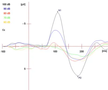
Another dipole model was computed for the 80, 90 and 100 dB intensities and a third regional source was added in the frontal region. A total of six equivalent dipoles for each evoked potential was computed. The tangential dipoles 1 and 3 represented the generators of the evoked potentials localizing in the primary auditory cortex. Dipoles 2 and 4 represent the generators in the secondary auditory cortex. The source waveforms of dipole 1 and 3 were used to compute the loudness dependence. The methods have been published in detail (Hegerl and Juckel 1993; Hegerl 1994; Hegerl et al. 1994; Hegerl and Juckel 1994). Additionally, N1 and P2 peaks were determined at the Cz electrode with an average reference and re-referenced to linked mastoids (Fig.2).
LDAEP processing
The LDAEP is the median of all slopes of each possible connection between the five different N1/P2 amplitudes corresponding to the five different intensities. The LDAEPs of the tangential dipole activity as well the LDAEPs computed of the Cz electrodes were used as the main variables for statistical evaluation. A recent publication underlines the need for processing both the data from single electrode estimation and DSA (Hagenmuller et al. 2011). The data were un-blinded after processing of the DSA and the identification of the N1 and P2 peaks in the DSA- and single-electrode data.
Statistical analysis
The data of one participant of the male levodopa group was removed due to extreme values. The remaining variables were analyzed for normal distribution with the Kolmogorov– Smirnov test. Group differences (levodopa vs. placebo) were assessed by unpaired t tests. Differences between the two measurements within the groups were tested with paired t tests.
Results
Kolmogorov–Smirnov test revealed a normal distribution for LDAEP measurements at baseline for the single electrode estimation data (mean 0.11 μV, SD 0.075 μV, p .458) as well as the DSA data (left tangential source: mean, 1.039 nA/m; SD, 0.767 nA/m; p .87; right tangential source mean, 1.212 nA/m; SD, 0.949 nA/m; p .54).
After unblinding the data, it turned out that there were 11 females and nine males in the levodopa group and ten females and 11 males in the placebo group.
No significant differences within the groups were found, independently of the application of levodopa or placebo, gender or the use of single electrode estimation or DSA (Table 1; Figs. 3and4).
Fig. 1 Symmetrical dipole sources of auditory evoked potentials following stimulation with 70 dB localized in Brodmann area 41 (primary
auditory cortex, Talairach coordinates−38, −25, 11 and 38, −25, 11)
Fig. 2 Example of single subject auditory evoked potentials following stimulation with different sound pressure levels (60 to 100 dB). Single electrode data from Cz
Table 1 Within-group differences of N1/P2 slopes (expressed as mean ± SD): paired t test (two-tailed) for within-group differences (within the levodopa and placebo groups) between baseline and post intervention measurement
Dipole ((nA/m)/10 dB) Cz (uV/10 dB)
Right Left
Levodopa group 0.074±1.07 0.098±0.58 0.008±0.34
Placebo group 0.009±0.36 0.004±0.56 0.005±0.04
Cz indicates data from single electrode estimation, dipole (e.g. left dipole) means the tangential dipoles
Irrespective of the estimation method or the time point of the measurement (Table2), no significant differences in the LDAEP were found between the groups.
The test–retest reliability (Cronbachs Alpha) between the baseline measurement and the intervention group was 0.964 (levodopa) and 0.9 (placebo) for the Cz-estimation and 0.911 (levodopa left tangential dipole), 0.67 (levodopa right tangential dipole), 0.759 (placebo left tangential dipole) 0.923 (placebo right tangential dipole).
Discussion
We had hypothesized that the acute synaptic dopamine excess caused by levodopa intake is associated with an intraindividual decrease of the LDAEP. In contrast to this hypothesis, a difference between the baseline measurement and the intervention was neither found in the levodopa nor in the placebo group. Hence, a synaptic dopaminergic excess caused by levodopa intake did not lead to an acute LDAEP alteration. Furthermore, the test–retest reliability between the baseline measure-ment and the intervention group states a high correlation between the two measurement conditions. Despite our negative result, the high test–retest reliability between the baseline and intervention group strengthens the LDAEP as a reliable neurophysiologic parameter with high individual stability.
We can only speculate about the cause for the increase in variance of the left dipole source after challenge with levodopa (Fig. 3). In the levodopa intervention group, some volunteers presented with nausea, which may have affected the data quality. However, this does not explain the exclusive increase in the left dipole. Another trial of our group (Wyss 2009) shows similar findings; in patients with schizophrenia, a larger variance in the amplitudes of left source waveforms was found. The healthy control group, however, showed no such increased variance. It is conceivable that a dopaminergic influence on the LDAEP exists in single subjects which only shows in a variance increase and not in the mean of the data. Assuming such an individual sensitivity exists, an examination of parameters influenc-ing the LDAEP such as the COMT polymorphism would be interesting. Our result stands in line with other findings in the literature; O'Neill et al. (O'Neill et al.2006,2008b) did not find any effect of acute dopamine receptor stimulation with pergolide or bromocriptine on the LDAEP. In the studies using dopamine receptor agonists and in our trial an iatrogenic condition as a state factor is used. So far studies suggesting a dopaminergic influence on the LDAEP (Juckel et al.2008b; Kawohl et al.2008a; Pogarell et al. 2004) show an association of the LDAEP with trait factors, i.e. genetic variants in dopamine path-ways. The LDAEP may rather present a chronic condition dependent on genetic polymorphisms associated with the 5-HT/dopamine system (O'Neill et al.2008a). In line with this is a recent study by Lee (Lee et al. 2010) using SPECT to approximate the availability of DATs and SERTs. It provided further evidence for the possible involvement of dopamine and serotonin in the genesis of LDAEP. The correlation between monoamine transporter availability and LDAEP could reflect the long-term monoaminergic activity.
Fig. 4 Boxplot showing dipole source strengths [nA/m] of levodopa and placebo group for the right dipole source (mad levodopa, pla placebo, nat baseline measurement, int intervention, dsar dipole source analysis right)
Fig. 3 Boxplot showing dipole source strengths [nA/m] of levodopa and placebo group for the left dipole source (mad levodopa, pla placebo, nat baseline measurement, int intervention, dsal dipole source analysis left)
Another recent study revealed an effect of a chronic modulation of serotonergic neurotransmission on the LDAEP (Simmons et al.2011).
The LDAEP may therefore be considered to be a chronic indicator of the 5-HT system and possibly the dopamine system. Further studies are needed to determine if this also applies for the dopamine system. This condition may be robust against short-term changes in neurotransmission, particularly dosages used in neuropsychopharmacology (Norra et al. 2008; O'Neill et al. 2008a). Thus, it seems questionable whether the LDAEP can actually be influ-enced by short challenge trials in humans. Considering the LDAEP as a chronic parameter, there is evidence support-ing a positive relationship between personality traits such as sensation seeking as well as impulsivity and the LDAEP; higher scores on measures of sensation seeking have been reported to be associated with an increased LDAEP and possibly a lower 5-HT function (Brocke et al.2000). Norra et al. found a correlation between an increased LDAEP and aspects of impulsiveness in patients with borderline personality (Norra et al.2003). Interestingly, dopaminergic dysfunction seems to play an important role in borderline personality disorder, affecting traits such as impulsivity. Dopamine receptor polymorphism have been associated with traits such as novelty seeking (Ebstein et al. 2000; O'Neill et al. 2008a). In line with the aforementioned supposition of the LDAEP acting as a chronic marker of 5-HT functioning is the finding of the individual LDAEP in depressed patients not being altered after medical treatment with a selective serotonin reuptake inhibitor (SSRI) (Gallinat et al. 2000; Lee et al. 2005) after clinical improvement of depressed symptoms. It has been shown that general anxiety disorder (GAD) patients with a stronger pretreatment LDAEP show a better response to a treatment with the SSRI escitalopram (Park et al.2011). Measurement of the LDAEP was said to provide useful clinical information for predicting treatment responses in patients with GAD. Juckel et al. (Juckel et al.2008a) postulate that the LDAEP may be more closely related to genetic variants
controlling the 5-HT release (5-HT auto receptors) and synthesis (tryptophan hydroxylase) rather than the reuptake. In a study by the same study group, 5-HT1B
alleles were reported to be associated with an increased LDAEP (Juckel et al. 2008a). The 5-HT1B receptor is
located presynaptically on serotonergic axon terminals. It occurs in high densities in the primary auditory cortex and in the brainstem raphe nuclei (Moret and Briley2000). In addition to controlling 5-HT release in terminal areas, this receptor inhibits the release and synthesis of 5-HT and reduces the firing rate of serotonergic neurons via a negative feedback loop in the raphe nuclei (Moret and Briley 2000). Interestingly, 5-HT1B receptors can also
function as a heteroreceptor at non-serotonergic neurons controlling the release of neurotransmitters other than 5-HT (such as dopamine, acetylcholine and glutamate) (Moret and Briley 2000). Thus, the 5-HT and dopamine interactions and their possibly influence on the LDAEP may be complex.
Our study is not without limitations. Ideally, blood samples should have been taken to estimate the individual levodopa blood levels at its estimated tmax
of 60 min after oral intake. Due to practical reasons, we refrained from conducting a crossover study. Nevertheless, we do not expect any additional outcome by such a design. Other limitations regarding the statistical power are the rather small sample size and low dosage of levodopa. Possibly a higher dosage of levodopa or a bigger sample size could have shown different results. Nevertheless, based upon our data, a number of 3,124 persons per group would be needed (power 0.9) in order to show significance within group differences given these results. This is clearly not practicable. On the other hand, our sample size would have been large enough to detect a moderate effect of 0.5 with a power of 48.2% or a larger effect of 0.8 with a power of 82% (Cohen 1992). Additionally, other dopaminergic influences associated with the LDAEP like the COMT polymorphism could have been examined.
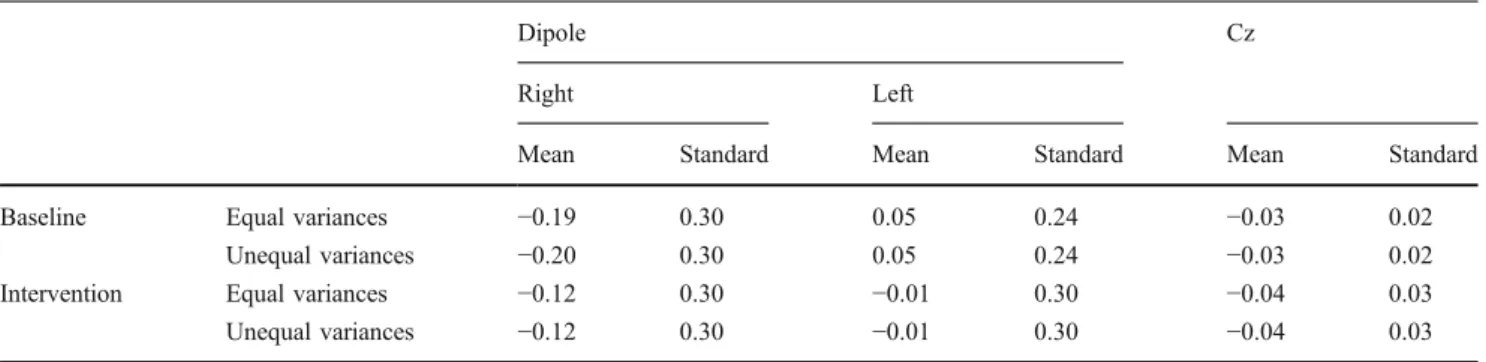
Table 2 Between-group differences (expressed as mean: mean difference, standard: standard error difference); unpaired t test (two-tailed) for differences between the placebo and the levodopa group (baseline and intervention)
Dipole Cz
Right Left
Mean Standard Mean Standard Mean Standard
Baseline Equal variances −0.19 0.30 0.05 0.24 −0.03 0.02
Unequal variances −0.20 0.30 0.05 0.24 −0.03 0.02
Intervention Equal variances −0.12 0.30 −0.01 0.30 −0.04 0.03
Unequal variances −0.12 0.30 −0.01 0.30 −0.04 0.03
Conclusion
Our study shows an absence of acute dopaminergic influence due to a single intake of levodopa on the LDAEP. Our findings are in agreement with the findings of O`Neill et al. (O'Neill et al. 2006, 2008b) stating the absence of acute dopaminergic influence on the LDAEP. To our knowledge, our study is the first trial having computed the data via dipole source analysis and Cz estimation.
As dopamine possibly may be a chronic parameter concerning the LDAEP, studies with, e.g. longer dopamine intake in healthy volunteers may be needed to examine a dopaminergic influence on the LDAEP.
Acknowledgements The authors would like to thank Johannes
Schley for support with the voluntary server of the University of Zürich and Barbara Rauscher and her team for taking the blood samples. This study has been funded by an unrestricted grant of Bristol-Myers Squibb. The authors disclose any further conflict of interest.
References
Axelrod J, Tomchick R (1958) Enzymatic O-methylation of epinephrine
and other catechols. J Biol Chem 233:702–705
Baribeau JC, Laurent JP (1987) The effect of selective attention on augmenting/intensity function of the early negative waves of
AEPs. Electroencephalogr Clin Neurophysiol Suppl 40:68–75
Black KJ, Mink JW (2000) Response to levodopa challenge in
Tourette syndrome. Mov Disord 15:1194–1198
Brocke B, Beauducel A, John R, Debener S, Heilemann H (2000) Sensation seeking and affective disorders: characteristics in the intensity dependence of acoustic evoked potentials. Neuropsy-chobiology 41:24–30
Cohen J (1992) A power primer. Psychol Bull 112(1):155–159 de la Fuente-Fernandez R, Sossi V, Huang Z, Furtado S, Lu JQ, Calne
DB, Ruth TJ, Stoessl AJ (2004) Levodopa-induced changes in synaptic dopamine levels increase with progression of Parkinson's
disease: implications for dyskinesias. Brain 127:2747–2754
Ebstein RP, Benjamin J, Belmaker RH (2000) Personality and polymorphisms of genes involved in aminergic
neurotransmis-sion. Eur J Pharmacol 410:205–214
Floel A, Breitenstein C, Hummel F, Celnik P, Gingert C, Sawaki L, Knecht S, Cohen LG (2005) Dopaminergic influences on
formation of a motor memory. Ann Neurol 58:121–130
Funke B, Malhotra AK, Finn CT, Plocik AM, Lake SL, Lencz T, DeRosse P, Kane JM, Kucherlapati R (2005) COMT genetic variation confers risk for psychotic and affective disorders: a case control study. Behav Brain Funct 1:19
Gallinat J, Bottlender R, Juckel G, Munke-Puchner A, Stotz G, Kuss HJ, Mavrogiorgou P, Hegerl U (2000) The loudness dependency of the auditory evoked N1/P2-component as a predictor of the acute SSRI response in depression. Psychopharmacology (Berl) 148:404–411
Hagenmuller F, Hitz K, Darvas F, Kawohl W (2011) Determination of the Loudness Dependence of Auditory Evoked Potentials (LDAEP): single electrode estimation versus dipole source
analysis. Hum Psychopharmacol Clin Exp 26:147–154
Hegerl U (1994) Event-related potentials in psychiatry: a new
perspective. Pharmacopsychiatry 27:47–48
Hegerl U, Juckel G (1993) Intensity dependence of auditory evoked potentials as an indicator of central serotonergic neurotransmission: a new hypothesis. Biol Psychiatry 33:173–187
Hegerl U, Juckel G (1994) Auditory evoked dipole source activity: indicator of central serotonergic dysfunction in psychiatric patients? Pharmacopsychiatry 27:75–78
Hegerl U, Juckel G (2000) Identifying psychiatric patients with serotonergic dysfunctions by event-related potentials. World J
Biol Psychiatry 1:112–118
Hegerl U, Gallinat J, Mrowinski D (1994) Intensity dependence of auditory evoked dipole source activity. Int J Psychophysiol
17:1–13
Hegerl U, Gallinat J, Juckel G (2001) Event-related potentials. Do they reflect central serotonergic neurotransmission and do they predict clinical response to serotonin agonists? J Affect Disord
62:93–100
Juckel G, Molnar M, Hegerl U, Csepe V, Karmos G (1997)
Auditory-evoked potentials as indicator of brain serotonergic activity—first
evidence in behaving cats. Biol Psychiatry 41:1181–1195
Juckel G, Hegerl U, Giegling I, Mavrogiorgou P, Wutzler A, Schuhmacher C, Uhl I, Brune M, Mulert C, Pogarell O, Rujescu D (2008a) Association of 5-HT1B receptor polymorphisms with the loudness dependence of auditory evoked potentials in a community-based sample of healthy volunteers. Am J Med Genet
B Neuropsychiatr Genet 147B:454–458
Juckel G, Kawohl W, Giegling I, Mavrogiorgou P, Winter C, Pogarell O, Mulert C, Hegerl U, Rujescu D (2008b) Association of catechol-O-methyltransferase variants with loudness depen-dence of auditory evoked potentials. Hum Psychopharmacol
23:115–120
Kawohl W, Waberski TD, Darvas F, Norra C, Gobbele R, Buchner H (2007) Comparative source localization of electrically and pressure-stimulated multichannel somatosensory evoked potentials.
J Clin Neurophysiol 24:257–262
Kawohl W, Giegling I, Mavrogiorgou P, Pogarell O, Mulert C, Moller HJ, Hegerl U, Rujescu D, Juckel G (2008a) Association of functional polymorphisms in NOS1 and NOS3 with loudness dependence of auditory evoked potentials. Int J Neuropsycho-pharmacol 11:477–483
Kawohl W, Hegerl U, Muller-Oerlinghausen B, Juckel G (2008b) Insights in the central serotonergic function in patients with affective disorders. Neuropsychiatry 22:23–27
Khor SP, Hsu A (2007) The pharmacokinetics and pharmacodynamics of levodopa in the treatment of Parkinson's disease. Curr Clin
Pharmacol 2:234–243
Kumakura Y, Danielsen EH, Reilhac A, Gjedde A, Cumming P (2004) Levodopa effect on [18F]fluorodopa influx to brain: normal volunteers and patients with Parkinson's disease. Acta Neurol
Scand 110:188–195
Lee TW, Yu YW, Chen TJ, Tsai SJ (2005) Loudness dependence of the auditory evoked potential and response to antidepressants in Chinese patients with major depression. J Psychiatry Neurosci
30:202–205
Lee IH, Yang YK, Chen PS, Huang HC, Yeh TL, Lu RB, Chiu NT, Yao WJ, Lin SH (2010) Loudness dependence of auditory evoked potentials (LDAEP) correlates with the availability of dopamine transporters and serotonin transporters in healthy volunteers—a two isotopes SPECT study. Psychopharmacology (Berl) 214:617–624
Meyer-Lindenberg A, Weinberger DR (2006) Intermediate phenotypes and genetic mechanisms of psychiatric disorders. Nat Rev
Neurosci 7:818–827
Micallef-Roll J, Rihet P, Hasbroucq T, Possamai C, Blin O (2001) Levodopa-induced drowsiness in healthy volunteers: results of a choice reaction time test combined with a subjective evaluation
Moret C, Briley M (2000) The possible role of 5-HT(1B/D) receptors in psychiatric disorders and their potential as a target for therapy.
Eur J Pharmacol 404:1–12
Nicodemus KK, Kolachana BS, Vakkalanka R, Straub RE, Giegling I, Egan MF, Rujescu D, Weinberger DR (2007) Evidence for statistical epistasis between catechol-O-methyltransferase (COMT) and polymorphisms in RGS4, G72 (DAOA), GRM3, and DISC1: influence on risk of schizophrenia. Hum Genet
120:889–906
Norra C, Mrazek M, Tuchtenhagen F, Gobbele R, Buchner H, Sass H, Herpertz SC (2003) Enhanced intensity dependence as a marker of low serotonergic neurotransmission in borderline personality
disorder. J Psychiatr Res 37:23–33
Norra C, Becker S, Brocheler A, Kawohl W, Kunert HJ, Buchner H (2008) Loudness dependence of evoked dipole source activity during acute serotonin challenge in females. Hum
Psychophar-macol 23:31–42
O'Neill BV, Croft RJ, Leung S, Guille V, Galloway M, Phan KL, Nathan PJ (2006) Dopamine receptor stimulation does not modulate the loudness dependence of the auditory evoked potential in humans. Psychopharmacology (Berl) 188:92–99 O'Neill BV, Croft RJ, Nathan PJ (2008a) The loudness dependence of
the auditory evoked potential (LDAEP) as an in vivo biomarker of central serotonergic function in humans: rationale, evaluation and review of findings. Hum Psychopharmacol 23:355–370 O'Neill BV, Guille V, Croft RJ, Leung S, Scholes KE, Phan KL,
Nathan PJ (2008b) Effects of selective and combined serotonin and dopamine depletion on the loudness dependence of the
auditory evoked potential (LDAEP) in humans. Hum
Psycho-pharmacol 23:301–312
Park YM, Kim DW, Kim S, Im CH, Lee SH (2011) The loudness dependence of the auditory evoked potential (LDAEP) as a predictor of the response to escitalopram in patients with generalized anxiety disorder. Psychopharmacology (Berl) 213:625–632
Pogarell O, Tatsch K, Juckel G, Hamann C, Mulert C, Popperl G, Folkerts M, Chouker M, Riedel M, Zaudig M, Moller HJ, Hegerl U (2004) Serotonin and dopamine transporter availabilities correlate with the loudness dependence of auditory evoked
potentials in patients with obsessive–compulsive disorder.
Neuro-psychopharmacology 29:1910–1917
Simmons JG, Nathan PJ, Berger G, Allen NB (2011) Chronic modulation of serotonergic neurotransmission with sertraline attenuates the loudness dependence of the auditory evoked potential in healthy participants. Psychopharmacology (Berl)
217:101–110
Uhl I, Gorynia I, Gallinat J, Mulert C, Wutzler A, Heinz A, Juckel G (2006) Is the loudness dependence of auditory evoked potentials modulated by the selective serotonin reuptake inhibitor citalopram in healthy subjects? Hum Psychopharmacol 21:463–471
Waberski TD, Gobbele R, Kawohl W, Cordes C, Buchner H (2003) Immediate cortical reorganization after local anesthetic block of the thumb: source localization of somatosensory evoked potentials
in human subjects. Neurosci Lett 347:151–154
Wyss C (2009) The role of central serotonergic activity in schizo-phrenic negative symptoms. Master Thesis, University of Zurich
![Fig. 4 Boxplot showing dipole source strengths [nA/m] of levodopa and placebo group for the right dipole source (mad levodopa, pla placebo, nat baseline measurement, int intervention, dsar dipole source analysis right)](https://thumb-eu.123doks.com/thumbv2/123doknet/14844798.626804/5.892.77.437.76.369/boxplot-strengths-levodopa-levodopa-baseline-measurement-intervention-analysis.webp)