Antibodies and methods for immunohistochemistry
of extracellular matrix proteins
The MIT Faculty has made this article openly available.
Please share
how this access benefits you. Your story matters.
Citation
Rickelt, Steffen and Richard O. Hynes. "Antibodies and methods
for immunohistochemistry of extracellular matrix proteins." 71-72
(October 2018): 10-27. © 2018 Elsevier B.V.
As Published
http://dx.doi.org/10.1016/j.matbio.2018.04.011
Publisher
Elsevier BV
Version
Author's final manuscript
Citable link
https://hdl.handle.net/1721.1/126498
Terms of Use
Creative Commons Attribution-NonCommercial-NoDerivs License
Antibodies and Methods for Immunohistochemistry of Extracellular
Matrix Proteins
Steffen Rickelta and Richard O. Hynesa,b,c
aDavid H. Koch Institute for Integrative Cancer Research, Massachusetts Institute of Technology, Cambridge, MA 02139, USA
bHoward Hughes Medical Institute, Chevy Chase, MD 20815, USA
cDepartment of Biology, Massachusetts Institute of Technology, Cambridge, MA 02139, USA
Correspondence to:
Richard O. Hynes, David H. Koch Institute for Integrative Cancer Research, Massachusetts Institute of Technology, 77 Massachusetts Avenue, Room 76-361D, Cambridge, MA 02139,
Steffen Rickelt, David H. Koch Institute for Integrative Cancer Research, Massachusetts Institute of Technology, 77 Massachusetts Avenue, Room 76-343, Cambridge, MA 02139,
Highlights
• Advice and literature on validation, characterization and use of antibodies in immunohistochemistry (IHC)
• Lists of ~200 tested and validated antibodies to ~100 ECM proteins
• Illustrative examples of the application of these antibodies and methods to FFPE tissue sections
Abstract
The diversity of extracellular matrix (ECM) proteins encoded in mammalian genomes and detected by proteomic analyses generates a need for well validated antibodies against these proteins. We present characterization of a large number of antibodies against ECM proteins, from both commercial and academic sources, together with discussion of methods and strategies for their effective use in immunohistochemistry and illustrations of their efficacy. These data should be of value to investigators seeking well validated antibodies to ECM proteins of interest and save significant time and money tracking down effective reagents.
Keywords
extracellular matrix antibodies, antibody validation, immunohistochemistry
Abbreviations
Ab, antibody; BL, basal lamina; CRC, colorectal cancer; ECM, extracellular matrix; FFPE, formaldehyde-fixed and paraffin-embedded; HIER, heat-induced epitope-retrieval; TMA, tissue microarray
Introduction
The extracellular matrix (ECM) is a fundamental component that surrounds or underlies most cells and tissues of metazoan organisms [1,2].ECM provides both biophysical and biochemical signals important for cell proliferation, cell apoptosis, differentiation and migration [3,4]. Moreover, the ECM also serves as a depot for soluble growth factors and
ECM-remodeling enzymes, whose release can be triggered by different physiological conditions [5,6]. These functions of ECM play crucial roles in embryonic development, homeostasis and in many diseases, such as fibroses, wound healing and scar formation, skeletal disorders and cancer [7-9].
Because of the importance of ECM, it is necessary to understand the composition and dynamics of ECM in various normal and disease states. This has been a challenge because of the
insolubility of ECM but recent proteomic approaches have revealed in detail the complexity of the ECM in vivo, typically comprising at least 150-200 proteins in any tissue [e.g., 10-20]. These studies have provided lists of potential biomarkers for disease states such as tumors,
metastases or fibroses. However, proteomic analyses are not practical for routine use as biomarkers for diagnostic or prognostic applications and there is a need for higher throughput methods to expand screens to larger numbers of samples than are feasible by proteomics. Furthermore, standard proteomic analyses do not provide information about distributions of the proteins detected. Both of these requirements can, in principle, be addressed by antibody-mediated detection methods. However, for many ECM proteins, validated antibodies are limited, although some do exist for well studied major ECM proteins, although by no means all. Genomic and proteomic studies have revealed that there are around 300 ECM proteins
encoded in mammalian genomes plus many other proteins that are incorporated into or bound by ECM, such as ECM modulators (modifying enzymes, crosslinkers, proteases) and growth factors and cytokines; collectively the “matrisome” [21]. This complexity necessitates a much broader range of well validated anti-ECM antibodies than has been available.
Immunohistochemistry (IHC) is a widely used technique and can be performed on
formaldehyde-fixed and paraffin-embedded (FFPE) tissues allowing access to archival material [22-24]. A key advantage of IHC is that it allows analysis not only of the anatomy of the tissue of interest but also visualization of the spatial distribution and expression of specific antigens or cellular components in a variety of tissue sections. However, the availability of well validated anti-ECM antibodies applicable to FFPE samples is even more limited. For all these reasons,
we have tested numerous antibodies to ECM proteins and developed reliable IHC staining protocols for use on FFPE tissue samples. Here we first provide general insights and
recommendations for appropriate antibody characterization needed to set up robust validation and reproducible IHC staining protocols. We then summarize our results on the applicability of over 200 antibodies (most of them commercially available) to over 100 matrisome proteins. In Table 1 we list all antibodies which yielded positive results and in Supplementary Table 1 all antibodies which, in our experiments, either did not yield positive stains or gave uncertain staining results. We further show examples of data for ECM proteins of particular interest in order to illustrate some key aspects relevant to effective application of IHC to ECM proteins (Figures1-8). In addition, we provide a summary of antibodies that are more broadly used in tumor biology and histopathology to determine specific tissue distribution or subcellular localization (Supplementary Table 2). However, we wish to note that while we have tested antibody specificity at the IHC level by the criteria discussed below, we have only tested the molecular specificity of antibodies generated in our own laboratory and some others but not the majority of the commercial antibodies reported here. Finally, we provide useful online references which will assist antibody searching, IHC methodology and troubleshooting guidelines as well as tissue expression data of protein-coding genes in normal tissue and tumor samples from
humans and, in some cases, mice (Supplementary Table 3).
Antibody reproducibility issues
Although antibodies are among the most frequently used tools in basic science research and in clinical assays, standardized methods for antibody validation across research applications are not universally applied. As a result, the quality and consistency of data generated through the use of antibodies can vary greatly and increasing concerns have been raised about
reproducibility of scientific data using commercially available antibodies [25-30]. Indeed, the high failure rate of commercially available antibodies is concerning and it has been estimated that US$800 million are wasted annually on poorly performing antibodies and about US$350 million are lost in biomedical research because published results cannot be replicated, with underperforming antibodies being a contributor in many cases [31].
Antibody validation recommendations
Although the quality of diagnostic and research antibodies remains a problem [27, 32-34], several attempts to improve antibody validation have been made and very useful
recommendations have been published [e.g., 26, 30, 35-38]. Recently, the International Working Group for Antibody Validation (IWGAV) was established with the goal of improving the
standardization and validation of antibodies used in common research applications [28]. This consortium proposed a set of standard guidelines that should help to ensure better antibody reproducibility.
Five conceptual recommendations for antibody validation are: (1) genetic strategies, such as testing the antisera against mutant tissues or cells known to lack the protein of interest (2) orthogonal strategies, using an antibody-independent method such as mass spectrometry to compare the expression of a target protein (3) independent antibody strategies, that is, use of two or more antibodies, ideally from different host species and with non-overlapping epitopes (4) testing against expressed recombinant proteins, and (5) immunocapture followed by mass spectrometry (see [28] for detailed information). At least one of these approaches and
preferably more should be used as a minimum criterion for claiming that a particular antibody has been adequately validated for a specific application.
Additional methods for antibody validation include affinity measurements, epitope mapping, isotype or sequence determination and array-based specificity [28]. Another validation method commonly used for IHC applications is absorption [39], in which the antigen is pre-incubated with the antibody before the immunoassay. Although this method is useful to show target binding, it will not rule out cross-reactivity with proteins containing epitopes similar to those in the intended target. To validate an antibody for IHC, it must be shown to be specific and reproducible in the tissue context in which it will be used. Ideally, experiments using several independent antibodies (if possible raised in different species) to stain for the same molecule should be sought to show specificity, selectivity and reliability on FFPE tissue samples. This approach is more convincing and the data more reliable than using a single antibody [26]. Finally, it is recommended to repeat validation experiments for each new antibody vial or lot to ensure reproducibility.
Immunohistochemistry standardization
Immunohistochemistry (IHC) can be summarized in three major steps: (1) binding of the primary antibody to a specific antigen, (2) formation of an antibody-antigen complex following the
addition of a secondary enzyme-conjugated detection antibody and finally, (3) a chromogenic enzyme substrate which leads to generation of a colored deposit at the site of the antibody-antigen complex. However, especially in the context of biomarker development on FFPE tissues
using IHC, standardization can be quite challenging. This is due to several factors known to affect antigenicity and influence subsequent staining procedures, including; varying fixation times, differences in fixative used as well as general handling variability throughout the labeling process. In this context, the need for improved standardization of IHC protocols has been addressed and recommendations and guidelines for IHC have been proposed to achieve reliable and consistent staining results [e.g., 25, 40, 41].
Antigen “recovery” or “retrieval”
Heat-induced epitope-retrieval (HIER) techniques, first developed by Shi et al. [42], are often employed to reveal epitopes that are otherwise difficult to detect following fixation in formalin and paraffin-embedding. Unfortunately, in practice, the application of IHC to routine processed FFPE sections has still proven to be challenging with significant inconsistency of results.
For this reason and to achieve the best staining results, each individual antibody presented here was tested following the recommendations and standardization guidelines for improved antibody validation and IHC optimization mentioned above. Following the proposed ‘test battery’
approach of Shi et al. [43], we screened the individual antibodies for the major variables involved in antigen retrieval. These include heating conditions (temperature and heating time), pH of the retrieval solution, and the retrieval buffer (for details see Supplementary Materials and Methods). Various pH values (pH 1, 6, and 9) of different buffer solutions (including acetate, citrate, phosphate and tris) and different temperatures (high, 120°C using decloaking
chamber/pressure cooker or lower using a microwave) can be tested to find optimal staining results.
For most ECM-related antibodies tested, our observations favored HIER for 2 min at 120°C in a decloaking chamber (i.e., pressure cooker) using one of two different buffer conditions, i.e., 10 mM sodium-citrate (pH6.0) or 10 mM Tris (pH9.0) containing 0.05% Tween-20. The preferred condition and dilutions for each antibody tested as well as control tissues used are listed in Table 1. However, we emphasize that each antibody vial, whether in use or newly purchased, should be tested to obtain the best possible results for the tissue of interest. An antibody validated in one buffer system will not necessarily perform similarly in another and may vary depending on the sample used. Moreover, to assess reproducibility of IHC, validated antibody should be run on a variety of tissue samples, either as whole tissue sections or represented on a tissue microarray (TMA; [44]). For the majority of antibodies, we used a panel of previously reported FFPE mouse and human control tissue samples and tested different antibody
concentrations to obtain optimal staining results. This allows comparison of antibody
localizations described in the literature with individual staining patterns obtained to assess the selectivity of each antibody as well as the reproducibility of localization data. Of most
importance, every experiment should include a positive and negative control to assess performance of antibody and reagents used, ideally using a set of tissue samples with known variable expression levels of the protein of interest [45].
Validation of antibodies to extracellular matrix molecules
Using the established IHC protocols, we have tested numerous antibodies against ECM proteins and summarize our results on the applicability of over 200 antibodies to over 100 matrisome proteins on FFPE samples (see Table 1 for antibodies which yield positive results and Supplementary Table 1 for antibodies with negative or uncertain staining results). Again, we wish to note that we have tested specificity at the IHC level and we have not tested the
molecular specificity of every antibody reported here. Readers should also refer to the results provided by the suppliers’ datasheets and in the references cited. We would recommend that users particularly interested in specific ECM proteins should also test by molecular methods such as immnunoblotting, immunoprecipitation or absorption.
Finally, since ECM molecules are frequently cross-linked in complexes mediated by multiple domains specialized for protein interactions or specific ECM assembly forms, we have found protease treatment of FFPE sections to be very helpful in improving IHC detection of ECM proteins. We tested several methods for enzyme digestion in order to enhance the antigen exposure and staining results. Commonly used enzymes include trypsin, proteinase K, pronase, ficin, and pepsin [for review see 23]. Enzyme digestion may destroy some epitopes and
increases the chance of damaged tissue morphology. However, in particular an additional enzyme digestion step using 0.2% pepsin in 0.2N HCl (Agilent) according to the manufacturers protocol prior to the immunostaining, improved the IHC results for several antibodies (see e.g., Figs. 3-5 and Table 1; see also Franciosi et al. [46]).
The Figures 1-8 which follow illustrate the application of the suggestions discussed above for a variety of ECM proteins and experimental situations and additional examples are included for reference in the Supplementary Material.
Figures and figure legends Figure 1
Figure 1. Antibodies to individual laminin chains in colorectal cancer (CRC) liver metastasis Images of representative CRC liver metastasis sections stained with hematoxylin and eosin (H&E, panel A) and the pan-laminin rabbit polyclonal antibody (Novus Biologicals, #NB300-144, panel B), reported to recognize Lm111 and Lm211. Several such polyclonal antibodies exist and stain most basal laminae (BL), as would be expected if they recognize 3-4 different laminin subunits, which among them occur in most laminins (see also Supplementary Figure Gallery pages 77-80). Such antibodies are valuable as markers of BL. However, in order to define precisely which of the 11 known laminin subunits (see [47] for laminin nomenclature) exist in any given BL, more specific reagents are necessary. We tested and validated ten monoclonal antibodies (mAb) and one rabbit polyclonal antibody to 10 of the subunits (see Table 1 and Supplementary Table 1) and
representative examples are shown here (the others are shown in Supplementary Figure Gallery pages 81-91). Panels C and D show that the B1 and B2 subunits co-localize in the same BL (B3, found only in Lam332, does not, data not shown). Panels E-H illustrate the diversity of expression in different BL of the A4 and A5 subunits; A5 is found in the same BL as B1 and B2 in one metastasis (panel F, cf. C and D, see *) but not in a metastasis from a different patient (H), whereas A4 is found in the second patient but not the first (E, I). Clearly, full analyses of laminin distributions will require diverse monoclonal antibodies as well as molecular methods. Similar variations in distributions of other BL markers in CRC metastases are shown in Fig. 2.
Figure 2
Figure 2. Diversity of basal lamina composition in normal liver and CRC liver metastasis IHC on sections of CRC liver metastases from two different patients exhibit different patterns of distribution of collagen IV (panels A, B) and perlecan (HSPG2, panels C, D), two canonical and well-studied basal laminae (BL) components found in healthy and diseased tissues [48,49]. One
metastasis shows the typical pattern of BL staining of collagen IV in both the metastasis (T) and the normal liver (L, panel A). In addition, however, the tumor edge of the same metastasis (*) and the metastasis of patient #2 (panel B) show, in addition, widespread staining for collagen IV in the extracellular matrix of the metastasis. In the case of perlecan, it is often seen elsewhere than in BL (see Supplementary Figure Gallery pages 73-74) and that is true for both the metastases shown here, although the patterns of localization differ both between metastasis and liver (C) and between the two patients’ metastases (C, D).
Figure 3
Figure 3. Use of pepsin treatment to enhance antibody detection of extracellular matrix proteins
As mentioned in the text, extracellular matrix proteins are highly complexed and crosslinked; this can impede access of antibodies to their epitopes and protease digestion can often help to reveal
extracellular matrix antigens that are otherwise occluded [46]. Figure 3 shows this to be the case for several different extracellular matrix proteins. Representative images of mouse and human tissue sections stained for collagen IV (A, B), collagen III (C, D), collagen VII (E, F), LTBP1 (G, H), HMCN1 (I, J) and AGRN (K, L) are shown. In each case, treatment with pepsin reveals the presence of the extracellular matrix protein, which is not, or only weakly, detected using heat-induced epitope-retrieval (HIER) alone. Such protease treatments need to be used with care since over-digestion can destroy epitopes and can lead to damage to the tissue sections. Different protease conditions need to be tested to obtain optimal staining results for each antibody and tissue (see also text). It is worth noting that panels I and J show an example where pepsin treatment reveals hemicentin (HMCN1) staining of the extracellular matrix (J), while at the same time removing cell-associated signal (I).
Figure 4
Figure 4. Differential detection of agrin using antibodies to different epitopes
The large extracellular matrix protein agrin is associated with many basal laminae (BL) and is
frequently upregulated in cancer. Antibodies to different epitopes of agrin have been reported to show differential distributions in kidney cryostat sections [e.g.,50,51]. However, IHC detection of agrin in formaldehyde-fixed and paraffin-embedded tissue sections has to date been challenging. As for the other extracellular matrix proteins described above, HIER combined with pepsin treatment greatly enhanced the staining efficacy of agrin antibodies (Fig. 3). However, even after pepsin treatment, anti-agrin antibodies to different epitopes (raised against N- and C-terminus of agrin) reveal diverse IHC staining patterns in several tissues (Fig. 4). Of particular note, although two different antibodies to agrin localize to all blood vessels in sections of mouse brain tissue (Fig. 4A and B), their staining pattern varies among a variety of tissues (Fig. 4C-H). Thus, while the BL of blood vessels (arrows in Fig. 4E, F), serving here as an internal positive control, are positive with both antibodies, the epithelial BLs appear negative for one antibody (panels E and G) while the other antibody clearly stains
positive (F and H). These results underline the value of using more than one antibody to detect extracellular matrix proteins reliably.
Figure 5
Figure 5. Uses of species-specific antibodies to define the sources of extracellular matrix proteins
In xenograft models, it is of particular interest to determine which extracellular matrix proteins are made by the transplanted cells or tissues and which by the host. In addition to the potential use of in
situ hybridization, species-specific antibody staining can provide valuable information. First,
antibodies that distinguish cells of the two species involved can be used to distinguish transplant from host (A, B). Here, antibodies to human-specific anti-Lamin are used to mark orthotopic transplants of human CRC organoids (T) to mouse colon (A) and their resulting liver metastases (B, see also [52]). Other human-specific antibodies, including anti-human vimentin or anti-human mitochondria
antibodies can similarly be used and we list several antibodies, useful for such analyses in
Supplementary Table 2. Second, species-specific antibodies to specific extracellular matrix proteins can be used to define their source as shown for agrin in panels C-F. Panels C and D show staining with an antibody recognizing both mouse and human agrin, which stains agrin in the basal laminae (BL, arrowheads) of the human CRC organoid transplant (T) and also in the blood vessels of the surrounding normal mouse colon and liver tissue (arrows). In contrast, antibody specific for mouse agrin (E, F) recognizes only the host blood vessels but not the BL of the human CRC organoid transplant (T). Clearly in this context the majority of agrin is made by the transplanted organoids (boxes mark regions selected for inserts).
Figure 6
Figure 6. Insights from immunohistochemistry extend proteomic characterizations of tumor-related extracellular matrix proteins
Proteomic analyses have identified a set of extracellular matrix proteins predominantly expressed in human primary CRC tumors or metastases to liver and little or not at all in normal colon and liver samples [14]. Staining with antibodies to a number of these extracellular matrix proteins not only confirms their presence in CRC metastases to liver (Fig. 6A) but also provides additional information (Fig. 6B). Many core matrix proteins are detected in the extracellular matrix as expected (e.g., LTBP2, THBS2, HMCN1) but some, including surfactant protein D (SFTPD), are intracellular and others such as SERPINE2 and TIMP1 show distributions either in the extracellular matrix or intracellularly, or both (B). The proteomic definition of these proteins as extracellular matrix-associated matrisome proteins
relies on their insolubility, the IHC results show that is not always a reliable indicator that they are actually bound to the extracellular matrix.
Figure 7
Figure 7. Spatial distributions of extracellular matrix proteins
Although many matrix molecules are detected by proteomics in CRC metastases to liver, IHC shows that tumor-associated extracellular matrix proteins (e.g., LTBP2 as shown here) can be located in different parts of the metastases. LTBP2 can be widely distributed throughout the extracellular matrix of the metastasis (A, B), or only in the surrounding capsule (C, D) or in localized parts of another metastasis (E, F). These different distributions are likely to have different biological consequences. Figure 8
Figure 8. Differential localization and expression patterns of extracellular matrix proteins during tumor progression
We also validated antibodies against proteins or specific splice variants of proteins often found during tumor development and metastasis. This figure shows two such proteins; tenascin C (B, E) and the EIIIB splice isoform of fibronectin (C, F) in human CRC-derived liver metastases (A-C) and in orthotopic CRC organoid transplants. Both proteins are clearly expressed in the tumor tissue (T) of the CRC liver metastases and at the invasive front (B, C) but are absent in the surrounding normal liver (L). Both proteins also show positive staining in the surrounding extracellular matrix of the CRC-derived organoids (T) and are present in the invading edges (inserts in D-F) of this CRC mouse model. Given the defined species specificity on human and mouse tissues for many antibodies (see Table 1), such antibodies can be used to study differential localizations in human tissue samples in comparison to available mouse models to decipher their potential involvement during tumor formation, progression and metastasis.
Useful links and resources
Since only limited numbers of antibodies to ECM proteins applied to FFPE samples have been described in the literature and we certainly do not cover all of them in our validation attempts, we have compiled a list of valuable online resources that will assist researchers in tracking down additional antibodies (Supplementary Table 3). Antibody product information and
validation data can sometimes be difficult to decipher and, with hundreds of vendors to choose from, it becomes difficult to know when a search has been exhaustive. The summarized search engines will help to find and identify antibodies that could work for a particular application, and in some instances, compare antibodies from many different vendors. This saves valuable time otherwise spent visiting each vendor’s website and allows extension of the search to novel suppliers. In addition, we provide useful online references relevant for proper antibody characterization, IHC standardization and methodology as well as troubleshooting guidelines (Supplementary Table 3).
Moreover, thanks to large open-access knowledge-based international efforts, such as the Human Protein Atlas (HPA, https://www.proteinatlas.org; [54-56]) or the GTEx consortium (https://www.gtexportal.org/, G.T. Consortium [57]) it is now possible to explore the expression of protein-coding genes in normal human tissues. In addition, the recently launched new open-access resource: Human Pathology Atlas as part of the HPA (www.proteinatlas.org/pathology), allows researchers to explore the possible prognostic value of all human protein-coding genes related to expression levels in different forms of human cancer, presenting Kaplan-Meier survival plots for all protein-coding genes in 17 different tumor types.
We also wish to highlight the Matrixome project,
(http://dbarchive.biosciencedbc.jp/archive/matrixome/bm/home.html; [58,59]), which contains the mouse basement membrane bodymap, a database of body-wide localizations of basal lamina proteins in developing mouse embryos. Finally, the Matrisome project
(http://matrisomeproject.mit.edu; [12,14]) provides, in addition to the methods and datasets available in current publications, valuable protocols and resources relevant to research on ECM proteins. In particular, the 2016 release of the ECM Atlas, an effort aimed at compiling mass spectrometry data from studies designed specifically to characterize the composition of the ECM of normal and diseased samples [14] is a useful resource to search for the distribution of ECM and ECM-associated molecules in a variety of tissues.
Conclusions
It is our hope that the guidance on reagents and methods and the illustrative examples of their application in studies of the ECM and its constituent and associated proteins will prove to be helpful to ECM scientists in their investigations. Not least, we hope that the data on well validated antibodies will save time and funds that would otherwise be expended finding appropriate antibodies for future investigations.
References
[1] R.P. Mecham, The Extracellular Matrix: an Overview. Springer Heidelberg., 2011. [2] R.O. Hynes, K.M. Yamada, Extracellular Matrix Biology. Cold Spring Harbor
Perspectives in Biology. New York., 2012.
[3] G. Charras, E. Sahai, Physical influences of the extracellular environment on cell migration, Nat Rev Mol Cell Biol 15(12) (2014) 813-24.
[4] J.D. Humphrey, E.R. Dufresne, M.A. Schwartz, Mechanotransduction and extracellular matrix homeostasis, Nat Rev Mol Cell Biol 15(12) (2014) 802-12.
[5] R.O. Hynes, The extracellular matrix: not just pretty fibrils, Science 326(5957) (2009) 1216-9.
[6] C.M. Nelson, M.J. Bissell, Of extracellular matrix, scaffolds, and signaling: tissue architecture regulates development, homeostasis, and cancer, Annu Rev Cell Dev Biol 22 (2006) 287-309.
[7] A. Aszodi, K.R. Legate, I. Nakchbandi, R. Fassler, What mouse mutants teach us about extracellular matrix function, Annu Rev Cell Dev Biol 22 (2006) 591-621.
[8] J.F. Bateman, R.P. Boot-Handford, S.R. Lamande, Genetic diseases of connective tissues: cellular and extracellular effects of ECM mutations, Nat Rev Genet 10(3) (2009) 173-83.
[9] C. Bonnans, J. Chou, Z. Werb, Remodelling the extracellular matrix in development and disease, Nat Rev Mol Cell Biol 15(12) (2014) 786-801.
[10] A. Didangelos, X. Yin, K. Mandal, A. Saje, A. Smith, Q. Xu, M. Jahangiri, M. Mayr, Extracellular matrix composition and remodeling in human abdominal aortic aneurysms: a proteomics approach, Mol Cell Proteomics 10(8) (2011) M111 008128.
[11] J. Barallobre-Barreiro, A. Didangelos, F.A. Schoendube, I. Drozdov, X. Yin, M. Fernandez-Caggiano, P. Willeit, V.O. Puntmann, G. Aldama-Lopez, A.M. Shah, N. Domenech, M. Mayr, Proteomics analysis of cardiac extracellular matrix remodeling in a porcine model of ischemia/reperfusion injury, Circulation 125(6) (2012) 789-802.
[12] A. Naba, K.R. Clauser, S. Hoersch, H. Liu, S.A. Carr, R.O. Hynes, The matrisome: in silico definition and in vivo characterization by proteomics of normal and tumor extracellular matrices, Mol Cell Proteomics 11(4) (2012) M111 014647.
[13] A. Naba, K.R. Clauser, J.M. Lamar, S.A. Carr, R.O. Hynes, Extracellular matrix
signatures of human mammary carcinoma identify novel metastasis promoters, Elife 3 (2014) e01308.
[14] A. Naba, K.R. Clauser, C.A. Whittaker, S.A. Carr, K.K. Tanabe, R.O. Hynes,
Extracellular matrix signatures of human primary metastatic colon cancers and their metastases to liver, BMC Cancer 14 (2014) 518.
[15] A. Naba, K.R. Clauser, H. Ding, C.A. Whittaker, S.A. Carr, R.O. Hynes, The extracellular matrix: Tools and insights for the "omics" era, Matrix Biol 49 (2016) 10-24.
[16] M.L. Decaris, M. Gatmaitan, S. FlorCruz, F. Luo, K. Li, W.E. Holmes, M.K. Hellerstein, S.M. Turner, C.L. Emson, Proteomic analysis of altered extracellular matrix turnover in bleomycin-induced pulmonary fibrosis, Mol Cell Proteomics 13(7) (2014) 1741-52.
[17] L. Krasny, A. Paul, P. Wai, B.A. Howard, R.C. Natrajan, P.H. Huang, Comparative proteomic assessment of matrisome enrichment methodologies, Biochem J 473(21) (2016) 3979-3995.
[18] V. Gocheva, A. Naba, A. Bhutkar, T. Guardia, K.M. Miller, C.M. Li, T.L. Dayton, F.J. Sanchez-Rivera, C. Kim-Kiselak, N. Jailkhani, M.M. Winslow, A. Del Rosario, R.O. Hynes, T. Jacks, Quantitative proteomics identify Tenascin-C as a promoter of lung cancer progression and contributor to a signature prognostic of patient survival, Proc Natl Acad Sci U S A 114(28) (2017) E5625-E5634.
[19] Y. Zhou, J.C. Horowitz, A. Naba, N. Ambalavanan, K. Atabai, J. Balestrini, P.B. Bitterman, R.A. Corley, B.S. Ding, A.J. Engler, K.C. Hansen, J.S. Hagood, F. Kheradmand, Q.S. Lin, E. Neptune, L. Niklason, L.A. Ortiz, W.C. Parks, D.J.
Tschumperlin, E.S. White, H.A. Chapman, V.J. Thannickal, Extracellular matrix in lung development, homeostasis and disease, Matrix Biol (2018).
[20] O.M.T. Pearce, R.M. Delaine-Smith, E. Maniati, S. Nichols, J. Wang, S. Bohm, V. Rajeeve, D. Ullah, P. Chakravarty, R.R. Jones, A. Montfort, T. Dowe, J. Gribben, J.L. Jones, H.M. Kocher, J.S. Serody, B.G. Vincent, J. Connelly, J.D. Brenton, C. Chelala, P.R. Cutillas, M. Lockley, C. Bessant, M.M. Knight, F.R. Balkwill, Deconstruction of a Metastatic Tumor Microenvironment Reveals a Common Matrix Response in Human Cancers, Cancer Discov 8(3) (2018) 304-319.
[21] R.O. Hynes, A. Naba, Overview of the matrisome--an inventory of extracellular matrix constituents and functions, Cold Spring Harb Perspect Biol 4(1) (2012) a004903. [22] C.A. Sullivan, G.G. Chung, Biomarker validation: in situ analysis of protein expression
using semiquantitative immunohistochemistry-based techniques, Clin Colorectal Cancer 7(3) (2008) 172-7.
[23] J.A. Ramos-Vara, M.A. Miller, When tissue antigens and antibodies get along: revisiting the technical aspects of immunohistochemistry--the red, brown, and blue technique, Vet Pathol 51(1) (2014) 42-87.
[24] C.R. Taylor, Predictive biomarkers and companion diagnostics. The future of
immunohistochemistry: "in situ proteomics," or just a "stain"?, Appl Immunohistochem Mol Morphol 22(8) (2014) 555-61.
[25] N.S. Goldstein, S.M. Hewitt, C.R. Taylor, H. Yaziji, D.G. Hicks, S. Members of Ad-Hoc Committee On Immunohistochemistry, Recommendations for improved standardization of immunohistochemistry, Appl Immunohistochem Mol Morphol 15(2) (2007) 124-33. [26] J. Bordeaux, A. Welsh, S. Agarwal, E. Killiam, M. Baquero, J. Hanna, V. Anagnostou, D.
Rimm, Antibody validation, Biotechniques 48(3) (2010) 197-209.
[27] M. Baker, Reproducibility crisis: Blame it on the antibodies, Nature 521(7552) (2015) 274-6.
[28] M. Uhlen, A. Bandrowski, S. Carr, A. Edwards, J. Ellenberg, E. Lundberg, D.L. Rimm, H. Rodriguez, T. Hiltke, M. Snyder, T. Yamamoto, A proposal for validation of antibodies, Nat Methods 13(10) (2016) 823-7.
[29] P. Acharya, A. Quinlan, V. Neumeister, The ABCs of finding a good antibody: How to find a good antibody, validate it, and publish meaningful data, F1000Res 6 (2017) 851. [30] M.G. Weller, Ten Basic Rules of Antibody Validation, Anal Chem Insights 13 (2018)
[31] A. Bradbury, A. Pluckthun, Reproducibility: Standardize antibodies used in research, Nature 518(7537) (2015) 27-9.
[32] C.B. Saper, P.E. Sawchenko, Magic peptides, magic antibodies: guidelines for appropriate controls for immunohistochemistry, J Comp Neurol 465(2) (2003) 161-3. [33] N.A. Vasilevsky, M.H. Brush, H. Paddock, L. Ponting, S.J. Tripathy, G.M. Larocca, M.A.
Haendel, On the reproducibility of science: unique identification of research resources in the biomedical literature, PeerJ 1 (2013) e148.
[34] J.L. Voskuil, The challenges with the validation of research antibodies, F1000Res 6 (2017) 161.
[35] R.D. Polakiewicz, Antibodies: The solution is validation, Nature 518(7540) (2015) 483. [36] L.P. Freedman, M.C. Gibson, A.R. Bradbury, A.M. Buchberg, D. Davis, M.P.
Dolled-Filhart, F. Lund-Johansen, D.L. Rimm, [Letter to the Editor] The need for improved education and training in research antibody usage and validation practices,
Biotechniques 61(1) (2016) 16-8.
[37] G. Roncador, P. Engel, L. Maestre, A.P. Anderson, J.L. Cordell, M.S. Cragg, V.C. Serbec, M. Jones, V.J. Lisnic, L. Kremer, D. Li, F. Koch-Nolte, N. Pascual, J.I.
Rodriguez-Barbosa, R. Torensma, H. Turley, K. Pulford, A.H. Banham, The European antibody network's practical guide to finding and validating suitable antibodies for research, MAbs 8(1) (2016) 27-36.
[38] A. Dove, Agreeable antibodies: antibody validation challenges and solutions., Science 357 (2017) 1165–1167.
[39] G. Jositsch, T. Papadakis, R.V. Haberberger, M. Wolff, J. Wess, W. Kummer, Suitability of muscarinic acetylcholine receptor antibodies for immunohistochemistry evaluated on tissue sections of receptor gene-deficient mice, Naunyn Schmiedebergs Arch Pharmacol 379(4) (2009) 389-95.
[40] F. D'Amico, E. Skarmoutsou, F. Stivala, State of the art in antigen retrieval for immunohistochemistry, J Immunol Methods 341(1-2) (2009) 1-18.
[41] W.J. Howat, A. Lewis, P. Jones, C. Kampf, F. Ponten, C.M. van der Loos, N. Gray, C. Womack, A. Warford, Antibody validation of immunohistochemistry for biomarker discovery: recommendations of a consortium of academic and pharmaceutical based histopathology researchers, Methods 70(1) (2014) 34-8.
[42] S.R. Shi, M.E. Key, K.L. Kalra, Antigen retrieval in formalin-fixed, paraffin-embedded tissues: an enhancement method for immunohistochemical staining based on microwave oven heating of tissue sections, J Histochem Cytochem 39(6) (1991) 741-8.
[43] S.R. Shi, R.J. Cote, C. Yang, C. Chen, H.J. Xu, W.F. Benedict, C.R. Taylor, Development of an optimal protocol for antigen retrieval: a 'test battery' approach exemplified with reference to the staining of retinoblastoma protein (pRB) in formalin-fixed paraffin sections, J Pathol 179(3) (1996) 347-52.
[44] R.L. Camp, V. Neumeister, D.L. Rimm, A decade of tissue microarrays: progress in the discovery and validation of cancer biomarkers, J Clin Oncol 26(34) (2008) 5630-7. [45] S.M. Hewitt, D.G. Baskin, C.W. Frevert, W.L. Stahl, E. Rosa-Molinar, Controls for
immunohistochemistry: the Histochemical Society's standards of practice for validation of immunohistochemical assays, J Histochem Cytochem 62(10) (2014) 693-7.
[46] S. Franciosi, R. De Gasperi, D.L. Dickstein, D.F. English, A.B. Rocher, W.G. Janssen, D. Christoffel, M.A. Sosa, P.R. Hof, J.D. Buxbaum, G.A. Elder, Pepsin pretreatment allows collagen IV immunostaining of blood vessels in adult mouse brain, J Neurosci Methods 163(1) (2007) 76-82.
[47] M. Aumailley, L. Bruckner-Tuderman, W.G. Carter, R. Deutzmann, D. Edgar, P. Ekblom, J. Engel, E. Engvall, E. Hohenester, J.C. Jones, H.K. Kleinman, M.P. Marinkovich, G.R. Martin, U. Mayer, G. Meneguzzi, J.H. Miner, K. Miyazaki, M. Patarroyo, M. Paulsson, V. Quaranta, J.R. Sanes, T. Sasaki, K. Sekiguchi, L.M. Sorokin, J.F. Talts, K. Tryggvason, J. Uitto, I. Virtanen, K. von der Mark, U.M. Wewer, Y. Yamada, P.D. Yurchenco, A simplified laminin nomenclature, Matrix Biol 24(5) (2005) 326-32.
[48] A. Pozzi, P.D. Yurchenco, R.V. Iozzo, The nature and biology of basement membranes, Matrix Biol 57-58 (2017) 1-11.
[49] M.J. Randles, M.J. Humphries, R. Lennon, Proteomic definitions of basement membrane composition in health and disease, Matrix Biol 57-58 (2017) 12-28.
[50] C.J. Raats, M.A. Bakker, W. Hoch, W.P. Tamboer, A.J. Groffen, L.P. van den Heuvel, J.H. Berden, J. van den Born, Differential expression of agrin in renal basement membranes as revealed by domain-specific antibodies, J Biol Chem 273(28) (1998) 17832-8.
[51] H. Suleiman, L. Zhang, R. Roth, J.E. Heuser, J.H. Miner, A.S. Shaw, A. Dani, Nanoscale protein architecture of the kidney glomerular basement membrane, Elife 2 (2013)
e01149.
[52] J. Roper, T. Tammela, N.M. Cetinbas, A. Akkad, A. Roghanian, S. Rickelt, M.
Almeqdadi, K. Wu, M.A. Oberli, F.J. Sanchez-Rivera, Y.K. Park, X. Liang, G. Eng, M.S. Taylor, R. Azimi, D. Kedrin, R. Neupane, S. Beyaz, E.T. Sicinska, Y. Suarez, J. Yoo, L. Chen, L. Zukerberg, P. Katajisto, V. Deshpande, A.J. Bass, P.N. Tsichlis, J. Lees, R. Langer, R.O. Hynes, J. Chen, A. Bhutkar, T. Jacks, O.H. Yilmaz, In vivo genome editing and organoid transplantation models of colorectal cancer and metastasis, Nat Biotechnol 35(6) (2017) 569-576.
[53] M. Uhlen, L. Fagerberg, B.M. Hallstrom, C. Lindskog, P. Oksvold, A. Mardinoglu, A. Sivertsson, C. Kampf, E. Sjostedt, A. Asplund, I. Olsson, K. Edlund, E. Lundberg, S. Navani, C.A. Szigyarto, J. Odeberg, D. Djureinovic, J.O. Takanen, S. Hober, T. Alm, P.H. Edqvist, H. Berling, H. Tegel, J. Mulder, J. Rockberg, P. Nilsson, J.M. Schwenk, M. Hamsten, K. von Feilitzen, M. Forsberg, L. Persson, F. Johansson, M. Zwahlen, G. von Heijne, J. Nielsen, F. Ponten, Proteomics. Tissue-based map of the human proteome, Science 347(6220) (2015) 1260419.
[54] P.J. Thul, L. Akesson, M. Wiking, D. Mahdessian, A. Geladaki, H. Ait Blal, T. Alm, A. Asplund, L. Bjork, L.M. Breckels, A. Backstrom, F. Danielsson, L. Fagerberg, J. Fall, L. Gatto, C. Gnann, S. Hober, M. Hjelmare, F. Johansson, S. Lee, C. Lindskog, J. Mulder, C.M. Mulvey, P. Nilsson, P. Oksvold, J. Rockberg, R. Schutten, J.M. Schwenk, A. Sivertsson, E. Sjostedt, M. Skogs, C. Stadler, D.P. Sullivan, H. Tegel, C. Winsnes, C. Zhang, M. Zwahlen, A. Mardinoglu, F. Ponten, K. von Feilitzen, K.S. Lilley, M. Uhlen, E. Lundberg, A subcellular map of the human proteome, Science 356(6340) (2017). [55] P.J. Thul, C. Lindskog, The human protein atlas: A spatial map of the human proteome,
Protein Sci 27(1) (2018) 233-244.
[57] E. Chautard, L. Ballut, N. Thierry-Mieg, S. Ricard-Blum, MatrixDB, a database focused on extracellular protein-protein and protein-carbohydrate interactions, Bioinformatics 25(5) (2009) 690-1.
[58] G. Launay, R. Salza, D. Multedo, N. Thierry-Mieg, S. Ricard-Blum, MatrixDB, the extracellular matrix interaction database: updated content, a new navigator and expanded functionalities, Nucleic Acids Res 43(Database issue) (2015) D321-7.
Acknowledgements
The authors wish to thank our laboratory colleagues, Drs. G. Abbruzzese, N. Jailkhani and C. Tian for images of mammary and pancreatic tissue samples, and former lab member Amy McMahon for the monoclonal AM3 antibody against FN-EIIIB domain, and all current members of the Hynes laboratory for advice and discussions. We would like to thank the members of the Hope Babette Tang (1983) Histology Facility at the Swanson Biotechnology Center at the Koch Institute, specifically, C. Condon and K. Cormier for technical support. We thank Drs. R.
Bronson (Harvard Medical School) and O. Yilmaz (Koch Institute) for pathological assistance and Drs. V. Deshpande, G. Lauwers, M. Mino-Kenudson, K. Tanabe (Massachusetts General Hospital) and S. Ogino (Dana-Farber Cancer Institute) for providing a variety of human normal and tumor specimens.
Funding
This work was supported by NIH grant U54-CA163109 (Tumor Microenvironment Network), the MIT Ludwig Center for Molecular Oncology and the Howard Hughes Medical Institute, of which R.O.H is an investigator. Facility support was provided by the Koch Institute Swanson
Biotechnology Center (Cancer Center Support Grant NIH-P30CA014051). S.R. was supported by postdoctoral fellowships from the Deutsche Forschungsgemeinschaft (DFG) RI2408/1-1 and the MIT Ludwig Center for Molecular Oncology.
Author Contributions
Conceived and designed the experiments: SR, ROH. Performed the experiments: SR.
Analyzed the data: SR, ROH. Wrote the paper: SR, ROH.
Disclosures
Table 1 - Legend
Extracellular matrix-related antibodies
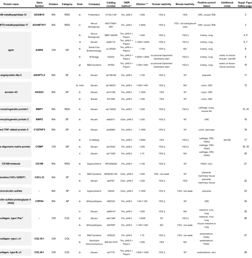
Antibodies to extracellular matrix proteins tested on formaldehyde-fixed and paraffin-embedded tissue samples using immunohistochemistry (IHC). This table provides an overview of all tested antibodies that showed positive staining signals. Presented are our IHC validation information and results, however, we have not validated the molecular specificity of every antibody (see text).
Matrisome division (CM, core matrisome; MA, matrisome-associated); matrisome category (COL, collagens; GP, ECM glycoproteins; PG, proteoglycans; REG, ECM regulators; SF, secreted factors; AP, ECM-affiliated proteins). Antibody host species (gt, goat; m, mouse; rb, rabbit; rt, rat; sh, sheep); mAb monoclonal antibody.
Heat-induced epitope retrieval (HIER) method used (0.05% Tween in Citric acid, pH6.0 or Tris, pH9.0) or protease treatment (Tris, pH9.0 + Pepsin treatment); for details see Supplementary Materials and Methods. Individual antibody dilutions recommended, the human and mouse reactivity as well as a positive control tissue, additional notes and references are listed.
Additional abbreviations used: CRC, colorectal cancer; NT, not tested; PDAC, pancreatic ductal adenocarcinoma; TEB terminal end buds.
* Indicates the preferred HIER method applied for the individual antibody, however, other conditions also might work.
** Indicates the antibody dilution we have tested; however, each antibody vial should be tested for individual application and tissue since lot-specific variability can occur. For additional details on the individual antibodies see also the manufacturers datasheets.
‡ A representative image of the staining with the antibody is shown in the Supplementary Figure Gallery.
References to antibodies listed in Table 1
[59] A. Eusebio, F. Oliveri, P. Barzaghi, M.A. Ruegg, Expression of mouse agrin in normal, denervated and dystrophic muscle, Neuromuscul Disord 13(5) (2003) 408-15.
[60] E. Hedbom, P. Antonsson, A. Hjerpe, D. Aeschlimann, M. Paulsson, E. Rosa-Pimentel, Y. Sommarin, M. Wendel, A. Oldberg, D. Heinegard, Cartilage matrix proteins. An acidic oligomeric protein (COMP) detected only in cartilage, J Biol Chem 267(9) (1992) 6132-6. [61] J.M. Gardner, R.O. Hynes, Interaction of fibronectin with its receptor on platelets, Cell
42(2) (1985) 439-48.
[62] R.O. Hynes, A.T. Destree, Relationships between fibronectin (LETS protein) and actin, Cell 15(3) (1978) 875-86.
[63] J.H. Peters, L.A. Sporn, M.H. Ginsberg, D.D. Wagner, Human endothelial cells
synthesize, process, and secrete fibronectin molecules bearing an alternatively spliced type III homology (ED1), Blood 75(9) (1990) 1801-8.
[64] J.H. Peters, J.E. Trevithick, P. Johnson, R.O. Hynes, Expression of the alternatively spliced EIIIB segment of fibronectin, Cell Adhes Commun 3(1) (1995) 67-89.
[65] M. Hyytiainen, J. Taipale, C.H. Heldin, J. Keski-Oja, Recombinant latent transforming growth factor beta-binding protein 2 assembles to fibroblast extracellular matrix and is susceptible to proteolytic processing and release, J Biol Chem 273(32) (1998) 20669-76. [66] L.F. Brown, B. Berse, L. Van de Water, A. Papadopoulos-Sergiou, C.A. Perruzzi, E.J.
Manseau, H.F. Dvorak, D.R. Senger, Expression and distribution of osteopontin in human tissues: widespread association with luminal epithelial surfaces, Mol Biol Cell 3(10) (1992) 1169-80.
Supplementary Materials and Methods
Human and mouse tissue samples
All human samples were kindly provided by collaborators from the Department of Pathology and the Cancer Center of the Massachusetts General Hospital (Boston, MA; V. Deshpande, G. Lauwers, M. Mino-Kenudson, K. Tanabe) as well as the Department of Medical Oncology of the Dana-Farber Cancer Institute (Boston, MA; S. Ogino). In addition, formaldehyde-fixed and paraffin-embedded (FFPE) human tissue blocks were purchased from Genvelop Life Science (Westborough, MA). Various mouse tissue samples were obtained from the animal facilities of the David H. Koch Institute for Integrative Cancer Research (Cambridge, MA) and processed for FFPE following standard procedures or snap-frozen and stored at -80°C until use.
Antibodies
All antibodies to extracellular matrix (ECM) and ECM-associated molecules used in this study are listed in Table 1 and Supplementary Table 1. In Table 1 we list all antibodies which yield positive staining results and in Supplementary Table 1 all antibodies which, in our experiments, either did not yield positive stains or gave uncertain staining results. Additional antibodies that are more broadly used in tumor biology and histopathology to determine specific tissue distribution or subcellular localization are listed in Supplementary Table 2.
Commercially available antibodies were from: Abcam (Cambridge, MA), ABclonal (Woburn, MA), Abgent (San Diego, CA), Agilent (Santa Clara, CA), Atlas Antibodies (Bromma, Sweden), Bethyl Laboratories (Montgomery, TX), BD Biosciences (San Jose, CA), Biocare Medical (Pecheco, CA), Biolegend (San Diego, CA), biorbyt (San Francisco, CA), Boster (Pleasanton, CA), Cell Signaling (Danvers, MA), Developmental Studies Hybridoma Bank (DSHB, Iowa City, IA), Emfret Analytics (Eibelstadt, Germany), GeneTex (Irvine, CA), Leica Biosystems (Nussloch, Germany), LSBio (Seattle, WA), MilliporeSigma (Burlington, MA), Novus Biologicals (Littleton, CO), PROGEN Biotechnik (Heidelberg, Germany), Proteintech (Rosemont, IL), OriGene (Rockville, MD), R&D Systems (Minneapolis, MN), Rockland Immunochemicals (Limerick, PA), Santa Cruz Biotechnology (Santa Cruz, CA), Sigma-Aldrich (St. Louis, MO), Sino Biological (Wayne, PA), sdix (Newark, DE), Thermo Fisher Scientific (Fremont, CA),
Several well-established antibodies for validation on mouse and human FFPE samples were kindly provided by the following: rabbit anti-Agrn antibody (M.A. Ruegg, Biozentrum, University
of Basel, (Basel, Switzerland), rabbit anti-Agrn antibody (J.H. Miner, Washington University, St. Louis, MO), rabbit anti-LOXL2 antibodies (G. Neufeld, Technion, Israel Institute of Technology, Haifa, Israel), rabbit anti-COMP antibodies (Å. Oldberg Lund University, Lund, Sweden), rabbit anti-LTBP2 antibodies (M. Hyytiäinen, University of Helsinki, Helsinki, Finland) and several rabbit anti-SPP1 antibodies (D.R. Senger, Beth Israel Deaconess Medical Center, Boston, MA). Anti-FN monoclonal and polyclonal antibodies generated in the Hynes laboratory are also listed in Table 1.
Anti-fibronectin-EIIIB monoclonal antibody generation
Mice null for an alternatively spliced domain of fibronectin, FN-EIIIB domain (also known as ED-B), were immunized with recombinant EIIIB segment coupled to keyhole limpet hemocyanin (KLH). Lymphoblasts from seropositive mice were used to generate hybridomas, screened against recombinant FN fragments to identify clones selective for EIIIB and tested against cultured endothelial cells and mouse embryos (WT or EIIIB-null) by immunofluorescence microscopy (McMahon, A., unpublished).
Immunohistochemistry
All tissues used in this study were processed for FFPE following standard procedures.
Consecutive sections (4-6 μm) were prepared using a Leica RM2255 rotary microtome (Leica Biosystems, Nussloch, Germany), dried at 60°C for 1h and stored at room temperature. The individual sections were dewaxed, rehydrated and either stained with hematoxylin and eosin (H&E) following standard procedures or treated with heat-induced antigen-retrieval (HIER) using a decloaking chamber (Biocare Medical, Pecheco, CA) prior to immunostaining. To achieve the best staining results, each individual antibody was tested following recommendations and guidelines for improved antibody validation and immunohistochemistry (IHC) optimization [e.g., 1-13].
We screened different antibody concentrations and adapted our protocols according to the ‘test battery’ approach of Shi et al. [1] to optimize the conditions for the major variables involved in antigen retrieval. These include the heating conditions, temperature and heating time, the pH of the retrieval solution, and the retrieval buffer. Various pH values (pH 1, 6, and 9) of different buffer solution (including acetate, citrate, phosphate, and tris) and different temperatures of super high 120°C (decloaking chamber, pressure cooker) or high 100°C (microwave, MW) and 98°C (MW) can be tested to find optimal results [see also 1,2,6,11].
For most ECM-related antibodies tested we found best results with heat-induced
epitope-retrieval (HIER) at 120°C for 2 min using either of two buffer conditions (0.05% Tween in 10 mM sodium-citrate at pH6.0 or pH9.0). However, we also tested the UNI-TRIEVE (Innovex
Biosciences, Richmond, CA) mild temperature retrieval solution formulated for non-boiling and gentle retrieval of antigens in ‘fragile’ FFPE tissue sections. UNI-TRIEVE solution is
pH-independent and formulated to unmask antigens at 60°C-80°C using a water bath or a hot plate as the heating source. An incubation of 30 minutes yielded good tissue morphology and was sufficient to validate some antibodies including, e.g., BMP1, COMP, LOXL2, SPP1
(Supplementary Figure Gallery), on mouse and human cartilage tissue sections.
After the HIER step, the sections were cooled to room temperature in phosphate-buffered saline (PBS) for 10 min before further processing. To inactivate all endogenous peroxidase and
alkaline phosphatase activity in the tissue, sections were subsequently pretreated using
BLOXALL endogenous enzyme blocking solution (Vector Laboratories, Burlingame, CA) for 10 min. Prior to application of primary antibodies, incubation with normal serum to prevent non-specific binding of the antibodies to the tissue or to Fc receptors is recommended. Proteins such as BSA or casein may also be used as blocking solutions. In addition, the Mouse on Mouse (M.O.M.) basic kit (Vector Laboratories) can be applied to reduce the endogenous mouse Ig staining when using mouse primary antibodies on mouse tissue sections to yield cleaner staining results. Subsequently, the sections were incubated with the individual primary
antibodies for 1h, followed by the secondary detection system according to the manufacturers’ protocol.
Although the Avidin-Biotin Complex (ABC) method using 3,3'-diaminobenzidine (DAB) as substrate is widely used as a technique for IHC, we repeatedly noticed varying background staining intensities probably due to the fact that many of the antibodies are polyclonal antisera. To simplify and optimize the staining conditions and to increase the IHC sensitivity, we
optimized a variety of secondary detection systems as well as enzyme substrates. Polymerized reporter enzyme staining systems have recently been developed which provide greater
sensitivity than conventional antibody conjugates. This approach is based on a new method of attaching a micropolymer with a high density of active enzyme to a secondary antibody. This results in strong staining signal with low IHC background staining and reduced non-specific binding. We predominantly used the ImmPRESS polymer detection systems (Vector
incubation this approach also shortens the general assay time and we observed improved staining outcomes with high sensitivity and signal intensity and less non-specific background staining. For primary antibodies generated in sheep, horseradish-peroxidase-conjugated rabbit anti-sheep secondary antibodies (Thermo Fisher Scientific) were used instead of the
ImmPRESS polymer detection system. Subsequently, the Vulcan Fast Red Chromogen Kit 2 (red staining; Biocare Medical, Concord, CA) or the DAB Quanto System (brown staining; Thermo Fisher Scientific) were applied as substrates. Hematoxylin was used as final counterstain.
To obtain consistent and reliable staining results on all tissues investigated, we adapted our protocols and used an automated staining system (LabVision Autostainer 360, Thermo Fisher Scientific) for most of the immunostainings presented. For image documentation the Leica Aperio AT2 slide scanner system was used. Appropriate positive and negative controls were performed for each batch of slides using a variety of mouse and human tissue sections. The preferred antigen retrieval conditions and dilutions for each antibody tested are listed in Table 1, Supplementary Tables 1 and 2.
Although we have not attempted quantification of the IHC staining we strongly recommend that all samples to be compared with one another should be processed using consistent staining reagents and conditions, ideally processed together whenever possible. There are available software programs for quantification of IHC staining but we have not tested them. We note that the large number of variables involved in IHC staining of FFPE tissues (often obtained from different sources using variable and often unknown fixation and embedding methods) can render quantification very challenging and rigorous control of fixation and staining conditions would be necessary to obtain reliable quantification.
Supplementary Table 1 - Legend
Additional extracellular matrix-related antibodies
Antibodies to extracellular matrix proteins tested on formaldehyde-fixed and paraffin-embedded tissue samples using immunohistochemistry (IHC), which, in our experiments, either did not yield positive stains or gave uncertain staining results. However, other antigen-retrieval or heat-induced epitope retrieval (HIER) approaches or tissue samples might yield appropriate staining results.
This table provides an overview of additional antibodies for which none of the HIER conditions (for details see Supplementary Materials and Methods) we tested showed a positive staining reactivity or we observed a ‘non-specific’ staining pattern. Each antibody was tested for a range of dilutions on a variety of tissues. However, since lot-specific variability can occur, other
antibody samples should be tested for the individual application and tissue of interest. For additional details see also the antibody manufacturers datasheets.
Matrisome division (CM, core matrisome; MA, matrisome-associated); matrisome category (COL, collagens; GP, ECM glycoproteins; PG, proteoglycans; REG, ECM regulators; SF, secreted factors; AP, ECM-affiliated proteins). Antibody host species (gt, goat; m, mouse; rb, rabbit); rt, rat; mAb monoclonal antibody. Additional abbreviations used: NT not tested; DSHB, Developmental Studies Hybridoma Bank
Supplementary Table 2 - Legend Reference antibodies
List of antibodies broadly used in tumor biology and histopathology to identify specific tissue distribution or subcellular localization on formaldehyde-fixed and paraffin-embedded tissue samples using immunohistochemistry.
This table provides an overview of all antibodies which reproducibly showed positive staining signals in our hands. Presented are our validation information and results, however, we have not tested each antibody for molecular specificity.
Indicated are the individual: protein name, gene symbol, antibody host species (gt, goat; m, mouse; rb, rabbit; rb mAb, rabbit monoclonal antibody; rt, rat), commercial antibody source and catalog number and heat-induced epitope retrieval (HIER) methods used (see also
Supplementary Materials and Methods), recommended antibody dilutions, human and mouse reactivity as well as a positive control tissue and, if applicable, the targets of each antibody. * Indicates the preferred HIER method applied for the individual antibody, however, other conditions also might work. For many antibodies both HIER conditions work alike.
** Indicates the antibody dilution we have tested; however, each antibody vial should be tested for individual application and tissue accordingly since lot specific variability can occur.
Supplementary Table 3 - Legend Online resources
Lists of useful online resources relevant for antibody searching, antibody characterization, IHC standardization and methodology, troubleshooting guidelines as well as links to tissue
expression data of protein-coding genes in normal and tumor tissues.
Supplementary Figure Gallery - Legend
Illustrative examples for observed staining patterns of many antibodies to extracellular matrix proteins. The images are alphabetically arranged by gene name and show the reactivity of the individual antibodies on representative human tissue samples, in some cases also for mouse tissues. In addition, the antibody host, supplier and catalog numbers are indicated. All images presented here are indicated in Table 1, which contains the individual staining procedure used for each antibody.
Supplementary Materials and Methods - References
[1] S.R. Shi, R.J. Cote, C. Yang, C. Chen, H.J. Xu, W.F. Benedict, C.R. Taylor, Development of an optimal protocol for antigen retrieval: a 'test battery' approach exemplified with reference to the staining of retinoblastoma protein (pRB) in formalin-fixed paraffin sections, J Pathol 179(3) (1996) 347-52.
[2] S.R. Shi, G. Gu, C.R. Taylor, Antigen Retrieval Techniques: Immunohistochemistry and Molecular Morphology. Eaton Publishing, Natick., 2000.
[3] C.B. Saper, P.E. Sawchenko, Magic peptides, magic antibodies: guidelines for appropriate controls for immunohistochemistry, J Comp Neurol 465(2) (2003) 161-3. [4] N.S. Goldstein, S.M. Hewitt, C.R. Taylor, H. Yaziji, D.G. Hicks, S. Members of Ad-Hoc
Committee On Immunohistochemistry, Recommendations for improved standardization of immunohistochemistry, Appl Immunohistochem Mol Morphol 15(2) (2007) 124-33. [5] C.A. Sullivan, G.G. Chung, Biomarker validation: in situ analysis of protein expression
using semiquantitative immunohistochemistry-based techniques, Clin Colorectal Cancer 7(3) (2008) 172-7.
[6] F. D'Amico, E. Skarmoutsou, F. Stivala, State of the art in antigen retrieval for immunohistochemistry, J Immunol Methods 341(1-2) (2009) 1-18.
[7] J. Bordeaux, A. Welsh, S. Agarwal, E. Killiam, M. Baquero, J. Hanna, V. Anagnostou, D. Rimm, Antibody validation, Biotechniques 48(3) (2010) 197-209.
[8] W.J. Howat, A. Lewis, P. Jones, C. Kampf, F. Ponten, C.M. van der Loos, N. Gray, C. Womack, A. Warford, Antibody validation of immunohistochemistry for biomarker discovery: recommendations of a consortium of academic and pharmaceutical based histopathology researchers, Methods 70(1) (2014) 34-8.
[9] F. Lin, Z. Chen, Standardization of diagnostic immunohistochemistry: literature review and geisinger experience, Arch Pathol Lab Med 138(12) (2014) 1564-77.
[10] G. O'Hurley, E. Sjostedt, A. Rahman, B. Li, C. Kampf, F. Ponten, W.M. Gallagher, C. Lindskog, Garbage in, garbage out: a critical evaluation of strategies used for validation of immunohistochemical biomarkers, Mol Oncol 8(4) (2014) 783-98.
[11] J.A. Ramos-Vara, M.A. Miller, When tissue antigens and antibodies get along: revisiting the technical aspects of immunohistochemistry--the red, brown, and blue technique, Vet Pathol 51(1) (2014) 42-87.
[12] N.R. Smith, C. Womack, A matrix approach to guide IHC-based tissue biomarker development in oncology drug discovery, J Pathol 232(2) (2014) 190-8.
[13] C.R. Taylor, Predictive biomarkers and companion diagnostics. The future of
immunohistochemistry: "in situ proteomics," or just a "stain"?, Appl Immunohistochem Mol Morphol 22(8) (2014) 555-61.
Table 1 - Extracellular matrix-related antibodies
Protein name Gene
Symbol Division Category Host Company
Catalog number
HIER
method * Dilution ** Human reactivity Mouse reactivity
Positive control tissue Additional notes Suppl. Figure Gallery page # ADAM metallopeptidase 12 ADAM12 MA REG rb Proteintech 14139-1-AP Tris, pH9.0 1:200 YES ‡ YES CRC, mouse TEB 2
ADAMTS metallopeptidase 17 ADAMTS17 MA REG m Novus Biologicals
H00170691-M01 Tris, pH9.0 1:3000 YES ‡
YES - but background
stain CRC, mouse TEB 3 rb Novus
Biologicals NBP1-90209
Tris, pH9.0 +
Pepsin 1:400 YES ‡ YES ‡ kidney, lung 4, 5 rb Abcam ab85174 Tris, pH9.0 +
Pepsin 1:1000-1:2000 YES ‡ YES ‡ kidney, lung 6, 7 rb Santa Cruz
Biotechnology sc-25528
Tris, pH9.0 +
Pepsin 1:100 YES ‡ NO kidney, lung 8 rb M.Ruegg AS204 Tris, pH9.0 +
Pepsin 1:2000-1:3000
occasional basement
membrane stain YES ‡ kidney, lung
works on frozen tissues / [ref.59] 9 gt R&D Systems AF550 Tris, pH9.0 +
Pepsin 1:250-1:500
occasional basement
membrane stain YES ‡ kidney, lung
works on frozen tissue sections 10 angiopoietin-like 4 ANGPTL4 MA SF rb Abcam ab196746 Tris, pH9.0 1:100 YES ‡ NT placenta 11
rb, mAb Abcam ab108321 Tris, pH9.0 1:200-1:400 YES ‡ NO colon, CRC 12
rb Abcam ab14196 Tris, pH9.0 1:1000 YES NT colon, CRC rb Boster PA1008 Tris, pH9.0 1:200 YES NT colon, CRC
bone morphogenetic protein1 BMP1 MA REG rb Abcam ab118520 Tris, pH9.0 1:200 YES ‡ YES ‡ cartilage, ovary,
mouse tail 13, 14 bone morphogenetic protein 2 BMP2 MA SF rb Abcam ab82511 Citric, pH6.0 1:250 YES ‡ NT CRC 15
C1q and TNF related protein 5 C1QTNF5 MA AP rb Abcam ab36893 Tris, pH9.0 1:1500 YES ‡ NT colon, pancreas 16
rb A.Oldberg - Tris, pH9.0 1:2000 YES YES ‡ cartilage, CRC,
PDAC [ref.60] 17 rb Abcam ab74524 Tris, pH9.0 1:250 YES ‡ YES ‡ cartilage, CRC,
PDAC 18, 19 rt Abcam ab11056 Tris, pH9.0 1:10 YES ‡ NO cartilage, CRC,
PDAC 20
CD109 molecule CD109 MA REG rb Sigma-Aldrich HPA009292 Tris, pH9.0 1:100 YES ‡ NT PDAC, skin 21
m R&D Systems MAB350-100 Citric, pH6.0 1:250 YES - but weak NT placenta, mammary tissue rb Abcam ab9797 Citric, pH6.0 1:200 YES ‡ YES placenta,
mammary tissue 22 chondroitin sulfate - MA AP m Sigma-Aldrich C8035 Citric, pH6.0 1:1000 YES ‡ YES - but weak placenta 23
chondroitin sulfate proteoglycan 4
(NG2) CSPG4 MA AP rb MilliporeSigma AB5320 Tris, pH9.0 1:50-1:100 YES ‡ NT CRC 24 m Abcam ab88147 Tris, pH9.0 1:500 YES ‡ NO intestine, liver,
lung 25
rb Abcam ab21286 Tris, pH9.0 1:2000 NT YES ‡ intestine, liver,
lung 26
rb MilliporeSigma AB765P Tris, pH9.0 1:100-1:250 NO YES - but weak mouse intestine or lung "collagen, type I Pan" - CM COL
Heat-induced epitope retrieval (HIER) method used (0.05% Tween in Citric acid, pH6.0 or Tris, pH9.0) or protease treatment (Tris, pH9.0 + Pepsin treatment); for details see Supplementary Materials and Methods. Individual antibody dilutions recommended, the human and mouse reactivity as well as a positive control tissue, additional notes and references are listed.
* Indicates the preferred HIER method applied for the individual antibody, however, other conditions also might work.
Additional abbreviations used: CRC, colorectal cancer; NT, not tested; PDAC, pancreatic ductal adenocarcinoma; TEB terminal end buds.
‡A representative image of the staining with the antibody is shown in the Supplementary Figure Gallery.
** Indicates the antibody dilution we have tested; however, each antibody vial should be tested for individual application and tissue since lot-specific variability can occur. For additional details on the individual antibodies see also the manufacturers datasheets.
annexin A5 ANXA5
agrin AGRN CM GP
AP MA
Antibodies to extracellular matrix proteins tested on formaldehyde-fixed and paraffin-embedded tissue samples using immunohistochemistry (IHC). This table provides an overview of all tested antibodies that showed positive staining signals. Presented are our IHC validation information and results, however, we have not validated the molecular specificity of every antibody (see text).
Matrisome division (CM, core matrisome; MA, matrisome-associated); matrisome category (COL, collagens; GP, ECM glycoproteins; PG, proteoglycans; REG, ECM regulators; SF, secreted factors; AP, ECM-affiliated proteins). Antibody host species (gt, goat; m, mouse; rb, rabbit; rt, rat; sh, sheep); mAb monoclonal antibody.
Matrisome
chemokine CXCL12/SDF1 CXCL12
cartilage oligomeric matrix protein COMP CM GP
SF MA