Copyright © 1998, American Society for Microbiology
Degradation of Morpholine by an Environmental Mycobacterium
Strain Involves a Cytochrome P-450
P. POUPIN,1N. TRUFFAUT,1* B. COMBOURIEU,2P. BESSE,2M. SANCELME,2
H. VESCHAMBRE,2ANDA. M. DELORT2
Laboratoire de Ge´ne´tique Microbienne, Universite´ de Technologie de Compie`gne, 60206 Compie`gne,1 and Laboratoire de Synthe`se, Electrosynthe`se et Etude de Syste`mes a` Inte´reˆt Biologique,
UMR 6504 CNRS, Universite´ Blaise Pascal, 63177 Aubie`re Cedex,2France
Received 23 June 1997/Accepted 31 August 1997
A Mycobacterium strain (RP1) was isolated from a contaminated activated sludge collected in a wastewater treatment unit of a chemical plant. It was capable of utilizing morpholine and other heterocyclic compounds, such as pyrrolidine and piperidine, as the sole source of carbon, nitrogen, and energy. The use of in situ1H nuclear
magnetic resonance (1H NMR) spectroscopy allowed the determination of two intermediates in the
biodegra-dative pathway, 2-(2-aminoethoxy)acetate and glycolate. The inhibitory effects of metyrapone on the degrada-tive abilities of strain RP1 indicated the involvement of a cytochrome P-450 in the biodegradation of morpho-line. This observation was confirmed by spectrophotometric analysis and1H NMR. Reduced cell extracts from
morpholine-grown cultures, but not succinate-grown cultures, gave rise to a carbon monoxide difference spec-trum with a peak near 450 nm, which indicated the presence of a soluble cytochrome P-450.1H NMR allowed
the direct analysis of the incubation medium containing metyrapone, a specific inhibitor of cytochrome P-450. The inhibition of morpholine degradation was dependent on the morpholine/metyrapone ratio. The heme-containing monooxygenase was also detected in pyrrolidine- and piperidine-grown cultures. The abilities of different compounds to support strain growth or the induction of a soluble cytochrome P-450 were assayed. The results suggest that this enzyme catalyzes the cleavage of the CON bond of the morpholine ring.
Morpholine (C4H9NO) is a simple heterocyclic compound
(Fig. 1) with great industrial importance. It is used as an anti-corrosive agent in water boiling systems, as a chemical inter-mediate (in catalysts, solvents, antioxidants, pharmaceuticals, bactericides, and pesticides), in the textile industry, in photo-graphic developers, in hair conditioners, in waxes, and in the preservation of book paper. Because of its solubility in water, significant amounts of this chemical compound could be re-leased, via industrial effluents, in the environment. It would then move with soil moisture and running water and would not sorb sediment or organic matter (12a). Morpholine, like other secondary amines, is subject to N nitrosation. This reaction can be catalyzed by various bacteria from secondary amines and nitrites or nitrates at neutral pH (3) but can also take place, in vivo, in mice by the action of NO2on morpholine (29). Nitroso
compounds are of particular concern because they are muta-gens and carcinomuta-gens. N-Nitrosomorpholine was reported to be mutagenic in microbial gene mutation assays with
Salmo-nella typhimurium (32), and recent studies assessing the
carci-nogenic activities of nitroso compounds have shown that this compound could enhance, even at low doses, the development of early stages of hepatocarcinogenesis in rats (8).
Removal of this pollutant from contaminated wastewater and the environment is possible by biological treatment, since Knapp et al. (12) have clearly established that morpholine is biodegradable. A Mycobacterium strain, MorG, could utilize this compound as the sole source of carbon, nitrogen, and energy via an inducible pathway. Some bacteria that are capa-ble of aerobic degradation of morpholine have been isolated from activated sludge, soils, and water. Except for two bacteria
belonging to the genus Arthrobacter (7), all morpholine-de-grading bacteria are mycobacteria (2, 4).
Biochemical studies of morpholine catabolism have been limited to those carried out with the environmental
Mycobac-terium strain MorG by Knapp et al. (12) and Swain et al. (24).
These authors proposed a pathway for morpholine degrada-tion by this strain in which the later stages of catabolism gave two C2-unit products: glycolate and ethanolamine. However,
the early reaction mechanisms were not elucidated.
In the companion paper (5) our results on the pathway of biodegradation of morpholine by another strain,
Mycobacte-rium aurum MO1, are presented. For that study, in situ 1H
nuclear magnetic resonance (1H NMR) was used. This method
allows direct detection and quantification of the metabolites formed, by analyzing the incubation medium at different times. We showed that M. aurum completely degraded morpholine (10 mM) in 10 h. Two intermediary compounds, 2-(2-aminoe-thoxy)acetate and glycolate, were identified.
In this paper, we report the isolation and the characteriza-tion of a Mycobacterium strain that metabolizes morpholine as the sole source of carbon, nitrogen, and energy. Experiments performed with inhibitors and spectrophotometric analysis implicated a cytochrome P-450 in this degradation.1H NMR
methodology, as described in the companion paper (5), was used to quantitatively monitor the degradation of morpholine and to identify the intermediates. The results were used to propose a pathway for morpholine degradation by this strain.
MATERIALS AND METHODS
Bacterial strain and growth conditions.Strain RP1 was isolated, by enrich-ment culture with morpholine as the sole source of carbon and energy, from an activated sludge collected in a chemical wastewater treatment plant. The strain was deposited in the Institut Pasteur collection (CIP105337). This bacterium was grown and maintained on peptone-beef (PB) medium (casein peptone, 3 gz liter21; beef extract, 5 g z liter21), Trypticase soy broth (bioMe´rieux, Marcy
l’Etoile, France), or a mineral salts medium (11) which contained (per liter) 1 g
* Corresponding author. Mailing address: Laboratoire de Ge´ne´t-ique Microbienne, Universite´ de Technologie de Compie`gne, B.P. 529, 60206 Compie`gne, France. Phone: 33 3 44 23 44 52. Fax: 33 3 44 20 48 13. E-mail: nicole.truffaut@utc.fr.
of KH2PO4, 1 g of K2HPO4, 0.04 g of MgSO4z 7H2O, 0.004 g of FeCl3z 6H2O,
and 1 g of (NH4)2SO4(when the carbon source did not contain nitrogen or when
the amine was only used as a source of carbon). The pH was adjusted to 7.0 with NaOH or HCl after the addition of the carbon source. On mineral salts medium the following carbon sources were used: thiomorpholine, tetrahydrofuran, and tetrahydropyran at 5 mM and morpholine, piperidine, pyrrolidine, and succinate at 10 mM. Solid medium was prepared from mineral salts and PB media by the addition of 1.5% (wt/vol) Noble agar (Difco, Detroit, Mich.) or agar, respec-tively.
The assays of growth inhibition were performed in test tubes at 30°C under agitation on a gyratory shaker (180 rpm). Bacterial growth was determined by monitoring the optical density at 600 nm (OD600).
Chemicals.Morpholine, glycolic acid, and ethanolamine were purchased from Aldrich Chemical (Sigma Aldrich Sarl, St. Quentin Fallavier, France); piperi-dine, pyrrolipiperi-dine, thiomorpholine, tetrahydrofuran, and tetrahydropyran were from Fluka (Sigma Aldrich Sarl); methimazole and metyrapone were obtained from Sigma; and tetradeuterated sodium trimethylsilylpropionate (TSPd4) was
purchased from EurisoTop (St. Aubin, France).
Analytical methods.The morpholine concentration was routinely estimated spectrophotometrically by the method of Stevens and Skov (23) as modified by Knapp et al. (12). In situ1H NMR spectroscopy was used when a greater
precision in morpholine concentration was required and to identify degradation intermediates. The protein concentration was determined by the Bradford method (1).
Spectrophotometric analysis of cytochrome P-450.Mycobacterial cells were harvested by centrifugation (8,0003 g, 10 min) at 4°C, washed, and resuspended in 50 mM phosphate buffer (pH 7.4) containing 0.3 mM phenylmethylsulfonyl fluoride. Bacterial walls were broken by three passages through a French pres-sure cell (SLM-Aminco) at 18,000 lb/in2and centrifuged at 30,0003 g at 4°C for
30 min. The supernatant was reduced by dithionite sodium and divided equally between two optically matched cuvettes, and the difference spectrum was re-corded in the presence of CO (19). An extinction coefficient of 91 mM21z cm21
was used to determine the content of cytochrome P-450 in crude extracts (14). To determine the induction of a cytochrome P-450 by tetrahydrofuran, tetra-hydropyran, or thiomorpholine, the cells were grown in PB medium to obtain sufficient biomass, washed, and resuspended in mineral salts medium supple-mented with one of these compounds at 5 mM.
All photometric analyses were done with a Shimadzu UV 160 A spectropho-tometer.
1H NMR spectroscopy. (i) Microorganism growth.For these experiments, strain RP1 cultures were grown in 100 ml of Trypticase soy broth (bioMe´rieux) in 500-ml Erlenmeyer flasks incubated at 30°C at 200 rpm. They were harvested after 48 h of culture.
(ii) Incubation with xenobiotics.Incubations of cells with morpholine (10 mM), ethanolamine (15 mM), and glycolate (10 mM) were carried out as de-scribed in the companion paper (5).
For the assay of inhibition with metyrapone, different concentrations (5 and 10 mM) were tested. This inhibitor was added to the flasks containing the cells (5 g of wet cells in 50 ml of buffer) and morpholine (10 mM). Samples were taken every hour for 12 h and then from time to time until 72 h.
(iii) Preparation of the samples for1H NMR.The preparation of the samples for NMR, the materials used, and the quantification of the metabolites were as described elsewhere (5).
RESULTS
Enrichment and isolation of bacteria. An enrichment cul-ture was established with activated sludge collected from a chemical wastewater treatment plant (18). A subenrichment culture inoculated with an aliquot of this culture was made in mineral salts medium containing 10 mM morpholine. The cul-ture was incubated on a gyratory shaker at 180 rpm and 30°C. Samples were regularly recovered, centrifuged, and assayed for morpholine content. When this amine was totally degraded, appropriate dilutions of the cell suspension were spread onto solid mineral salts medium supplemented with morpholine. Isolated colonies were selected, and their ability to degrade morpholine was determined in liquid mineral salts medium containing 10 mM morpholine.
Identification of a morpholine-degrading bacterium. Enrich-ment and isolation procedures yielded only one bacterial strain (designated RP1) from this mixed culture that was able to utilize morpholine as a growth substrate. This culture was estimated to be pure after microscopic observation and several subcultures onto solid mineral salts agar medium containing morpholine and onto PB agar medium. Strain RP1 formed red, convex, slightly mucoid colonies on solid medium, and no dif-fusible pigment was observed. The coloration was affected nei-ther by the growth conditions (PB agar medium, solid mineral salts medium supplemented with different compounds, and temperature) nor by the presence of light. The cells were non-motile and exhibited an elementary rod-coccus life cycle. RP1 was gram positive, partially acid fast, catalase positive, and oxi-dase negative. On the basis of analysis of mycolic acids, polar lipids, wall sugars, and total proteins (Institut Pasteur, Paris, France), strain RP1 was assigned to the genus Mycobacterium: the cell wall peptidoglycan was based on meso-diaminopimelic acid, the major cell wall sugars were glucose and arabinose, and the wall envelope contained mycolic acids, phosphatidyl-ethanolamine, phosphatidylglycerol, phosphatidylinositol, and phosphatidylinositol mannosides. Comparison of mycolic acids
FIG. 1. Structures of the compounds used.
FIG. 2. Growth of Mycobacterium sp. strain RP1 on liquid mineral salts medium (å) and morpholine degradation (}).
and total proteins of this bacterium with those of reference strains indicated that RP1 was closely related to
Mycobacte-rium chlorophenolicum. Experiments to determine the 16S
ribosomal DNA sequence of this microorganism and to estab-lish its phylogenetic relationship to other mycobacteria are in progress.
Degradation of morpholine. The ability of Mycobacterium sp. strain RP1 to degrade morpholine in pure culture was determined in liquid mineral salts medium (Fig. 2). When morpholine (10 mM) was used as the sole source of carbon, nitrogen, and energy, it was rapidly degraded. At the end of the logarithmic growth phase (50 h), no morpholine could be de-tected in the culture medium. During this time, the turbidity of the culture (OD600) increased from 0.018 to 0.62 and ammonia
accumulated (data not shown). The medium had a uniformly turbid appearance, and no clumps or aggregates were visible.
The natural clumping of the cells is a growth characteristic widely shared among members of the genus Mycobacterium, making it difficult to follow the growth and the degradation abilities of these bacteria (20). In contrast to M. aurum MO1,
Mycobacterium sp. strain RP1 did not present this
inconve-nience. In addition, the doubling time in liquid medium sup-plemented with morpholine was about 9 h for strain RP1 versus about 12 h for M. aurum MO1, making it easier to study morpholine degradation with strain RP1.
In order to monitor the degradation of morpholine more precisely, we used the in situ1H NMR spectroscopy
method-ology presented in the companion paper (5) for the study of the pathway of biodegradation of morpholine by M. aurum MO1. With this technique, the incubation medium is directly analyzed both qualitatively and quantitatively. It allows the identification of the intermediates formed during the degrada-tion of morpholine.
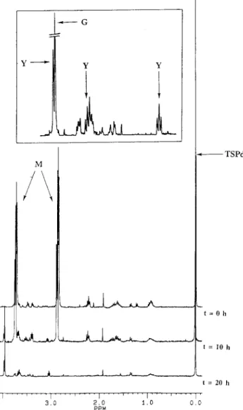
FIG. 3. Kinetics of morpholine degradation by Mycobacterium sp. strain RP1. Resting cells (5 g of wet cells in 50 ml of Knapp buffer) were incubated with 10 mM morpholine at 30°C with agitation (200 rpm) for 72 h. Samples (1 ml) were collected every hour for 12 hours and from time to time until 72 h; after centrifugation, the supernatants of these samples were analyzed by1H NMR
spectroscopy at 300.13 MHz. TSPd4was used as a reference for chemical shifts
and quantification. The inset corresponds to an expanded scale, from 2.60 to 4.00 ppm, of the 20-h spectrum. M, morpholine; Y, 2-(2-aminoethoxy)acetate; G, glycolic acid.
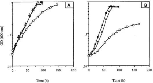
FIG. 4. (A) Time courses for the concentrations of morpholine (3), glycolate (h), and 2-(2-aminoethoxy)acetate (‚) during the degradation of morpholine (10 mM) by Mycobacterium sp. strain RP1 cells at 100 gz liter21. (B) Kinetics of
degradation of ethanolamine (15 mM) (1) and glycolate (10 mM) (h) by Mycobacterium sp. strain RP1 (100 gz liter21). The quantification was done by
integrating the signals in1H NMR spectra relative to the area of the reference
Resting RP1 cells were incubated with 10 mM morpholine at 30°C with agitation (200 rpm) for 72 h. The conditions found to be best for M. aurum MO1 (100 g of wet cells in 1 liter of Knapp buffer) were used. Samples of the incubation medium were taken periodically and centrifuged, and the supernatants were analyzed by1H NMR after adjustment of the pH to 10 (to
avoid changes in chemicals shifts) and addition of a reference (TSPd4) for chemical shifts (0 ppm) and quantification.
Figure 3 shows the kinetics of morpholine degradation as monitored by1H NMR spectroscopy; only spectra recorded at
0, 10, and 20 h are plotted. The two pseudotriplets at 2.88 and 3.72 ppm correspond to the CH2 of morpholine. In the
ex-panded region of the spectrum recorded at 20 h, different intermediates are observed: the three signals, named Y, at 3.96 (singlet) and 3.67 and 3.05 (pseudotriplets) have been identi-fied as 2-(2-aminoethoxy)acetate after synthesis of this com-pound (5), and the singlet G at 3.95 ppm corresponds to glycolate, the second observed intermediate. The last singlet, at 0 ppm, corresponds to the CH3of the TSPd4, our reference.
For quantification, the concentrations of morpholine and of the different intermediates were calculated by measuring the areas of the peaks and comparing them with that of the peak for TSPd4. The equation used for the calculation is described
in the companion paper (5). Figure 4A shows the time courses for the concentrations of morpholine, 2-(2-aminoethoxy)ac-etate, and glycolate.
Under these conditions, morpholine was degraded in 13 h at a rate of about 0.8 mM/h. The two intermediary compounds, 2-(2-aminoethoxy)acetate and glycolate, appeared after about 10 h of incubation. In contrast to the case for M. aurum MO1 (5), glycolate was not completely degraded, and its concentra-tion in the supernatant increased.
Degradation of ethanolamine and glycolic acid.The degra-dation of ethanolamine and glycolic acid, supposed intermedi-ates of the biodegradation pathway according to Swain et al. (24), was also tested with this strain. The concentrations of ethanolamine and glycolic acid were, respectively, 15 and 10 mM. The kinetics of these two degradations are presented in Fig. 4B. Ethanolamine and glycolic acid were completely de-graded within 10 h. No metabolite was detected in the medium. Inhibition of morpholine degradation by selective inhibi-tors. Degradation of a saturated heterocycle ring is likely to
begin by the breakage of a bond between the heteroatom and an adjacent carbon atom. It was demonstrated that xenobiotic compounds bearing amine and ether functional groups could serve as substrates for flavin-containing monooxygenase or cy-tochrome P-450 (9, 21, 31). Previous work (12) showed that morpholine degradation was associated with oxygen consump-tion. According to these results and the chemical structure of morpholine, it was possible that the enzyme responsible for the ring cleavage was a monooxygenase.
In order to check the involvement of such enzymes in the first steps of morpholine degradation by Mycobacterium sp. strain RP1, the influence of selected inhibitors on the degrada-tion ability of this strain was tested. Metyrapone (2-methyl-1,2-di-3-pyridyl-1-propanone) was chosen as a specific cytochrome P-450 inhibitor (25), and methimazole (2-mercapto-1-methyl-imidazole) was chosen as a competitive inhibitor of flavin-containing monooxygenase (26).
As shown in Fig. 5A, the growth of Mycobacterium sp. strain RP1 on liquid mineral salts medium supplemented with succi-nate was slightly affected by the presence of metyrapone and was not affected by the addition of an equivalent concentration of methimazole. The same experiments performed with mor-pholine as the sole source of carbon, nitrogen, and energy (Fig. 5B) indicated that without inhibitors in the medium or with methimazole, the stationary phase (OD600of 0.7) was reached
within 60 h. After the same time, the OD600in the culture with
metyrapone was only 0.06, thus indicating that this compound inhibited the growth of strain RP1 on morpholine. Metyrapone did not affect the viability of Mycobacterium sp. strain RP1, since the addition of this chemical to succinate-containing me-dium did not prevent cell growth. Consequently, the observed effects of metyrapone on the growth of the bacterium RP1 in morpholine-containing medium were due to its inhibitory properties. These results suggest that a cytochrome P-450 is involved in the oxidative catabolism of this amine. However, no flavin-containing monooxygenase seems to be implicated in morpholine degradation by strain RP1.
To obtain direct evidence of the inhibitory effect of metyrap-one, experiments with different concentrations of this com-pound were carried out, and the results were analyzed by1H
NMR. To the flasks containing the cells (100 gz liter21) were FIG. 5. Influence of methimazole (‚) and metyrapone (E) on the growth of Mycobacterium sp. strain RP1 on succinate (A) and on morpholine (B). The inhibitors were added to the medium at a final concentration of 300mg z ml21. Control cultures without an inhibitor were grown on succinate (h) and morpholine (■).
added metyrapone (5 and 10 mM) and morpholine (10 mM). The kinetics of morpholine degradation are reported in Fig. 6. The addition of metyrapone led to a concentration-depen-dent inhibition of the morpholine degradation reactions. When the concentration of metyrapone was increased, the following effects were observed: (i) the rates of morpholine degradation (Fig. 6A) and 2-(2-aminoethoxy)acetate formation (Fig. 6B and C) were decreased, (ii) the appearance of the intermedi-ates was delayed (Fig. 6B and C), and (iii) the final concen-tration of 2-(2-aminoethoxy)acetate was decreased, while that of glycolate was increased.
These results confirm the presence of an activity due to a cytochrome P-450 in the morpholine degradation pathway. In addition, the effects of metyrapone on 2-(2-aminoethoxy)ace-tate formation indicate that the oxidation leading to the open-ing of the heterocycle takes place duropen-ing the early stages of morpholine degradation. The accumulation of glycolate was unexpected, and it shows that morpholine is not completely mineralized. This interesting phenomenon is not yet explained. Spectrophotometric evidence of the induction of a cyto-chrome P-450.Cytochrome P-450s contain a ferriprotoporphy-rin IX prosthetic group (heme). The heme is anchored in the active site by an axial iron-sulfur bond between the heme ferric iron and a cysteine sulfydryl group. Under reduced conditions, the ferrous iron can bind carbon monoxide. This binding gives rise to a distinctive absorption band at approximately 450 nm. Cell extracts from cultures grown on morpholine and on suc-cinate were treated with sodium dithionite and CO. In the spectrum of the CO-treated reduced extract, but not in that of the nontreated reduced extract, of morpholine-grown bacteria, a peak at 449 nm was observed (Fig. 7, spectrum A). This result demonstrates the presence of a soluble cytochrome P-450 in this cell extract. The cytochrome P-450 content was about 90 pmol per mg of protein. Such a monooxygenase was not de-tected (but could be present at a low level) in the protein extracts of succinate-grown (Fig. 7, spectrum B) or acetate-grown (data not shown) bacteria. This indicated that the pres-ence of a soluble cytochrome P-450 in Mycobacterium sp. strain RP1 was induced by growth on morpholine.
Growth of Mycobacterium sp. strain RP1 on other substrates and induction of a cytochrome P-450.The abilities of different substrates to support the growth of Mycobacterium sp. strain
RP1 and to induce a cytochrome P-450 were tested (Table 1).
Mycobacterium sp. strain RP1 was not able to grow on
thio-morpholine. Pyrrolidine and piperidine could support growth of RP1 and could be used as the sole source of carbon, nitro-gen, and energy. These compounds, and also thiomorpholine, induced the production of a cytochrome P-450. No induction of the synthesis of a cytochrome P-450 occurred with tetrahy-drofuran and tetrahydropyran. These two last molecules could support slow growth (OD6005 0.15 in 12 days).
DISCUSSION
An actinomycete that grew on morpholine as the sole source of carbon, nitrogen, and energy was isolated from an activated sludge. This microorganism was identified as Mycobacterium sp. strain RP1 and found to be related to M. chlorophenolicum on the basis of physiological and biochemical characteristics.
Mycobacterium sp. strain RP1 was also able to grow on two
other cyclic amines, pyrrolidine and piperidine. Other morpho-line-degrading bacteria, such as M. aurum MO1, can use these two compounds (13).
FIG. 6. Incubation of Mycobacterium sp. strain RP1 cells (100 gz liter21) with morpholine (10 mM) in the presence of 5 mM (h) or 10 mM (‚) metyrapone or
in the absence of metyrapone (1). Time courses for the concentrations of morpholine (A), glycolic acid (B), and 2-(2-aminoethoxy)acetate (C) are shown.
FIG. 7. Carbon monoxide difference spectra of crude extracts of morpholine-grown (spectrum A) and succinate-morpholine-grown (spectrum B) cells of Mycobacterium sp. strain RP1. The protein concentration was 10 mgz ml21.
The use of1H NMR showed the complete degradation of
morpholine in 13 h at a rate of 0.8 mM/h and unambiguously identified two intermediates, 2-(2-aminoethoxy)acetate and glycolate. This suggests that Mycobacterium sp. strain RP1 cleaves the CON bond of the morpholine ring. The morpho-line biodegradation pathway observed with this strain seems to be very similar to that obtained with M. aurum MO1.
We have also shown that the enzymes required for the bio-degradation of ethanolamine and glycolic acid are present in strain RP1. Both of these possible intermediates were de-graded by this strain within 10 h. The pathway via ethanol-amine has not been evidenced, as ethanolethanol-amine has not been detected during the biodegradation of morpholine. However, this intermediate might have built up below the limit for NMR detection (50mM). Only glycolate and 2-(2-aminoethoxy)ace-tate were demonstrated to be intermediary compounds in this process.
Metyrapone and not methimazole inhibited the growth of
Mycobacterium sp. strain RP1 on morpholine, strongly
suggest-ing that a cytochrome P-450 is involved in the degradation of this compound. The same results were obtained for growth on pyrrolidine and piperidine. Purification of cytochrome P-450 is being considered in order to obtain conclusive evidence on this point. In parallel experiments, we have directly shown that morpholine is a substrate for cytochrome P-450: when me-tyrapone was added in morpholine-containing mineral salts medium, analysis by1H NMR indicated that the degradation
of morpholine was inhibited, the kinetics of formation of 2-(2-aminoethoxy)acetate were slowed, and no other compound (except glycolate) was detected. The appearance of the inter-mediary metabolites was delayed with increasing amounts of
metyrapone. In addition, there was an unexpected accumula-tion of glycolate. Further knowledge of the biodegradaaccumula-tion pathway of Mycobacterium sp. strain RP1 is needed to under-stand this phenomenon.
The presence of a soluble heme-containing monooxygenase was confirmed by the CO difference spectrum of cell extracts of
Mycobacterium sp. strain RP1 grown on liquid mineral salts
medium amended with morpholine. A cytochrome P-450 was also detected when this bacterium was grown on pyrrolidine, piperidine, and thiomorpholine but not tetrahydrofuran, tetra-hydropyran, or succinate. Among the different compounds tested, only the cyclic amines induced the synthesis of a heme-containing monooxygenase, suggesting the cleavage of the CON bond.
The involvement of a cytochrome P-450 in the degradation of morpholine represents another example of the intervention of these enzymes in xenobiotic metabolism (15), in which ac-tinomycetes have a significant role (16, 17, 22). Although the presence of different inducible cytochrome P-450s in the same microorganism has been demonstrated, it seems probable that the synthesis of a single cytochrome P-450 could be induced by these cyclic amines: (i) morpholine, thiomorpholine, pyrroli-dine, and piperidine are closely related compounds which have common properties; (ii) Knapp et al. (12) have noticed that morpholine-grown, but not acetate-grown, Mycobacterium sp. strain MorG was capable of immediately oxidizing pyrrolidine and piperidine; and (iii) all morpholine-negative mutants of MorG obtained by Swain et al. (24) failed to utilize pyrrolidine. To our knowledge, the implication of cytochrome P-450 in degradation mediated by mycobacteria has been demonstrated only for halogenated phenols. These monooxygenases were membrane associated (27, 28).
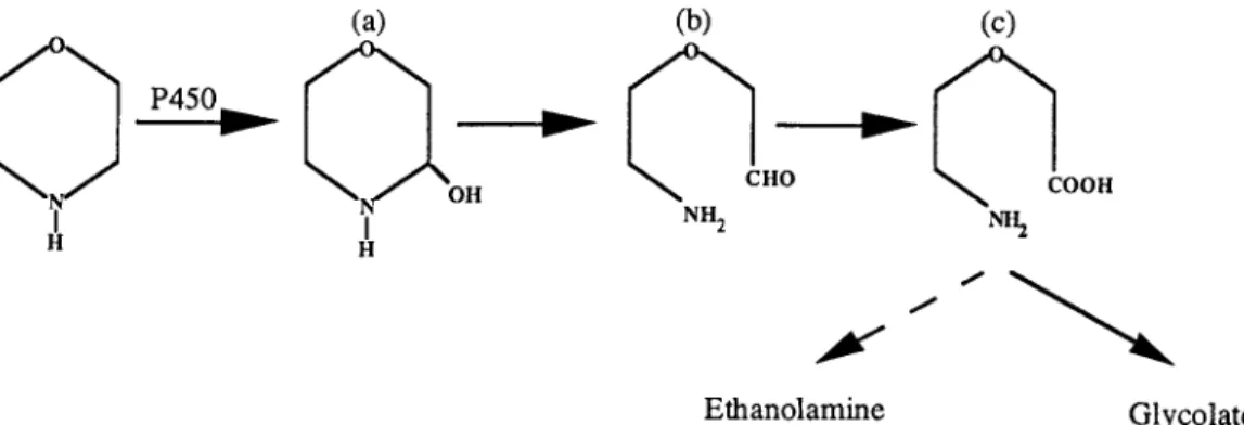
The most probable reaction catalyzed by this enzyme on these cyclic amines is CON bond cleavage by a mecha-nism similar to an N dealkylation (Fig. 8). The monooxygenase catalyzes a hydroxylation on a C atom adjacent to the amine group which gives an unstable compound (compound a). This mechanism (rather than N oxygenation) is favored when a-protons are available (10). The compound so formed could be linearized to give 2-(2-aminoethoxy)acetaldehyde (compound b),whichcouldundergooxidationtoform2-(2-aminoethoxy)ace-tate (compound c).
In conclusion, the present results show that Mycobacterium sp. strain RP1 catalyzes the degradation of morpholine, pyr-rolidine, and piperidine. This strain can use these compounds, which are the secondary amines most used in industry, as the sole source of carbon, nitrogen and energy. These degrada-tions involve a soluble cytochrome P-450. The growth of strain
FIG. 8. Hypothetical pathway for morpholine degradation by Mycobacterium sp. strain RP1. a, 2-hydroxymorpholine; b, 2-(2-aminoethoxy)acetaldehyde; c, 2-(2-aminoethoxy)acetate.
TABLE 1. Growth of Mycobacterium sp. strain RP1 on different substrates and induction of a cytochrome P-450
Substrate Growtha Cytochrome P-450(pmolz mg of protein21) Acetate 1 NDb Succinate 1 ND Morpholine 1 90 Thiomorpholine 2 80 Pyrrolidine 1 140 Piperidine 1 132 Tetrahydrofuran 2/1 ND Tetrahydropyran 2/1 ND
a1, growth; 2/1, slow growth; 2, no growth. bND, not detected.
RP1 on different substrates and the induction of a heme-containing monooxygenase suggest that this enzyme attacks morpholine at the CON position. This reaction could be fol-lowed by ring cleavage to form 2-(2-aminoethoxy)acetate which is further degraded to glycolic acid, as shown by the detection of these intermediates in the1H NMR spectra. This
study, together with previous work (6, 11, 30), underlines the importance of mycobacteria in the degradation of xenobiotic compounds. Further investigations to identify metabolites of the early reactions in the catabolism of morpholine by
Myco-bacterium sp. strain RP1 are in progress.
ACKNOWLEDGMENTS
This work was supported by interdisciplinary programs of CNRS “PIRSEM” and “ECOTECH.” P. Poupin and B. Combourieu were recipients of a fellowship from the Ministe`re de la Recherche et de l’Enseignement Supe´rieur.
We acknowledge Anne-Lise Etienne, coordinator of the CNRS pro-grams.
REFERENCES
1. Bradford, M. M. 1976. A rapid and sensitive method for the quantitation of microgram quantities of protein utilizing the principle of protein-dye bind-ing. Anal. Biochem. 72:248–254.
2. Brown, V. R., and J. S. Knapp. 1990. The effect of withdrawal of morpholine from the influent and its reinstatement on the performance and microbial ecology of a model activated sludge plant treating a morpholine-containing influent. J. Appl. Microbiol. 69:43–53.
3. Calmels, S., H. Ohshima, and H. Bartsch. 1988. Nitrosamine formation by denitrifying and non-denitrifying bacteria: implication of nitrite reductase and nitrate reductase in nitrosation catalysis. J. Gen. Microbiol. 134:221–226. 4. Cech, J. S., P. Hartman, M. Slosarek, and J. Chudoba. 1988. Isolation and identification of a morpholine-degrading bacterium. Appl. Environ. Micro-biol. 54:619–621.
5. Combourieu, B., P. Besse, M. Sancelme, H. Veschambre, A. M. Delort, P. Poupin, and N. Truffaut.1998. Morpholine degradation pathway of Myco-bacterium aurum MO1: direct evidence of intermediates by in situ1H nuclear
magnetic resonance. Appl. Environ. Microbiol. 64:153–158.
6. Dean-Ross, D., and C. E. Cerniglia. 1996. Degradation of pyrene by Myco-bacterium flavescens. Appl. Microbiol. Biotechnol. 46:307–312.
7. Dmitrenko, G. N., P. I. Gvozdyak, and V. M. Udod. 1987. Selection of destructor microorganisms for heterocyclic xenobiotics. Khim. Tekhnol. Vody 9:442–445.
8. Enzmann, H., H. Zerban, A. Kopp-Schneider, E. Loser, and P. Bannasch. 1995. Effects of low doses of N-nitrosomorpholine on the development of early stages of hepatocarcinogenesis. Carcinogenesis 16:1513–1518. 9. Guengerich, F. P. 1990. Enzymatic oxidation of xenobiotic chemicals.
Bio-chem. Mol. Biol. 25:97–153.
10. Guengerich, F. P., and T. L. Macdonald. 1990. Mechanisms of cytochromes P450 catalysis. FASEB J. 4:2453–2459.
11. Knapp, J. S., and V. R. Brown. 1988. Morpholine biodegradation. Int. Bio-deterior. 25:299–306.
12. Knapp, J. S., A. G. Callely, and J. Mainprize. 1982. The microbial degrada-tion of morpholine. J. Appl. Bacteriol. 52:5–13.
12a.Lewis, R. J., Sr. (ed.). 1995. Chemical review: morpholine. Danger. Prop. Ind. Mater. Rep. 15:270–297.
13. Mazure, N., and N. Truffaut. 1994. Degradation of morpholine by Mycobac-terium aurum MO1. Can. J. Microbiol. 40:751–765.
14. Miles, J. S., A. W. Munro, B. N. Rospendowski, W. E. Smith, J. McKnight, and A. J. Thomson.1992. Domains of the catalytically self-sufficient cyto-chrome P450 BM-3. Genetic construction, overexpression, purification and spectroscopic characterization. Biochem. J. 288:503–509.
15. Munro, A. W., and J. G. Lindsay. 1996. Bacterial cytochromes P450. Mol. Microbiol. 20:1115–1125.
16. Nagy, I., G. Schoofs, F. Compernolle, P. Proost, J. Vanderleyden, and R. De Mot. 1995. Degradation of the thiocarbamate herbicide EPTC (S-ethyl dipropylcarbamothioate) and biosafening by Rhodococcus sp. strain NI86/21 involve an inducible cytochrome P-450 system and aldehyde dehydrogenase. J. Bacteriol. 177:676–687.
17. O’Keefe, D. P., and P. A. Harder. 1991. Occurrence and biological function of cytochrome P450 monooxygenases in the actinomycetes. Mol. Microbiol. 5:2099–2105.
18. Penaud, F. 1989. Ph.D. thesis. University of Compie`gne, Compie`gne, France. 19. Peterson, J. A., and J.-Y. Lu. 1991. Bacterial cytochromes P450: isolation and
identification. Methods Enzymol. 206:612–620.
20. Poupin, P., N. Mazure, and N. Truffaut. 1996. Morpholine degradation by strain Mycobacterium aurum MO1: improvement of cells growth and mor-pholine degradation rate by cell immobilization, p. 770–776. In R. H. Wij-ffels, R. M. Buitlaar, C. Blucke, and J. Tramper (ed.), Progress in biotech-nology, vol. 11. Immobilized cells: basis and applications. Elsevier Science, B.V., Amsterdam, The Netherlands.
21. Sariaslani, S. F. 1991. Microbial cytochromes P450 and xenobiotic metabo-lism. Adv. Appl. Microbiol. 36:133–178.
22. Sariaslani, S. F., and C. A. Omer. 1992. Actinomycetes cytochromes P450 involved in oxidative metabolism: biochemistry and molecular biology. Crit. Rev. Plant Sci. 11:1–16.
23. Stevens, W. H., and K. Skov. 1965. A rapid spectrophotometric method for determining parts per million of morpholine in boiler water. Analyst 90:182– 183.
24. Swain, A., K. V. Waterhouse, W. A. Venables, A. G. Callely, and S. E. Lowe. 1991. Biochemical studies of morpholine catabolism by an environmental mycobacterium. Appl. Microbiol. Biotechnol. 35:110–114.
25. Testa, B., and P. Jenner. 1981. Inhibitors of cytochrome P450s and their mechanism of action. Drug Metab. 12:1–117.
26. Tomasi, I., I. Artaud, Y. Bertheau, and D. Mansuy. 1995. Metabolism of polychlorinated phenols by Pseudomonas cepacia AC1100: determination of the first two steps and specific inhibitory effect of methimazole. J. Bacteriol. 177:307–311.
27. Uotila, J. S., M. S. Salkinoja-Salonen, and J. H. A. Apajalahti. 1991. Deg-radation of pentachlorophenol by membrane bound enzymes from Rhodo-coccus chlorophenolicus PCP-1. Biodegradation 2:25–31.
28. Uotila, J. S., V. H. Kitunen, T. Saastamoinen, T. Coote, M. M. Ha¨ggblom, and M. S. Salkinoja-Salonen.1992. Characterization of aromatic dehaloge-nases of Mycobacterium fortuitum CG-2. J. Bacteriol. 174:5669–5675. 29. Van Stee, E. W., R. A. Slone, J. E. Simmons, M. P. Moorman, and K. D.
Brunnemann.1995. Endogenous formation of N-nitrosomorpholine in mice from15NO
2by inhalation and morpholine gavage. Carcinogenesis 16:89–92.
30. Wang, R. F., W. W. Cao, and C. E. Cerniglia. 1995. Phylogenetic analysis of polycyclic aromatic hydrocarbon degrading mycobacteria by 16S rRNA se-quencing. FEMS Microbiol. Lett. 130:75–80.
31. White, G. F., N. J. Russel, and E. C. Tidswell. 1996. Bacterial scission of ether bonds. Microbiol. Rev. 60:216–232.
32. World Health Organization. 1995. Morpholine: health and safety guide. World Health Organization, Geneva, Switzerland.