Review
The ubiquitin/26S proteasome system in plant–pathogen
interactions: a never-ending hide-and-seek game
ANNE-SOPHIE DIELEN
1, SALOUA BADAOUI
2, THIERRY CANDRESSE
1AND
SYLVIE GERMAN-RETANA
1,*
1Interactions Plante-Virus, UMR GDPP 1090, INRA Université de Bordeaux 2, BP 81, F-33883 Villenave d'Ornon Cedex, France 2UMR GDEC 1095, INRA/Université Blaise Pascal, 234 avenue du Brézet, 63100 Clermont-Ferrand, France
SUMMARY
The ubiquitin/26S proteasome system (UPS) plays a central role in plant protein degradation. Over the past few years, the impor-tance of this pathway in plant–pathogen interactions has been increasingly highlighted. UPS is involved in almost every step of the defence mechanisms in plants, regardless of the type of pathogen. In addition to its proteolytic activities, UPS, through its 20S RNase activity, may be part of a still unknown antiviral defence pathway. Strikingly, UPS is not only a weapon used by plants to defend themselves, but also a target for some patho-gens that have evolved mechanisms to inhibit and/or use this system for their own purposes. This article attempts to summa-rize the current knowledge on UPS involvement in plant– microbe interactions, a complex scheme that illustrates the never-ending arms race between hosts and microbes.
INTRODUCTION
All cell processes, from division to death, include essential protein degradation steps. In eukaryotes, most of the protein degradation events are controlled by the ubiquitin/26S protea-some system (UPS) (Dreher & Callis, 2007). As sessile organisms that face environmental variations, plants have evolved a number of mechanisms to protect themselves against abiotic and biotic stresses, such as preformed physical barriers, antimi-crobial compounds, and the perception and recognition of pathogen-associated molecular patterns (PAMPs) or pathogen race-specific effectors. Proteomic plasticity is a crucial element in these plant responses to abiotic and biotic stresses. UPS there-fore plays a central role in plant defence by altering the pro-teome and thus increasing the chances of survival (Dreher & Callis, 2007). UPS involvement in plant defence mechanisms
occurs at different levels, from ubiquitin to 26S proteasome. Changes in ubiquitin and E1 and/or E2 enzyme levels may have broad effects on cell reprogramming during plant defence, and many research groups have shown recently that E3 ubiquitin ligases, the key components of UPS targeting specificity, are implicated in plant–pathogen interactions, including early defence reactions, gene-for-gene interactions and induced disease resistance (Delauré et al., 2008; Zeng et al., 2006).
This article attempts to place this new information in perspec-tive, highlighting the crucial role played by UPS in plant disease responses, involving both the ubiquitin conjugating system and 26S proteasome. It also shows that pathogens are able to hijack this pathway, to counteract plant defence or to utilize host UPS components to their own advantage, revealing a complex UPS– pathogen interaction scheme.
UBIQUITIN/26S PROTEASOME: A POWERFUL SYSTEM TO REGULATE PROTEIN STABILITY
Ubiquitin conjugation pathway
Most UPS substrate proteins are covalently bound to ubiquitin, a 76-amino-acid protein, by a three-step energy-dependent mechanism prior to degradation by 26S proteasome (Fig. 1A). Ubiquitin is activated for transfer by the formation of a thiolester bond with a ubiquitin-activating enzyme (E1). Activated ubiq-uitin is then transferred to a ubiqubiq-uitin-conjugating enzyme (E2), again through a thiolester bond. Finally, a ubiquitin ligase enzyme (E3) recruits and transfers ubiquitin to the target protein. The ubiquitin C-terminal glycine residue is linked to a substrate amino acid (generally a lysine residue) via an isopeptide bond (Vierstra, 2009). This mechanism is repeated to obtain polyubiq-uitinated target proteins (Dreher & Callis, 2007). The shortest polyubiquitin chain capable of activating proteasomal degrada-tion is four monomers long (Thrower et al., 2000), although many substrate proteins appear to be tagged with longer chains (Hanna & Finley, 2007).
The major role of polyubiquitination is to address the sub-strate protein to the 26S proteasome, where its peptide bonds are hydrolysed, reducing a folded protein into oligopeptides that are released in the cytoplasm (Hanna & Finley, 2007). However, ubiquitination not only targets proteins for degradation, but is involved in the regulation of post-translational modifications. The impact of ubiquitination depends on many factors, including the ubiquitin chain length and the particular amino acid of the ubiquitin chain through which it is attached to the target protein. Thus, proteins containing lysine-48 (Lys-48)-linked poly-ubiquitin tags are targeted for proteasomal degradation. In con-trast, monoubiquitinated or polyubiquitinated proteins linked via ubiquitin Lys-63 are not degraded but involved in nonproteolytic events, such as subcellular localization or functional post-transductional modification, protein activation or protein– protein interactions (Deng et al., 2000; Manzano et al., 2008; Schnell & Hicke, 2003). Notably, it has also been demonstrated that some proteins can be addressed to the 26S proteasome in a ubiquitin-independent manner, such as calmodulin or p53 (Orlowski & Wilk, 2003), but the associated mechanism(s) still remains unknown.
Different types of E3 ubiquitin ligase
The E3s, responsible for the final tagging of proteins, provide specificity to the UPS process. Large E3 ligase families are encoded in plant genomes, each family member controlling the ligation of ubiquitin to only one or a small subset of substrate
proteins, implying that a wide variety of targets may be recog-nized (Vierstra, 2003). Over 6% of the predicted Arabidopsis thaliana genome encodes UPS proteins (Dreher & Callis, 2007), including only two E1, 37 predicted E2 (Downes & Vierstra, 2005) and at least 1400 predicted E3 proteins. Plant E3s fall into different families on the basis of their subunit composition and mode of action (Lechner et al., 2006). These families can be classified into two major groups, which covalently [HECT (homol-ogy to E6-associated protein C-terminus) E3s] and noncovalently [U-box domain and RING (really interesting new gene) E3s] bind to ubiquitin (Fig. 1B). HECT E3 ligases establish a covalent bond with ubiquitin before its transfer to the target protein (Dreher & Callis, 2007). To date, seven HECT E3s have been identified in the A. thaliana genome, among which UPL3 (HECT-containing ubiq-uitin protein ligase 3) is involved in trichome shape control (Downes et al., 2003). U-box and RING E3s are thought to be structurally related and functionally similar, using hydrogen bonds/salt bridges or zinc chelation, respectively, to transfer ubiquitin to the substrate (Downes & Vierstra, 2005; Stone et al., 2005). About 61 U-box domain E3s are predicted in the A. thaliana genome, but at least 475 proteins (about 2% of the predicted A. thaliana proteins) contain one or more RING motifs (Stone et al., 2005, 2006). Two types of RING-type E3 ubiquitin ligase exist: single subunit E3 and RING-finger proteins as sub-units of multiprotein E3 complexes. One of the most conserved multisubunit RING E3 families in eukaryotes is the cullin RING ligases (CRL), among which the modular SCF group is the largest and best characterized because of its roles in many cellular
Fig. 1 An overview of the ubiquitin/26S proteasome system (UPS). (A) The UPS pathway begins with the ATP-dependent activation of ubiquitin by an E1
enzyme. Ubiquitin is then transferred to an E2 enzyme, and finally attached to the target protein via an E3 enzyme. Multiple cycles of ubiquitin conjugation lead to a polyubiquitinated substrate that is degraded by the 26S proteasome complex, releasing short peptides. (B) Types of E3 ubiquitin ligase enzymes. See text for details. (C) Structure of the 26S proteasome. The 20S core protease is composed of four heptameric rings, forming three cavities. The outer rings containa subunits and the inner ringsb subunits. Three of the b subunits (namely b1, b2 and b5) harbour a protease activity. One or two 19S regulatory particles can be attached to the outer rings. The 19S regulatory particle is composed of two complexes, the lid and the base. The base contains six proteasomal ATPases attached to thea rings of the 20S proteasome. The Rpn10 subunit can interact either with the lid or the base and stabilizes the complex. Adapted from Delauré
processes. Arabidopsis thaliana SCF complexes are composed of four major subunits: cullin-1 (CUL1), SKP1 (S-phase kinase-associated protein)-like protein (ASK1/2), RBX1 (RING box protein) and F-box protein (Dreher & Callis, 2007). CUL1 acts as a scaffold in assembling the different subunits of the SCF complex by interacting at its C-terminal region with RBX1 and at its N-terminus with SKP1. RBX1 and SKP1 are linked to E2-ubiquitin and F-box protein, respectively. The RBX1–E2 association mediates the ubiquitin transferase activity, and the SKP1–F-box protein complex confers substrate specificity (Lechner et al., 2006). The A. thaliana genome encodes about 700 F-box proteins (Devoto et al., 2003; Gagne et al., 2002), and 687 potential F-box proteins have been identified in Oryza sativa (Jain et al., 2007), suggesting a high targeting potential. Inter-estingly, the number of F-box proteins in plants is significantly higher than in other eukaryotes, but the reason for such an expansion remains unclear to date (Thomann et al., 2005).
The 26S proteasome
The structure of the 26S proteasome is highly conserved in eukaryotes and can be divided into two distinct particles: the 20S core proteasome (CP) and the 19S regulatory particle (RP) (Fig. 1C) (Vierstra, 2003). The CP is a broad-spectrum, ATP- and ubiquitin-independent protease, composed of four stacked rings, defining a barrel-shaped structure. The outer rings are composed of sevena subunits and the inner rings are composed of seven b subunits, which give the 20S complex the general structure of a1–7b1–7b1–7a1–7. Three proteolytic activities, carried by theb1,b2 andb5subunits, are housed by the inner rings, defining a cata-lytic chamber. Access to this chamber is controlled by thea rings, which only allow unfolded proteins to enter (Kurepa & Smalle, 2007; Lorentzen & Conti, 2006; Vierstra, 2003). One or both CP outer rings can be capped by a 19S RP, composed of two com-plexes, the lid and the base (Kurepa & Smalle, 2007). The base contains three non-ATPase subunits (RPN) and a ring of six ATPase subunits (RPT) that interacts with the CPa subunits, and is probably involved in target unfolding and gate opening via ATP-dependent mechanisms. The lid, which binds to the base, generally contains nine RPN subunits, although some other sub-units may also bind to the lid subcomplex in regulating 26S proteasome activity (Glickman & Raveh, 2005). Little is known about the lid subunit functions, except for RPN11, which has a deubiquitinating activity, and RPN5, 6 and 7, which are essential for subparticle assembly (Vierstra, 2003).
Interestingly, it has been demonstrated in vitro that animal and plant 20S proteasomes also harbour RNase activity. This activity was first observed with chicken liver proteasomes that degrade 18S rRNA in vitro (Tsukahara et al., 1989). The authors showed that this activity was inactivated by heat treatment and by the addition of low sodium dodecylsulphate concentrations,
thus excluding the hypothesis of a contamination by low-molecular-mass RNases. Using Tobacco mosaic virus (TMV, Tobamovirus) RNA as a substrate, it has been demonstrated further that calf liver 20S proteasome RNase activity is an endo-nuclease activity, associated with thea5subunit (named zeta), and that this activity is not influenced by 20S integrity, as strong dissociating conditions (6Murea) do not impair RNA degrada-tion (Petit et al., 1997; Pouch et al., 1995). Petit et al. (1997) also showed that a lower endonuclease activity is associated with the a1subunit (named iota), but, to date, this observation has not been confirmed. Some RNAs containing tRNA-like structures, such as the Human immunodeficiency virus (HIV, Retrovirus) TAR (transactivation response) RNA, are also degraded (Gautier-Bert et al., 2003). Furthermore, 20S RNA complex reconstruction in vitro and RNA residual fragment identification in purified pro-teasome fractions have confirmed that the animal 20S protea-some harbours an RNA endonuclease activity (Gautier-Bert et al., 2003). Recently, the existence of a similar 20S proteasome endonuclease activity has also been demonstrated in purified sunflower proteasome, using TMV and Lettuce mosaic virus (LMV, Potyvirus) RNA substrates (Ballut et al., 2003), indicating that the existence of such an activity is not specific to animal proteasomes.
THE UBIQUITIN/PROTEASOME PATHWAY IN PLANT DEFENCE REACTIONS
UPS plays a central role in the cell and targets two major types of protein: misfolded/damaged proteins and functional proteins carrying specific destruction signals. Therefore, UPS acts both as a quality control and regulatory system (Goldberg, 2003; Kurepa & Smalle, 2007). As a regulatory system, UPS is involved in mechanisms as essential as cell cycle control (Jurado et al., 2008; Hershko, 2005; del Pozo et al., 2006), programmed cell death (PCD) (Endo et al., 2001; Jin et al., 2006; Stone & Callis, 2007) and plant development (Brukhin et al., 2005; Downes et al., 2003; Kevany et al., 2007; Schwager et al., 2007; Sheng et al., 2006), and is also implicated in self-incompatibility (Hua & Kao, 2008) and signal transduction cascades following light (Hoecker, 2005; Moon et al., 2007), sucrose (Ellis et al., 2002) or hormone (Bostick et al., 2004; Manzano et al., 2008; Stone et al., 2006) signalling. Thus, UPS plays a role in plant responses to the majority of external environment changes. For example, a hot pepper U-box ubiquitin ligase mRNA is rapidly and highly induced in response to various environmental stress factors, including dehydration, high salinity and cold temperature (Cho et al., 2006), proteasomal function is required to trigger PCD in heat-shocked plants (Vacca et al., 2007) and 20S proteasome is involved in heavy metal resistance in cadmium- and nickel-stressed maize plants by scavenging metal-oxidized proteins (Forzani et al., 2002; Pena et al., 2007).
Plants face biotic stress in the form of insects, nematodes, fungi, bacteria or virus attacks on a daily basis. However, most plants are resistant to most pathogens because of the complex mechanisms they have developed to inhibit or limit damage. Plant defence can, in particular, be mediated by specific interac-tions between pathogen avirulence (Avr) and plant resistance (R) genes, triggering the deployment of active defence mechanisms and the containment of pathogens in restricted areas (Dangl & Jones, 2001). During the past few years, a growing body of evidence has indicated that UPS is not only implicated in crucial cellular surveillance mechanisms, but also that it can be involved in the defence of plants against pathogens. UPS appears to be required in various steps of the jasmonate, salicylic acid and ethylene signalling pathways (Binder et al., 2007; Devoto et al., 2002; Thines et al., 2007; Yaeno & Iba, 2008), all three of which are involved in defence reactions against bioaggressors. Never-theless, hormone signalling does not seem to be the only way in which UPS is involved in plant defence mechanisms. Indeed, UPS is an emerging actor in plant–microbe interactions.
Role of ubiquitination in plant defence
About 20% of A. thaliana gene expression changes under pathogen attack (Rietz & Parker, 2007) and the up-regulation of general components of UPS have commonly been observed in various plant–pathogen interactions. Protein (poly)ubiquitina-tion may play a role in basal host resistance: in powdery mildew (Blumeria graminis)-attacked barley epidermis, depletion of cel-lular ubiquitin by transient-induced gene silencing (TIGS), con-trary to the targeting of the 19S RP by RNA interference (RNAi), induces high susceptibility towards the compatible strain B. graminis f. sp. hordei (Dong et al., 2006). The ubiquitin conjuga-tion pathway is thus necessary for the enhanced protein degra-dation observed, but the 26S proteasome is not (Dong et al., 2006). Ubiquitination may also be involved in gene-for-gene resistance: sunflower hypocotyls accumulate high levels of ubiq-uitin transcripts when challenged with an incompatible strain of powdery mildew (Plasmopara halstedii), but not with a compat-ible strain (Mazeyrat et al., 1999). Furthermore, in A. thaliana, the mos5 (modifier of snc1 5) suppressor of the gain-of-function snc1 (suppressor of npr1-1 constitutive 1) mutant has been shown to be affected in UPS (Goritschnig et al., 2007). The snc1 mutant corresponds to a point mutation in an R gene, resulting in the constitutive activation of defence responses without inter-action with pathogens. Interestingly, the mos5 mutant carries a 15-bp deletion in AtUBA1 (ubiquitin-activating enzyme 1) (Goritschnig et al., 2007), which is thus able to block the signal-ling cascade downstream of an R gene, through an as yet unknown mechanism. In fact, the mos5 mutation only affects the resistance response conferred by a small subset of R proteins, suggesting that the ubiquitination pathway is essential for the
activation and downstream signalling of some, but possibly not all, R gene-mediated responses (Goritschnig et al., 2007). UPS also appears to be implicated in compatible interactions. The altered response to viral infection of tobacco plants perturbed in UPS (Becker et al., 1993), and the up-regulation of two tobacco ubiquitin-activating enzymes (NtE1A and NtE1B) by infection with Tomato mosaic virus (ToMV, Tobamovirus) and TMV (Tak-izawa et al., 2005), support the idea that UPS participates in the molecular dialogue between viruses and compatible host plants.
E3 ubiquitin ligases mediating plant defence signalling
SGT1, an E3 ubiquitin ligase ubiquitous in plant defence The first clue indicating that E3-mediated proteolysis contributes to R gene defence was provided by the rapid turnover of A. thaliana RPM1 (resistance to Pseudomonas syringae pv. macu-licola 1), a nucleotide binding site leucine-rich repeat (NBS-LRR) R protein, coincident with the onset of the hypersensitive response (HR) (Boyes et al., 1998). As RPM1 confers resistance to some P. syringae strains by triggering an HR, the authors sug-gested that this observation could correspond to a negative feedback loop used by the plant to control HR lesion size and response amplitude at the site of infection (Boyes et al., 1998). The disappearance of RPM1 seems to be mediated by UPS as it is suppressed after treatment with a proteasome inhibitor (Kawasaki et al., 2005). To date, the two identified RING-finger E3 proteins, RIN2 (RPM1-interacting protein 2) and RIN3, which interact directly with RPM1, are not required for its degradation, which suggests that other unidentified partners are involved (Kawasaki et al., 2005) (Fig. 2A). Interestingly, it has been reported that RPM1 is undetectable in A. thaliana rar1 (required for Mla12 resistance 1) mutants, suggesting that RAR1 regulates RPM1 stability (Tornero et al., 2002). RAR1 encodes a predicted cytosolic protein of unknown function, conserved in all eukary-otes except yeast (Shirasu et al., 1999). It seems that RAR1 associates in A. thaliana with cytosolic HSP90 to act as a co-chaperone and to stabilize some NBS-LRR resistance proteins, such as RPM1 (Holt et al., 2005; Hubert et al., 2003). A yeast two-hybrid screen has shown that the A. thaliana orthologue of RAR1 interacts with AtSGT1 (suppressor of G2 allele of skp1), a plant orthologue of the yeast E3 protein SGT1, involved in cell cycle control, which associates with the SKP1 and cullin subunits of the SCF complex (Azevedo et al., 2002). It has been shown by mutational analysis that AtSGT1b is required for A. thaliana resistance against Peronospora parasitica (Austin et al., 2002; Tör et al., 2002).
The involvement of RAR1 and SGT1 in defence mechanisms has also been highlighted in several other plants. In barley, RAR1 is required for powdery mildew resistance triggered by the Mla6 and Mla12 R proteins (Shirasu et al., 1999). Indeed,
RAR1 positively regulates Mla gene steady-state levels in barley (Bieri et al., 2004). SGT1 is also implicated in barley disease resistance as its silencing alters powdery mildew resistance (Azevedo et al., 2002). SGT1, but not RAR1, is also involved in potato resistance to Phytophthora infestans triggered by the RB (resistance to P. infestans-bulbocastanum1) R gene (Bhaskar et al., 2008), whereas the Nicotiana benthamiana SGT1 ortho-logue (NbSGT1), which interacts with both NbRAR1 and NbSKP1, is required for N gene-mediated resistance to TMV (Liu et al., 2002). NbSGT1 silencing causes N. benthamiana to lose R gene-mediated resistance against Potato virus X (PVX, Potex-virus), Cladosporium fulvum, Phytophthora infestans and P. syringae, and the loss of some (against P. syringae pv. maculi-cola or Xanthomonas axonopodis pv. vesicatoria), but not all [Cauliflower mosaic virus (CaMV), Caulimovirus; X. campestris pv. campestris], nonhost resistances, highlighting the role of SGT1 in the molecular control of a wide range of pathogens (Peart et al., 2002). Furthermore, the silencing of NbSGT1 causes a reduction in the levels of the Rx protein, suggesting a role in the accumulation of some R proteins (Azevedo et al., 2006). These studies suggest a crucial role for the ubiquitination pathway in R gene-mediated disease resistance, and SGT1 may be a general factor involved in plant defence mechanisms (Fig. 2B). In agreement with this hypothesis, a recent study has shown that SGT1 and RAR1 play a role in soybean resistance to P. syringae, as they are required for the induction of R gene-mediated defence mechanisms, systemic acquired resistance and basal defence (Fu et al., 2009).
Other E3 ubiquitin ligases involved in resistance mechanisms In addition to the important role of SGT1, several examples underline the involvement of other E3 ligases in plant defence mechanisms, regardless of the type of pathogen. Table 1 sum-marizes some of these examples, highlighting the diversity of targeted pathogens and citing the corresponding substrates (if known).
OsRHC1 (RING HC domain 1), a rice RING zinc finger protein, confers improved resistance to P. syringae pv. tomato DC3000 to A. thaliana. This resistance is proteasome dependent as it is abolished by MG132, a known proteasome inhibitor (Cheung et al., 2007). Overexpression of OsDRF1 (defence-related F-box 1), an F-box protein whose expression is induced by benzothia-diazole, a chemical inducer of plant defence responses, also enhances disease resistance to P. syringae pv. tabaci and ToMV in transgenic tobacco (Cao et al., 2008). OsDRF1 seems to be implicated only in responses to biotic stresses, as no evidence of OsDRF1 implication in abiotic stress tolerance has been found. In Solanum lycopersicum cv. Micro-Tom, the expression of E3 ligase LeATL6 (Arabidopsis toxico para levadura-toxic to fungi-6) has also been found to be elicited in roots using a cell wall protein fraction of Pythium oligandrum. Overexpression of LeATL6, driven by the CaMV 35S promoter, activates the transcription of some members of the pathogenesis-related (PR) gene family in wild-type tomato, but not in jai1-1 (jasmonate insensitive 1-1) mutants, in which the jasmonate signalling pathway is impaired, suggesting that LeATL6 may play a role in elicitor-induced resis-tance via a jasmonate-dependent pathway (Hondo et al., 2007).
Fig. 2 SGT1 (suppressor of G2 allele of skp1) involvement in plant defence mechanisms. (A) RPM1 (resistance to Pseudomonas syringae pv. maculicola 1)
degradation during the onset of the hypersensitive response (HR). To date, the E3 ligase mediating RPM1 degradation still remains unknown. As SGT1 interacts with RAR1 (required for Mla12 resistance 1), a protein that regulates RPM1 stability, SGT1 may be required in this degradation mechanism. (B) Examples of the crucial role played by SGT1 in plant defence mechanisms (R gene products are indicated by green shading). PVX, Potato virus X; TMV, Tobacco mosaic virus; (1), Azevedo et al., 2006; (2), Liu et al., 2002; (3), Azevedo et al., 2002; (4), Fu et al., 2009; (5), Bhaskar et al., 2008; (6), Tör et al., 2002.
The Nicotiana tabacum ACIF1 (Avr9/Cf9-INDUCED F-BOX 1) F-box protein, which associates with ASK1 and CUL1 in the SCF complex, is required for elicitor-induced HR and is involved in N gene-mediated resistance to TMV (van den Burg et al., 2008). Moreover, the function of A. thaliana ACIF1 homologues is linked to abiotic and biotic stress responses by regulating absci-sic acid- and jasmonate-responsive genes (van den Burg et al., 2008).
The Arabidopsis RING E3 ligase HISTONE MONOUBIQUITINA-TION 1 (HUB1) is also a regulatory component of plant defence against necrotrophic fungal pathogens via the regulation of gene expression. Indeed, loss-of-function A. thaliana hub1 mutants display extreme susceptibility to Botrytis cinerea and Alternaria brassicicola (Dhawan et al., 2009). As HUB1 is involved in H2B histone monoubiquitination, which does not lead to degradation by the 26S proteasome but to chromatin regulation and modulation of gene expression, the action of HUB1 might be mediated by a direct effect on gene expression rather than through the UPS pathway (Dhawan et al., 2009).
Involvement of the 26S proteasome in plant defence Both 26S RP and CP could play a role in defence against patho-gens, but the demonstration so far remains indirect. A screen for TMV-induced nuclear proteins in Capsicum annuum cv. Bugang has shown that RPN7 expression is increased at both the tran-scriptional and translational levels during incompatible interac-tion (TMV-P0), but not during compatible interacinterac-tion (TMV-P1.2), and may thus be involved in PCD (Lee et al., 2006). Three
tobacco 20S subunits,a3,a6andb1, are rapidly and specifically induced at the transcriptional level by cryptogein, a fungal elicitor of defence reactions. The expression of these defence-induced (din) subunits (a3din,a6din andb1din) is correlated with the induction of systemic acquired resistance (SAR), an inducible immune response against a broad spectrum of pathogens, and the production of reactive oxygen species (ROS) (Dahan et al., 2001). It has been postulated that these subunits could replace the corresponding constitutive 20S subunits, leading to a newly reassembled ‘plant defence proteasome’ (Suty et al., 2003), which could be the plant equivalent to the animal immunopro-teasome, induced by interferon-g (Kloetzel, 2004), even though the proteolytic activities of the plant defence proteasome remain unchanged (Suty et al., 2003). Theb1din subunit may contribute to ROS regulation during plant defence reactions by inhibiting NADPH oxidase at the transcriptional level (Lequeu et al., 2005), but these data need to be confirmed.
The proteasome-mediated degradation of NPR1 (nonexpres-sor of pathogenesis-related genes 1), a transcription coactivator critical for SAR, has been shown recently to play a dual role in the regulation of plant immunity. Indeed, in noninduced cells (no SAR), the nuclear proteasome-mediated turnover of NPR1 pre-vents the inappropriate activation of SAR, whereas in SAR-induced cells, phosphorylation of NPR1 facilitates its recruitment to a cullin-3-based ubiquitin ligase and its proteasomal degra-dation, stimulating NPR1 target gene expression (Spoel et al., 2009). The authors have hypothesized that such a turnover of ‘exhausted’ phosphorylated NPR1 from the target promoter allows ‘fresh’ NPR1 to reinitiate the transcription cycle.
Table 1 Some plant E3-ligases involved in defence mechanisms.
Protein Organism E3 ligase type Predicted substrates Pathways Pathogen References
ACIF1 N. tabacum F-box TMV (van den Burg et al., 2008) ACRE276 N. tabacum U-box Cf-genes mediated HR TMV (Yang et al., 2006)
ATL1 S. lycopersicum RING (Hondo et al., 2007)
BAH1/NLA A. thaliana RING SA signalling P. syringae (Yaeno & Iba, 2008)
COI1 A. thaliana F-box JAZ proteins JA signalling (Devoto et al., 2002, Thines
et al., 2007)
DRF1 O. sativa F-box P. syringae
ToMV
(Cao et al., 2008) EBF1, EBF2 A. thaliana F-box EIN3 ethylene signalling (Binder et al., 2007) PUB22, 23, 24 A. thaliana U-box PAMP-triggered resistance
(negative regulation)
(Trujillo et al., 2008)
RHC1 O. sativa RING P. syringae (Cheung et al., 2007)
SGT1 A. thaliana F-box P. parasitica (Austin et al., 2002, Tör et al.,
2002)
SGT1 A. thaliana F-box P. syringae (Kawamura et al., 2009)
SGT1 A. thaliana F-box R. solanacearum (Kawamura et al., 2009)
SGT1 G. max F-box P. syringae (Fu et al., 2009)
SGT1 N. benthamiana F-box TMV (Liu et al., 2002)
SGT1 Potato F-box P. infestans (Bhaskar et al., 2008)
SON1 A. thaliana F-box P. parasitica
P. syringae
(Kim & Delaney, 2002)
SPL11 O. sativa U-box M. grisea
X. oryzae
The 20S a5 subunit may also play a crucial role in plant defence. The expression of thea5subunit from Oryza grandiglu-mis (OgPAE1) is induced during defence responses, and the overexpression of OgPAE1 in A. thaliana leads to resistance against Bo. cinerea, but the underlying mechanism(s) remains unknown (Jeon et al., 2008). The implication of the proteasome a5subunit in plant defence might be more important in resis-tance to viruses, as it has been shown to specifically cleave in vitro TMV RNA (Ballut et al., 2003; Petit et al., 1997). To date, a key question remains unresolved, that of an in vivo contribution to plant antiviral defence for this catalytic RNase activity iden-tified in vitro. In such a scenario, plants would have evolved two antiviral mechanisms based on RNA degradation: RNAi and 20S-mediated degradation (Ballut et al., 2005). 20S-20S-mediated RNA degradation could be considered as a first component of plant antiviral defence, targeting nonhost RNAs, a less fine-tuned mechanism than RNAi. Interestingly, calf liver proteasomes hydrolyse HIV-TAR, a tRNA-like structure at the 5′-end of HIV mRNAs (Gautier-Bert et al., 2003). The authors have suggested that this degradation may be part of a defence mechanism, as HIV mRNAs lacking their 5′-end are poorly translated.
Viral proteins themselves are the target of UPS. Indeed, the degradation of virus movement proteins (MPs) by the 26S pro-teasome seems to be a common event during plant–virus inter-actions. TMV MP was first shown to be degraded in vivo by the 26S proteasome. Proteasome inhibitors lead to an increased stability of this protein, which then accumulates in the endoplas-mic reticulum in a polyubiquitinated form (Reichel & Beachy, 2000). The Turnip yellow mosaic virus (TYMV, Tymovirus) 69K MP is also specifically degraded by the 26S proteasome via a ubiquitin-dependent mechanism (Drugeon & Jupin, 2002). More recently, the PVX TGBp3 (triple gene block protein 3) protein, required for virus cell-to-cell movement, and the Potato leafroll virus (PLRV, Luteovirus) MP, have also been shown to be degraded by the 26S proteasome pathway (Ju et al., 2008; Vogel et al., 2007). This degradation may be a common feature of viral MPs, regulating virus spread and limiting host cell damage: Gillepsie et al. (2002) showed that impairment of TMV MP deg-radation using proteasome inhibitors results in improved viral transport (Gillepsie et al., 2002). Interestingly, viral protein deg-radation may not only play a role in plant defence: viruses may regulate their cycle by targeting abnormal or excess proteins for degradation. Indeed, mutant and denatured, but not wild-type, TMV capsid proteins are massively targeted by the ubiquitin-conjugating pathway in tobacco. As functional proteins are not targeted, this ubiquitination event may not be related to defence mechanisms (Jockusch & Wiegand, 2003). However, whether these degradation events are obligatory steps in the virus cycle to ensure effective invasion, or whether they play a role in defence responses, still remains unknown. Interestingly, about 1/2000 of functional TMV capsid proteins undergo
monoubiq-uitination (Dunigan et al., 1988). Such a modification may play a role, so far unknown, during the TMV cycle.
Ubiquitin conjugation of viral MPs may sometimes more prob-ably reflect a misfolded protein pathway activation than a plant defence mechanism (Jockusch & Wiegand, 2003), supporting the idea that viruses use UPS to control the quality of their own proteins and illustrating the hijacking of this system by gens. However, there is now evidence that a number of patho-gens have evolved specific systems to inhibit and/or harness UPS to their means by mimicking host proteins to promote their own survival.
SHUTTING OFF THE PROTEASOME CATALYTIC ACTIVITIES
Bacterial proteasome inhibitors
Polyketides and nonribosomal peptides are large classes of natural compounds, including important agrochemical and phar-maceutical products, such as antifungals, antibiotics and antitu-mour agents (Cane & Walsh, 1999). Pseudomonas syringae pv. syringae secretes syringolin A (SylA), a peptide derivative synthesized by a mixed nonribosomal peptide/polyketide syn-thetase. SylA is a virulence factor in the Phaseolus vulgaris–P. syringae pv. syringae interaction, as a SylA-negative mutant strain is less virulent than the wild-type. SylA irreversibly inhibits, both in vitro and in vivo, all three A. thaliana 20S proteolytic activities by covalently binding to the catalytic subunits (Groll et al., 2008). Glidobactin, a molecule structurally related to SylA, is another nonribosomal peptide/polyketide synthetase product isolated from the soil bacterium Burkholderia cepacia, which inhibits in vitro two of the three 20S catalytic activities (Groll et al., 2008). The gene cluster involved in glidobactin synthesis is also found in Burkholderia pseudomallei, the agent causing melioidosis, an important public health disease. As a result of its proteasome inhibitory activity, glidobactin could be involved in Bu. pseudomallei virulence (Schellenberg et al., 2007).
Virus inhibition of 26S proteasome activities
LMV affects the 20S proteasome activity: during infection, high-molecular-weight proteasome-containing complexes are found in pea, suggesting that 20S aggregates in complexes that do not exist in healthy plants and may include other molecules of host or viral origin (Ballut et al., 2005). The viral protein HcPro (helper component proteinase), a key potyviral protein involved in various steps of the viral cycle (replication, cell-to-cell move-ment, aphid transmission) and already known as a suppressor of post-transcriptional gene silencing (PTGS) (Lakatos et al., 2006; Merai et al., 2006; Plisson et al., 2003), associates in vivo and in vitro with purified 20S proteasome and is probably part of the
aggregates observed in planta. This hypothesis has recently been reinforced by the demonstration that the Potato virus Y (PVY, Potyvirus) HcPro physically interacts with three A. thaliana pro-teasome subunits:a1,b2andb5(Jin et al., 2007). Unpublished results from our laboratory indicate, in addition, that the LMV HcPro interacts in vivo with the A. thalianaa5subunit. Further-more, HcPro has been shown in vitro to inhibit the 20S-associated RNase activity, as pre-incubation of cauliflower purified 20S proteasomes with HcPro significantly inhibits the degradation of TMV RNA (Ballut et al., 2005). By contrast, HcPro seems to stimulate slightly 20S chymotrypsin and trypsin-like proteolytic activities (Ballut et al., 2005). To date, the role of such modulations of proteasome enzymatic activities remains unclear, but they represent prime candidates for the harnessing of cellu-lar functions for the virus’ own ends or for the targeting of host cell defence mechanisms by the virus.
Although little is known about proteasome inhibition by plant viruses, there are numerous examples of such a strategy for animal viruses, with HIV-1 being a well-studied case. The HIV Tat protein interacts in vitro and in vivo with thea3anda7subunits, with sixb subunits and with the interferon-g-inducible b2iandb5i subunits, leading to the inhibition of proteasome proteolytic activities in vivo (Apcher et al., 2003). Tat also interferes with immunoproteasome assembly by inhibiting 20S–11S RP complex formation (Seeger et al., 1997). The E1A (early region1A) aden-ovirus protein also inhibits proteasome activity by interacting with the 19S RP subunits S2, S4 and S8, leading to an increased expression of p53 (Zhang et al., 2004). This may be a way in which adenoviruses regulate the host cell cycle, as p53 controls cell cycle checkpoints (Bertrand et al., 2004).
As shown above, some plant pathogens have evolved strate-gies to inhibit UPS. However, others reprogram it by using pro-teins that mimic host UPS propro-teins. This trick enables pathogens to circumvent the host’s defence responses or to harness the host UPS for their own purposes.
UPS USURPATION BY PATHOGENS
There is growing evidence that pathogens hijack the host UPS and that these mechanisms are potentially involved in virulence.
Table 2 summarizes some of these examples, including the cor-responding cellular targets (if known).
Bacterial usurpation of the host UPS
There are numerous indications that bacterial pathogens are able to harness or reprogram their host’s UPS. The type III and type IV secretion systems (T3SS and T4SS) are macromolecular protein complexes of major importance for the virulence of pathogenic bacteria infecting either plants or animals. They allow pathogens to inject type III- or type IV-secreted effectors (T3SE or T4SE) directly from the bacterial cell into the host cell cytoplasm. T3SE and T4SE are often the primary weapons used to target host defence signalling (McCann & Guttman, 2008; Juhas et al., 2008). During host transformation, Agrobacterium exports to the host cell, via its T4SS, a T-DNA–protein complex composed of a single-stranded T-DNA packaged by several VirE2 proteins and a single VirD2 protein (Tzfira & Citovsky, 2002). VirE2 inter-acts with A. thaliana VIP1 (VirE2 interacting protein 1) protein, which facilitates the nuclear uptake of VirE2 and thus may par-ticipate in the nuclear import of the entire T-DNA complex (Tzfira et al., 2001). Before integration in the host genome, the T-DNA must be uncoated by a process that is so far unknown. However, VirF, another exported bacterial protein, may be involved in T-DNA uncoating as it contains an F-box motif domain and interacts with A. thaliana ASK1 and ASK2 in vitro. VirF may therefore be incorporated into an SCF complex including host components (Schrammeijer et al., 2001). VirF interacts in planta with VIP1, leading to its intranuclear destabilization via targeted proteasome degradation. The observation that 26S proteasome inhibitors block T-DNA–protein complex uncoating (Tzfira et al., 2004) led to the proposition of a model in which VirE2 degrada-tion by the host cell UPS would be a consequence of the ASK1/ 2–VirF–VIP1–VirE2 network of interactions.
Some T3SE proteins act as molecular mimics of eukaryotic proteins and target essential cell mechanisms. Ralstonia solan-acearum encodes seven GALA (GAxALA domain proteins) T3SEs which contain F-box domains (Angot et al., 2006). In a two-hybrid system, GALA1, GALA5, GALA6 and GALA7 interact physically with A. thaliana ASK1 and ASK2. Furthermore, a
Table 2 Some pathogen E3 ligases involved in virulence mechanisms.
Protein Organism E3 ligase type Interacting proteins Reference
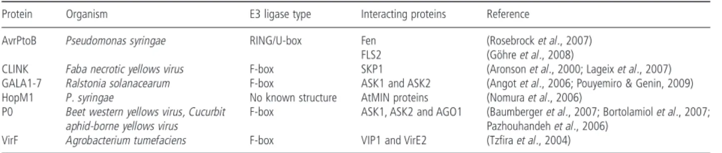
AvrPtoB Pseudomonas syringae RING/U-box Fen (Rosebrock et al., 2007) FLS2 (Göhre et al., 2008)
CLINK Faba necrotic yellows virus F-box SKP1 (Aronson et al., 2000; Lageix et al., 2007) GALA1-7 Ralstonia solanacearum F-box ASK1 and ASK2 (Angot et al., 2006; Pouyemiro & Genin, 2009) HopM1 P. syringae No known structure AtMIN proteins (Nomura et al., 2006)
P0 Beet western yellows virus, Cucurbit
aphid-borne yellows virus
F-box ASK1, ASK2 and AGO1 (Baumberger et al., 2007; Bortolamiol et al., 2007; Pazhouhandeh et al., 2006)
GALA7 knockout (KO) mutant strain shows a dramatic reduction in virulence on Medicago truncatula, but not on A. thaliana or tomato. Simultaneous deletion of all seven GALA genes leads to a loss of virulence of R. solanacearum on A. thaliana and tomato. These proteins thus seem to be an essential, if partially redun-dant, virulence factor acting via an SCF-dependent mechanism, where GALA effectors are involved as F-box-containing proteins to interfere with the host UPS (Angot et al., 2006). The host proteins targeted for degradation by SCFGALA have yet to be identified, but could be crucial elements of the plant defence response (Pouyemiro & Genin, 2009).
Another well-studied case of T3SE mimicking and directly interfering with the host UPS is the P. syringae pv. tomato AvrPtoB. This small triple-helix protein is targeted to the plasma membrane and recognized by the Pto R protein, mediating HR-based PCD in resistant plants via the Prf (Pseudomonas resistance and fenthion sensitivity) protein (Mucyn et al., 2006). The AvrPtoB C-terminal region shows homology to RING-finger and U-box E3 proteins (Janjusevic et al., 2006; Zipfel & Rathjen, 2008). Mutations within this domain result in reduced virulence of the bacterium in vivo (Janjusevic et al., 2006). As P. syringae does not have an endogenous UPS, this observation suggests that this bacterium has acquired an E3 domain protein, allowing it to harness the host UPS to target host proteins for degradation and thus a gain in virulence (Abramovitch et al., 2006; Block et al., 2008). AvrPtoB addresses the host R protein Fen (fenthion
sensitivity), a serine/threonine kinase, for proteasomal degrada-tion, as proteasome inhibitors cause Fen accumulation when expressed with full-length AvrPtoB, but not with truncated AvrPtoB1–387lacking the C-terminal E3 domain (Rosebrock et al., 2007). Interestingly, Fen recognizes truncated AvrPtoB1–387, leading to PCD-mediated resistance in Pto-lacking tomato vari-eties via the activation of Prf-mediated immunity (Rosebrock et al., 2007). This complex relationship illustrates the co-evolution between pathogen virulence and plant R proteins, and how the plant UPS can be exploited by pathogens to promote their virulence (Fig. 3) (Craig et al., 2009; Rosebrock et al., 2007). In such a co-evolution scenario, several steps are envi-sioned. First, the pathogen AvrPtoB1–387 evolves to overcome plant basal defences. The plant Fen kinase then evolves to rec-ognize AvrPtoB1–387, triggering a PCD-mediated resistance. Next, P. syringae ‘incorporates’ an E3 domain into AvrPtoB, targeting Fen for degradation. Finally, the Pto kinase evolves to recognize AvrPtoB and to be impervious to AvrPtoB ubiquitination activity, thus restoring plant immunity through PCD-based mechanisms (Delauré et al., 2008; Rosebrock et al., 2007). Pto polyubiquiti-nation avoidance has recently been elucidated: Pto, via its kinase activity, phosphorylates AvrPtoB, inactivating its E3 domain and thus triggering plant defence mechanisms (Ntoukakis et al., 2009). Interestingly, AvrPtoB also targets the A. thaliana FLS2 (flagellin sensing 2) protein for proteasomal degradation, impair-ing PAMP perception and subsequent plant defence mechanisms
Fig. 3 Evolution of the Pseudomonas syringae virulence factor AvrPtoB and the host R proteins Fen (fenthion sensitivity) and Pto. (A) AvrPtoB1–387overcomes
plant basal defences, causing disease. (B) Fen recognizes AvrPtoB1–387and triggers programmed cell death (PCD)-based plant resistance. (C) AvrPtoB acquires an
E3 ligase domain, causing ubiquitin/26S proteasome system (UPS)-dependent Fen degradation, thus enhancing disease. (D) Pto, impervious to E3 ligase activity (via its kinase activity and phosphorylation of AvrPtoB), evolves to recognize AvrPtoB, restoring plant immunity. Adapted from Rosebrock et al., 2007.
(Göhre et al., 2008). AvrPtoB-mediated degradation of host pro-teins involved in different defence steps thus seems to be a crucial mechanism for P. syringae virulence.
HopM1 is another P. syringae virulence protein translocated into the host cell via the T3SS. Using transgenic plants, it has been shown that HopM1 constructs lacking the C-terminal region interact with 21 A. thaliana AtMIN (A. thaliana HopM1 interactors) proteins, but no interactors are found when using full-length HopM1 (Nomura et al., 2006). HopM1-dependent destabilization of AtMIN proteins is not affected by protease inhibitors, but is blocked by proteasome inhibitors, leading to the accumulation of polyubiquitinated proteins. Among AtMIN pro-teins, AtMIN7 encodes one of the eight members of the adenos-ine diphosphate (ADP) ribosylation factor (ARF) guanadenos-ine nucleotide exchange factor (GEF) protein family, key components of vesicle trafficking that may play a role in the plant immune system by triggering callose deposition on the plant cell wall. Pseudomonas syringae may thus have evolved a mechanism to suppress cell wall-associated host defence mechanisms, facilitat-ing bacterial infection (Nomura et al., 2006).
Hijacking of the host UPS by plant viruses
The Faba bean necrotic yellows virus (FBNYV, Nanovirus), a single-stranded DNA virus, encodes a small protein, CLINK (cell cycle LINK), harbouring an F-box domain, which interacts with a Medicago sativa SKP1 homologue both in vitro and in vivo (Aronson et al., 2000), and may deregulate the host cell cycle by targeting an A. thaliana retinoblastoma-related protein (pRB) for degradation (Lageix et al., 2007). The Beet severe curly top virus (BSCTV, Geminivirus) encodes a C4 protein that enhances host cell division to which viral replication is coupled. Arabidopsis thaliana E3 RKP (related to KPC1) protein expression is induced by the BSCTV C4 protein, and A. thaliana rkp mutants are less susceptible to BSCTV infection (Lai et al., 2009). As RKP has been shown to act as a cell cycle regulator in Arabidopsis, these results suggest that this E3 ligase may affect geminivirus repli-cation by the regulation of the host cell cycle (Lai et al., 2009). Plant viruses are well known as potent inducers and targets of RNAi, as replicating viruses generate double-stranded RNA (dsRNA) (Hannon, 2002). dsRNAs are first cleaved into double-stranded short-interfering RNA (siRNA, 21–23 nucleotides long) by DICER, a specific type III RNase (Ketting, 2006; ur-Rahman et al., 2008). siRNAs are then incorporated into RISC, a macro-molecular complex that destroys target mRNA. The RISC complex contains a member of the Argonaute (AGO) gene family, which binds to siRNA and is implicated in small RNA-mediated regu-latory mechanisms (Hannon, 2002; Vaucheret, 2008; Voinnet, 2008). Two of the 10 A. thaliana AGO proteins, AGO1 and AGO4, have been shown to be implicated in plant defence mechanisms. ago4 mutants show enhanced susceptibility to virulent and
avirulent P. syringae pv. tomato DC3000 strains, suggesting a role for AGO4 (involved in RNA-directed RNA methylation, RdRM) in the limitation of the bacterial spread in the host (Agorio & Vera, 2007). Interestingly, AGO4 acts independently of RdRM, as mutants altered in components upstream/downstream of AGO4 in RdRM do not show compromised resistance to P. syringae (Agorio & Vera, 2007). Hypomorphic ago1 mutants show increased susceptibility to Cucumber mosaic virus (CMV, Cucumovirus) (Morel et al., 2002), confirming the essential role played by the RISC complex in plant antiviral immunity. To coun-teract this resistance mechanism, viruses encode silencing sup-pressor proteins. To date, more than 35 supsup-pressor proteins have been identified in plant viruses, including the potyviral HcPro (Ding & Voinnet, 2007; Lakatos et al., 2006; Merai et al., 2006). Some of these suppressors interact with the UPS, such as the Beet western yellows virus and Cucurbit aphid-borne yellows virus (BWYV and CABYV, Polerovirus) P0 proteins. P0 is a potent silencing suppressor, containing a minimal N-terminal F-box-like domain that enables it to interact in vivo with A. thaliana ASK1 and ASK2, leading to CUL1-containing complex formation (Pazhouhandeh et al., 2006). P0 F-box domain-lacking viruses are hypovirulent, and recombinant PVX expressing P0 shows more severe symptoms in N. benthamiana when compared with the mild wild-type PVX. P0 thus seems to be involved in virus pathogenicity through its F-box domain (Pazhouhandeh et al., 2006). Recently, it has been shown that P0 targets AGO1 for degradation, thus impairing PTGS (Baumberger et al., 2007; Bor-tolamiol et al., 2007), but the underlying mechanisms still remain unclear, as proteasomal degradation may not be involved (Baumberger et al., 2007).
CONCLUDING REMARKS
In the past few years, UPS has emerged as an essential protago-nist in plant–pathogen interactions. Ubiquitination and subse-quent protein degradation seem to occur at several levels of the plant defence mechanism, from basal responses to R gene-mediated resistance (Fig. 4). Ubiquitin-dependent protein degra-dation is known to regulate a wide range of key cellular processes; it is thus not surprising that crucial plant defence elements are found to be UPS substrates. In this respect, it is noteworthy that protein ubiquitination not only triggers degra-dation, but has also been shown to potentially modify protein localization or function (Deng et al., 2000; Manzano et al., 2008; Schnell & Hicke, 2003). Although not demonstrated so far, these modifications may also play an important role in plant– pathogen interactions.
Protein degradation and/or modifications do not seem to be the only way in which UPS is involved in plant defence. The 20S RNase activity demonstrated in vitro, which specifically targets viral substrates, and the inhibitory role of the potyviral protein
HcPro, which interacts with at least four 20S subunits, suggest that both catalytic activities (RNase and protease) harboured by the 20S proteasome could be part of a new antiviral defence pathway. These findings clearly require deeper investigations, but may represent another exciting role for the UPS in plant defence. UPS is, in many cases, targeted and/or used by pathogens that have evolved to inhibit or interfere with its normal functioning (Kisselev, 2008). The finding that pathogens use E3 ligases for their own purposes provides new insights into the complex molecular interactions between host and pathogen (Fig. 4). The
identification and characterization of the host substrates will show how important is this mimicry tactic in blocking pathogen recognition and/or the activation of defence mechanisms (Angot et al., 2007).
UPS inhibition by pathogens does not only represent an agro-nomical and ecoagro-nomical challenge. It may be considered as a chance for human health. Any alteration of UPS components may have disastrous consequences in eukaryotic cells: proteaso-mal dysfunction is involved in many human diseases, such as cancers, neurodegenerative and autoimmune diseases and
Fig. 4 The ubiquitin/26S proteasome system (UPS), a central element in plant defence and pathogen virulence mechanisms. A summary figure. BSCTV, Beet
severe curly top virus; BWYV, Beet western yellows virus; CABYV, Cucurbit aphid-borne yellows virus; FBNYV, Faba bean necrotic yellows virus; LMV, Lettuce mosaic virus; TMV, Tobacco mosaic virus; ToMV, Tomato mosaic virus.
cardiac dysfunction (Dahlmann, 2007). Pseudomonas syringae pv. syringae SylA irreversibly inhibits the three 20S proteolytic activities (Groll et al., 2008), induces apoptosis and inhibits the proliferation of neuroblastoma and ovarian cancer cells, suggest-ing that derivatives of this natural compound may serve for the development of novel anticancer drugs (Coleman et al., 2006). Furthermore, proteasome inhibitors also sensitize cancer cells to chemotherapy: proteasome inhibitors trigger oxidative damage, enhancing the chemotherapeutic efficiency in human leukaemia cells (Dasmahapatra et al., 2006; Lü et al., 2008). Such results may thus be a chance: why not use pathogen inhibitors to counteract UPS dysfunction? Proteasome inhibitors may also be used against parasite development. Recently, novel microbial compounds have been identified as proteasome inhibitors: marine Salinispora tropica salinosporamide A inhibits 20S activ-ity in vitro and protects mice against Plasmodium infection (Prudhomme et al., 2008). Some of the newly discovered plant– pathogen proteasome inhibitors may thus lead to novel thera-peutic applications in the next few years.
REFERENCES
Abramovitch, R., Janjusevic, R., Stebbins, E. and Martin, G. (2006)
Type III effector AvrPtoB requires intrinsic E3 ubiquitin ligase activity to suppress plant cell death and immunity. Proc. Natl. Acad. Sci. USA, 103, 2851–2856.
Agorio, A. and Vera, P. (2007) ARGONAUTE4 is required for resistance to
Pseudomonas syringae in Arabidopsis. Plant Cell, 19, 3778–3790.
Angot, A., Peeters, N., Lechner, E., Vailleau, F., Baud, C., Gentzbittel, L., Sartorel, E., Genschik, P., Boucher, C. and Genin, S. (2006)
Ralstonia solanacearum requires F-box-like domain-containing type III effectors to promote disease on several host plants. PNAS, 103, 14620– 14625.
Angot, A., Vergunst, A., Genin, S. and Peeters, N. (2007) Exploitation
of eukaryotic ubiquitin signaling pathways by effectors translocated by bacterial type III and type IV secretion systems. PLoS Pathog. 3, 1–13.
Apcher, G.S., Heink, S., Zantopf, D., Kloetzel, P.-M., Schmid, H.-P., Mayer, R.J. and Kruger, E. (2003) Human immunodeficiency virus-1
Tat protein interacts with distinct proteasomal [alpha] and [beta] sub-units. FEBS Letters, 553, 200–204.
Aronson, M.N., Meyer, A.D., Gyorgyey, J., Katul, L., Vetten, H.J., Gronenborn, B. and Timchenko, T. (2000) Clink, a Nanovirus-Encoded
Protein, Binds both pRB and SKP1. J. Virol. 74, 2967–2972.
Austin, M., Muskett, P., Kahn, K., Feys, B., Jones, J. and Parker, J.
(2002) Regulatory role of SGT1 in early gene-mediated plant defences.
Science, 295, 2077–2080.
Azevedo, C., Sadanandom, A., Kitagawa, K., Freialdenhoven, A., Shirasu, K. and Schulze-Lefert, P. (2002) The RAR1 interactor SGT1,
an essential component of R gene-triggered disease resistance. Science,
295, 2073–2076.
Azevedo, C., Betsuyaku, S., Peart, J., Takahashi, A., Noe, N., Sadanandom, A., Casais, C., Parker, J. and Shirasu, K. (2006) Role
of SGT1 in resistance protein accumulation in plant immunity. The
EMBO Journal, 25, 2007–2016.
Ballut, L., Petit, F., Mouzeyar, S., Le Gall, O., Candresse, T., Schmid, P., Nicolas, P. and Badaoui, S. (2003) Biochemical identification of
proteasome-associated endonuclease activity in sunflower. Biochimica
et Biophysica Acta, 1645, 30–39.
Ballut, L., Drucker, M., Pugniere, M., Cambon, F., Blanc, S., Roquet, F., Candresse, T., Schmid, H.-P., Nicolas, P., Gall, O. L. and Badaoui, S.
(2005) HcPro, a multifunctional protein encoded by a plant RNA virus, targets the 20S proteasome and affects its enzymic activities. J. Gen.
Virol. 86, 2595–2603.
Baumberger, N., Tsai, C.H., Li, M., Havecker, E. and Baulcombe, D.
(2007) The polerovirus silencing suppressor P0 targets ARGONAUTE proteins for degradation. Curr. Biol. 17, 1609–1614.
Becker, F., Buschfeld, E., Schell, J. and Bachmair, A. (1993) Altered
response to viral infection by tobacco plants perturbed in ubiquitin system. Plant J. 3, 875–881.
Bertrand, P., Saintigny, Y. and Lopez, B. (2004) p53’s double life:
transactivation-independent repression of homologous recombination.
Trends Genet. 20, 235–243.
Bhaskar, P., Raasch, J., Kramer, L., Neumann, P., Wielgus, S., Austin-Phillips, S. and Jiang, J. (2008) Sgt1, but not Rar1, is essential for the
RB-mediated broad-spectrum resistance to potato late blight. BMC Plant Biology, 8, 8–16.
Bieri, S., Mauch, S., Shen, Q.H., Peart, J., Devoto, A., Casais, C., Ceron, F., Schulze, S., Steinbiß, H.H., Shirasu, K. and Schulze-Leferta, P.
(2004) RAR1 Positively Controls Steady State Levels of Barley MLA Resistance Proteins and Enables Sufficient MLA6 Accumulation for Effective Resistance. The Plant Cell, 16, 3480–3495.
Binder, B., Walker, J., Gagne, J., Emborg, T., Hemmann, G., Bleecker, A. and Vierstra, R. (2007) The Arabidopsis EIN3 Binding F-Box Proteins
EBF1 and EBF2 Have Distinct but Overlapping Roles in Ethylene Signal-ing. The Plant Cell, 19, 509–523.
Block, A., Li, G., Fu, Z.Q. and Alfano, J. (2008) Phytopathogen type III
effector weaponry and their plant targets. Curr. Opin. Plant Biol. 11, 1–8.
Bortolamiol, D., Pazhouhandeh, M., Marrcco, K., Genschik, P. and Ziegler-Graff, V. (2007) The polerovirus F box protein P0 targets
ARGO-NAUTE1 to suppress RNA silencing. Curr. Biol. 17, 1615–1621.
Bostick, M., Lochhead, S.R., Honda, A., Palmer, S. and Callis, J. (2004)
Related to ubiquitin 1 and 2 are redundant and essential and regulate vegetative growth, auxin signaling, and ethylene production in Arabi-dopsis. Plant Cell, 16, 2418–2432.
Boyes, D.C., Nam, J. and Dangl, J.L. (1998) The Arabidopsis thaliana
RPM1 disease resistance gene product is a peripheral plasma membrane
protein that is degraded coincident with the hypersensitive response.
Proc. Natl. Acad. Sci. USA, 95, 15849–15854.
Brukhin, V., Gheyselinck, J., Gagliardini, V., Genschik, P. and Grossniklaus, U. (2005) The RPN1 subunit of the 26S proteasome in
Arabidopsis is essential for embryogenesis. Plant Cell, 17, 2723– 2737.
van den Burg, H.A., Tsitsigiannis, D.I., Rowland, O., Lo, J., Rallapalli, G., MacLean, D., Takken, F.L.W. and Jones, J. (2008) The F-Box
protein ACRE189/ACIF1 regulates cell death and defense responses activated during pathogen recognition in tobacco and tomato. The Plant
Cell, 20, 697–719.
Cane, D. and Walsh, C. (1999) The parallel and convergent universes of
polyketide synthases and nonribosomal peptide synthetases. Chem.
Cao, Y., Yang, Y., Zhang, H., Li, D., Zheng, Z. and Song, F. (2008)
Overexpression of a rice defense-related F-box protein gene OsDRF1 in tobacco improves disease resistance through potentiation of defense gene expression. Physiologia Plantarum, 134, 440–452.
Cheung, M.-Y., Zeng, N.-Y., Tong, S.-W., Li, F.W.-Y., Zhao, K.-J., Zhang, Q., Sun, S.S.-M. and Lam, H.M. (2007) Expression of a RING-HC
protein from rice improves resistance to Pseudomonas syringae pv.
tomato DC3000 in transgenic Arabidopsis thaliana. Journal of Experi-mental Botany, 58, 4147–4159.
Cho, S.K., Chung, H.S., Ryu, M.Y., Park, M.J., Lee, M.M., Bahk, Y.-Y., Kim, J., Pai, H.-S. and Kim, W.T. (2006) Heterologous expression
and molecular and cellular characterization of CaPUB1 encoding a hot pepper U-Box E3 ubiquitin ligase homolog. Plant Physiology, 142, 1664–1682.
Coleman, C.S., Rocetes, J.P., Park, D.J., Wallick, C.J., Warn-Cramer, B.J., Michel, K., Dudler, R. and Bachmann, A.S. (2006) Syringolin A,
a new plant elicitor from the phytopathogenic bacterium Pseudomonas syringae pv. syringae, inhibits the proliferation of neuroblastoma and ovarian cancer cells and induces apoptosis. Cell Proliferation, 39, 599– 609.
Craig, A., Ewan, R., Mesmar, J., Gudipati, V. and Sadanandom, A.
(2009) E3 ubiquitin ligases and plant innate immunity. J. Exp. Bot. 60, 1123–1132.
Dahan, J., Etienne, P., Petitot, A.S., Houot, V., Blein, J.P. and Suty, L.
(2001) Cryptogein affects expression of alpha3, alpha6 and beta1 20S proteasome subunits encoding genes in tobacco. J. Exp. Bot. 52, 1947– 1948.
Dahlmann, B. (2007) Role of proteasomes in disease. BMC Biochemistry, 8, S3.
Dangl, J.L. and Jones, J. (2001) Plant pathogens and integrated defence
responses to infection. Nature, 411, 826–833.
Dasmahapatra, G., Rahmani, M., Dent, P. and Grant, S. (2006)
The tyrphostin adaphostin interacts synergistically with proteasome inhibitors to induce apoptosis in human leukemia cells through a reactive oxygen species (ROS)-dependent mechanism. Blood, 107, 232–240.
Delauré, S., Van Hemelrijck, W., De Bolle, M., Cammue, B. and De Coninck, B. (2008) Building up plant defenses by breaking down
pro-teins. Plant Sci. 174, 375–385.
Deng, L., Wang, C., Spencer, E., You, J., Slaughter, C., Pickart, C. et al.
(2000) Activation of the IkappaB kinase complex by TRAF6 requires a dimeric ubiquitin-conjugating enzyme complex and a unique polyubiq-uitin chain. Cell, 103, 351–361.
Devoto, A., Nieto-Rostro, M., Xie, D., Ellis, C., Harmston, R., Patrick, E., Davis, J., Sherratt, L., Coleman, M. and Turner, J.G. (2002) COI1
links jasmonate signalling and fertility to the SCF ubiquitin–ligase complex in Arabidopsis. The Plant Journal, 32, 457–466.
Devoto, A., Muskett, P. and Shirasu, K. (2003) Role of ubiquitination in
the regulation of plant defence against pathogens. Curr. Opin. Plant
Biol. 6, 307–311.
Dhawan, R., Luo, H., Foerster, A.M., AbuQamar, S., Du, H.-N., Briggs, S., Scheld, O.M. and Mengiste, T. (2009) Histone monoubiquitination
1 interacts with a subunit of the mediator complex and regulates defense against necrotrophic fungal pathogens in Arabidopsis. The
Plant Cell, 21, 1000–1019.
Ding, S.W. and Voinnet, O. (2007) Antiviral immunity directed by small
RNAs. Cell, 130, 413–426.
Dong, W., Nowara, D. and Schweizer, P. (2006) Protein
polyubiquitina-tion plays a role in basal host resistance of barley. Plant Cell, 18, 3321–3331.
Downes, B. and Vierstra, R.D. (2005) Post-translational regulation in
plants employing a diverse set of polypeptide tags. J. Biochem. Soc.
Trans. 33, 393–399.
Downes, B.P., Stupar, R.M., Gingerich, D.J. and Vierstra, R.D. (2003)
The HECT ubiquitin-protein ligase (UPL) family in Arabidopsis: UPL3 has a specific role in trichome development. Plant J. 35, 729–742.
Dreher, K. and Callis, J. (2007) Ubiquitin, hormones and biotic stress in
plants. Annals of Botany, 99, 787–822.
Drugeon, G. and Jupin, I. (2002) Stability in vitro of the 69K movement
protein of Turnip yellow mosaic virus is regulated by the ubiquitin-mediated proteasome pathway. J. Gen. Virol. 83, 3187–3197.
Dunigan, D.D., Dietzgen, R.G., Schoelz, J.E. and Zaitlin, M. (1988)
Tobacco mosaic virus particles contain ubiquitinated coat protein sub-units. Virology, 165, 310–312.
Ellis, C., Turner, J.G. and Devoto, A. (2002) Protein complexes mediate
signalling in plant responses to hormones, light, sucrose and pathogens.
Plant Mol. Biol. 50, 971–980.
Endo, S., Demura, T. and Fukuda, H. (2001) Inhibition of proteasome
activity by the TED4 protein in extracellular space: a novel mechanism for protection of living cells from injury caused by dying cells. Plant Cell
Physiol. 42, 9–19.
Forzani, C., Lobreaux, S., Mari, S., Briat, J.F. and Lebrun, M. (2002)
Metal resistance in yeast mediated by the expression of a maize 20S proteasome [alpha] subunit. Gene, 293, 199–204.
Fu, D.Q., Ghabrial, S. and Kachroo, A. (2009) GmRAR1 and GmSGT1 Are
Required for Basal, R Gene-Mediated and Systemic Acquired Resistance in Soybean. Mol. Plant Microbe Interact., 22, 86–95.
Gagne, J., Downes, B.P., Shiu, S.H., Durski, A. and Vierstra, R.D. (2002)
The F-box subunit of the SCF E3 complex is encoded by a diverse superfamily of genes in Arabidopsis. Proc. Natl. Acad. Sci. USA, 99, 11519–11524.
Gautier-Bert, K., Murol, B., Jarrousse, A.S., Ballut, L., Badaoui, S., Petit, F. and Schmid, H.P. (2003) Substrate affinity and substrate
specificity of proteasomes with RNase activity. Molecular Biology
Reports, 30, 1–7.
Gillepsie, T., Boevink, P., Haupt, S., Roberts, A., Toth, R., Valentine, T., Chapman, S. and Oparka, K. (2002) Functional analysis of a
DNA-shuffled movement protein reveals that microtubules are dispensable for the cell-to-cell movement of Tobacco mosaic virus. The Plant Cell,
14, 1207–1222.
Glickman, M. and Raveh, D. (2005) Proteasome plasticity. FEBS Lett. 579, 3214–3223.
Göhre, V., Spallek, T., Häweker, H., Mersmann, S., Mentzel, T., Boller, T., de Torres, M., Mansfield, J.W. and Robatzek, S. (2008)
Plant pattern-recognition receptor FLS2 is directed for degradation by the bacterial ubiquitin ligase AvrPtoB. Current Biology, 18, 1824– 1832.
Goldberg, A. (2003) Protein degradation and protection against
mis-folded or damaged proteins. Nature, 426, 895–899.
Goritschnig, S., Zhang, Y. and Li, X. (2007) The ubiquitin pathway is
required for innate immunity in Arabidopsis. The Plant Journal, 49, 540–551.
Groll, M., Schellenberg, B., Bachmann, A., Archer, C., Huber, R., Powell, T., Lindow, S., Kaiser, M. and Dudler, R. (2008) A plant