The MIT Faculty has made this article openly available. Please share how this access benefits you. Your story matters.
Citation Zhang, Xiangyu, Joshua Kim & Susumu Tonegawa. “Amygdala Reward Neurons Form and Store Fear Extinction Memory.” Neuron 105 (2020): 1077-1093.e7
As Published 10.1016/j.neuron.2019.12.025
Publisher Elsevier BV
Version Original manuscript
Citable link https://hdl.handle.net/1721.1/124954
Terms of Use Creative Commons Attribution-NonCommercial-NoDerivs License
1
Amygdala Reward Neurons Form and Store Fear Extinction Memory
1Xiangyu Zhang
1, Joshua Kim
1& Susumu Tonegawa
1,2* 23
1RIKEN–MIT Laboratory for Neural Circuit Genetics at the Picower Institute for Learning and 4
Memory, Department of Biology and Department of Brain and Cognitive Sciences, 5
Massachusetts Institute of Technology, Cambridge, MA 02139, USA 6
2Howard Hughes Medical Institute at Massachusetts Institute of Technology, Cambridge, MA 7 02139, USA 8 9 *Correspondence: tonegawa@mit.edu 10
2 Summary
1
The ability to extinguish conditioned fear memory is critical for adaptive control of fear response, 2
and its impairment is a hallmark of emotional disorders like post-traumatic stress disorder (PTSD). 3
Fear extinction is thought to take place when animals form a new memory that suppresses the 4
original fear memory. However, little is known about the nature and the site of formation and 5
storage of the new extinction memory. Here, we demonstrate that a fear extinction memory engram 6
is formed and stored in a genetically distinct basolateral amygdala (BLA) neuronal population that 7
drive reward behaviors and antagonize the BLA’s original fear neurons. The activation of the fear 8
extinction engram neurons and natural reward-responsive neurons overlap extensively in the BLA. 9
Furthermore, these two neuron subsets are mutually interchangeable in driving reward behaviors 10
and fear extinction behaviors. Thus, fear extinction memory is a newly formed reward memory. 11
12
Keywords: Fear extinction, memory, engram, reward, amygdala
3 Introduction
1
The inability to extinguish fear is a hallmark of many psychiatric disorders, such as PTSD 2
and generalized anxiety disorder (Shalev et al., 2017; Stein and Sareen, 2015). Following 3
Pavlovian fear conditioning (Pavlov and Anrep, 1927), repeated or prolonged presentations of the 4
conditioned stimulus (CS) without an expected aversive unconditioned stimulus (US) diminishes 5
the conditioned fear response, a phenomenon called fear extinction (Herry et al., 2010; Myers and 6
Davis, 2007; Quirk and Mueller, 2008). Fear extinction has been proposed to involve the formation 7
of a new memory in competition with the original fear memory (Bouton, 2004; Quirk and Mueller, 8
2008; Quirk et al., 2010). The amygdala is a key structure for fear memory (Davis, 1992; Duvarci 9
and Pare, 2014; Ehrlich et al., 2009; Maren and Fanselow, 1996), and is also involved in the fear 10
extinction memory (Amano et al., 2010; Grewe et al., 2017; Herry et al., 2008). However, it is 11
unknown whether the amygdala is the site for the storage of fear extinction memory, and if so, 12
which subset of amygdala neuron stores this memory. 13
Excitatory neurons in the mouse basolateral amygdala (BLA) respond to both positive and 14
negative valence stimuli (Beyeler et al., 2016; Davis and Whalen, 2001; Kim et al., 2016; Namburi 15
et al., 2015; Redondo et al., 2014), and more than 90% of these neurons are composed of two 16
genetically, functionally and anatomically distinct neuronal populations (Kim et al., 2016; Kim et 17
al., 2017). R-spondin-2-expressing (Rspo2+) neurons located in the anterior BLA respond to 18
negative valence stimuli and control negative behaviors and memories, whereas protein-19
phosphatase-1-regulatory-inhibitor-subunit-1B-expressing (Ppp1r1b+) neurons located in the 20
posterior BLA (pBLA) respond to positive valence stimuli and control appetitive behaviors and 21
4
memories (Kim et al., 2016). Furthermore, these two neuronal populations antagonize each other 1
through feed-forward inhibitions mediated by local inhibitory interneurons (Kim et al., 2016). 2
In this study, we investigated a potential role of BLA Ppp1r1b+ neurons in fear extinction 3
using a contextual fear extinction paradigm. We found that fear extinction memory engram cells 4
are formed and stored within the BLA Ppp1r1b+ neuronal population, and these engram cells are 5
necessary for suppressing the original fear memory. Furthermore, fear extinction engram cells and 6
natural reward-responsive cells in the pBLA Ppp1r1b+ neurons are mutually interchangeable in 7
driving reward functions and fear extinction behaviors. 8
9
Results
10
BLA Ppp1r1b+ neurons are activated during contextual fear extinction
11
Fear extinction phenomena have been observed and studied in both cue-dependent fear 12
conditioning and context-dependent fear conditioning paradigms (Amano et al., 2010; Baldi and 13
Bucherelli, 2014; Herry et al., 2008; Trouche et al., 2013; Zushida et al., 2007). Since the BLA 14
plays a critical role in contextual fear conditioning (Calandreau et al., 2005; Goosens and Maren, 15
2001; Huff and Rudy, 2004; Redondo et al., 2014) and a substantial amount of information is 16
available regarding the excitatory neuronal subsets in BLA (see Introduction) (Kim et al., 2016), 17
we employed a contextual fear extinction paradigm in this study. On Day 1, the Extinction group 18
received contextual fear conditioning (CFC) in a box where the context served as the CS and three 19
rounds of footshocks served as the US. On Day 2, mice were returned to the conditioned box for 20
45 min in the absence of footshocks to receive contextual fear extinction training (FET). On Day 21
3, the mice were tested for 5 min extinction memory retrieval in the conditioned box and then 22
5
sacrificed (Figure 1A). Two control groups were set up as follows. The Non-Extinction group went 1
through the same protocol as the Extinction group, except that on Day 2 the mice stayed in their 2
home cage (HC) and didn’t receive any extinction training (Figure 1A). The Non-Shock group 3
received the same protocol as the Extinction group, but received no footshocks on Day 1 (Figure 4
1A). The Extinction group and Non-Extinction group, but not the Non-Shock group, displayed 5
robust freezing behavior on Day 1 that increased as more footshocks were presented (Figure 1B). 6
On Day 3, the Extinction group froze much less than the Non-Extinction group (Figure 1C). 7
Double smFISH (single molecular Fluorescence in situ Hybridization) was performed to 8
detect the activity-dependent expression of the immediate early gene, Fos, in Rspo2+ and 9
Ppp1r1b+ cells (see Methods). The proportions of Fos+ cells of Rspo2+ cells and Fos+ cells of 10
Ppp1r1b+ cells were quantified across the anterior-posterior (A/P) axis for the aBLA and pBLA, 11
respectively (Kim et al., 2016). The proportion of Fos+/Rspo2+ was lower in the Extinction group 12
compared to the Non-Extinction group (Figures 1D, 1E and 1H). In contrast, the proportion of 13
Fos+/Ppp1r1b+ was higher in the Extinction group compared to the Non-Extinction group while 14
no difference was observed between the Non-Extinction and Non-Shock groups (Figures 1F, 1G 15
and 1I). When BLA Ppp1r1b+ neurons were optogenetically inhibited during extinction retrieval, 16
the neuronal activity of Rspo2+ cells (Fos+/Rspo2+) as well as the freezing level increased during 17
extinction retrieval (Figures S1). This result suggests that Ppp1r1b+ neurons suppress Rspo2+ 18
neurons during fear extinction retrieval, which is consistent with the previously reported feed-19
forward inhibition of BLA Rspo2+ cells by BLA Ppp1r1b+ cells (Kim et al., 2016). 20
21
Dynamics of individual BLA neurons throughout CFC and fear extinction
6
Next, we performed in vivo calcium imaging with a microendoscope to directly track 1
individual BLA neuronal activity during CFC followed by contextual fear extinction. The 2
genetically encoded calcium indicator GCaMP6f was expressed in BLA Rspo2+ and Ppp1r1b+ 3
neurons by injecting adeno-associated virus 5 (AAV5)-human synapsin (hsyn):double-floxed 4
inverse open reading frame (DIO)-GCaMP6f into the aBLA of Rspo2-Cre mice and the pBLA of 5
Ppp1r1b-Cre mice, respectively (Figures 2A-2C). The efficiency of Cre-loxP recombination in
6
Rspo2-Cre and Ppp1r1b-Cre mice lines are around 90% and 75%, respectively. (Kim et al., 2016). 7
We used an automated sorting algorithm to identify individual neurons and tracked their 8
longitudinal activity across days (Figures 2D, S2A, and S2B; see Methods) (Kitamura et al., 2017; 9
Mukamel et al., 2009; Okuyama et al., 2016). Neuronal activity was analyzed under five 10
conditions: during habituation to the conditioning chamber before CFC (Hab), after footshocks 11
during CFC (Shock), fear retrieval (FR), extinction training (FET) and extinction retrieval (ER) 12
(Figures 2D; see Methods). In order to quantify how neuronal activity changed across these 13
conditions, the activity in each condition was explicitly compared to activity in another condition 14
that served as a baseline, i.e. shock compared to habituation, fear retrieval compared to habituation, 15
FET compared to fear retrieval, and extinction retrieval compared to fear retrieval (Figures 2E and 16
2F; see Methods). The cells with increased activity were referred to as Up cells, those with 17
decreased activity were referred to as Down cells, and cells whose activity did not change were 18
referred to as No change cells. 19
BLA Rspo2+ cells were predominantly activated by footshocks during CFC and fear 20
retrieval, and inhibited during extinction training and extinction retrieval (Figures 2E, 2G, and 21
S2C-S2E). Conversely, BLA Ppp1r1b+ neurons were predominantly activated during extinction 22
training and extinction retrieval, and inhibited by footshocks and fear retrieval (Figures 2F, 2G, 23
7
and S2F-S2H). A larger percentage of BLA Rspo2+ cells were activated during fear retrieval 1
compared to extinction retrieval, whereas a larger percentage of BLA Ppp1r1b+ cells were 2
activated during extinction retrieval compared to fear retrieval (Figure 2G). Compared to BLA 3
Rspo2+ cells whose activity did not change in response to footshocks (Shock-No Change), shock-4
activated Rspo2+ cells (Shock-Up) were preferentially inhibited during extinction training and 5
extinction retrieval (Figures 2H, 2J and 2K). Specifically, 30% of the shock-activated Rspo2+ cells 6
were also activated during fear retrieval and inhibited during both extinction training and 7
extinction retrieval (Figures 2J and 2K). Compared to BLA Ppp1r1b+ neurons whose activity did 8
not change by extinction training (E.T.-No Change), Ppp1r1b+ neurons that were activated during 9
extinction training (E.T.-Up) were preferentially reactivated during extinction retrieval (Figures 10
2I, 2L and 2M). Combined with the c-Fos quantification data (Figure 1), these results show that 11
pBLA Ppp1r1b+ neurons are recruited during fear extinction training and reactivated during 12
extinction memory retrieval whereas shock-activated aBLA Rspo2+ neurons are suppressed during 13
these processes. 14
15
BLA Ppp1r1b+ neurons drive fear extinction learning
16
We next investigated the roles of Ppp1r1b+ and Rspo2+ neurons in fear extinction at the 17
behavioral level using optogenetic manipulations. We injected a Cre-dependent AAV carrying 18
ChR2 (channelrhodopsin-2) or ArchT (archaerhodopsin) into the aBLA of the Rspo2-Cre mice 19
(Figure 3A and S3A) or the pBLA of the Ppp1r1b-Cre mice (Figure 3B and S3B). Mice underwent 20
the three-day contextual fear extinction protocol as before (Figure 1A). Starting 5 min after the 21
onset of extinction training on Day 2, 3 min of blue or green laser light was delivered during FET 22
8
repeatedly at 2 min intervals (Figure 3C). Inhibition of Rspo2+ neurons and activation of Ppp1r1b+ 1
neurons both facilitated fear extinction learning (Figures 3E and 3F). In contrast, activation of 2
Rspo2+ neurons and inhibition of Ppp1r1b+ neurons both impaired fear extinction and extinction 3
memory retrieval (Figures 3D and 3G). These results show that pBLA Ppp1r1b+ neurons are 4
necessary and sufficient to form fear extinction memory, whereas aBLA Rspo2+ neurons 5
antagonize it. 6
7
Fear extinction memory engram cells are formed in BLA Ppp1r1b+ neuronal population
8
As memories are stored in specific engram cell circuits (Tonegawa et al., 2015), we then 9
investigated whether the engram for fear extinction memory can be identified in the pBLA 10
Ppp1r1b+ neurons. By using our previously established engram technology (Liu et al., 2012; 11
Ramirez et al., 2013; Redondo et al., 2014; Ryan et al., 2015), we injected AAV9-c-Fos-tTA 12
(tetracycline transactivator) together with AAV9-TRE (tetracycline response element)-ChR2-13
EYFP (Ext-ChR2 group) or AAV9-TRE-EYFP (Ext-EYFP group) into the pBLA of C57BL/6 mice 14
(Figures 4A and 4B; see Methods), and subjected them to a series of engram labeling and 15
behavioral steps (Figure 4D; see Methods). This genetic manipulation permits labeling of engram 16
neurons during memory formation or memory retrieval (Figures S4A-S4C) (Khalaf et al., 2018; 17
Liu et al., 2012; Ramirez et al., 2013). During fear extinction training, formation of the putative 18
fear extinction memory engram cells occurs in parallel with retrieval of the original fear memory. 19
In order to isolate the putative fear extinction memory engram neurons, we used the retrieval stage 20
of the fear extinction memory for their labelling (Day3) as fear memory expression will have 21
subsided to the background level by then (Figure 4D). 22
9
A large fraction (79 ± 2%) of pBLA neurons activated by fear extinction retrieval were 1
Ppp1r1b+ (Figure 4C). In order to access the contribution of food and water supplied in the home 2
cage (HC) to the baseline activity of Ppp1r1b+ neurons, we set up another negative control group, 3
HC-ChR2 group (Figure 4D). As expected, ChR2-EYFP+ cells in the HC-ChR2 group was very
4
sparse compared to those in the ChR2-EYFP group (Figure 4B, S4C-S4D). Three groups of mice, 5
Ext-ChR2 group, Ext-EYFP group and HC-ChR2 group, exhibited indistinguishable freezing 6
levels during the first round of contextual fear extinction protocol from Day 1 to Day 3 (Figure 7
4E). To examine the roles of these labeled neurons in contextual fear extinction, we subjected the 8
mice to another round of CFC in the same box for fear reinstatement (Day 5). When these neurons 9
were optogenetically activated during the second round of fear extinction training on Day 6 (see 10
Methods), the Ext-ChR2 group showed accelerated extinction compared to the Ext-EYFP and HC-11
ChR2 groups (Figure 4F). This reactivation-induced accelerated extinction was not observed when 12
the laser was off (Figures S4E-S4F). These results show that the extinction memory engram is 13
formed in the BLA Ppp1r1b+ neuron population. 14
15
BLA Ppp1r1b+ extinction engram cells are crucial for suppressing Rspo2+ fear cells
16
We then investigated whether pBLA Ppp1r1b+ extinction engram neurons are not only 17
capable of driving fear extinction, but also necessary for maintaining extinction memory and for 18
suppressing Rspo2+ fear cells. We labeled the pBLA extinction engram cells with ArchT-mCherry 19
during fear extinction memory retrieval (Figures 5A-5C and S5A). As expected, a large proportion 20
(75 ± 2%) of the pBLA neurons activated by fear extinction retrieval were PPP1R1B+ (Figures 21
S5B-S5C). Optogenetic inhibition of these ArchT-labeled neurons specifically impaired the 22
10
retrieval of fear extinction memory on Day 4 (Figure 5D) and failed to suppress BLA Rspo2+ fear 1
neurons (Figures 5E-5G). The later is consistent with the feed-forward inhibition of Rspo2+ 2
neurons by Ppp1r1b+ extinction neurons within the BLA (Kim et al., 2016). Together, these results 3
demonstrate a causal role of pBLA Ppp1r1b+ engram cells in extinction memory. These fear 4
extinction engram cells: i) are reactivated during extinction memory retrieval (Figure 2M), ii) have 5
the capacity to drive fear extinction (Figure 4F), and iii) are necessary for suppressing the original 6
fear memory (Figures 5D-5G). 7
8
Reward neurons and fear extinction engram neurons overlap extensively in the amygdala
9
Our previous study demonstrated that pBLA Ppp1r1b+ neurons responded to a variety of
10
appetitive stimuli (Kim et al., 2016), whereas our present study shows that fear extinction memory 11
is stored in the pBLA Ppp1r1b+ neuronal population (Figures 1-5). Therefore, we investigated the 12
relationship between these two subsets of Ppp1r1b+ neurons. To label reward-responding pBLA 13
neurons, AAV9-c-Fos-tTA and AVV9-TRE-EYFP virus were injected into pBLA of C57BL/6 mice 14
on Dox-ON diet (Figure 6A). One week post-surgery, all mice were partially deprived of water for 15
24 hrs on Dox-ON diet (Day 1) and water was given as a reward on Dox-OFF diet (Day 2) (Figure 16
6B). Mice were then divided into W/W (Water/Water), W/F (Water/Food), W/Ext 17
(Water/Extinction), and W/NonExt (Water/NonExtinction) groups, and kept on Dox-ON diet 18
throughout the remainder of the experiment (Figure 6B; see Methods). 19
Neurons activated by water reward on Day 2 were labeled with EYFP, and their proportions 20
of all neurons (EYFP+/DAPI+) were similar across all four groups (Figures 6C, 6F and S6A). 21
Neurons activated by the stimulus delivered on Day 5, namely the second delivery of water reward 22
11
(W/W group), food reward (W/F group), extinction memory retrieval (W/Ext group), and fear 1
memory retrieval (W/NonExt group), were detected by immunohistochemistry for endogenous c-2
Fos. The proportions of c-Fos+ cells of all neurons (c-Fos+/DAPI+) were similar between W/W 3
group, W/F group and W/Ext group, and were substantially higher compared to the W/NonExt 4
group (Figures 6D, 6F, and S6A). The proportion of the c-Fos and EYFP double-positive cells of 5
EYFP+ cells (c-Fos+EYFP+/EYFP+) was used to measure the degree of similarity between the cells 6
responding to the pair of stimuli used in each mouse group. This proportion reached as high as 7
40% in the W/W group, which represented the ceiling effect of a repeated presentation of the same 8
reward. The proportion of W/Ext group was lower compared to W/W group but it did not differ 9
significantly from the proportion of W/F group, and both of these groups and W/W group gave 10
greater proportions than W/NonExt group (Figures 6E, 6F, and S6A). These results show that the 11
overlap of pBLA Ppp1r1b+ neurons activated by fear extinction and water reward is as high as the 12
overlap of pBLA Ppp1r1b+ neurons activated by two bona fide rewards (water and food). 13
14
Ppp1r1b+ fear extinction engram cells and natural reward cells are interchangeable
15
We then examined whether this observed cellular overlap could also be demonstrated at 16
the behavioral level. To test whether optogenetic activation of fear extinction engram cells can 17
drive appetitive behavior, four groups of C57BL/6 mice were set up (Figures 7A and 7B). In the 18
Ext-ChR2 and Ext-EYFP groups, extinction engram cells were labeled with ChR2-EYFP and 19
EYFP during extinction retrieval on Day 3, respectively (Figures 7B, S7A, and S7B). As controls, 20
neurons activated by water reward and fear memory retrieval were labeled with ChR2 in Water-21
ChR2 group and CFC-ChR2 group, respectively (Figures 7B, S7C, and S7D). On Day 4, all mice 22
12
were subjected to an optogenetic self-stimulation behavior test or optogenetic place preference test 1
(Figures 7B, S7E and S7F; see Methods). 50% of Ext-ChR2 group mice and 67% of Water-ChR2 2
group mice displayed self-stimulation behavior, but not the Ext-EYFP group or CFC-ChR2 group 3
(Figure 7C). In the optogenetic real-time place preference test, mice in the Ext-ChR2 group and 4
Water-ChR2 group spent significantly more time on the stimulated side, whereas Ext-EYFP and 5
CFC-ChR2 groups spent equal times on both sides of the chamber (Figures 7D, 7E, and S7G). 6
Reciprocally, optogenetic activation of ChR2-expressing water reward cells accelerated fear 7
extinction learning (Figures 7F and 7G). Thus, activation of fear extinction engram cells generates 8
appetitive behaviors and activation of reward neurons facilitates fear extinction. 9
10
Discussion
11
In this study, we have shown that fear extinction requires new memory engram cells that 12
are formed and stored within BLA Ppp1r1b+ reward neuronal population. The fear extinction 13
engram cells are defined by their reactivation during extinction retrieval, and their sufficiency and 14
necessity to drive fear extinction. The overlap of neuronal activation of extinction memory engram 15
neurons and natural reward-responsive neurons is extensive and these two types of neurons are 16
mutually interchangeable in driving reward functions and fear extinction behaviors. Thus, the shift 17
from fear to safety in the fear extinction process is mediated by newly formed Ppp1r1b+ reward 18
memory neurons and their inhibition of the original Rspo2+ fear memory neurons, both present 19
within the BLA. 20
The inhibition of Rspo2+ fear neurons by Ppp1r1b+ extinction neurons is likely to be 21
mediated by the local inhibitory interneurons in BLA (Kim et al., 2016), supporting the emerging 22
13
notion that the switch between fear expression and fear extinction takes place within the BLA 1
(Ehrlich et al., 2009; Grewe et al., 2017; Herry et al., 2008; Pare and Duvarci, 2012). Consistent 2
with previous recording studies (Grewe et al., 2017; Herry et al., 2008), our study shows fear 3
extinction is due to new learning rather than a loss of the original fear memory (Herry et al., 2010; 4
Myers and Davis, 2007; Pavlov, 1927). The Ppp1r1b+ extinction neurons may correspond to the 5
previously reported BLA neurons identified by in vivo recording, which were activated throughout 6
extinction learning after cue fear conditioning (Herry et al., 2008). However, our findings extend 7
beyond that study as we further demonstrated that the newly formed extinction memory is an 8
appetitive memory and inhibits the original fear memory. 9
10
Fear extinction recruits the brain’s reward circuit
11
The finding that the fear extinction memory is a rewarding memory indicates that the 12
emotional valence associated with CS is switched from negative to positive during fear extinction. 13
Notably, the finding that activation of fear extinction engram cells can elicit a reward effect (Figure 14
7) supports the notion that the absence of expected punishments is in itself a reward. Therefore, 15
fear extinction is positively reinforced by recruiting the brain’s reward circuitry. Previous studies 16
correlated the reward system to fear extinction (Correia et al., 2016; Felsenberg et al., 2018), but 17
their causal relationship was never demonstrated. In contrast, our results show a causal role of the 18
reward system in fear extinction: the pBLA reward neuron population is a necessary component 19
of the fear extinction memory. Furthermore, these findings provide strong evidence of the 20
antagonistic interactions between aversive and appetitive systems that have been observed in 21
Pavlovian fear conditioning (Nasser and McNally, 2012). 22
14
1
Multiple parallel circuits in fear extinction
2
In addition to direct feed-forward inhibition within BLA, BLA Ppp1r1b+ extinction 3
neurons could also drive fear extinction through their interactions with other brain regions. As 4
downstream targets of BLA, both intercalated cells (ITC) and central amygdala (CeA) have been 5
shown to play important roles in fear extinction (Likhtik et al., 2008). Our previous study showed 6
that BLA Ppp1r1b+ neurons provide monosynaptic projections to the Prkcd+ neurons located in 7
the lateral subnuclei of CeA (CeL), which are also activated during fear extinction retrieval (Kim 8
et al., 2017). Therefore, the fear extinction behavior can also be modulated via BLA Ppp1r1b+ -9
CeA monosynaptic projection or BLA Ppp1r1b+-ITC-CeA di-synaptic projection. The infralimbic 10
region (IL) of the prefrontal cortex is another critical cite for fear extinction (Burgos-Robles et al., 11
2007; Laurent and Westbrook, 2009; Vidal-Gonzalez et al., 2006) and its connections with BLA 12
Ppp1r1b+ neurons may also be involved in this process (Cho et al., 2013; Kim et al., 2016; Likhtik 13
et al., 2005; Quirk et al., 2006; Sotres-Bayon et al., 2012). 14
15
Intrinsic reinforcement signals during fear extinction learning
16
How Ppp1r1b+ cells are activated for the acquisition of fear extinction memory is another 17
question that remains to be elucidated. Reinforcement learning theories would predict that 18
acquisition of extinction memory is instructed by negative prediction error, in which the predictive 19
value of the CS with respect to the occurrence of the US is compromised (McNally et al., 2011; 20
Schultz, 2016). Given there is no obvious external US in the case of fear extinction, this stimulus 21
15
may be provided internally and preferentially to Ppp1r1b neurons in the BLA. The identification 1
of the agent and the circuit that provide this stimulus would be of fundamental importance. 2
Accumulating evidence suggests that dopamine reinforces fear extinction learning by 3
signaling the omission of expected aversive US in both animals and humans (de Jong et al., 2019; 4
Kalisch et al., 2019; Matsumoto and Hikosaka, 2009; Raczka et al., 2011; Ray Luo, 2018; Salinas-5
Hernandez et al., 2018). In the mesolimbic dopaminergic system, amygdala receives dopaminergic 6
inputs from the ventral tegmental area (VTA) and substantia nigra (SNc) in the midbrain (Brinley-7
Reed and McDonald, 1999; Pezze and Feldon, 2004). Future studies are needed to investigate how 8
dopamine functions as an instructive signal onto BLA neurons for fear extinction learning. 9
10
In conclusion, our study provides the empirical evidence for the key concept derived from 11
learning theory that an omission of the expected punishment is rewarding(Felsenberg et al., 2018; 12
Kim et al., 2006; Nasser and McNally, 2012), pinpointing potential targets for effective 13
interventions of fear-related disorders (Shalev et al., 2017; Stein and Sareen, 2015). 14
16 References
1
Amano, T., Unal, C.T., and Pare, D. (2010). Synaptic correlates of fear extinction in the amygdala. 2
Nat Neurosci 13, 489-494. 3
Baldi, E., and Bucherelli, C. (2014). Entorhinal cortex contribution to contextual fear conditioning 4
extinction and reconsolidation in rats. Neurobiol Learn Mem 110, 64-71. 5
Beyeler, A., Namburi, P., Glober, G.F., Simonnet, C., Calhoon, G.G., Conyers, G.F., Luck, R., 6
Wildes, C.P., and Tye, K.M. (2016). Divergent Routing of Positive and Negative Information from 7
the Amygdala during Memory Retrieval. Neuron 90, 348-361. 8
Bouton, M.E. (2004). Context and behavioral processes in extinction. Learn Mem 11, 485-494. 9
Brinley-Reed, M., and McDonald, A.J. (1999). Evidence that dopaminergic axons provide a dense 10
innervation of specific neuronal subpopulations in the rat basolateral amygdala. Brain Res 850, 11
127-135. 12
Burgos-Robles, A., Vidal-Gonzalez, I., Santini, E., and Quirk, G.J. (2007). Consolidation of fear 13
extinction requires NMDA receptor-dependent bursting in the ventromedial prefrontal cortex. 14
Neuron 53, 871-880. 15
Calandreau, L., Desmedt, A., Decorte, L., and Jaffard, R. (2005). A different recruitment of the 16
lateral and basolateral amygdala promotes contextual or elemental conditioned association in 17
Pavlovian fear conditioning. Learn Mem 12, 383-388. 18
Cho, J.H., Deisseroth, K., and Bolshakov, V.Y. (2013). Synaptic encoding of fear extinction in 19
mPFC-amygdala circuits. Neuron 80, 1491-1507. 20
Correia, S.S., McGrath, A.G., Lee, A., Graybiel, A.M., and Goosens, K.A. (2016). Amygdala-21
ventral striatum circuit activation decreases long-term fear. Elife 5. 22
Davis, M. (1992). The role of the amygdala in fear and anxiety. Annu Rev Neurosci 15, 353-375. 23
Davis, M., and Whalen, P.J. (2001). The amygdala: vigilance and emotion. Mol Psychiatry 6, 13-24
34. 25
de Jong, J.W., Afjei, S.A., Pollak Dorocic, I., Peck, J.R., Liu, C., Kim, C.K., Tian, L., Deisseroth, 26
K., and Lammel, S. (2019). A Neural Circuit Mechanism for Encoding Aversive Stimuli in the 27
Mesolimbic Dopamine System. Neuron 101, 133-151 e137. 28
Duvarci, S., and Pare, D. (2014). Amygdala microcircuits controlling learned fear. Neuron 82, 29
966-980. 30
Ehrlich, I., Humeau, Y., Grenier, F., Ciocchi, S., Herry, C., and Luthi, A. (2009). Amygdala 31
inhibitory circuits and the control of fear memory. Neuron 62, 757-771. 32
Felsenberg, J., Jacob, P.F., Walker, T., Barnstedt, O., Edmondson-Stait, A.J., Pleijzier, M.W., 33
Otto, N., Schlegel, P., Sharifi, N., Perisse, E., et al. (2018). Integration of Parallel Opposing 34
Memories Underlies Memory Extinction. Cell 175, 709-722 e715. 35
Goosens, K.A., and Maren, S. (2001). Contextual and auditory fear conditioning are mediated by 36
the lateral, basal, and central amygdaloid nuclei in rats. Learn Mem 8, 148-155. 37
Grewe, B.F., Grundemann, J., Kitch, L.J., Lecoq, J.A., Parker, J.G., Marshall, J.D., Larkin, M.C., 38
Jercog, P.E., Grenier, F., Li, J.Z., et al. (2017). Neural ensemble dynamics underlying a long-term 39
associative memory. Nature. 40
Herry, C., Ciocchi, S., Senn, V., Demmou, L., Muller, C., and Luthi, A. (2008). Switching on and 41
off fear by distinct neuronal circuits. Nature 454, 600-606. 42
Herry, C., Ferraguti, F., Singewald, N., Letzkus, J.J., Ehrlich, I., and Luthi, A. (2010). Neuronal 43
circuits of fear extinction. Eur J Neurosci 31, 599-612. 44
17
Huff, N.C., and Rudy, J.W. (2004). The amygdala modulates hippocampus-dependent context 1
memory formation and stores cue-shock associations. Behav Neurosci 118, 53-62. 2
Kalisch, R., Gerlicher, A.M.V., and Duvarci, S. (2019). A Dopaminergic Basis for Fear Extinction. 3
Trends Cogn Sci. 4
Khalaf, O., Resch, S., Dixsaut, L., Gorden, V., Glauser, L., and Graff, J. (2018). Reactivation of 5
recall-induced neurons contributes to remote fear memory attenuation. Science 360, 1239-1242. 6
Kim, H., Shimojo, S., and O'Doherty, J.P. (2006). Is avoiding an aversive outcome rewarding? 7
Neural substrates of avoidance learning in the human brain. PLoS Biol 4, e233. 8
Kim, J., Pignatelli, M., Xu, S., Itohara, S., and Tonegawa, S. (2016). Antagonistic negative and 9
positive neurons of the basolateral amygdala. Nat Neurosci 19, 1636-1646. 10
Kim, J., Zhang, X., Muralidhar, S., LeBlanc, S.A., and Tonegawa, S. (2017). Basolateral to Central 11
Amygdala Neural Circuits for Appetitive Behaviors. Neuron 93, 1464-1479 e1465. 12
Kitamura, T., Ogawa, S.K., Roy, D.S., Okuyama, T., Morrissey, M.D., Smith, L.M., Redondo, 13
R.L., and Tonegawa, S. (2017). Engrams and circuits crucial for systems consolidation of a 14
memory. Science 356, 73-78. 15
Laurent, V., and Westbrook, R.F. (2009). Inactivation of the infralimbic but not the prelimbic 16
cortex impairs consolidation and retrieval of fear extinction. Learn Mem 16, 520-529. 17
Likhtik, E., Pelletier, J.G., Paz, R., and Pare, D. (2005). Prefrontal control of the amygdala. J 18
Neurosci 25, 7429-7437. 19
Likhtik, E., Popa, D., Apergis-Schoute, J., Fidacaro, G.A., and Pare, D. (2008). Amygdala 20
intercalated neurons are required for expression of fear extinction. Nature 454, 642-645. 21
Liu, X., Ramirez, S., Pang, P.T., Puryear, C.B., Govindarajan, A., Deisseroth, K., and Tonegawa, 22
S. (2012). Optogenetic stimulation of a hippocampal engram activates fear memory recall. Nature 23
484, 381-385.
24
Maren, S., and Fanselow, M.S. (1996). The amygdala and fear conditioning: has the nut been 25
cracked? Neuron 16, 237-240. 26
Matsumoto, M., and Hikosaka, O. (2009). Two types of dopamine neuron distinctly convey 27
positive and negative motivational signals. Nature 459, 837-841. 28
McNally, G.P., Johansen, J.P., and Blair, H.T. (2011). Placing prediction into the fear circuit. 29
Trends Neurosci 34, 283-292. 30
Mukamel, E.A., Nimmerjahn, A., and Schnitzer, M.J. (2009). Automated analysis of cellular 31
signals from large-scale calcium imaging data. Neuron 63, 747-760. 32
Myers, K.M., and Davis, M. (2007). Mechanisms of fear extinction. Mol Psychiatry 12, 120-150. 33
Namburi, P., Beyeler, A., Yorozu, S., Calhoon, G.G., Halbert, S.A., Wichmann, R., Holden, S.S., 34
Mertens, K.L., Anahtar, M., Felix-Ortiz, A.C., et al. (2015). A circuit mechanism for 35
differentiating positive and negative associations. Nature 520, 675-678. 36
Nasser, H.M., and McNally, G.P. (2012). Appetitive-aversive interactions in Pavlovian fear 37
conditioning. Behav Neurosci 126, 404-422. 38
Okuyama, T., Kitamura, T., Roy, D.S., Itohara, S., and Tonegawa, S. (2016). Ventral CA1 neurons 39
store social memory. Science 353, 1536-1541. 40
Pare, D., and Duvarci, S. (2012). Amygdala microcircuits mediating fear expression and 41
extinction. Curr Opin Neurobiol 22, 717-723. 42
Pavlov, I.P., and Anrep, G.V. (1927). Conditioned reflexes; an investigation of the physiological 43
activity of the cerebral cortex (London: Oxford University Press: Humphrey Milford). 44
18
Pavlov, P.I. (1927). Conditioned reflexes: An investigation of the physiological activity of the 1
cerebral cortex. Ann Neurosci 17, 136-141. 2
Pezze, M.A., and Feldon, J. (2004). Mesolimbic dopaminergic pathways in fear conditioning. Prog 3
Neurobiol 74, 301-320. 4
Quirk, G.J., Garcia, R., and Gonzalez-Lima, F. (2006). Prefrontal mechanisms in extinction of 5
conditioned fear. Biol Psychiatry 60, 337-343. 6
Quirk, G.J., and Mueller, D. (2008). Neural mechanisms of extinction learning and retrieval. 7
Neuropsychopharmacology 33, 56-72. 8
Quirk, G.J., Pare, D., Richardson, R., Herry, C., Monfils, M.H., Schiller, D., and Vicentic, A. 9
(2010). Erasing fear memories with extinction training. J Neurosci 30, 14993-14997. 10
Raczka, K.A., Mechias, M.L., Gartmann, N., Reif, A., Deckert, J., Pessiglione, M., and Kalisch, 11
R. (2011). Empirical support for an involvement of the mesostriatal dopamine system in human 12
fear extinction. Transl Psychiatry 1, e12. 13
Ramirez, S., Liu, X., Lin, P.A., Suh, J., Pignatelli, M., Redondo, R.L., Ryan, T.J., and Tonegawa, 14
S. (2013). Creating a false memory in the hippocampus. Science 341, 387-391. 15
Ray Luo, A.U., Adam Weitemier, Luca Aquili, Jenny Koivumaa, Thomas J. McHugh and Joshua 16
P. Johansen (2018). A dopaminergic switch for fear to safety transitions. Nature Communication. 17
Redondo, R.L., Kim, J., Arons, A.L., Ramirez, S., Liu, X., and Tonegawa, S. (2014). Bidirectional 18
switch of the valence associated with a hippocampal contextual memory engram. Nature 513, 426-19
430. 20
Ryan, T.J., Roy, D.S., Pignatelli, M., Arons, A., and Tonegawa, S. (2015). Memory. Engram cells 21
retain memory under retrograde amnesia. Science 348, 1007-1013. 22
Salinas-Hernandez, X.I., Vogel, P., Betz, S., Kalisch, R., Sigurdsson, T., and Duvarci, S. (2018). 23
Dopamine neurons drive fear extinction learning by signaling the omission of expected aversive 24
outcomes. Elife 7. 25
Schultz, W. (2016). Dopamine reward prediction error coding. Dialogues Clin Neurosci 18, 23-26
32. 27
Shalev, A., Liberzon, I., and Marmar, C. (2017). Post-Traumatic Stress Disorder. N Engl J Med 28
376, 2459-2469.
29
Sotres-Bayon, F., Sierra-Mercado, D., Pardilla-Delgado, E., and Quirk, G.J. (2012). Gating of fear 30
in prelimbic cortex by hippocampal and amygdala inputs. Neuron 76, 804-812. 31
Stein, M.B., and Sareen, J. (2015). CLINICAL PRACTICE. Generalized Anxiety Disorder. N Engl 32
J Med 373, 2059-2068. 33
Tonegawa, S., Liu, X., Ramirez, S., and Redondo, R. (2015). Memory Engram Cells Have Come 34
of Age. Neuron 87, 918-931. 35
Trouche, S., Sasaki, J.M., Tu, T., and Reijmers, L.G. (2013). Fear extinction causes target-specific 36
remodeling of perisomatic inhibitory synapses. Neuron 80, 1054-1065. 37
Vidal-Gonzalez, I., Vidal-Gonzalez, B., Rauch, S.L., and Quirk, G.J. (2006). Microstimulation 38
reveals opposing influences of prelimbic and infralimbic cortex on the expression of conditioned 39
fear. Learn Mem 13, 728-733. 40
Zushida, K., Sakurai, M., Wada, K., and Sekiguchi, M. (2007). Facilitation of extinction learning 41
for contextual fear memory by PEPA: a potentiator of AMPA receptors. J Neurosci 27, 158-166. 42
19 Acknowledgments
1
We thank X. Zhou, K. Marshall, B. Bane, F.J. Bushard and W. Yu for technical assistance; C. Sun 2
and C.J. MacDonald for discussion on the calcium imaging data analysis; M. Pignatelli, S. 3
Muralidhar, C. Sun and C.J. MacDonald for comments and discussion on the manuscript; and all 4
members of the Tonegawa laboratory for their support. 5
Funding
6
This work was supported by the RIKEN Brain Science Institute, the Howard Hughes Medical 7
Institute, and the JPB Foundation. 8
Author contributions:
9
X.Z., J.K. and S.T. contributed to the study design. X.Z. contributed to the data collection and 10
interpretation. X.Z. conducted the surgeries, behavior experiments and histological analyses. X.Z. 11
and S.T. wrote the paper. All authors discussed and commented on the manuscript. 12
Competing interests
13
The authors declare no competing financial interests. Correspondence and requests for materials 14
should be addressed to Susumu Tonegawa. 15
Data and material availability
16
AAV9-c-Fos-tTA, AAV9-TRE-ChR2-EYFP, AAV9-TRE-eArchT-mCherry, and AAV9
-TRE-17
EYFP were developed at the Massachusetts Institute of Technology by the group of S.T.; virus 18
plasmids are available through a material transfer agreement. The Rspo2-Cre transgenic mouse 19
line was developed at RIKEN BSI by the group of Shigeyoshi Itohara and is available through a 20
material transfer agreement. All data is available in the main text or the supplementary materials. 21
20
21
Figure 1. BLA Ppp1r1b+ neurons are activated during contextual fear extinction
1
(A) Experimental design of the Extinction group, NonExtinction group and NonShock group. (B 2
and C) Freezing levels of all three groups on Day 1 (B) and Day 3 (C). One-way ANOVA. 3
Extinction n = 4; NonExt n = 4; NonShock n = 4. (D) Percentages of Fos+ neurons within BLA 4
Rspo2+ neurons across A/P axis, -0.8 mm to -2.0 mm. (E) Average of the percentage of 5
Fos+/Rspo2+ shown in (D). One-way ANOVA. Extinction n = 4; NonExt n = 4; NonShock n = 4. 6
(F) Percentages of Fos+ neurons within BLA Ppp1r1b+ neurons across A/P axis, -1.6 mm to -2.6 7
mm. (G) Average of the percentage of Fos+/Ppp1r1b+ shown in (F). One-way ANOVA. 8
Extinction n = 4; NonExt n = 4; NonShock n = 4. (H and I) Double smFISH of Fos (red) and 9
Rspo2 (green, H) in aBLA or Ppp1r1b (green, I) in pBLA. *P < 0.05, **P < 0.01, ***P < 0.001, 10
****P < 0.0001. Data are presented as mean ± SEM. Data are presented as mean ± SEM. Scale 11
bars: 200 µm (H and I) 12
22
23
Figure 2. Longitudinal Ca2+ imaging of BLA Rspo2+ and Ppp1r1b+ neurons during fear
1
extinction
2
(A) Cre-dependent expression of GCamp6f and implantation of microendoscope above the aBLA 3
of Rspo2-Cre and pBLA of Ppp1r1b-Cre mice. (B) aBLA of Rspo2-Cre mice. Left: Representative 4
image showing GCamp6f expression and GRIN lens implant; Right: Stacked FOV (Field of View) 5
image. (C) pBLA of Ppp1r1b-Cre mice. Left: Representative image showing GCamp6f expression 6
and GRIN lens implant; Right: Stacked FOV image. (D) Behavior protocol for Ca2+ recording. (E) 7
From left to right: percentages of BLA Rspo2+ neurons with increased (Up, orange), decreased 8
(Down, blue) or No change (grey) responses to shock (vs. habituation), fear retrieval (vs. 9
habituation), extinction training (vs. fear retrieval) and extinction retrieval (vs. fear retrieval). 169 10
cells total; 4 Rspo2-Cre mice. F.R. represents fear retrieval. (F) From left to right: percentages of 11
BLA Ppp1r1b+ neurons with increased (Up, orange), decreased (Down, blue) or No change (grey) 12
responses to shock (vs. habituation), fear retrieval (vs. habituation), extinction training (vs. fear 13
retrieval) and extinction retrieval (vs. fear retrieval). 179 cells total; 5 Ppp1r1b-Cre mice. F.R. 14
represents fear retrieval. (G) Percentages of activated BLA Rspo2+ and Ppp1r1b+ neurons during 15
fear retrieval on Day 2 and extinction retrieval on Day 3 per animal. (H) Compared to BLA Rpso2+ 16
neurons with stable responses to footshocks (Shock-No change, grey), BLA Rpso2+ neurons with 17
increased response to shock (Shock-Up, orange) were preferentially inhibited during extinction 18
training (45.9 ± 3.9% vs. 24.6 ± 8.2%) and extinction retrieval (40.5 ± 2.1% vs. 24.8 ± 12.0%). 19
Paired t-test. (I) Compared to BLA Ppp1r1b+ neurons with stable activity during extinction 20
training (E.T.-No Change), BLA Ppp1r1b+ neurons with increased activity during extinction 21
training (E.T.-Up) were strongly reactivated during extinction retrieval (58.2 ± 5.3% vs. 15.1 ± 22
24
4.2%). Paired t-test. (J) Relative fluorescence change (ΔF/F) of BLA Rpso2+ neurons that showed 1
increased activity during shock, increased activity during fear retrieval, decreased activity during 2
extinction training and extinction retrieval (n=13). Each row represents the same cell that was 3
tracked across three days. (K) Ca2+ traces of six representative BLA Rspo2+ neurons that are shown 4
in (J) with corresponding cell numbers. (L) Relative fluorescence change (ΔF/F) of BLA Ppp1r1b+ 5
neurons that showed increased activity during both extinction training and extinction retrieval 6
(n=16). Each row represents the same cell that was tracked across three days. (M) Ca2+ traces of 7
six representative BLA Ppp1r1b+ neurons that are shown in (L) with corresponding cell numbers. 8
*P < 0.05, **P < 0.01, ***P < 0.001, ****P < 0.0001. Data are presented as mean ± SEM. 9
25
26
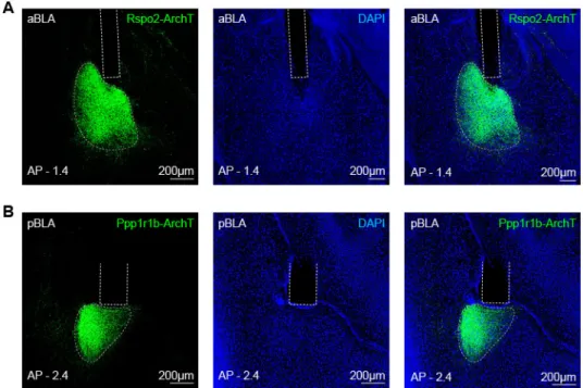
Figure 3. Activation of BLA Ppp1r1b+ neurons and inhibition of BLA Rspo2+ neurons
1
facilitate contextual fear extinction
2
(A) Left: diagram of bilateral injection of AAV-DIO-ChR2-EYFP or AAV-DIO-eArchT-EYFP 3
virus and optical fiber implant in the aBLA of Rspo2-Cre mice. Right: representative histology. 4
(B) Left: diagram of bilateral injection of AAV-DIO-ChR2-EYFP or AAV-DIO-eArchT-EYFP 5
virus and optical fiber implant in the pBLA of Ppp1r1b-Cre mice. Right: representative histology. 6
(C) Experimental protocol of optogenetic manipulation during fear extinction. (D) Optogenetic 7
activation of BLA Rspo2+ neurons impaired fear extinction training and memory. Rspo2-Cre-: n 8
= 14, Rspo2-ChR2: n = 13. Two-way RM (Repeated Measures) ANOVA. Single-phase 9
exponential decay fit: τRspo2-Cre- = 14.06, τRspo2-ChR2 NA. (E) Optogenetic inhibition of BLA Rspo2+ 10
neurons facilitated early stage of fear extinction training. Rspo2 Cre-: n = 12, Rspo2-ArchT: n = 11
13. Two-way RM ANOVA. Single-phase exponential decay fit: τRspo2-Cre- = 12.43, τRspo2-ArchT = 12
1.65. (F) Optogenetic activation of BLA Ppp1r1b+ neurons facilitated fear extinction training. 13
Ppp1r1b Cre-: n = 13, Ppp1r1b-ChR2: n = 9. Two-way RM ANOVA. Single-phase exponential 14
decay fit: τ Ppp1r1b-Cre- = 11.13, τPpp1r1b-ChR2 = 3.18. (G) Optogenetic inhibition of BLA Ppp1r1b+ 15
neurons impaired fear extinction training and memory. Ppp1r1b Cre-: n = 10, Ppp1r1b-ArchT: n 16
= 11. Two-way RM ANOVA. Single-phase exponential decay fit: τ Ppp1r1b-Cre- = 11.34, τPpp1r1b-ArchT 17
= 20.36. *P < 0.05, **P < 0.01, ***P < 0.001, ****P < 0.0001. Data are presented as mean ± 18
SEM. 19
27
28
Figure 4. Activation of BLA fear extinction memory engram cells promotes fear extinction
1
(A) Virus-based activity-dependent labeling scheme. C56BL/6 mice were injected with AAV9 -c-2
fos-tTA and AAV9-TRE-ChR2-EYFP or AAV9-TRE-EYFP virus and implanted with optical 3
fibers that bilaterally target pBLA. (B) Representative images of EYFP expression under three 4
different conditions: Dox OFF + Extinction (left), Dox OFF + Home Cage (middle) and Dox ON 5
+ Extinction (right). (C) Image showing 79% of EYFP-expressing cells (green) were PPP1R1B+ 6
(red) in pBLA. (D) Experimental protocol to label fear extinction engram cells during extinction 7
retrieval in Ext-ChR2 and Ext-EYFP groups. Fear extinction engram neurons were reactivated on 8
Day 6 after reinstatement. In HC-ChR2 control group, ChR2-expressing neurons were labeled 9
when mice were in home cage on Day 4. (E) From Day 1 to Day 3, Ext-EYFP, Ext-ChR2 and HC-10
ChR2 group showed similar freezing level. Two way RM ANOVA test for Day 1 and Day 2. One-11
way ANOVA test for Day 3. (F) On Day 6, reactivation of fear extinction engram cells facilitated 12
fear extinction in Ext-ChR2 group. Bar graph showing freezing levels at 5-10 min, 10-15min and 13
15-20 min time slots on Day 6 (marked with red dashed line). Two-way RM ANOVA. Ext-EYFP 14
group n = 12; Ext-ChR2 group n = 16; HC-ChR2 group n = 12. Interaction: F (16, 296) = 1.566; 15
P = 0.077. Time: F (8, 296) = 69.65; P < 0.001. Column factor: F (2, 37) = 4.02; P = 0.0263. 16
Subject (matching): F (37, 296) = 7.869; P < 0.001. P (EYPF vs. ChR2) = 0.0020; P (EYPF vs. 17
HC) = 0.9977; P (ChR2 vs. HC) = 0.0025. 18
29
30
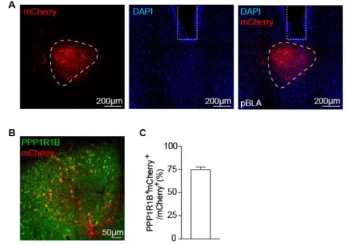
Figure 5. BLA Ppp1r1b+ fear extinction memory engram cells suppress Rspo2+ fear cells
1
(A) Virus-based activity-dependent labeling scheme. C56BL/6 mice were injected with AAV9 -c-2
fos-tTA and AAV9-TRE-ArchT-mCherry or AAV9-TRE-mCherry virus and implanted with 3
optical fibers bilaterally targeting pBLA. (B) Experimental protocol. Fear extinction engram was 4
labeled on Day 3 during extinction retrieval and inhibited on the following day. (C) Representative 5
image showing activity-dependent expression of ArchT-mCherry and optical fiber implant in 6
pBLA. (D) Inhibition of fear extinction engram cells in pBLA caused significant recovery of fear 7
response on Day 4. Unpaired t-test. mCherry group n = 13; ArchT group n = 12. (E) When pBLA 8
extinction engram neurons were inhibited during extinction retrieval on Day 4, percentage of Fos+ 9
neurons within BLA Rspo2+ neurons across A/P axis, -0.8 mm to -2.0 mm. (F) Average of the 10
percentage of Fos+/Rspo2+ shown in (E). Unpaired t-test. Ext-mCherry group n=4; Ext-ArchT 11
group n=4. (G) Double smFISH of Fos (red) and Rspo2 (green) in aBLA of Ext-mCherry mice 12
(left) and Ext-ArchT mice (right). *P < 0.05, **P < 0.01, ***P < 0.001, ****P < 0.0001. Data 13
are presented as mean ± SEM. 14
31
32
Figure 6. Fear extinction engram neurons and reward neurons overlap extensively on the
1
cellular level
2
(A) Virus-based activity-dependent labeling scheme. C56BL/6 mice were injected with AAV9 -c-3
fos-tTA and AAV9-TRE-EYFP. (B) Experimental protocol for activity-dependent labeling of 4
reward-responsive neurons and detect extinction-activated neurons (W/Ext, blue). Water stimulus 5
(W/W, black), food stimulus (W/F, orange) and fear memory retrieval (W/NonExt, red) were used 6
as controls. (C) Similar proportions of neurons were labeled by water reward in all four groups. 7
One-way ANOVA. W/W n = 5; W/F n = 5; W/Ext n = 5; W/NonExt n = 5. (D) Percentage of c-8
Fos+ cells in pBLA that were activated by water, food, extinction retrieval or fear retrieval. One-9
way ANOVA. (E) Reactivation levels (c-Fos+EYFP+/EYFP+) in W/W group, W/F group and 10
W/Ext group were significantly higher than that in W/NonExt group. No significant difference 11
was observed between W/F group and W/Ext group. One-way ANOVA. (F) Double 12
immunofluorescence staining for EYFP (green) and c-Fos (red) in pBLA. *P < 0.05, **P < 0.01, 13
****P < 0.0001. Data are presented as mean ± SEM. 14
33
1
DOX ON DOX OFF DOX ON
Day 1 Day 2 Day 3 Day 4 Day 5
Extinction Retrieval CFC Water deprivation Water Opto Extinction A E
EYFP Light-ON ChR2 Light-ON ChR2 Light-OFF
C S3 Day 3 Hab S1 S2 0 15 30 45 60 Freezing (% )
Day 4 Opto Extinction
Time (min) 0 15 30 45 60 Freezing (% ) 0 5 10 15 20 25 30 35 40 45 Day 5 0 15 30 45 60 Freezing (% )
EYFP-ONChR2-ONChR2-OFF EYFP-ON or ChR2-ONgroup Extinction Retrieval CFC Water deprivation Water Extinction ChR2-OFF group
DOX ON DOX OFF DOX ON
Day 1 Day 2 Day 3 Day 4
Extinction ExtinctionRetrieval Opto Self-stimOptoPP
Water Water deprivation Water deprivation Ext-ChR2 or Ext-EYFP Water-ChR2 group CFC
HomeCage RetrievalFear CFC-ChR2 group CFC B 0 50 100 150 Nose Poke s # Active port Inactive port
Ext-ChR2 Ext-EYFP Water-ChR2CFC-ChR2
AAV c-fos tTA ITR ITR AAV + Virus-mediated activity-dependent labeling TRE
ITR ChR2-EYFP ITR
OR
TRE
ITR EYFP ITR
AAV -sp pBLA -sp C57BL/6 mice G
5-10min 10-15min 15-20min 0 15 30 45 60 Freezing (% ) ** ** ** * * Day 4 Opto Extinction
D 0 20 40 60 80 % T ime in chamber s
Stim paired side Non-stim paired side
Ext-ChR2 Ext-EYFP Water-ChR2CFC-ChR2
n.s. n.s.
** **
F
Ext-ChR2 Ext-EYFP Water-ChR2 CFC-ChR2
Opto Self-stim OptoPP Opto Self-stim
34
Figure 7. Fear extinction engram neurons and reward neurons are behaviorally
1
interchangeable
2
(A) Virus-based activity-dependent labeling scheme. C56BL/6 mice were injected with AAV9 -c-3
fos-tTA and AAV9-TRE-ChR2-EYFP or AAV9-TRE-EYFP virus and implanted with optical 4
fibers bilaterally targeting pBLA. (B) Experimental protocol to label and activate fear extinction 5
engram neurons (Ext-ChR2 or Ext-EYFP group), water-reward neurons (Water-ChR2 group) and 6
fear retrieval engram neurons (CFC-ChR2 group) in optogenetic self-stimulation behavior or 7
optogenetic place preference (OptoPP) behavior on Day 4. (C) 50% of Ext-ChR2 group mice and 8
67% of Water-ChR2 group mice exhibited self-stimulation behavior, with a threshold of (active 9
ports–inactive ports) 20. No self-stimulation behavior was exhibited in Ext-EYFP or CFC-ChR2 10
groups. Ext-ChR2 n = 12; Ext-EYFP n =12; Water-ChR2 n = 11; CFC-ChR2 n = 8. (D) Ext-ChR2 11
and Water-ChR2 group showed preference towards stimulation paired side. ChR2 n = 12; Ext-12
EYFP n =11; Water-ChR2 n = 12; CFC-ChR2 n = 11. Paired t-test. (E) Heatmaps showing time 13
spent in each side of the chamber. Blue bar showing the stimulation paired side. (F) Experimental 14
protocol to activate reward neurons during fear extinction (Day 4). Optical laser was off for ChR2-15
OFF group during extinction training on Day 4. Optogenetic activation of water-reward neurons 16
accelerated fear extinction learning. EYFP ON, n = 8; ChR2 ON, n = 11; ChR2 Light-17
Off, n = 10. Two-way RM ANOVA. *P < 0.05, **P < 0.01, ****P < 0.0001. Data are presented 18
as mean ± SEM. 19
1
Supplementary Materials for
1 2
Amygdala Reward Neurons Form and Store Fear Extinction Memory
34
Xiangyu Zhang1, Joshua Kim1 & Susumu Tonegawa1,2* 5
6 7
Correspondence to: tonegawa@mit.edu 8
2
Materials and Methods
1
Subjects
2
The Ppp1r1b-Cre (also called Cartpt-Cre) mice were obtained from GENSAT (stock 3
number: 036659-UCD). The Rspo2-Cre mice were generated in house. Cre-expressing mice were 4
genetic knock-in mice or have been previously been validated for genetic specificity (16). All Cre 5
transgenic mice were bred using a heterozygous male with females of C57BL/6 background and 6
maintained as heterozygous. Mice had access to food and water ad libitum and were socially 7
housed in numbers of two to five littermates until surgery. After surgery, mice were singly 8
housed. 2- to 6-month old male mice were used for all experiments. For engram labeling, C57/B6 9
mice were raised on food containing 40 mg kg−1 DOX 1 week before stereotaxic surgery and were 10
maintained on Dox diet for the remaining experiment period except for the target Dox-OFF days. 11
All mice were maintained and cared in accordance with protocols approved by the Massachusetts 12
Institute of Technology (MIT) Committee on Animal Care (CAC) and guidelines by the National 13
Institutes of Health (NIH). 14
15
Viral constructs
16
AAV5-Ef1a-DIO-eArch3.0-EYFP (AV5257), AAV5-Ef1a-DIO-ChR2-EYFP (AV5226B),
17
and AAV5-Ef1a-DIO-eYFP (AV4310D) were obtained from the University of North Carolina at 18
Chapel Hill Vector Core. AAV9-c-fos-tTA, AAV9-TRE-EYFP, AAV9-TRE-ChR2-EYFP, AAV9 -19
TRE-mCherry, and AAV9-TRE-eArchT-mCherry were generated by and acquired from the Gene
20
Therapy Center and Vector Core at the University of Massachusetts Medical School as previously 21
described (Kitamura et al., 2017; Liu et al., 2012; Redondo et al., 2014). The AAV5 -hsyn:DIO-22
GCaMP6f virus was generated by and acquired from the University of Pennsylvania Vector Core. 23
3
Stereotactic surgery
1
Mice undergoing stereotactic injections were anesthetized under isoflurane. Standard 2
stereotactic procedures were used. Viruses were injected using a mineral oil–filled glass 3
micropipette attached to a 1 µl Hamilton microsyringe. Virus was injected at a speed of 80 nl/min, 4
which was controlled by a microsyringe pump. The needle was lowered to the target site and 5
remained for 5 min after the injection. The incision was closed with sutures. Mice were given 0.1 6
mg/kg slow release Buprenex as analgesic and remained on a heating pad until full recovery from 7
anesthesia. The histology of all injections was verified after behavior experiments. 8
For optogenetic behavior experiment, 200 µL of AAV5 Cre-dependent virus was bilaterally 9
injected into the anterior BLA of Rspo2-Cre mice (AP −1.4 mm, ML ±3.4 mm, DV −4.9 mm) and 10
the posterior BLA of Ppp1r1b-Cre mice (AP −2.0 mm, ML ±3.4 mm, DV −4.9 mm) and incubated 11
for 3 – 4 weeks before behavioral experiments. 12
For activity-dependent labeling and activation/inhibition of pBLA engram cells, 200 nL 13
of 2.0 × 10⁹ GC of AAV9-cfos-tTA and AAV9-TRE-ChR2-EYFP or AAV9 -TRE-eArch3.0-14
mCherry (1:1 mixture) were bilaterally injected into the pBLA (AP −2.0 mm, ML ±3.4 mm, DV 15
−4.9 mm) of doxycycline (Dox)-fed C57/B6 mice and incubated for 7 days before subsequent 16
experiments. AAV9-TRE-EYFP or AAV9-TRE-mCherry was used as corresponding controls. 17
For optical implant surgery, two optic fibers (200 mm core diameter, 5.5 mm length; Doric 18
Lenses) were bilaterally lowered above the injection sites (aBLA: −1.4 mm AP, ±3.4 mm ML, 19
−4.7 mm DV; pBLA: −2.0 mm AP, ± 3.4 mm ML, −4.7 mm DV). The implants were secured to 20
the skull with two jewelry screws and adhesive cement (C&B Metabond). A protective cap, made 21
using a 1.5 mL black Eppendorf tube, was fixed onto the implant using dental cement (Kim et al., 22
2016; Kim et al., 2017). 23
4 For Ca2+ imaging surgery, AAV5-hsyn:DIO-GCaMP6f was unilaterally injected into right 1
aBLA of Rspo2-Cre mice and right pBLA of Ppp1r1b-Cre mice with the following coordinates: 2
aBLA was targeted at −1.4 mm AP, +3.4 mm ML, −4.9 mm DV and pBLA was targeted at 3
−2.0 mm AP, +3.4 mm ML, −4.9 mm DV. Two weeks following AAV5-hsyn:DIO-GCaMP6f virus
4
injection, a GRIN lens (0.5 mm diameter, 8 mm length; Inscopix) was implanted targeting aBLA 5
or pBLA (aBLA: −1.4 mm AP, +3.4 mm ML, −4.7 mm DV; pBLA: −2.0 mm AP, +3.4 mm ML, 6
−4.7 mm DV). 7
8
Contextual fear extinction behavior protocol
9
Behavioral context was 29 × 25 × 22 cm chambers (Med Associates) with grid floors, 10
opaque ceilings, white lighting, and scented with 5% benzaldehyde. Before fear conditioning, mice 11
were habituated to investigator handling for 5 min on three consecutive days in the holding room 12
where the mice were housed. On Day 1, mice were habituated in the behavioral context for 3 min, 13
followed by three footshocks (0.60 mA, 2 s) delivered at 180 s, 250 s and 320 s. Mice remained in 14
the behavior chamber for 80 s after the third footshock and then returned to their home cages in 15
the holding room. 24 hrs after fear conditioning, mice returned to the fear conditioning chambers 16
to receive 45 min extinction training without any footshocks. Then mice were put back to their 17
home cages for another 24 hrs. On Day 3, mice were placed in the conditioned context for 5 min 18
extinction retrieval test. Behavior videos were recorded with VideoFreeze software and freezing 19
level was scored manually by experimenters who were blinded to conditions. 20
21
Single molecular fluorescent in situ hybridization
5 To examine the expression of Fos gene (Figures 1 and 5), single molecule fluorescence in 1
situ hybridization (smFISH) was performed using RNAscope Fluorescent Multiplex Kit
2
(Advanced Cell Diagnostics, ACDBio) as previously described (16, 18). Isoflurane anesthetized 3
mice were decapitated, their brains harvested and flash frozen on aluminum foil on dry ice. Brains 4
were stored at −80 °C. Prior to sectioning, brains were equilibrated to −16 °C in a cryostat for 5
30 min. Brains were coronally sectioned at 20 µm with cryostat and thaw-mounted onto Superfrost 6
Plus slides (25 x 75 mm, Fisherbrand). Sections from a single brain were serially thaw-mounted 7
onto 10 slides through the entire BLA (anterior-posterior distance from Bregma, −0.8 mm to 8
−2.6 mm). Slides were air-dried for 60 min at room temperature prior to storage at −80 °C. smFISH 9
probes for all genes examined were obtained from ACDBio, Fos (Cat #421981), Ppp1r1b (Cat 10
#405901) and Rspo2 (Cat #402001). Slides were counterstained for the nuclear marker DAPI using 11
ProLong Diamond Antifade mounting medium with DAPI (ThermosFisher) (Kim et al., 2016; 12 Kim et al., 2017). 13 14 Immunohistochemistry 15
Mice were perfused transcardially with phosphate buffered saline (PBS), followed by 4% 16
paraformaldehyde (PFA). Brains were post-fixed with 4% PFA solution overnight and then 17
sectioned coronally 50 µm at with a vibratome. Free floating brain sections were washed in PBST 18
(PBS, 3% TritonX) three times for 10 min, blocked for 1 hr in blocking buffer (PBST, 5% normal 19
goat serum), and incubated in primary antibody in blocking buffer overnight at 4 °C. On the next 20
day, brains were washed with three 10 min washes of PBST and incubated in secondary antibody 21
in blocking buffer at room temperature for 2 hrs. Primary antibodies used were chicken anti-GFP 22
(Invitrogen, A10262, 1:1,000), rabbit c-Fos (Santa Cruz, sc-52, 1:1,000) and rabbit anti-23
6 PPP1R1B (Abcam, ab40801, 1:1,000). Secondary antibodies used were goat anti-chicken Alexa 1
Fluor 488 (Invitrogen, A11039, 1:1,000) and goat anti-rabbit Alexa Fluor 555 (Invitrogen, 2
A21428, 1: 1,000). After three more 10-min PBST washes, slides were covered with coverslips 3
and mounted using VectaShield mounting solution containing DAPI (Vector Laboratories). 4
5
Calcium imaging
6
Calcium imaging of BLA Rspo2+ neurons and Ppp1r1b+ neurons was performed on 7
Rspo2-Cre and Ppp1r1b-Cre mice, respectively. AAV5-hsyn:DIO-GCaMP6f was unilaterally 8
injected into right aBLA of Rspo2-Cre mice and right pBLA of Ppp1r1b-Cre mice with the 9
following coordinates: aBLA was targeted at −1.4 mm AP, +3.4 mm ML, −4.9 mm DV and pBLA 10
was targeted at −2.0 mm AP, +3.4 mm ML, −4.9 mm DV. Two weeks following AAV5- hsyn:DIO-11
GCaMP6f virus injection, a GRIN lens (0.5 mm diameter, 8 mm length; Inscopix) was implanted 12
targeting aBLA or pBLA (aBLA: −1.4 mm AP, +3.4 mm ML, −4.7 mm DV; pBLA: −2.0 mm AP, 13
+3.4 mm ML, −4.7 mm DV). Two weeks after GRIN lens implant, a baseplate for the miniaturized 14
microscope camera was attached above the GRIN lens using ultraviolet-light curable glue (Loctite 15
4305) (Mukamel et al., 2009; Ziv et al., 2013). 16
For contextual fear extinction behavior, recording was performed during the light cycle. 17
Inscopix endoscope and calcium events were captured at 20 Hz with an Inscopix miniature 18
microscope (nVista HD). Inscopix endoscope was synchronized with Med Associate behavior 19
chamber via a TTL signal, which was used for all the other fear extinction behavior experiments 20
in this study. Mice underwent the three-day contextual fear extinction procedure as previously 21
described (Figure 1). During contextual fear conditioning (CFC) on Day 1, freezing behavior and 22
Ca2+ signals of subjects were recorded for 3 min habituation and 3 min when three footshocks were 23
7 delivered. During 45 min fear extinction training, Ca2+ signals was recorded during the 0-3 min, 1
5-8 min, 10-13 min, 15-18 min, 20-23 min, 25-28 min, 30-33 min, 35-38 min and 40-43 min 2
timestamps. The 0-3 min epoch was considered as fear retrieval stage. The other eight epochs 3
together were considered as extinction training stage. In the heatmap and Ca2+ traces (Figure 2E-4
2I), extinction training referred referred to 40-43 min time bin recording on Day 2. Recorded 5
calcium-imaging movies were first processed with ImageJ to subtract base line activity (dividing 6
each image by a low-pass, r = 20 pixels, and high pass, r = 1000 pixels), followed by motion 7
correction using Inscopix Mosaic software (Mukamel et al., 2009). Subsequently, ΔF/F ((F-8
F0)/F0) signal was calculated in Mosaic software (Inscopix), in which F0 was calculated over the 9
entire recording session. A stacked image of ΔF/F recording movie was acquired by maximum 10
intensity projection. Cell identifications were semi-automatically identified and cross-registered 11
for longitudinal recording by visual inspection (Kitamura et al., 2017). The corresponding Ca2+ 12
activity was extracted by applying PCA/ICA analysis in Mosaic software (Inscopix). In total, we 13
recorded 179 BLA Ppp1r1b+ cells from 5 Ppp1r1b-Cre mice and 169 BLA Rspo2+ cells from 4 14 Rspo2-Cre mice. 15 16 Data analysis 17
Ca2+ events were detected with a threshold 15% ΔF/F. We calculated the average time of 18
Ca2+ events, i.e., the sum of all Ca2+ events duration/recording time. To identify cells with 19
increased or decreased activity in response to footshocks during CFC on Day 1, we compared the 20
mean ΔF/F of 3 min after delivery of footshocks with that of 3 min habituation stage on Day1. To 21
identify cells with changed neuronal activities during fear retrieval, we compared the mean ΔF/F 22
of the fear retrieval stage (0-3 min) on Day 2 with that of the 3 min habituate period before 23