HAL Id: inserm-00332530
https://www.hal.inserm.fr/inserm-00332530
Submitted on 21 Oct 2008
HAL is a multi-disciplinary open access
archive for the deposit and dissemination of
sci-entific research documents, whether they are
pub-lished or not. The documents may come from
teaching and research institutions in France or
abroad, or from public or private research centers.
L’archive ouverte pluridisciplinaire HAL, est
destinée au dépôt et à la diffusion de documents
scientifiques de niveau recherche, publiés ou non,
émanant des établissements d’enseignement et de
recherche français ou étrangers, des laboratoires
publics ou privés.
A Hybrid System for the Semantic Annotation of
Sulco-Gyral Anatomy in MRI Images.
Ammar Mechouche, Xavier Morandi, Christine Golbreich, Bernard Gibaud
To cite this version:
Ammar Mechouche, Xavier Morandi, Christine Golbreich, Bernard Gibaud. A Hybrid System for
the Semantic Annotation of Sulco-Gyral Anatomy in MRI Images.. Medical Image Computing and
Computer-Assisted Intervention - MICCAI 2008, 11th International Conference., Sep 2008, United
States. pp.807-814. �inserm-00332530�
A Hybrid System for the Semantic Annotation
of Sulco-Gyral Anatomy in MRI Images
Ammar Mechouche1, Xavier Morandi1,2, Christine Golbreich3,4, and Bernard
Gibaud1
1
Unit/Project VisAGeS U746, INSERM - INRIA - CNRS - Univ-Rennes 1, France {Ammar.Mechouche,Bernard.Gibaud}@irisa.fr
http://www.irisa.fr/visages/
2
University Hospital of Rennes, Department of Neurosurgery, France Xavier.Morandi@chu-rennes.fr
3
University of Versailles, Versailles - Christine.Golbreich@uvsq.fr
4
LIRMM UMR 5506, Montpellier, France - http://www.lirmm.fr/tatoo/
Abstract. This paper presents an interactive system for the annotation of brain anatomical structures in Magnetic Resonance Images. The sys-tem is based on hybrid knowledge and techniques. First, it exploits both numerical knowledge from atlases and symbolic knowledge from a rule-extended ontology represented in OWL, the Web ontology language, and combines them with graphical data about cortical sulci, automatically ex-tracted from the images. Second, the annotations of the parts of gyri and of sulci located in a region of interest are obtained with different reason-ing techniques: Constraint Satisfaction Solvreason-ing and Description Logics techniques. Preliminary experiments have been achieved on normal and also pathological data. The results obtained so far are very promising.
1
Introduction
Semantic annotation associates meaningful labels to images, in order to high-light their information content. Semantic annotation is becoming a major issue regarding wide scale sharing of information on the web and is of critical im-portance in biomedical research, notably for translational research aiming at facilitating the exploitation of experimental data across several disciplines and scales [1]. This is however a very challenging topic, due to the difficulty of defining consensual reference knowledge models, on the one hand, and to use them in image annotation tools, on the other hand. Such models, usually called ontologies, are often classified into two categories referred as lightweight and heavyweight ontologies, respectively [2]: 1) simple term lists, thesauri or tax-onomies, 2) highly expressive knowledge models, according to which instances are created, involving complex assertions and constraints. Our primary appli-cation field is the preparation of surgical procedures in neurosurgery. Actually, the precise identification of gyri and sulci around the lesional area is of pri-mary importance because they provide useful landmarks for the surgeon during surgery, especially in eloquent cortex [3]. The annotation of images, in our case
anatomical MRI images, has two objectives: 1) to help to identify precisely the gyri and sulci around the lesion, and to provide the detailed annotation data re-lating anatomical conceptual entities to graphical primitives extracted from the images; 2) to use these metadata for cases retrieval in an images database. Our approach significantly differs from classical approaches of parcellation of cortex in MRI images [4–7]. Those are generally global (i.e. they analyze the whole cortex), have limited precision (i.e. they usually address gross anatomy), refer to simple term lists, are entirely automatic, mostly based on numerical priors (i.e. atlases), and primarily suited to normal anatomy. In contrast, our approach is local (i.e. focuses on a particular region in the image), may provide labels with high anatomical precision, can involve a human user participation, is based on symbolic prior knowledge (provided by a highly expressive ontology), and may be relevant to interpret pathological images as well as normal cases. The work presented here concerns a hybrid system designed according to the previous ap-proach, i.e. a semantic apap-proach, rather than morphometric or statistical. The knowledge supporting the labelling process consists of the mereo-topological re-lations between the different cortical features. This knowledge is described in an
ontology of cortical gyri and sulci represented in OWL DL5, according to the
description logics (DL) paradigm [8]. The whole reasoning aims at producing in-stances of the ontology concerning the parts of sulci and of gyri involved in a set of graphical primitives extracted from the images (segments of cortical sulci, and parts of gyri called “patches“), that satisfy the axioms and constraints defined in the ontology. The very large number of possible combinations led us to adopt a hybrid approach, consisting in selecting a reasonable number of hypotheses for the labelling of patches, based on an atlas, and to select the valid combina-tions of such hypotheses, based on existing prior knowledge about the spatial arrangement of the gyri. This part constitutes a major extension of the method presented in [13]. Section 2 provides an overview of the method and further details of the system components; section 3 describes our first experiments with both normal and pathological data; section 4 discusses these preliminary results and highlights the capabilities and current limitations of the system.
2
Method
2.1 Definitions
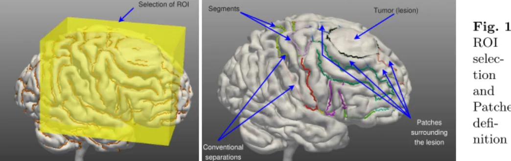
Definition 1:A segment (figure 1) is a part of an external trace of a sulcus. The
segments are organized in a graph describing the connections between them.
Definition 2:A conventional separation (figure 1) is a fictitious line added by
the user in order to connect two segments separated by a gyrus.
Definition 3:A patch (figure 1) is a subset of the brain surface, corresponding
to a part of a gyrus, and delimited by a set of continuous segments and conven-tional separations.
Definition 4: An interpretation consists of a set of labels associated with the
5
patches and segments of a Region Of Interest (ROI); each patch and each seg-ment has one label only.
Definition 5:A consistent interpretation is an interpretation, where each patch
label and each segment label is consistent with our prior knowledge about the sulco-gyral anatomy and with the information supplied by the user.
2.2 System Overview
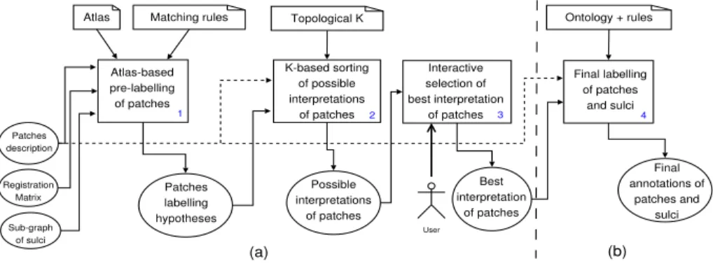
The overall labelling process involves three steps (figure 2): (1) the brain is segmented, the external traces of the sulci are extracted [9], the ROI is selected by the user, and the patches are defined (section 2.3); (2) patches identification: this is done by using Constraint Satisfaction Problem techniques (CSP) allowing to infer all the consistent interpretations for the patches with respect to prior knowledge about the spatial arrangement of the gyri (section 2.4). For that, the system uses a set of hypotheses (possible labels) derived from the matching of patch information with an atlas (SPAMs [10]); (3) sulci identification is done using the best interpretation computed for the patches and the logical definitions of the sulci in the ontology (section 2.5). The final annotations for the patches and the segments are generated in standard web languages to facilitate their exploitation by semantic web technologies. The paper focuses on (2) and (3).
Tumor (lesion) Patches surrounding the lesion Segments Conventional separations Selection of ROI Fig. 1. ROI selec-tion and Patches defi-nition
2.3 Delimitation and Extraction of the Patches
The brain is automatically segmented from a T1-MRI scan and the graph of the external traces of the sulci is automatically extracted. Then, the user selects
Segmentation, Selection of ROI, Definition of the patches
1 Identification of the patches 2 Identification of the sulci 3 Final annotations of patches and sulci
Atlas-based pre-labelling of patches 1 Patches description
Atlas Matching rules
K-based sorting of possible interpretations of patches 2 Topological K Interactive selection of best interpretation of patches 3 Final labelling of patches and sulci 4 Ontology + rules Registration Matrix Sub-graph of sulci Patches labelling hypotheses Possible interpretations of patches Best interpretation of patches User Final annotations of patches and sulci (a) (b)
Fig. 3. The complete labelling process
a ROI and the corresponding sub-graph is automatically extracted as shown on figure 1. Next, the user introduces a number of conventional separations, in order to produce a partition of the selected cortex area into a set of contiguous patches. Finally, the system computes the description of the topological relations and orientations between neighboring patches, and between the patches and the segments which form them. This description is represented in OWL (f ile1).
2.4 Identification of the Patches
Generation of Patches Labelling Hypotheses. The segments delimiting the
patches within the ROI are realigned into the stereotaxic space (SPAMs space) thanks to the registration matrix produced during the segmentation process (figure 3). The position of each segment with respect to each SPAM is analyzed in order to determine whether it bounds this SPAM or not, and with which orientation. This analysis also provides a confidence index. All this information is represented in OWL (f ile2). The matching of information from f ile1 and f ile2 in the case of normal subjects is done by rules of the following form: Bounds(x, y) ∧ SulcusPart(x) ∧ Patch(y) ∧ anteriorTo(x, y) ∧ Bounds(x, z) ∧ Gyrus(z)
∧ anteriorTo(x, z) → partOf(y, z). This rule, for example, expresses that if we
have a part of sulcus which bounds a given patch with an anterior orientation, and at the same time this part of sulcus bounds a given SPAM with the same orientation, then this patch is inferred to be part of the gyrus corresponding to this SPAM in the ontology. Six matching rules of this kind, corresponding to the six spatial orientations, are defined. Slightly different rules are used in pathological cases, taking into account only adjacency between the parts of sulci and the SPAMs, not orientations, because they might lead to erroneous decisions due to displacements related to the pathology. These rules assign to each patch a set of hypotheses. The correct label is assumed to belong to this set.
Determining the Consistent Interpretations Using a CSP Solver. A
number of constraints. A variable is defined by its domain, i.e. the set of values that can be assigned to this variable. A constraint relates several variables and restricts the involved variables values to legal assignments. Constraint reasoning is the process of computing a solution to the given CSP, i.e. an assignment of values to the variables that satisfy all the given constraints on the variables [11]. In our case (figure 3-a), the patches represent the variables, the hypotheses computed for the patches represent the domains of the variables, and the spatial relations between the patches represent the constraints. The solutions of the CSP problem provide all possible interpretations for the patches with respect to our prior knowledge on the spatial arrangement of the gyri and parts of gyri. In order to find the best consistent interpretation for the patches among those computed by the CSP solver, the system allows interactions with the user. The latter is invited to select a preferred label for patches exhibiting different labels in different interpretations. Then the system eliminates those interpretations that are not consistent with the user information. The interactions are repeated until only one interpretation remains for the patches.
2.5 Identification of the Segments
To label the sulci segments the system uses the patches description, the best interpretation of the patches and the logical definitions of the sulci in the ontol-ogy (figure 3-b). The ontolontol-ogy contains the mereo-topological knowledge about the sulci and the gyri. To model the knowledge about the brain we have mainly
used the Ono atlas [12], the Foundational Model of Anatomy (FMA6), and the
expertise of a neuroanatomist. The concepts describing the sulci and gyri and their relations are defined in an ontology in OWL DL. An example of a concept
definition of the ontology is the following:RightCentralSulcusPart ≡ (∃ Bounds ((∃
hasEntity (∃ partOf RightPostCentralGyrus)) u (∃ hasOrientation Anterior))) u (∃ Bounds ((∃ hasEntity (∃ partOf RightPreCentralGyrus)) u (∃ hasOrientation Poste-rior))) u (∀ Bounds ((∃ hasEntity (∃ partOf RightPreCentralGyrus)) t (∃ hasEntity (∃ partOf RightPostCentralGyrus))). This logical expression expresses that a part of the central sulcus of the right hemisphere bounds at least one part of the right postcentral gyrus with an anterior orientation and one part of the precen-tral gyrus with a posterior orientation, and only parts of such gyri. Our brain ontology contains for each hemisphere logical definitions of 49 gyri, 5 lobes, 3 operculum, 17 gyri parts, 44 sulci, 44 sulci parts (segments), and 31 relations. The ontology is enriched by some rules increasing its expressivity (see [13] for more details).
3
Results
3.1 Material
Preliminary experiments were made using T1-MRI images obtained with a 3T scanner (Philips Achieva) from three normal subjects and two patients
(patho-6
logical subjects). In the two pathological cases the lesion was located in the right frontal lobe. The brain segmentation and the extraction of the external traces of
the sulci were done with Brainvisa7tools and Vistal8respectively. We have used
44 SPAMs corresponding to the gyri. The system is implemented in C++ and Java, the connection between C++ and Java programs is done thanks to JNI
(Java Native Interface). The ontology is edited and created with the Prot´eg´e9
software. The rules are edited and created with the SWRL10Plugin. The results
were obtained with the Java Constraint Library11, and the KAON212reasoner.
3.2 Evaluation
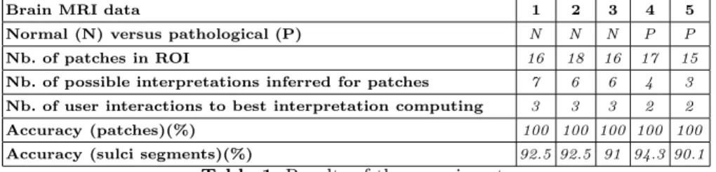
The evaluation is done on a ROI defined by an expert neuroanatomist. It includes the superior frontal gyrus, the middle frontal gyrus, the inferior frontal gyrus, the precentral gyrus, the postcentral gyrus, the central sulcus, the superior precentral sulcus, the intermediate precentral sulcus, the inferior precentral sulcus, the superior frontal sulcus, and the inferior frontal sulcus regions. We compared the results provided by the system to the labels given by the expert considered as a gold-standard. The same procedure was applied to the five MRI datasets except for the matching rules, since orientations were not considered in case of pathological data. For each case, we report the type of the image (normal versus pathological), the number of patches extracted from the selected ROI, the number of consistent interpretations inferred for the patches, the number of user interactions needed to reach the best interpretation for the patches, the accuracy of patch labels (defined as the ratio of the accurately labelled patches over the total number of patches in ROI), and finally the accuracy for the sulci segments (defined as the ratio of the accurately labelled sulci segments over the total number of sulci segments in the ROI).
Brain MRI data 1 2 3 4 5
Normal (N) versus pathological (P) N N N P P Nb. of patches in ROI 16 18 16 17 15 Nb. of possible interpretations inferred for patches 7 6 6 4 3 Nb. of user interactions to best interpretation computing 3 3 3 2 2 Accuracy (patches)(%) 100 100 100 100 100 Accuracy (sulci segments)(%) 92.5 92.5 91 94.3 90.1
Table 1. Results of the experiments
7 http://brainvisa.info/index_f.html 8 http://www.irisa.fr/vista/Themes/Logiciel/VIsTAL/VIsTAL.html 9 http://protege.stanford.edu/ 10 http://www.w3.org/Submission/SWRL/ 11 http://liawww.epfl.ch/JCL/ (a CSP solver) 12
Table 1 reports the results for the five cases. We observe that the identifica-tion of the patches is complete after a reasonable number of interacidentifica-tions with the user (about 3). The sulci identification is quite accurate, although some segments are not well classified due to the insufficient precision in the computation of the orientations between the patches and the segments. It is important to notice that the system performance is not decreased in pathological cases in spite of signif-icant structures displacement according to the expert. An example of inferred annotations is reported figure 4-a. We observe on figure 4-(b,c,d) that if we add conventional separations where it is not necessary, then the system becomes less accurate. Regarding the time computing, the Atlas-based pre-labelling of the patches, the CSP reasoning, and the KAON2 reasoning take about 1 minute.
PoCG PoCG PoCG PreCG PreCG PreCG SupFG SupFG SupFG SupFG MidFG MidFG MidFG MidFG MidFG InfFG InfFG CS CS CS SupPrCS IntPrCS InfPrCS SupFS SupFS SupFS SupFS InfFS InfFS New-added conventional separation PreCG InfFG PreCG (a) (b) (c) (d) MidFG
Fig. 4. (a) Inferred labels for the ROI and (b)(c)(d) in-fluence of irrelevant conventional separa-tions.
4
Conclusion
We have presented a hybrid system developed to semi-automatically label brain MRI images with semantic annotations and the first results obtained so far. The presented approach is novel with respect to several aspects: (1) the use of a CSP solver to select consistent interpretations of the gyri, (2) the easy generation of semantically-rich annotations of gyral/sulcal structures, (3) the use of explicit prior knowledge described in a formal ontology, (4) its represention in OWL, the Ontology Web Language, to facilitate knowledge sharing. Our first experiments using both normal and pathological data are very promising. We plan to still im-prove the system in the future with respect to several aspects. The definition of the conventional separations is defined manually in the current implementation, and could be automated. The use of atlases as a means to produce the initial hypotheses could be optimized, e.g. by using different types of numerical atlases, more adapted to the particular case under study. The orientations’ computation could be improved, e.g. based on [14]. Finally, the ontology could be refined, in order to include more fine-grained gyri and sulci, and could be directly used
to derive the orientations’ definitions in the CSP problem. Moreover, the de-pendence of the system performance on initial processing steps (especially the definition of conventional separations) should be further investigated.
References
1. Ruttenberg, A., Clark, T., Bug, W., Samwald, M., Bodenreider, O., Chen, H., Do-herty, D., Forsberg, K., Gao, Y., Kashyap, V., Kinoshita, J., Luciano, J., Marshall, M., Ogbuji, C., Rees, J., Stephens, S., Wong, G., Wu, E., Zaccagnini, D., Hongser-meier, T., Neumann, E., Herman, I., Cheung, K.: Advancing translational research with the Semantic Web. BMC Bioinformatics 8(Suppl 3):S2 (2007)
2. Corcho, O.: Ontology based document annotation: trends and open research prob-lems. Int. Journal of metadata, semantics and ontologies 1(1)(47-57) (2006) 3. Jannin, P., Morandi, X., Fleig, O., Le Rumeur, E., Toulouse, P., Gibaud, B.,
Scara-bin, J.: Integration of sulcal and functional information for multimodal neuronav-igation. Journal of Neurosurgery 96(4)(713-723) (2002)
4. Tzourio-Mazoyer, N., Landeau, B., Papathanassiou, D., Crivello, F., Etard, O., Delcroix, N., Mazoyer, B., Joliot, M.: Automated anatomical labeling of acti-vations in SPM using a macroscopic anatomical parcellation of the MNI MRI Single-Subject brain. Neuroimage 15(1)(273-289) (2002)
5. Cachia, A., Mangin, J., Rivi`ere, D., Papadopoulos-Orfanos, D., Kherif, F., Bloch, I., R´egis, J.A.: A generic framework for parcellation of the cortical surface into gyri using geodesic Vorono¨ı diagrams. Medical Image Analysis 7(4)(403-416) (2003) 6. Fischl, B., van der Kouwe, A., Destrieux, C., Halgren, E., S´egonne, F., Salat, D.,
Busa, E., Seidman, L., Goldstein, J., Kennedy, D., Caviness, V., Makris, N., Rosen, B., Dale, A.: Automatically parcellating the human cerebral cortex. Cerebral Cortex 14(11-22) (2004)
7. Clouchoux, C., Coulon, O., Anton, J., Mangin, J., R´egis, J.: A new cortical surface parcellation model and its automatic implementation. In: MICCAI 2006. LNCS 4191, Springer (2006) 193–200
8. Franz, B., Diego, C., Deborah, L., Daniele, N., F.P.S., P., eds.: The Description Logic Handbook: Theory, Implementation, and Applications. In Franz, B., Diego, C., Deborah, L., Daniele, N., F.P.S., P., eds.: Description Logic Handbook, Cam-bridge University Press (2003)
9. Le Goualher, G., Barillot, C., Bizais, Y.: Modeling cortical sulci with active rib-bons. IJPRAI 11(8) (1997) 1295–1315
10. Collins, D.L., Zijdenbos, A.P., Baar´e, W.F.C., Evans, A.C.: INSECT: Improved cortical structure segmentation. In: IPMI99. LNCS 1613 (1999) 210–223
11. Krzysztof, A.: Principles of Constraint Programming. (2003)
12. Ono, M., Kubik, S., Abernathey, C.: Atlas of the Cerebral Sulci. (1990)
13. Mechouche, A., Golbreich, C., Gibaud, B.: Towards a hybrid system using an ontology enriched by rules for the semantic annotation of brain MRI images. In: RR 2007. LNCS 4524, Springer (2007) 219–228
14. Yann, H., Aline, D.: Qualitative spatial relationships for image interpretation by using semantic graph. In Escolano, F., Vento, M., eds.: GbRPR. Volume 4538 of Lecture Notes in Computer Science., Springer (2007) 240–250
Acknowledgement We thank Louis Collins from the Montreal Neurological
Insti-tute for providing us with the SPAMs database, Christian Barillot for his revision of the manuscript, and the Regional Council of Brittany for supporting this project.