Key Words: AHA Scientific Statements ◼ arrhythmia ◼ cardiopulmonary resuscitation ◼ defibrillation ◼ heart arrest ◼ pediatrics ◼ post–cardiac arrest care
Alexis A. Topjian, MD,
MSCE, Chair
Tia T. Raymond, MD,
Vice-Chair
Dianne Atkins, MD
Melissa Chan, MD
Jonathan P. Duff, MD,
MEd
Benny L. Joyner Jr, MD,
MPH
Javier J. Lasa, MD
Eric J. Lavonas, MD, MS
Arielle Levy, MD, MEd
Melissa Mahgoub, PhD
Garth D. Meckler, MD,
MSHS
Kathryn E. Roberts, MSN,
RN
Robert M. Sutton, MD,
MSCE
Stephen M. Schexnayder,
MD
On behalf of the Pediatric
Basic and Advanced Life
Support Collaborators
© 2020 American Heart Association, Inc.
Part 4: Pediatric Basic and Advanced Life
Support
2020 American Heart Association Guidelines for Cardiopulmonary
Resuscitation and Emergency Cardiovascular Care
Circulation
https://www.ahajournals.org/journal/circ
TOP 10 TAKE-HOME MESSAGES
1. High-quality cardiopulmonary resuscitation (CPR) is the foundation of
resuscitation. New data reaffirm the key components of high-quality CPR:
providing adequate chest compression rate and depth, minimizing
inter-ruptions in CPR, allowing full chest recoil between compressions, and
avoiding excessive ventilation.
2. A respiratory rate of 20 to 30 breaths per minute is new for infants and
children who are (a) receiving CPR with an advanced airway in place or (b)
receiving rescue breathing and have a pulse.
3. For patients with nonshockable rhythms, the earlier epinephrine is
adminis-tered after CPR initiation, the more likely the patient is to survive.
4. Using a cuffed endotracheal tube decreases the need for endotracheal tube
changes.
5. The routine use of cricoid pressure does not reduce the risk of regurgitation
during bag-mask ventilation and may impede intubation success.
6. For out-of-hospital cardiac arrest, bag-mask ventilation results in the same
resuscitation outcomes as advanced airway interventions such as
endotra-cheal intubation.
7. Resuscitation does not end with return of spontaneous circulation (ROSC).
Excellent post–cardiac arrest care is critically important to achieving the best
patient outcomes. For children who do not regain consciousness after ROSC,
this care includes targeted temperature management and continuous
elec-troencephalography monitoring. The prevention and/or treatment of
hypo-tension, hyperoxia or hypoxia, and hypercapnia or hypocapnia is important.
8. After discharge from the hospital, cardiac arrest survivors can have physical,
cognitive, and emotional challenges and may need ongoing therapies and
interventions.
9. Naloxone can reverse respiratory arrest due to opioid overdose, but there is
no evidence that it benefits patients in cardiac arrest.
10. Fluid resuscitation in sepsis is based on patient response and requires
fre-quent reassessment. Balanced crystalloid, unbalanced crystalloid, and colloid
fluids are all acceptable for sepsis resuscitation. Epinephrine or
norepineph-rine infusions are used for fluid-refractory septic shock.
PREAMBLE
More than 20 000 infants and children have a cardiac arrest per year in the
United States.
1–4In 2015, emergency medical service–documented
out-of-hos-pital cardiac arrest (OHCA) occurred in more than 7000 infants and children.
4Approximately 11.4% of pediatric OHCA patients
survived to hospital discharge, but outcomes varied
by age, with survival rates of 17.1% in adolescents,
13.2% in children, and 4.9% in infants. In the same
year, pediatric in-hospital cardiac arrest (IHCA)
inci-dence was 12.66 events per 1000 infant and child
hospital admissions, with an overall survival to
hospi-tal discharge rate of 41.1%.
4Neurological outcomes
remain difficult to assess across the pediatric age
spectrum, with variability in reporting metrics and
time to follow-up across studies of both OHCA and
IHCA. Favorable neurological outcome has been
re-ported in up to 47% of survivors to discharge.
5De-spite increases in survival from IHCA, there is more
to be done to improve both survival and neurological
outcomes.
6The International Liaison Committee on
Resusci-tation (ILCOR) Formula for Survival emphasizes 3
es-sential components for good resuscitation outcomes:
guidelines based on sound resuscitation science,
ef-fective education of the lay public and resuscitation
providers, and implementation of a well-functioning
Chain of Survival.
7These guidelines contain recommendations for
pediatric basic and advanced life support, excluding
the newborn period, and are based on the best
avail-able resuscitation science. The Chain of Survival
(Sec-tion 2), which is now expanded to include recovery
from cardiac arrest, requires coordinated efforts from
medical professionals in a variety of disciplines and,
in the case of OHCA, from bystanders, emergency
dispatchers, and first responders. In addition, specific
recommendations about the training of resuscitation
providers are provided in Part 6: Resuscitation
Educa-tion Science, and recommendaEduca-tions about systems of
care are provided in Part 7.
INTRODUCTION
Scope of Guidelines
These guidelines are intended to be a resource for lay
rescuers and healthcare providers to identify and treat
infants and children in the prearrest, intra-arrest, and
postarrest states. These apply to infants and children
in multiple settings; the community, prehospital, and
the hospital environment. Prearrest, intra-arrest, and
postarrest topics are reviewed, including cardiac arrest
in special circumstances, such as in patients with
con-genital heart disease.
For the purposes of the pediatric advanced life
sup-port guidelines, pediatric patients are infants, children,
and adolescents up to 18 years of age, excluding
new-borns. For pediatric basic life support (BLS), guidelines
apply as follows:
• Infant guidelines apply to infants younger than
approximately 1 year of age.
• Child guidelines apply to children approximately
1 year of age until puberty. For teaching
pur-poses, puberty is defined as breast development in
females and the presence of axillary hair in males.
• For those with signs of puberty and beyond, adult
basic life support guidelines should be followed.
Resuscitation of the neonate is addressed in “Part 5:
Neonatal Resuscitation” and applies to the newborn
typically only during the first hospitalization following
birth. Pediatric basic and advanced life support
guide-lines apply to neonates (less than 30 days old) after
hos-pital discharge.
Coronavirus Disease 2019 Guidance
Together with other professional societies, the American
Heart Association (AHA) has provided interim guidance
for basic and advanced life support in adults, children,
and neonates with suspected or confirmed coronavirus
disease 2019 (COVID-19). Because evidence and
guid-ance are evolving with the COVID-19 situation, this
in-terim guidance is maintained separately from the
emer-gency cardiovascular care (ECC) guidelines. Readers are
directed to the AHA website for the most recent
guid-ance.
8Organization of the Pediatric Writing
Committee
The Pediatric Writing Group consisted of pediatric
clini-cians including intensivists, cardiac intensivists,
cardiolo-gists, emergency medicine physicians, medical
toxicolo-gists, and nurses. Volunteers with recognized expertise
in resuscitation are nominated by the writing group chair
and selected by the AHA ECC Committee. The AHA has
rigorous conflict of interest policies and procedures to
minimize the risk of bias or improper influence during
development of the guidelines.
9Prior to appointment,
writing group members and peer reviewers disclosed all
commercial relationships and other potential (including
intellectual) conflicts. Writing group members whose
re-search led to changes in guidelines were required to
de-clare those conflicts during discussions and abstain from
voting on those specific recommendations. This process
is described more fully in “Part 2: Evidence Evaluation
and Guidelines Development.” Disclosure information
for writing group members is listed in Appendix 1.
Methodology and Evidence Review
These pediatric guidelines are based on the extensive
evidence evaluation performed in conjunction with the
ILCOR and affiliated ILCOR member councils. Three
dif-ferent types of evidence reviews (systematic reviews,
scoping reviews, and evidence updates) were used in
the 2020 process.
10,11After review by the ILCOR
Sci-ence Advisory Committee Chair, the evidSci-ence update
worksheets were included in Appendix C of the 2020
ILCOR Consensus on CPR and ECC Science With
Treat-ment Recommendations.
11aEach of these resulted in a
description of the literature that facilitated guideline
de-velopment. This process is described more fully in “Part
2: Evidence Evaluation and Guidelines Development.”
12Class of Recommendation and Level of
Evidence
The writing group reviewed all relevant and current AHA
Guidelines for Cardiopulmonary Resuscitation (CPR) and
ECC and all relevant 2020 ILCOR Consensus on CPR and
ECC Science With Treatment Recommendations evidence
and recommendations to determine if current guidelines
should be reaffirmed, revised, or retired or if new
recom-mendations were needed. The writing group then
draft-ed, reviewdraft-ed, and approved recommendations, assigning
to each a Class of Recommendation (COR; ie, strength)
and Level of Evidence (LOE; ie, quality, certainty). Criteria
for each COR and LOE are described in Table 1.
Guideline Structure
The 2020 Guidelines are organized in discrete modules
of information on specific topics or management issues.
13Each modular “knowledge chunk” includes a table of
recommendations using standard AHA nomenclature of
Table 1. Applying Class of Recommendation and Level of Evidence to Clinical Strategies, Interventions, Treatments, or Diagnostic Testing in Patient Care (Updated May 2019)*
This table defines the Classes of Recommendation (COR) and Levels of Evidence (LOE). COR indicates the strength the writing group assigns the recommendation, and the LOE is assigned based on the quality of the scientific evidence. The outcome or result of the intervention should be specified (an improved clinical outcome or increased diagnostic accuracy or incremental prognostic information). Classes of Recommendation COR designations include Class 1, a strong recommendation for which the potential benefit greatly outweighs the risk; Class 2a, a moderate recommendation for which benefit most likely outweighs the risk; Class 2b, a weak recommendation for which it’s unknown whether benefit will outweigh the risk; Class 3: No Benefit, a moderate recommendation signifying that there is equal likelihood of benefit and risk; and Class 3: Harm, a strong recommendation for which the risk outweighs the potential benefit. Suggested phrases for writing Class 1 recommendations include • Is recommended • Is indicated/useful/ effective/beneficial
• Should be performed/ administered/other
Comparative-effectiveness phrases include treatment/strategy A is recommended/indicated in preference to treatment B, and treatment A should be chosen over treatment B. Suggested phrases for writing Class 2a recommendations include • Is reasonable • Can be useful/effective/ beneficial
Comparative-effectiveness phrases include treatment/strategy A is probably recommended/indicated in preference to treatment B, and it is reasonable to choose treatment A over treatment B. For comparative-effectiveness recommendations (COR 1 and 2a; LOE A and B only), studies that support the use of comparator verbs should involve direct comparisons of the treatments or strategies being evaluated. Suggested phrases for writing Class 2b recommendations include • May/might be reasonable
• May/might be considered
• Usefulness/effectiveness is unknown/unclear/uncertain or not well-established
Suggested phrases for writing Class 3: No Benefit recommendations (generally, LOE A or B use only) include • Is not recommended • Is not indicated/useful/ effective/beneficial
• Should not be performed/administered/other Suggested phrases for writing Class 3: Harm recommendations include • Potentially harmful • Causes harm • Associated with excess morbidity/mortality
• Should not be performed/administered/other Levels of Evidence For LOEs, the method of assessing quality is evolving, including the application of standardized, widely-used, and preferably validated evidence grading tools; and for systematic reviews, the incorporation of an Evidence Review Committee. LOE designations include Level A, Level B-R, Level B-NR, Level C-LD, and Level C-EO. Those categorized as Level A are derived from • High-quality evidence from more than 1 randomized clinical trial, or RCT • Meta-analyses of high-quality RCTs
• One or more RCTs corroborated by high-quality registry studies Those categorized as Level B-R (randomized) are derived from
• Moderate-quality evidence from 1 or more RCTs
• Meta-analyses of moderate-quality RCTs
Those categorized as Level B-NR (nonrandomized) are derived from
• Moderate-quality evidence from 1 or more well-designed, well-executed nonrandomized studies, observational studies, or
registry studies
• Meta-analyses of such studies
Those categorized as Level C-LD (limited data) are derived from • Randomized or nonrandomized observational or registry studies with limitations of design or execution • Meta-analyses of such studies
• Physiological or mechanistic studies in human subjects Those categorized as Level C-EO (expert opinion) are derived from • Consensus of expert opinion based on clinical experience COR and LOE are determined independently (any COR may be paired with any LOE). A recommendation with LOE C does not imply that the recommendation is weak. Many important clinical questions addressed in guidelines do not lend themselves to clinical trials. Although RCTs are unavailable, there may be a very clear clinical consensus that a particular test or therapy is useful or effective.
COR and LOE. Recommendations are presented in
or-der of COR: most potential benefit (Class 1), followed
by lesser certainty of benefit (Class 2), and finally
poten-tial for harm or no benefit (Class 3). Following the COR,
recommendations are ordered by the certainty of
sup-porting LOE: Level A (high-quality randomized controlled
trials) to Level C-EO (expert opinion). This order does not
reflect the order in which care should be provided.
A brief introduction or short synopsis is provided to
contextualize the recommendations with important
background information and overarching management
or treatment concepts. Recommendation-specific
sup-portive text clarifies the rationale and key study data
supporting the recommendations. When appropriate,
flow diagrams or additional tables are included.
Hyper-linked references are provided to facilitate quick access
and review.
Document Review and Approval
The guideline was submitted for blinded peer review to
5 subject matter experts nominated by the AHA. Peer
reviewer feedback was provided for guidelines in draft
format and again in final format. The guideline was also
reviewed and approved for publication by the AHA
Sci-ence Advisory and Coordinating Committee and AHA
Executive Committee. Disclosure information for peer
reviewers is listed in Appendix 2.
Abbreviations
Abbreviation Meaning/Phrase ACLS advanced cardiovascular life support AED automated external defibrillator
ALS advanced life support
AHA American Heart Association
BLS basic life support
COI conflict of interest
COR Class of Recommendation
CPR cardiopulmonary resuscitation
ECC emergency cardiovascular care
ECLS extracorporeal life support
ECMO extracorporeal membrane oxygenation
ECPR extracorporeal cardiopulmonary resuscitation
EO Expert Opinion
ETI endotracheal intubation
FBAO foreign body airway obstruction
IHCA in-hospital cardiac arrest
ILCOR International Liaison Committee on Resuscitation
LD limited data
LOE Level of Evidence
MCS mechanical circulatory support
NR nonrandomized
OHCA out-of-hospital cardiac arrest
PALS pediatric advanced life support
PICO population, intervention, comparator, outcome pVT pulseless ventricular tachycardia
RCT randomized clinical trial
ROSC return of spontaneous circulation
SGA supraglottic airway
TTM targeted temperature management
VF ventricular fibrillation
REFERENCES
1. Holmberg MJ, Ross CE, Fitzmaurice GM, Chan PS, Duval-Arnould J, Grossestreuer AV, Yankama T, Donnino MW, Andersen LW; American Heart Association’s Get With The Guidelines–Resuscitation Investigators. Annual Incidence of Adult and Pediatric In-Hospital Cardiac Arrest in the United States. Circ Cardiovasc Qual Outcomes. 2019;12:e005580.
2. Atkins DL, Everson-Stewart S, Sears GK, Daya M, Osmond MH, Warden CR, Berg RA; Resuscitation Outcomes Consortium Investigators. Epidemiol-ogy and outcomes from out-of-hospital cardiac arrest in children: the Resuscitation Outcomes Consortium Epistry-Cardiac Arrest. Circulation. 2009;119:1484–1491. doi: 10.1161/CIRCULATIONAHA.108.802678 3. Knudson JD, Neish SR, Cabrera AG, Lowry AW, Shamszad P, Morales DL,
Graves DE, Williams EA, Rossano JW. Prevalence and outcomes of pediat-ric in-hospital cardiopulmonary resuscitation in the United States: an anal-ysis of the Kids’ Inpatient Database*. Crit Care Med. 2012;40:2940–2944. doi: 10.1097/CCM.0b013e31825feb3f
4. Virani SS, Alonso A, Benjamin EJ, Bittencourt MS, Callaway CW, Carson AP, Chamberlain AM, Chang AR, Cheng S, Delling FN, et al: on behalf of the American Heart Association Council on Epidemiology and Prevention Statistics Committee and Stroke Statistics Subcommit-tee. Heart disease and stroke statistics—2020 update: a report from the American Heart Association. Circulation. 2020;141:e139–e596. doi: 10.1161/CIR.0000000000000757
5. Matos RI, Watson RS, Nadkarni VM, Huang HH, Berg RA, Meaney PA, Carroll CL, Berens RJ, Praestgaard A, Weissfeld L, Spinella PC; American Heart Association’s Get With The Guidelines–Resuscitation (Formerly the National Registry of Cardiopulmonary Resuscitation) Investigators. Dura-tion of cardiopulmonary resuscitaDura-tion and illness category impact survival and neurologic outcomes for in-hospital pediatric cardiac arrests.
Circula-tion. 2013;127:442–451. doi: 10.1161/CIRCULATIONAHA.112.125625
6. Girotra S, Spertus JA, Li Y, Berg RA, Nadkarni VM, Chan PS; American Heart Association Get With the Guidelines–Resuscitation Investigators. Survival trends in pediatric in-hospital cardiac arrests: an analysis from Get With the Guidelines-Resuscitation. Circ Cardiovasc Qual Outcomes. 2013;6:42–49. doi: 10.1161/CIRCOUTCOMES.112.967968
7. Søreide E, Morrison L, Hillman K, Monsieurs K, Sunde K, Zideman D, Eisenberg M, Sterz F, Nadkarni VM, Soar J, Nolan JP; Utstein Formula for Survival Collaborators. The formula for survival in resuscitation.
Resuscita-tion. 2013;84:1487–1493. doi: 10.1016/j.resuscitaResuscita-tion.2013.07.020
8. American Heart Association. CPR & ECC. https://cpr.heart.org/. Accessed June 19, 2020.
9. American Heart Association. Conflict of interest policy. https://www. heart.org/en/about-us/statements-and-policies/conflict-of-interest-policy. Accessed December 31, 2019.
10. International Liaison Committee on Resuscitation (ILCOR). Continuous evidence evaluation guidance and templates: 2020 evidence update pro-cess final. https://www.ilcor.org/documents/continuous-evidence-evalua-tion-guidance-and-templates. Accessed December 31, 2019.
11. Institute of Medicine (US) Committee of Standards for Systematic Reviews of Comparative Effectiveness Research. Finding What Works in Health
Care: Standards for Systematic Reviews. Eden J, Levit L, Berg A, Morton S,
eds. Washington, DC: The National Academies Press; 2011.
11a. Maconochie IK, Aickin R, Hazinski MF, Atkins DL, Bingham R, Couto TB, Guerguerian A-M, Nadkarni VM, Ng K-C, Nuthall GA, et al; on behalf of the Pediatric Life Support Collaborators. Pediatric life support: 2020 Interna-tional Consensus on Cardiopulmonary Resuscitation and Emergency Car-diovascular Care Science With Treatment Recommendations. Circulation. 2020;142(suppl 1):S140–S184. doi: 10.1161/CIR.0000000000000894
12. Magid DJ, Aziz K, Cheng A, Hazinski MF, Hoover AV, Mahgoub M, Panchal AR, Sasson C, Topjian AA, Rodriguez AJ, et al. Part 2: evidence evaluation and guidelines development: 2020 American Heart Asso-ciation Guidelines for Cardiopulmonary Resuscitation and Emergency Cardiovascular Care. Circulation. 2020;142(suppl 2):S358–S365. doi: 10.1161/CIR.0000000000000898
13. Levine GN, O’Gara PT, Beckman JA, Al-Khatib SM, Birtcher KK, Cigarroa JE, de Las Fuentes L, Deswal A, Fleisher LA, Gentile F, Goldberger ZD, Hlatky MA, Joglar JA, Piano MR, Wijeysundera DN. Recent Innova-tions, ModificaInnova-tions, and Evolution of ACC/AHA Clinical Practice Guidelines: An Update for Our Constituencies: A Report of the Ameri-can College of Cardiology/AmeriAmeri-can Heart Association Task Force on Clinical Practice Guidelines. Circulation. 2019;139:e879–e886. doi: 10.1161/CIR.0000000000000651
MAJOR CONCEPTS
The epidemiology, pathophysiology, and common
eti-ologies of pediatric cardiac arrest are distinct from adult
and neonatal cardiac arrest. Cardiac arrest in infants and
children does not usually result from a primary cardiac
cause; rather, it is the end result of progressive
respira-tory failure or shock. In these patients, cardiac arrest is
preceded by a variable period of deterioration, which
eventually results in cardiopulmonary failure,
bradycar-dia, and cardiac arrest. In children with congenital heart
disease, cardiac arrest is often due to a primary cardiac
cause, although the etiology is distinct from adults.
Outcomes for pediatric IHCA have improved over
the past 20 years, in part because of early
recogni-tion, high-quality CPR, postarrest care, and
extracor-poreal cardiopulmonary resuscitation (ECPR).
1,2In a
recent analysis of the Get With The Guidelines
Resus-citation Registry, a large multicenter, hospital-based
cardiac arrest registry, pediatric cardiac arrest survival
to hospital discharge was 19% in 2000 and 38% in
2018.
2Survival has increased on average by 0.67%
per year, though that increase has plateaued since
2010.
2New directions of research and therapy may
be required to improve cardiac arrest survival. More
cardiac arrest events now occur in an intensive care
unit (ICU) setting, suggesting that patients at risk for
cardiac arrest are being identified sooner and
trans-ferred to a higher level of care.
3Survival rates from OHCA remain less encouraging.
In a recent analysis of the Resuscitation Outcomes
Consortium Epidemiological Registry, a multicenter
OHCA registry, annual survival to hospital discharge
of pediatric OHCA between 2007 and 2012 ranged
from 6.7% to 10.2% depending on region and
pa-tient age.
4There was no significant change in these
rates over time, consistent with other national
reg-istries from Japan and from Australia and New
Zea-land.
5,6In the Resuscitation Outcomes Consortium
Epidemiological Registry, survival of OHCA was higher
in regions with more arrests that were witnessed by
emergency medical services and with higher
bystand-er CPR rates, stressing the importance of early
recog-nition and treatment of these patients.
4As survival rates from pediatric cardiac arrest
in-crease, there has been a shift with more focus on
neu-rodevelopmental, physical, and emotional outcomes
of survivors. Recent studies demonstrate that a
quar-ter of patients with favorable outcomes have global
cognitive impairment and that 85% of older children
who were reported to have favorable outcomes have
selective neuropsychological deficits.
7Figure 1. Pediatric Chains of Survival for in-hospital (top) and out-of-hospital (bottom) cardiac arrest. CPR indicates cardiopulmonary resuscitation.
Pediatric Chains of Survival for in-hospital and out-of-hospital cardiac arrest. (2; IHCA, OHCA) 2 horizontal chains for pediatrics, 1 for In-Hos-pital Cardiac Arrest and 1 for Out-of-Hospital Cardiac Ar-rest. On each chain, 6 links show icons for actions to help an adult in cardiac arrest.
The Pediatric Chain of Survival
Historically, cardiac arrest care has largely focused on
the management of the cardiac arrest itself,
highlight-ing high-quality CPR, early defibrillation, and effective
teamwork. However, there are aspects of prearrest
and postarrest care that are critical to improve
out-comes. As pediatric cardiac arrest survival rates have
plateaued, the prevention of cardiac arrest becomes
even more important. In the out-of-hospital
environ-ment, this includes safety initiatives (eg, bike helmet
laws), sudden infant death syndrome prevention, lay
rescuer CPR training, and early access to emergency
care. When OHCA occurs, early bystander CPR is
criti-cal in improving outcomes. In the in-hospital
environ-ment, cardiac arrest prevention includes early
recog-nition and treatment of patients at risk for cardiac
arrest such as neonates undergoing cardiac surgical
procedures, patients presenting with acute fulminant
myocarditis, acute decompensated heart failure, or
pulmonary hypertension.
Following resuscitation from cardiac arrest,
man-agement of the post–cardiac arrest syndrome (which
may include brain dysfunction, myocardial
dysfunc-tion with low cardiac output, and ischemia or
reperfu-sion injury) is important to avoid known contributors
to secondary injury, such as hypotension.
8,9Accurate
neuroprognostication is important to guide caregiver
discussions and decision-making. Finally, given the
high risk of neurodevelopmental impairment in
car-diac arrest survivors, early referral for rehabilitation
assessment and intervention is key.
To highlight these different aspects of cardiac
arrest management, the Pediatric Chain of
Surviv-al has been updated (Figure 1). A separate OHCA
Chain of Survival has been created to distinguish
the differences between OHCA and IHCA. In both
the OHCA and IHCA chains, a sixth link has been
added to stress the importance of recovery, which
focuses on short- and long-term treatment
evalu-ation, and support for survivors and their families.
For both chains of survival, activating the emergency
response is followed immediately by the initiation of
high-quality CPR. If help is nearby or a cell phone
is available, activating the emergency response and
starting CPR can be nearly simultaneous. However,
in the out-of-hospital setting, a single rescuer who
does not have access to a cell phone should begin
CPR (compressions-airway-breathing) for infants and
children before calling for help because respiratory
arrest is the most common cause of cardiac arrest
and help may not be nearby. In the event of sudden
witnessed collapse, rescuers should use an available
automatic external defibrillator (AED), because early
defibrillation can be lifesaving.
REFERENCES
1. Girotra S, Spertus JA, Li Y, Berg RA, Nadkarni VM, Chan PS; American Heart Association Get With the Guidelines–Resuscitation Investigators. Survival trends in pediatric in-hospital cardiac arrests: an analysis from Get With the Guidelines-Resuscitation. Circ Cardiovasc Qual Outcomes. 2013;6:42–49. doi: 10.1161/CIRCOUTCOMES.112.967968
2. Holmberg MJ, Wiberg S, Ross CE, Kleinman M, Hoeyer-Nielsen AK, Donnino MW, Andersen LW. Trends in Survival After Pediatric In-Hospital Cardiac Arrest in the United States. Circulation. 2019;140:1398–1408. doi: 10.1161/CIRCULATIONAHA.119.041667
3. Berg RA, Sutton RM, Holubkov R, Nicholson CE, Dean JM, Harrison R, Heidemann S, Meert K, Newth C, Moler F, Pollack M, Dalton H, Doctor A, Wessel D, Berger J, Shanley T, Carcillo J, Nadkarni VM; Eunice Kennedy Shriver National Institute of Child Health and Human Development Col-laborative Pediatric Critical Care Research Network and for the American Heart Association’s Get With the Guidelines-Resuscitation (formerly the National Registry of Cardiopulmonary Resuscitation) Investigators. Ratio of PICU versus ward cardiopulmonary resuscitation events is increasing. Crit
Care Med. 2013;41:2292–2297. doi: 10.1097/CCM.0b013e31828cf0c0
4. Fink EL, Prince DK, Kaltman JR, Atkins DL, Austin M, Warden C, Hutchison J, Daya M, Goldberg S, Herren H, Tijssen JA, Christenson J, Vaillancourt C, Miller R, Schmicker RH, Callaway CW; Resuscitation Out-comes Consortium. Unchanged pediatric out-of-hospital cardiac arrest in-cidence and survival rates with regional variation in North America.
Resus-citation. 2016;107:121–128. doi: 10.1016/j.resusResus-citation.2016.07.244
5. Kitamura T, Iwami T, Kawamura T, Nitta M, Nagao K, Nonogi H, Yonemoto N, Kimura T; Japanese Circulation Society Resuscitation Sci-ence Study Group. Nationwide improvements in survival from out-of-hospital cardiac arrest in Japan. Circulation. 2012;126:2834–2843. doi: 10.1161/CIRCULATIONAHA.112.109496
6. Straney LD, Schlapbach LJ, Yong G, Bray JE, Millar J, Slater A, Alexander J, Finn J; Australian and New Zealand Intensive Care Society Paediatric Study Group. Trends in PICU Admission and Survival Rates in Children in Aus-tralia and New Zealand Following Cardiac Arrest. Pediatr Crit Care Med. 2015;16:613–620. doi: 10.1097/PCC.0000000000000425
7. Slomine BS, Silverstein FS, Christensen JR, Page K, Holubkov R, Dean JM, Moler FW. Neuropsychological Outcomes of Children 1 Year After Pedi-atric Cardiac Arrest: Secondary Analysis of 2 Randomized Clinical Trials.
JAMA Neurol. 2018;75:1502–1510. doi: 10.1001/jamaneurol.2018.2628
8. Topjian AA, de Caen A, Wainwright MS, Abella BS, Abend NS, Atkins DL, Bembea MM, Fink EL, Guerguerian AM, Haskell SE, Kilgannon JH, Lasa JJ, Hazinski MF. Pediatric Post-Cardiac Arrest Care: A Scientific Statement From the American Heart Association. Circulation. 2019;140:e194–e233. doi: 10.1161/CIR.0000000000000697
9. Laverriere EK, Polansky M, French B, Nadkarni VM, Berg RA, Topjian AA. Association of Duration of Hypotension With Survival After Pediatric Cardiac Arrest. Pediatr Crit Care Med. 2020;21:143–149. doi: 10.1097/PCC.0000000000002119
SEQUENCE OF RESUSCITATION
Rapid recognition of cardiac arrest, immediate initiation of
high-quality chest compressions, and delivery of effective
ventilations are critical to improve outcomes from cardiac
arrest. Lay rescuers should not delay starting CPR in a child
with no “signs of life.” Healthcare providers may consider
assessing the presence of a pulse as long as the initiation
of CPR is not delayed more than 10 seconds. Palpation for
the presence or absence of a pulse is not reliable as the
sole determinant of cardiac arrest and the need for chest
compressions. In infants and children, asphyxial cardiac
ar-rest is more common than cardiac arar-rest from a primary
cardiac event; therefore, effective ventilation is important
during resuscitation of children. When CPR is initiated, the
sequence is compressions-airway-breathing.
High-quality CPR generates blood flow to vital
or-gans and increases the likelihood of return of
spon-taneous circulation (ROSC). The 5 main components
of high-quality CPR are (1) adequate chest
compres-sion depth, (2) optimal chest comprescompres-sion rate, (3)
minimizing interruptions in CPR (ie, maximizing chest
compression fraction or the proportion of time that
chest compressions are provided for cardiac arrest),
(4) allowing full chest recoil between compressions,
and (5) avoiding excessive ventilation. Compressions
of inadequate depth and rate,
1,2incomplete chest
re-coil,
3and high ventilation rates
4,5are common during
pediatric resuscitation.
Initiation of CPR
Recommendation-Specific Supportive Text
1. Lay rescuers are unable to reliably determine the
presence or absence of a pulse.
6–202. No clinical trials have compared manual pulse
checks with observations of “signs of life.”
However, adult and pediatric studies have
identi-fied a high error rate and harmful CPR pauses
dur-ing manual pulse checks by trained rescuers.
21–23In 1 study, healthcare provider pulse palpation
accuracy was 78%
21compared with lay rescuer
pulse palpation accuracy of 47% at 5 seconds
and 73% at 10 seconds.
63. One pediatric study demonstrated only a small
delay (5.74 seconds) in commencement of rescue
breathing with compressions-airway-breathing
compared with airway-breathing-compressions.
24Although the evidence is of low certainty,
con-tinuing to recommend
compressions-airway-breathing likely results in minimal delays in rescue
breathing and allows for a consistent approach to
cardiac arrest treatment in adults and children.
Components of High-Quality CPR
Recommendation-Specific Supportive Text
1. Large observational studies of children with OHCA
show the best outcomes with
compression-ven-tilation CPR, though outcomes for infants with
OHCA are often poor regardless of resuscitation
strategy.
25–292. Large observational studies of children with
OHCA show that compression-only CPR is
supe-rior to no bystander CPR, though outcomes for
infants with OHCA are often poor.
27,283. Allowing complete chest re-expansion improves
the flow of blood returning to the heart and
thereby blood flow to the body during CPR.
There are no pediatric studies evaluating the
effect of residual leaning during CPR, although
Recommendations for Initiation of CPR COR LOE Recommendations
1 C-LD
1. Lay rescuers should begin CPR for any victim who is unresponsive, not breathing normally, and does not have signs of life; do not check for a pulse.6–20
2a C-LD
2. In infants and children with no signs of life, it is reasonable for healthcare providers to check for a pulse for up to 10 s and begin compressions unless a definite pulse is felt.21–23
2b C-EO
3. It may be reasonable to initiate CPR with compressions-breathing over airway-breathing-compressions.24
Recommendations for Components of High-Quality CPR COR LOE Recommendations
1 B-NR
1. CPR using chest compressions with rescue breaths should be provided to infants and children in cardiac arrest.25–29
1 B-NR
2. For infants and children, if bystanders are unwilling or unable to deliver rescue breaths, it is recommended that rescuers should provide chest compressions only.27,28
1 C-EO 3. After each compression, rescuers should allow the chest to recoil completely.2,3,30
2a C-LD 4. It is reasonable to use a chest compression rate of ≈100–120/min for infants and children.31,32
2a C-LD
5. For infants and children, it is reasonable for rescuers to provide chest compressions that depress the chest at least one third the anterior-posterior diameter of the chest, which equates to approximately 1.5 inches (4 cm) in infants to 2 inches (5 cm) in children. Once children have reached puberty, it is reasonable to use the adult compression depth of at least 5 cm but no more than 6 cm.33–36
2a C-EO
6. For healthcare providers, it is reasonable to perform a rhythm check, lasting no more than 10 s, approximately every 2 min.
2a C-EO 7. It is reasonable to ventilate with 100% oxygen during CPR.
2a C-EO
8. When performing CPR without an advanced airway, it is reasonable for single rescuers to provide a compression-to-ventilation ratio of 30:2 and for 2 rescuers to provide a compression-to-ventilation ratio of 15:2.25
2b C-LD
9. When performing CPR in infants and children with an advanced airway, it may be reasonable to target a respiratory rate range of 1 breath every 2–3 s (20–30 breaths/min), accounting for age and clinical condition. Rates exceeding these recommendations may compromise hemodynamics.5
leaning during pediatric CPR is common.
2,3In 1
observational study of invasively monitored and
anesthetized children, leaning was associated
with elevated cardiac filling pressures, leading to
decreased coronary perfusion pressures during
sinus rhythm.
304. A small observational study found that a
com-pression rate of at least 100/min was associated
with improved systolic and diastolic blood
pres-sures during CPR for pediatric IHCA.
31One
mul-ticenter, observational study of pediatric IHCA
demonstrated increased systolic blood pressures
with chest compression rates between 100 and
120/min when compared with rates exceeding
120/min.
32Rates less than 100/min were
associ-ated with improved survival compared to rates
of 100 to 120/min; however, the median rate in
this slower category was approximately 95/min
(ie, very close to 100/min).
325. Three anthropometric studies have shown that
the pediatric chest can be compressed to one third
of the anterior-posterior chest diameter without
damaging intrathoracic organs.
33–35An
observa-tional study found an improvement in rates of
ROSC and 24-hour survival, when at least 60%
of 30-second epochs of CPR achieve an average
chest compression depth greater than 5 cm for
pediatric IHCA.
366. Current recommendations include a brief rhythm
check every 2 minutes when a monitor or AED is
available.
7. There are no human studies addressing the effect
of varying inhaled oxygen concentrations during
CPR on outcomes in infants and children.
8. The optimum compression-to-ventilation ratio is
uncertain. Large observational studies of children
with OHCA demonstrated better outcomes with
compression-ventilation CPR with ratios of either
15:2 or 30:2 compared with compression-only
CPR.
259. One small, multicenter observational study of
intubated pediatric patients found that
ventila-tion rates (at least 30 breaths/min in children
less than 1 year of age, at least 25 breaths/min
in older children) were associated with improved
rates of ROSC and survival.
5However, increasing
ventilation rates are associated with decreased
systolic blood pressure in children. The optimum
ventilation rate during continuous chest
compres-sions in children with an advanced airway is based
on limited data and requires further study.
Recommendations 1 and 2 were reviewed in the “2017
American Heart Association Focused Update on
Pediat-ric Basic Life Support and Cardiopulmonary
Resuscita-tion Quality: An Update to the American Heart
Associa-tion Guidelines for Cardiopulmonary ResuscitaAssocia-tion and
Emergency Cardiovascular Care.”
37Figure 2. 2-Finger compressions.
Side view of an infant lying faceup. A rescuer presses two fingers straight down against the infant’s chest.
CPR Technique
Recommendations for CPR Technique COR LOE Recommendations
1 C-LD
1. For infants, single rescuers (whether lay rescuers or healthcare providers) should compress the sternum with 2 fingers (Figure 2) or 2 thumbs placed just below the intermammary line.38–41
1 C-LD
2. For infants, the 2-thumb–encircling hands technique (Figure 3) is recommended when CPR is provided by 2 rescuers. If the rescuer cannot physically encircle the victim’s chest, compress the chest with 2 fingers.42–46
2b C-LD
3. For children, it may be reasonable to use either a 1- or 2-hand technique to perform chest compressions.47–49
2b C-EO
4. For infants, if the rescuer is unable to achieve guideline recommended depths (at least one third the anterior-posterior diameter of the chest), it may be reasonable to use the heel of 1 hand.
Recommendation-Specific Supportive Text
1. One anthropometric
38and 3 radiological
stud-ies
39–41found that optimal cardiac compressions
occur when fingers are placed just below the
intermammary line. One observational pediatric
study found that blood pressure was higher when
compressions were performed over the lower
third of the sternum compared to the
midster-num.
41See Figure 2 for the 2-finger technique.
2. Systematic reviews suggest that the 2-thumb–
encircling hands technique may improve CPR
quality when compared with 2-finger
compres-sions, particularly for depth.
42,43However, recent
manikin studies suggest that the
2-thumb–encir-cling hands technique may be associated with
lower chest compression fractions (percent of
car-diac arrest time that chest compression are
pro-vided)
44and incomplete chest recoil,
45,46especially
when performed by single rescuers. See Figure 3
for the 2-thumb–encircling hands technique.
3. There are no pediatric-specific clinical data to
determine if the 1-hand or 2-hand technique
pro-duces better outcomes for children receiving CPR.
In manikin studies, the 2-hand technique has been
associated with improved compression depth,
47compression force,
48and less rescuer fatigue.
494. There were no human studies comparing the
1-hand compression versus the
2-thumb–encir-cling hands technique in infants.
Support Surfaces for CPR
Recommendations for Support Surfaces for CPR COR LOE Recommendations
1 C-LD
1. During IHCA, when available, activate the bed’s “CPR mode” to increase mattress stiffness.50–53
2a C-LD 2. It is reasonable to perform chest compressions on a firm surface.53–59
2a C-LD
3. During IHCA, it is reasonable to use a back-board to improve chest compression depth.53,55,56,60–63
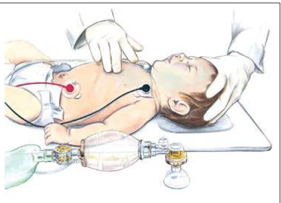
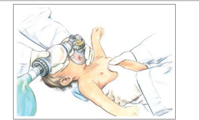
Figure 3. 2-Thumb–encircling hands compressions.
One rescuer holds bag-mask over nose and mouth of infant lying faceup on a table. Sec-ond rescuer has both thumbs centered on infant’s chest, fingers wrapped around infant’s torso.
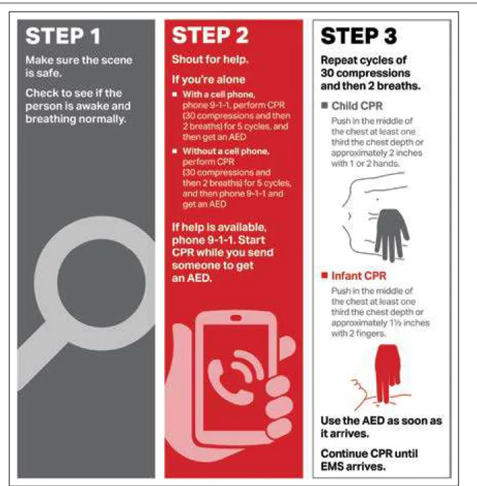
Figure 4. Pediatric BLS for lay rescuers.
AED indicates automated external defibrillator; BLS, basic life support; CPR, cardiopulmonary resuscitation; and EMS, emergency medical services.
3 vertical rectangles show the steps to help an unrespon-sive child or infant: Make sure the scene is safe, shout for help, repeat cycles of 30 compressions and then 2 breaths.
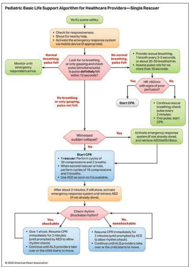
Figure 5. Pediatric Basic Life Support Algorithm for Healthcare Providers—Single Rescuer.
AED indicates automated external defibrillator; ALS, advanced life support; CPR, cardiopulmonary resuscitation; and HR, heart rate.
Text in cascading boxes describes the actions that a single rescuer should perform in sequence during a pediatric cardiac arrest. Arrows guide the rescuer from one box to the next as the rescuer performs the actions. Some boxes have 2 arrows that lead outward, each to a different pathway depending on the outcome of the most recent action taken. Pathways are hyperlinked. Box 1 Verify scene safety. Box 2 • Check for responsiveness. • Shout for nearby help. • Activate the emergency response system via mobile device (if appropriate). Box 3 Look for no breathing or only gasping and check pulse (simultaneously). Is a pulse definitely felt within 10 seconds? If there is normal breathing and a pulse is felt, proceed to Box 3a. If there is no normal breathing but a pulse is felt, proceed to Box 3b. If there is no breathing or there is only gasping and no pulse is felt, proceed to Box 5. Box 3a Monitor until emergency responders arrive. Box 3b • Provide rescue breathing, 1 breath every 2 to 3 seconds, or about 20 to 30 breaths per minute. • Assess pulse rate for no more than 10 seconds. Proceed to Box 4. Box 4 Is heart rate less than 60 per minute with signs of poor perfusion? If Yes, proceed to Box 4a. If No, proceed to Box 4b. Box 4a Start CPR. Box 4b • Continue rescue breathing: check pulse every 2 minutes. • If no pulse, start CPR. Box 5 Was the sudden collapse witnessed? If Yes, proceed to Box 5a. If No, proceed to Box 6. Box 5a Activate the emergency response system (if not already done) and retrieve the AED or defibrillator, then proceed to Box 6. Box 6 Start CPR. • 1 rescuer: Perform cycles of 30 compressions and 2 breaths. • When the second rescuer arrives, perform cycles of 15 compressions and 2 breaths. • Use the AED as soon as it is available. Proceed to Box 7. Box 7 After about 2 minutes, if still alone, activate the emergency response system and retrieve AED (if not already done). Proceed to Box 8. Box 8 Check rhythm. Is it a shockable rhythm? If Yes, it is shockable, proceed to Box 9. If No, it is nonshockable, proceed to Box 10. Box 9 • Give 1 shock. Resume CPR immediately for 2 minutes (until prompted by the AED to allow a rhythm check). • Continue until advanced life support providers take over or the child starts to move. Return to Box 8, if necessary. Box 10 • Resume CPR immediately for 2 minutes (until prompted by the AED to allow a rhythm check). • Continue until advanced life support providers take over or the child starts to move. Return to Box 8, if necessary.
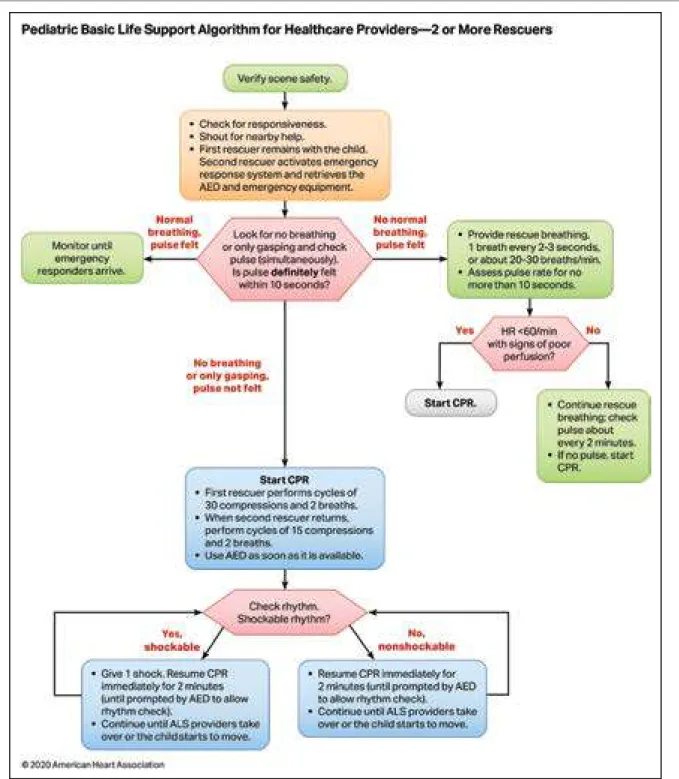
Figure 6. Pediatric Basic Life Support Algorithm for Healthcare Providers—2 or More Rescuers.
AED indicates automated external defibrillator; ALS, advanced life support; CPR, cardiopulmonary resuscitation; and HR, heart rate. Text in cascading boxes describes the actions that 2 or more rescuers should perform in sequence
during a pediatric cardiac arrest. Arrows guide the rescuers from one box to the next as they perform the actions. Some boxes have 2 arrows that lead outward, each to a different pathway depending on the outcome of the most recent action taken. Pathways are hyperlinked. Box 1 Verify scene safety. Box 2
• Check for responsiveness.
• Shout for nearby help.
• First rescuer remains with the child. Second rescuer activates the emergency response system and retrieves the AED and emergency equipment. Box 3 Look for no breathing or only gasping and check pulse (simultaneously). Is pulse definitely felt within 10 seconds? If there is normal breathing and a pulse is felt, proceed to Box 3a. If there is no normal breathing but a pulse is felt, proceed to Box 3b. If there is no breathing or there is only gasping and no pulse is felt, proceed to Box 5. Box 3a Monitor until emergency responders arrive. Box 3b
• Provide rescue breathing, 1 breath every 2 to 3 seconds, or about 20 to 30 breaths per minute. • Assess pulse rate for no more than 10 seconds. Proceed to Box 4. Box 4 Is heart rate less than 60 per minute with signs of poor perfusion? If Yes, proceed to Box 4a. If No, proceed to Box 4b. Box 4a Start CPR. Box 4b
• Continue rescue breathing: check pulse about every 2 minutes. • If no pulse, start CPR.
Box 5 Start CPR
• First rescuer performs cycles of 30 compressions and 2 breaths. • When second rescuer returns, perform cycles of 15 compressions and 2 breaths. • Use the AED as soon as it is available. Proceed to Box 6. Box 6 Check rhythm. Is it a shockable rhythm? If Yes, it is shockable, proceed to Box 7. If No, it is nonshockable, proceed to Box 8. Box 7
• Give 1 shock. Resume CPR immediately for 2 minutes (until prompted by the AED to allow a rhythm check). • Continue until advanced life support providers take over or the child starts to move. Return to Box 6, if necessary. Box 8
• Resume CPR immediately for 2 minutes (until prompted by the AED to allow a rhythm check). • Continue until advanced life support providers take over or the child starts to move. Return to Box 6, if necessary.
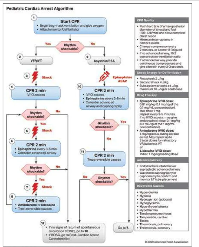
Figure 7. Pediatric Cardiac Arrest Algorithm.
ASAP indicates as soon as possible; CPR, cardiopulmonary resuscitation; ET, endotracheal; HR, heart rate; IO, intraosseous; IV, intravenous; PEA, pulseless electrical activity; and VF/pVT, ventricular fibrillation/pulseless ventricular tachycardia.
Text in cascading boxes describes the actions that a provider should perform in sequence during a pediatric cardiac arrest. Ar-rows guide providers from one box to the next as they perform the actions. Some boxes have 2 arrows that lead outward, each arrow to a different treatment pathway depending on the outcome of the most recent action taken. Pathways are hyperlinked. Box 1 Start CPR • Begin bag-mask ventilation and give oxygen • Attach monitor/defibrillator Is the rhythm shockable? If Yes, it is shockable, proceed to Box 2. If No, it is nonshockable, proceed to Box 9. Box 2 The patient has VF or pVT; proceed to Box 3. Box 3 Deliver shock. Box 4 CPR 2 minutes • IV or IO access Is the rhythm shockable? If Yes, it is shockable, proceed to Box 5. If No, it is nonshockable, proceed to Box 12. Box 5 Deliver shock. Box 6 CPR 2 minutes. • Epinephrine every 3 to 5 minutes • Consider advanced airway Is the rhythm shockable? If Yes, it is shockable, proceed to Box 7. If No, it is nonshockable, proceed to Box 12. Box 7 Deliver shock. Box 8 CPR 2 minutes • Amiodarone or lidocaine • Treat reversible causes Is the rhythm shockable? If Yes, it is shockable, return to Box 5. If No, it is nonshockable, proceed to Box 12. Box 9 The patient has asystole or PEA; give epinephrine ASAP. Box 10 CPR 2 minutes • IV or IO access • Epinephrine every 3 to 5 minutes • Consider advanced airway and capnography Is the rhythm shockable? If Yes, it is shockable, proceed to Box 7. If No, it is nonshockable, proceed to Box 11. Box 11 CPR 2 minutes Treat reversible causes. Is rhythm shockable? If Yes, it is shockable, proceed to Box 7. If No, it is nonshockable, proceed to Box 12. Box 12 • If there are no signs of return of spontaneous circulation, proceed to Box 10 • If return of spontaneous circulation is achieved, go to Post–Cardiac Arrest Care checklist Sidebar for the Pediatric Cardiac Arrest Algorithm CPR Quality • Push hard (at least one-third of the anteroposterior diameter of the chest) and fast (100 to 120 per minute) and allow complete chest recoil • Minimize interruptions in compressions • Change compressor every 2 minutes, or sooner if fatigued • If no advanced airway, 15 to 2 compression to ventilation ratio • If advanced airway, provide continuous compressions and give a breath every 2 to 3 seconds Shock Energy for Defibrillation • First shock: 2 Joules per kilogram • Second shock: 4 Joules per kilogram • Subsequent shocks: at least 4 Joules per kilogram, up to a maximum of 10 Joules per kilogram or adult dose Drug Therapy • Epinephrine IV or IO dose: 0.01 milligrams per kilogram (0.1 mil-liliter per kilogram of the 0.1 milligram per milmil-liliter concentration). Maximum dose: 1 milligram. Repeat every 3 to 5 minutes. If no IV or IO access, may give endotracheal dose of 0.1 milligrams per kilogram (0.1 milliliter per kilogram of the 1 milligram per milliliter concentration) • Amiodarone IV or IO dose: 5 milligrams per kilogram bolus during cardiac arrest. May repeat up to 3 total doses for refractory VF or pulseless VT or Lidocaine IV or IO dose: Initial: 1 milligram per kilogram loading dose Advanced Airway • Endotracheal intubation or supraglottic advanced airway • Waveform capnography or capnometry to confirm and monitor ET tube placement Reversible Causes • Hypovolemia • Hypoxia • Hydrogen ion (acidosis) • Hypoglycemia • Hypokalemia or hyperkalemia • Hypothermia • Tension pneumothorax • Tamponade, cardiac • Toxins • Thrombosis, pulmonary • Thrombosis, coronary
Recommendation-Specific Supportive Text
1. “CPR mode” is available on some hospital beds
to stiffen the mattress during CPR. Manikin
mod-els indicate that mattress compression ranges
between 12% and 57% of total compression
depth, with softer mattresses being compressed
the most.
50–53This can lead to reduced sternal
displacement and a reduction in effective chest
compression depth.
2. Manikin studies and 1 pediatric case series show
that effective compression depth can be achieved
even on a soft surface, providing the CPR provider
increases overall compression depth to
compen-sate for mattress compression.
53–593. Meta-analysis of 6 studies
53,56,60–63showed a 3-mm
(95% CI 1–4 mm) improvement in chest
compres-sion depth associated with backboard use when
CPR was performed on a manikin placed on a
mat-tress or bed.
Opening the Airway
Recommendations for Opening the Airway COR LOE Recommendations
1 C-LD 1. Unless a cervical spine injury is suspected, use a head tilt–chin lift maneuver to open the airway.64
1 C-EO
2. For the trauma patient with suspected cervical spinal injury, use a jaw thrust without head tilt to open the airway.
1 C-EO
3. For the trauma patient with suspected cervical spinal injury, if the jaw thrust does not open the airway, use a head tilt–chin lift maneuver.
Recommendation-Specific Supportive Text
1. No data directly address the ideal method to
open or maintain airway patency. One
retro-spective cohort study evaluated various head-tilt
angles in neonates and young infants
undergo-ing diagnostic MRI and found that the highest
proportion of patent airways was at a head-tilt
angle of 144 to 150 degrees based on a
regres-sion analysis.
642. While no pediatric studies evaluate jaw thrust
versus head tilt–chin lift to open the airway,
the jaw thrust is widely accepted as an
effec-tive way to open the airway, and this maneuver
theoretically limits cervical motion compared
with the head tilt–chin lift.
3. There are no pediatric studies evaluating the
impact of a head tilt–chin lift maneuver to open
the airway in a trauma patient with suspected
cervical spine injury. However, if providers are
unable to open the airway and deliver effective
ventilations using a jaw thrust, given the
impor-tance of a patent airway, using a head tilt–chin
lift maneuver is recommended.
Figures 4, 5, 6, and 7 show, respectively, an
info-graphic for pediatric BLS for lay rescuers, the current
pediatric BLS algorithms for healthcare
provider, sin-gle-rescuer CPR and 2-rescuer CPR, and the current
algorithm for pediatric cardiac arrest.
REFERENCES
1. Niles DE, Duval-Arnould J, Skellett S, Knight L, Su F, Raymond TT, Sweberg T, Sen AI, Atkins DL, Friess SH, de Caen AR, Kurosawa H, Sutton RM, Wolfe H, Berg RA, Silver A, Hunt EA, Nadkarni VM; pediatric Resuscitation Quality (pediRES-Q) Collaborative Investigators. Characterization of Pediatric In-Hospital Cardiopulmonary Resuscitation Quality Metrics Across an Inter-national Resuscitation Collaborative. Pediatr Crit Care Med. 2018;19:421– 432. doi: 10.1097/PCC.0000000000001520
2. Sutton RM, Niles D, Nysaether J, Abella BS, Arbogast KB, Nishisaki A, Maltese MR, Donoghue A, Bishnoi R, Helfaer MA, Myklebust H, Nadkarni V. Quantitative analysis of CPR quality during in-hospital resuscitation of old-er children and adolescents. Pediatrics. 2009;124:494–499. doi: 10.1542/ peds.2008-1930
3. Niles D, Nysaether J, Sutton R, Nishisaki A, Abella BS, Arbogast K, Maltese MR, Berg RA, Helfaer M, Nadkarni V. Leaning is common during in-hospital pediatric CPR, and decreased with automated corrective feedback.
Resus-citation. 2009;80:553–557. doi: 10.1016/j.resusResus-citation.2009.02.012
4. McInnes AD, Sutton RM, Orioles A, Nishisaki A, Niles D, Abella BS, Maltese MR, Berg RA, Nadkarni V. The first quantitative re-port of ventilation rate during in-hospital resuscitation of older children and adolescents. Resuscitation. 2011;82:1025–1029. doi: 10.1016/j. resuscitation.2011.03.020
5. Sutton RM, Reeder RW, Landis WP, Meert KL, Yates AR, Morgan RW, Berger JT, Newth CJ, Carcillo JA, McQuillen PS, Harrison RE, Moler FW, Pollack MM, Carpenter TC, Notterman DA, Holubkov R, Dean JM, Nadkarni VM, Berg RA; Eunice Kennedy Shriver National Institute of Child Health and Human Development Collaborative Pediatric Critical Care Research Network (CPCCRN). Ventilation Rates and Pediatric In-Hospital Cardiac Arrest Survival Outcomes. Crit Care Med. 2019;47:1627–1636. doi: 10.1097/CCM.0000000000003898
6. Bahr J, Klingler H, Panzer W, Rode H, Kettler D. Skills of lay people in checking the carotid pulse. Resuscitation. 1997;35:23–26. doi: 10.1016/s0300-9572(96)01092-1
7. Brearley S, Shearman CP, Simms MH. Peripheral pulse palpation: an unreli-able physical sign. Ann R Coll Surg Engl. 1992;74:169–171.
8. Cavallaro DL, Melker RJ. Comparison of two techniques for detect-ing cardiac activity in infants. Crit Care Med. 1983;11:189–190. doi: 10.1097/00003246-198303000-00009
9. Inagawa G, Morimura N, Miwa T, Okuda K, Hirata M, Hiroki K. A com-parison of five techniques for detecting cardiac activity in infants. Paediatr
Anaesth. 2003;13:141–146. doi: 10.1046/j.1460-9592.2003.00970.x
10. Kamlin CO, O’Donnell CP, Everest NJ, Davis PG, Morley CJ. Accuracy of clinical assessment of infant heart rate in the delivery room. Resuscitation. 2006;71:319–321. doi: 10.1016/j.resuscitation.2006.04.015
11. Lee CJ, Bullock LJ. Determining the pulse for infant CPR: time for a change? Mil Med. 1991;156:190–193.
12. Mather C, O’Kelly S. The palpation of pulses. Anaesthesia. 1996;51:189– 191. doi: 10.1111/j.1365-2044.1996.tb07713.x
13. Ochoa FJ, Ramalle-Gómara E, Carpintero JM, García A, Saralegui I. Com-petence of health professionals to check the carotid pulse. Resuscitation. 1998;37:173–175. doi: 10.1016/s0300-9572(98)00055-0
14. Owen CJ, Wyllie JP. Determination of heart rate in the baby at birth.
Re-suscitation. 2004;60:213–217. doi: 10.1016/j.reRe-suscitation.2003.10.002
15. Sarti A, Savron F, Casotto V, Cuttini M. Heartbeat assessment in infants: a comparison of four clinical methods. Pediatr Crit Care Med. 2005;6:212– 215. doi: 10.1097/01.PCC.0000154952.59176.E0
16. Sarti A, Savron F, Ronfani L, Pelizzo G, Barbi E. Comparison of three sites to check the pulse and count heart rate in hypotensive infants. Paediatr
Anaesth. 2006;16:394–398. doi: 10.1111/j.1460-9592.2005.01803.x
17. Tanner M, Nagy S, Peat JK. Detection of infant’s heart beat/pulse by care-givers: a comparison of 4 methods. J Pediatr. 2000;137:429–430. doi: 10.1067/mpd.2000.107188
18. Whitelaw CC, Goldsmith LJ. Comparison of two techniques for determin-ing the presence of a pulse in an infant. Acad Emerg Med. 1997;4:153– 154. doi: 10.1111/j.1553-2712.1997.tb03725.x
19. Dick WF, Eberle B, Wisser G, Schneider T. The carotid pulse check revisited: what if there is no pulse? Crit Care Med. 2000;28(suppl):N183–N185. doi: 10.1097/00003246-200011001-00002
20. Eberle B, Dick WF, Schneider T, Wisser G, Doetsch S, Tzanova I. Check-ing the carotid pulse check: diagnostic accuracy of first responders in pa-tients with and without a pulse. Resuscitation. 1996;33:107–116. doi: 10.1016/s0300-9572(96)01016-7
21. Tibballs J, Russell P. Reliability of pulse palpation by healthcare personnel to diagnose paediatric cardiac arrest. Resuscitation. 2009;80:61–64. doi: 10.1016/j.resuscitation.2008.10.002
22. Tibballs J, Weeranatna C. The influence of time on the accuracy of health-care personnel to diagnose paediatric cardiac arrest by pulse palpation.
Re-suscitation. 2010;81:671–675. doi: 10.1016/j.reRe-suscitation.2010.01.030
23. O’Connell KJ, Keane RR, Cochrane NH, Sandler AB, Donoghue AJ, Kerrey BT, Myers SR, Vazifedan T, Mullan PC. Pauses in compressions dur-ing pediatric CPR: Opportunities for improvdur-ing CPR quality. Resuscitation. 2019;145:158–165. doi: 10.1016/j.resuscitation.2019.08.015
24. Lubrano R, Cecchetti C, Bellelli E, Gentile I, Loayza Levano H, Orsini F, Bertazzoni G, Messi G, Rugolotto S, Pirozzi N, Elli M. Comparison of times of intervention during pediatric CPR maneuvers using ABC and CAB se-quences: a randomized trial. Resuscitation. 2012;83:1473–1477. doi: 10.1016/j.resuscitation.2012.04.011
25. Kitamura T, Iwami T, Kawamura T, Nagao K, Tanaka H, Nadkarni VM, Berg RA, Hiraide A; implementation working group for All-Japan Utstein Registry of the Fire and Disaster Management Agency. Conventional and chest-compression-only cardiopulmonary resuscitation by bystanders for children who have out-of-hospital cardiac arrests: a prospective, nationwide, population-based cohort study. Lancet. 2010;375:1347–1354. doi: 10.1016/S0140-6736(10)60064-5 26. Goto Y, Maeda T, Goto Y. Impact of dispatcher-assisted bystander
cardiopul-monary resuscitation on neurological outcomes in children with out-of-hospital cardiac arrests: a prospective, nationwide, population-based cohort study. J Am Heart Assoc. 2014;3:e000499. doi: 10.1161/JAHA.113.000499 27. Naim MY, Burke RV, McNally BF, Song L, Griffis HM, Berg RA, Vellano K,
Markenson D, Bradley RN, Rossano JW. Association of Bystander Cardiopul-monary Resuscitation With Overall and Neurologically Favorable Survival After Pediatric Out-of-Hospital Cardiac Arrest in the United States: A Report From the Cardiac Arrest Registry to Enhance Survival Surveillance Registry. JAMA
Pediatr. 2017;171:133–141. doi: 10.1001/jamapediatrics.2016.3643
28. Fukuda T, Ohashi-Fukuda N, Kobayashi H, Gunshin M, Sera T, Kondo Y, Yahagi N. Conventional Versus Compression-Only Ver-sus No-Bystander Cardiopulmonary ReVer-suscitation for Pediatric Out-of-Hospital Cardiac Arrest. Circulation. 2016;134:2060–2070. doi: 10.1161/CIRCULATIONAHA.116.023831
29. Ashoor HM, Lillie E, Zarin W, Pham B, Khan PA, Nincic V, Yazdi F, Ghassemi M, Ivory J, Cardoso R, Perkins GD, de Caen AR, Tricco AC; ILCOR Basic Life Support Task Force. Effectiveness of different compression-to-ventilation methods for cardiopulmonary resuscitation: A systematic review.
Resusci-tation. 2017;118:112–125. doi: 10.1016/j.resusciResusci-tation.2017.05.032
30. Glatz AC, Nishisaki A, Niles DE, Hanna BD, Eilevstjonn J, Diaz LK, Gillespie MJ, Rome JJ, Sutton RM, Berg RA, Nadkarni VM. Sternal wall pressure compa-rable to leaning during CPR impacts intrathoracic pressure and haemody-namics in anaesthetized children during cardiac catheterization.
Resuscita-tion. 2013;84:1674–1679. doi: 10.1016/j.resuscitaResuscita-tion.2013.07.010
31. Sutton RM, French B, Nishisaki A, Niles DE, Maltese MR, Boyle L, Stavland M, Eilevstjønn J, Arbogast KB, Berg RA, et al. American Heart As-sociation cardiopulmonary resuscitation quality targets are associated with improved arterial blood pressure during pediatric cardiac arrest.
Resuscita-tion. 2013;84:168–172. doi: 10.1016/j.resuscitaResuscita-tion.2012.08.335
32. Sutton RM, Reeder RW, Landis W, Meert KL, Yates AR, Berger JT, Newth CJ, Carcillo JA, McQuillen PS, Harrison RE, Moler FW, Pollack MM, Carpenter TC, Notterman DA, Holubkov R, Dean JM, Nadkarni VM, Berg RA; Eunice Kennedy Shriver National Institute of Child Health and Human Development Collabora-tive Pediatric Critical Care Research Network (CPCCRN) Investigators. Chest compression rates and pediatric in-hospital cardiac arrest survival outcomes.
Resuscitation. 2018;130:159–166. doi: 10.1016/j.resuscitation.2018.07.015
33. Kao PC, Chiang WC, Yang CW, Chen SJ, Liu YP, Lee CC, Hsidh MJ, Ko PC, Chen SC, Ma MH. What is the correct depth of chest compression for infants and children? A radiological study. Pediatrics. 2009;124:49–55. doi: 10.1542/peds.2008-2536
34. Sutton RM, Niles D, Nysaether J, Arbogast KB, Nishisaki A, Maltese MR, Bishnoi R, Helfaer MA, Nadkarni V, Donoghue A. Pediat-ric CPR quality monitoring: analysis of thoracic anthropometPediat-ric data.
Resuscitation. 2009;80:1137–1141. doi: 10.1016/j.resuscitation.
2009.06.031
35. Braga MS, Dominguez TE, Pollock AN, Niles D, Meyer A, Myklebust H, Nysaether J, Nadkarni V. Estimation of optimal CPR chest compression depth in children by using computer tomography. Pediatrics. 2009;124:e69– e74. doi: 10.1542/peds.2009-0153
36. Sutton RM, French B, Niles DE, Donoghue A, Topjian AA, Nishisaki A, Leffelman J, Wolfe H, Berg RA, Nadkarni VM, et al. 2010 American Heart Association recommended compression depths during pediatric in-hospital resuscitations are associated with survival. Resuscitation. 2014;85:1179– 1184. doi: 10.1016/j.resuscitation.2014.05.007
37. Atkins DL, de Caen AR, Berger S, Samson RA, Schexnayder SM, Joyner BL Jr, Bigham BL, Niles DE, Duff JP, Hunt EA, Meaney PA. 2017 American Heart Association Focused Update on Pediatric Basic Life Support and Cardiopulmonary Resuscitation Quality: An Update to the American Heart Association Guidelines for Cardiopulmonary Resuscita-tion and Emergency Cardiovascular Care. CirculaResuscita-tion. 2018;137:e1–e6. doi: 10.1161/CIR.0000000000000540
38. Clements F, McGowan J. Finger position for chest compressions in cardiac arrest in infants. Resuscitation. 2000;44:43–46. doi: 10.1016/s0300-9572(99)00165-3
39. Finholt DA, Kettrick RG, Wagner HR, Swedlow DB. The heart is under the lower third of the sternum. Implications for exter-nal cardiac massage. Am J Dis Child. 1986;140:646–649. doi: 10.1001/archpedi.1986.02140210044022
40. Phillips GW, Zideman DA. Relation of infant heart to sternum: its signifi-cance in cardiopulmonary resuscitation. Lancet. 1986;1:1024–1025. doi: 10.1016/s0140-6736(86)91284-5
41. Orlowski JP. Optimum position for external cardiac compression in in-fants and young children. Ann Emerg Med. 1986;15:667–673. doi: 10.1016/s0196-0644(86)80423-1
42. Douvanas A, Koulouglioti C, Kalafati M. A comparison between the two methods of chest compression in infant and neonatal resuscitation: a re-view according to 2010 CPR guidelines. J Matern Fetal Neonatal Med. 2018;31:805–816. doi: 10.1080/14767058.2017.1295953
43. Lee JE, Lee J, Oh J, Park CH, Kang H, Lim TH, Yoo KH. Comparison of two-thumb encircling and two-finger technique during infant cardiopulmo-nary resuscitation with single rescuer in simulation studies: a systematic review and meta-analysis. Medicine (Baltimore). 2019;98:e17853. doi: 10.1097/MD.0000000000017853
44. Lee SY, Hong JY, Oh JH, Son SH. The superiority of the two-thumb over the two-finger technique for single-rescuer infant cardiopul-monary resuscitation. Eur J Emerg Med. 2018;25:372–376. doi: 10.1097/MEJ.0000000000000461
45. Tsou JY, Kao CL, Chang CJ, Tu YF, Su FC, Chi CH. Biomechanics of two-thumb versus two-finger chest compression for cardiopulmonary resusci-tation in an infant manikin model. Eur J Emerg Med. 2020;27:132–136. doi: 10.1097/MEJ.0000000000000631
46. Pellegrino JL, Bogumil D, Epstein JL, Burke RV. Two-thumb-encircling advantageous for lay responder infant CPR: a ran-domised manikin study. Arch Dis Child. 2019;104:530–534. doi: 10.1136/archdischild-2018-314893
47. Kim MJ, Lee HS, Kim S, Park YS. Optimal chest compression technique for paediatric cardiac arrest victims. Scand J Trauma Resusc Emerg Med. 2015;23:36. doi: 10.1186/s13049-015-0118-y
48. Stevenson AG, McGowan J, Evans AL, Graham CA. CPR for chil-dren: one hand or two? Resuscitation. 2005;64:205–208. doi: 10.1016/j.resuscitation.2004.07.012
49. Peska E, Kelly AM, Kerr D, Green D. One-handed versus two-handed chest compressions in paediatric cardio-pulmonary resuscitation. Resuscitation. 2006;71:65–69. doi: 10.1016/j.resuscitation.2006.02.007
50. Lin Y, Wan B, Belanger C, Hecker K, Gilfoyle E, Davidson J, Cheng A. Reducing the impact of intensive care unit mattress compressibility dur-ing CPR: a simulation-based study. Adv Simul (Lond). 2017;2:22. doi: 10.1186/s41077-017-0057-y
51. Noordergraaf GJ, Paulussen IW, Venema A, van Berkom PF, Woerlee PH, Scheffer GJ, Noordergraaf A. The impact of compliant surfaces on in-hospital chest compressions: effects of common mattresses and a backboard. Resuscitation. 2009;80:546–552. doi: 10.1016/j. resuscitation.2009.03.023
52. Oh J, Chee Y, Song Y, Lim T, Kang H, Cho Y. A novel method to de-crease mattress compression during CPR using a mattress compres-sion cover and a vacuum pump. Resuscitation. 2013;84:987–991. doi: 10.1016/j.resuscitation.2012.12.027