The proprotein convertase PCSK9 ( 1 ) is the third gene
involved in autosomal dominant familial
hypercholester-olemia ( 2 ). Gain-of-function PCSK9 mutations result in
increased levels of plasma low density lipoprotein (LDL)
cholesterol ( 2–4 ). In contrast, gene disruption ( 5, 6 ) and
loss-of-function mutations in PCSK9 (
3, 7
) prevent the
degradation of the LDL receptor (LDLR), resulting in a
higher clearance of plasma LDL-cholesterol. These
semi-nal fi ndings led to the development of therapies based on
PCSK9 inhibition/silencing for the treatment of
hyper-cholesterolemia ( 8, 9 ). Although liver LDLR protein levels
are reduced in mice injected with PCSK9 ( 10, 11 ) or
over-expressing PCSK9 in hepatocytes ( 6 ), high quantities of
PCSK9 can also downregulate LDLR protein levels in
ex-trahepatic tissues such as the lung, adipose, and kidney
( 12, 13 ), suggesting that endogenous circulating PCSK9
that originates from hepatocytes ( 6, 13 ) may
downregu-late LDLR protein in others tissues. At adulthood, PCSK9
is highly expressed in liver and is also abundant in the
small intestine, as well as in the kidney and brain
through-out embryonic development ( 1 ). In mouse brain, PCSK9 is
transiently expressed in the telencephalon [maximal at
embryonic day (E)12.5] and cerebellum [from E17.5 to
postnatal day (P)19] ( 1 ). At adulthood, it is only
signifi-cantly expressed in the rostral extension of the
olfac-tory peduncle (RE-OP) ( 1 ). In addition, transgenic mice
Abstract Proprotein convertase subtilisin/kexin type 9
(PCSK9) plays a major role in cholesterol homeostasis
through enhanced degradation of the LDL receptor (LDLR)
in liver. As novel inhibitors/silencers of PCSK9 are now
be-ing tested in clinical trials to treat hypercholesterolemia, it is
crucial to defi ne the physiological consequences of the lack
of PCSK9 in various organs. LDLR regulation by PCSK9 has
not been extensively described during mouse brain
develop-ment and injury. Herein, we show that PCSK9 and LDLR are
co-expressed in mouse brain during development and at
adulthood. Although the protein levels of LDLR and
apoli-poprotein E (apoE) in the adult brain of Pcsk9
ⴚ / ⴚmice are
similar to those of wild-type (WT) mice, LDLR levels
in-creased and were accompanied by a reduction of apoE
lev-els during development. This suggests that the upregulation
of LDLR protein levels in Pcsk9
ⴚ / ⴚmice enhances apoE
deg-radation. Upon ischemic stroke, PCSK9 was expressed in
the dentate gyrus between 24 h and 72 h following brain
reperfusion. Although mouse behavior and lesion volume
were similar, LDLR protein levels dropped
ⵑ 2-fold less in
the Pcsk9
ⴚ / ⴚ-lesioned hippocampus, without affecting apoE
levels and neurogenesis.
Thus, PCSK9 downregulates
LDLR levels during brain development and following
tran-sient ischemic stroke in adult mice.
—Rousselet, E., J.
Marcinkiewicz, J. Kriz, A. Zhou, M. E. Hatten, A. Prat, and
N. G. Seidah. PCSK9 reduces the protein levels of the LDL
receptor in mouse brain during development and after
is-chemic stroke. J. Lipid Res. 2011.
52: 1383–1391.
Supplementary key words low density lipoprotein • apolipoprotein E • hypercholesterolemia • brain development • neurogenesis
This work was supported by Canadian Institutes of Health Research (CIHR) Team Grants CTP 82946 and MOP 102741, CIHR Canada Chair 216684, the Strauss Foundation, and a fellowship from the Canadian Heart and Stroke Foundation (E.R.).
Manuscript received 18 January 2011 and in revised form 13 April 2011. Published, JLR Papers in Press, April 25, 2011
DOI 10.1194/jlr.M014118
PCSK9 reduces the protein levels of the LDL
receptor in mouse brain during development and after
ischemic stroke
Estelle Rousselet , * Jadwiga Marcinkiewicz , * Jasna Kriz ,
†Ann Zhou ,
§Mary E. Hatten , **
Annik Prat , * and Nabil G. Seidah
1,*
Biochemical Neuroendocrinology,* Clinical Research Institute of Montréal (IRCM) , Montréal, Québec, Canada ; Department of Psychiatry and Neuroscience, † Laval University , Québec City, Québec, Canada ; Robert S. Dow Neurobiology Laboratories , § Portland, OR ; and Developmental Neurobiology,** Rockefeller University , New York, NY
Abbreviations: BBB, blood-brain barrier; BrdU, bromodeoxyuri-dine; CNS, central nervous system; CSF, cerebrospinal fl uid; E, embry-onic day; EGL, external granular layer; FCx, frontal cortex; IGL, internal granular layer; LDLR, LDL receptor; tMCAO, transient middle cerebral artery occlusion; ML, molecular layer; Ob, olfactory bulb; P, postnatal day; PCSK9, proprotein convertase subtilsin kexin 9; PFA, paraformaldehyde; RE-OP, rostral extension of the olfactory peduncle; WT, wild type.
1
To whom correspondence should be addressed. e-mail: seidahn@ircm.qc.ca
The online version of this article (available at http://www.jlr.org) contains supplementary data in the form of seven fi gures.
by guest, on November 9, 2019
www.jlr.org
Downloaded from
.html
immunostainings, P7 mice were transcardially perfused with 0.9% NaCl and then with 4% paraformaldehyde (PFA). Brains were frozen in isopentane at ⫺ 30°C and cut into coronal sections (12 m thick).
Mouse ischemic surgery
Transient middle cerebral artery occlusion (tMCAO) was per-formed on two- to three-month-old WT and Pcsk9 ⫺ / ⫺ mice (weight
20-25 g). Under temporary 2% isofl urane anesthesia, unilateral transient focal cerebral ischemia was induced by tMCAO for 1 h, followed by different reperfusion periods (6, 24, 72 h and 1 week) ( 19, 20 ). Body temperature was maintained at 37°C using a heat-ing pad and an infrared heatheat-ing lamp. Under an operatheat-ing mi-croscope, a 12 mm-long 6-0 silicon-coated monofi lament suture (Doccol Corporation) was inserted through the left proximal ex-ternal carotid artery into the inex-ternal carotid artery, and then into the circle of Willis, thus occluding the middle cerebral ar-tery. After 1 h of tMCAO, the fi lament was withdrawn, blood fl ow restored to normal, and wounds sutured. The same procedure was performed on sham-operated mice, where the monofi lament was not inserted. All animals were allowed ad libitum access to water and food before and after surgery. They were fasted for 3 h before euthanization to standardize LDLR levels in all mice.
For in situ hybridization, brains were removed after 6, 24, 72 h and 1 week of brain reperfusion. To quantify neurogenesis in the dentate gyrus, bromodeoxyuridine (50 mg/kg) (BrdU; Sigma-Aldrich) was injected intraperitoneally 24 h postsurgery, twice a day (with an 8 h interval) for two days. To quantify the lesion size and BrdU-positive cells, mouse brains were removed after 72 h reperfusion, perfused with 50 ml of PBS, frozen in isopentane at ⫺ 30°C, and cut into coronal cryosections (17 m thick).
Neurological scores
An expanded six-point scale was used as described previously ( 21 ). Behavioral assessments were made at 1, 24, 48, and 72 h after reperfusion. Neurological defi cits were scored as follows: 0 = nor-mal; 1 = mild turning behavior with or without inconsistent curling when picked up by the tail, <50% attempts to curl to the contralat-eral side; 2 = mild consistent curling, >50% attempts to curl to the contralateral side; 3 = strong and immediate consistent curling, mouse holds a curled position for more than 1-2 s, with its nose almost reaching tail; 4 = severe curling progressing into barreling, loss of walking or righting refl ex; 5 = comatose or moribund state. At least 8 mice per group were evaluated for each experiment, and scores were averaged for statistical analysis.
In situ hybridization
In situ hybridization on whole-mount embryos, brains, or liv-ers was performed as described previously ( 1 ). Briefl y, cryosec-tions were fi xed in 4% PFA for 1 h, and in situ hybridization was performed using mouse PCSK9 ( 1 ), LDLR ( 15 ), Furin and PC5/6 ( 22 ), mouse SKI-1/S1P ( 23 ), and mouse clusterin cRNA probes labeled with 35 S-UTP. To generate a mouse clusterin cRNA probe, clusterin cDNA was PCR amplifi ed from an Origene clone (MC203543) using sense 5 ′ - ACCGCCATGAAGATTCTCCTGCT-GTGCG-3 ′ and antisense 5 ′ - CAACCGCGGCTTCCGCACG-GCTTTTCC-3 ′ oligonucleotides. Hybridization was analyzed on X-ray fi lm.
Nissl staining
Brain infarct was assessed by Nissl staining. The relative size of the infarct was measured using image software from the Scion Corporation (Frederick, MD), calculated in arbitrary units (pix-els), and expressed as a percentage of the contralateral nonle-sioned area (100%) for each section.
expressing enhanced green fl uorescent protein (EGFP)
under the control of the Pcsk9 promoter revealed the
pres-ence of EGFP in nerve fi bers within the olfactory bulb,
which is innervated by the RE-OP (National Institutes of
Health GENSAT Project) ( 14 ).
Although the role of PCSK9 in the brain during mouse
development ( 1 ) has not been extensively investigated, its
overexpression in primary neuronal cultures obtained at
E12.5 has been shown to enhance the recruitment of
un-differentiated progenitor cells into the neuronal lineage
( 1 ). Contrary to PCSK9 knockdown in zebrafi sh, which
re-sults in early death and an extensive disorganization of the
central nervous system (CNS) ( 15 ), PCSK9 knockout mice
are viable ( 5, 6 ). Furthermore, we did not observe any
gross alterations in adult Pcsk9
⫺ / ⫺mice in the cerebellum,
hippocampus, or cortex ( 16 ). In pathological situations,
such as induction of neural apoptosis by serum withdrawal,
PCSK9 is upregulated ( 17 ), and overexpression of PCSK9
in cultured cerebellar granular neurons induces cell death
( 18 ), suggesting that PCSK9 may be involved in neural
apoptotic processes.
In the present study, we show that PCSK9 and LDLR
mRNAs are co-expressed in the same cell layer within the
telencephalon at E12.5, the cerebellum at P7, and at
adult-hood in the RE-OP. As in liver, PCSK9 also enhances LDLR
protein degradation during brain development. In
con-trast, at adulthood within the RE-OP and olfactory bulb,
LDLR protein levels are not affected by PCSK9. To
investi-gate the role of PCSK9 following brain injury, we induced a
transient ischemic stroke in adult mice ( 19, 20 ) and
ana-lyzed the expression of PCSK9 in the dentate gyrus from
6 h to 1 week following injury. The data showed that the
upregulated PCSK9 reduced LDLR protein levels in the
lesioned dentate gyrus without signifi cantly affecting de novo
neurogenesis. We also showed that protein levels of apoE
were decreased in Pcsk9
⫺ / ⫺mice during development but
not at adulthood or following transient ischemic stroke.
METHODS
Animals
Wild-type (WT) C57BL/6J mice, Ldlr ⫺ / ⫺ C57BL/6J mice
(#002207) and apoE ⫺ / ⫺ C57BL/6J mice (#002052) were obtained
from The Jackson Laboratory and bred in house. Pcsk9 ⫺ / ⫺ and
transgenic mice overexpressing V5-tagged PCSK9 in the liver were described previously ( 6 ) and were backcrossed for 10 gen-erations to the C57BL/6J genetic background. The mice were housed in the Clinical Research Institute of Montreal (IRCM) animal facility on a 12 h light/dark cycle. All mouse experimenta-tions were approved by the IRCM bioethics committee for animal care.
Tissue collection
E12.5, P7, and adult (three-month-old) mice were euthanized with 2% isofl urane. For Western blot analyses, mouse brains at E12.5, cerebella at P7, RE-OP, and adult olfactory bulbs were dis-sected and frozen in isopentane at ⫺ 30°C. For Nissl staining and LDLR immunofl uorescence, E12.5 embryonic, P7, and adult brains were frozen at ⫺ 30°C in isopentane and cut into sagital cryosections (8, 12, and 17 m thick, respectively). For other
by guest, on November 9, 2019
www.jlr.org
Downloaded from
( 1 ). To defi ne the role of PCSK9 in brain, we fi rst focused
on its expression during embryonic development. In situ
hybridization revealed that, at E12.5, PCSK9 was expressed
only in the liver, small intestine, and telencephalon ( Fig.
1A
), suggesting an early developmental role of PCSK9 in
these tissues. In telencephalon, PCSK9 was expressed in
the frontal cortex (FCx) ( Fig. 1A ). The expression of the
LDLR was also assessed in adjacent sections and was found
to colocalize with all PCSK9 expression sites (arrows in Fig.
1A ). Furthermore, immunofl uorescence analysis, under
nonpermeabilizing conditions that refl ect cell surface
ex-pression, showed that LDLR protein levels were higher in
Pcsk9
⫺ / ⫺telencephalon than in WT ones ( Fig. 1B ). LDLR
quantitation by Western blot in 10 WT and 13 Pcsk9
⫺ / ⫺extracts confi rmed this observation, and revealed a
ⵑ
2.7-fold increase in Pcsk9
⫺ / ⫺compared with WT
telencepha-lons ( Fig. 1C ). Nissl staining revealed that the telencephalon
organization was not grossly altered in Pcsk9
⫺ / ⫺mice ( Fig.
1D ), suggesting that at E12.5 the regulation of LDLR by
PCSK9 was not critical for tissue integrity.
PCSK9 enhances the degradation of the LDLR in mouse
cerebellum at P7
We previously showed that PCSK9 was expressed in the
cerebellum at P7 ( 1 ). As assessed by in situ hybridization,
PCSK9 mRNA was expressed in the external granular layer
(EGL), where it colocalized with the LDLR mRNA ( Fig.
2A
). As in E12.5 telencephalon, cell surface LDLR
label-ing was higher in Pcsk9
⫺ / ⫺external granular cells versus
WT ones ( Fig. 2B ). Western blot analysis of fi ve cerebella
for each genotype showed that the
ⵑ 2.5-fold increase was
signifi cant ( Fig. 2C ). Altogether, these data demonstrate
that, at P7, cerebellum PCSK9 locally downregulates the
protein levels of the LDLR.
We next determined whether the lack of PCSK9 can
al-ter the development of the cerebellum. Nissl staining
re-vealed a normal cerebellum organization in Pcsk9
⫺ / ⫺mice,
with no gross modifi cation of the EGL, molecular layer
(ML), or internal granular cell layer (IGL) ( Fig. 2D ). This
was confi rmed by the staining of Purkinje cells with
calbin-din (supplemental Fig. IA), and of granular cells with
anti-Pax-6 and anti-TAG-1, which label the granular cell bodies
( 24 ) and axons ( 25 ), respectively (supplemental Fig. IB).
Although PCSK9 induces ex vivo the recruitment of
undif-ferentiated neural progenitor cells into the neuronal
lin-eage ( 1 ), we demonstrated that in vivo the protein levels
of markers of cell proliferation (PCNA, Cyclin E), cell
dif-ferentiation (Calbindin), and synapse (synaptophysin,
GAP43) remain unchanged in Pcsk9
⫺ / ⫺mice
(supplemen-tal Fig. II).
At adulthood, PCSK9 does not induce LDLR degradation
in the RE-OP or olfactory bulb
In adult brain, PCSK9 mRNA was only expressed in the
RE-OP ( 1 ), where LDLR was also colocalized ( Fig. 3A ),
suggesting that herein PCSK9 could also regulate LDLR
protein levels. Because neurons of the RE-OP innervate
the olfactory bulb (Ob), we also measured LDLR protein
levels in this structure. However, Western blot analyses
Immunohistochemistry
For LDLR visualization, brain cryosections were fi xed for 1 h in 4% PFA, incubated with monoclonal mouse LDLR antibody (1:150; R and D Systems), and revealed with Alexa Fluor 488- labeled anti-goat IgGs (1:200). Nuclei were stained with Hoescht 33258 for 5 min (100 g/ml; Sigma-Aldrich). For staining of cell mark-ers, fi xed P7 brain cryosections were incubated with mouse monoclonal anti-calbindin (1:300, Sigma-Aldrich), mouse mono-clonal-TAG-1 (1:2, 4D7, kindly provided by Drs. J. Dodd and T. Jessell), and rabbit polyclonal anti-Pax-6 (1:300; Covance). Anti-gen antibody complexes were revealed by 1 h incubation with cor-responding species-specifi c Alexa Fluor (488 or 555)-tagged antibodies (1:300; Molecular Probes). Nuclei were stained with Hoescht 33258 for 5 min (100 g/ml; Sigma-Aldrich).
BrdU incorporation into DNA was visualized using a specifi c antibody (Roche Diagnostics). Brain cryosections were dried and then fi xed 5 min in methanol at ⫺ 20°C, then incubated for 45 min in 2N HCl at 37°C and neutralized in borate buffer for 10 min. Brain slices were incubated overnight with a mouse BrdU antibody (1:100), detected with a biotin-labeled secondary anti-body (PerkinElmer), and revealed using the Vectastain kit (Vec-tor Labora(Vec-tories) and DAB substrate (Zymed Labora(Vec-tories). BrdU-positive cells in the dentate gyrus were counted on four brain slices regularly spaced for at least fi ve mice for each group. BrdU counts following ischemic stroke were expressed as per-centages of counts in the nonlesioned side (fi xed at 100%).
Protein analyses
Tissue were homogenized in ice-cold precipitation assay buffer (50 mM Tris-HCl, pH 7.8, 150 mM NaCl, 1% Nonidet P-40, 0.5% sodium deoxycholate, 0.1% SDS) containing a mixture of prote-ase inhibitors (Roche Applied Science). Proteins were separated by 8% SDS-PAGE and visualized using mouse anti-LDLR (1:1,000; R and D Systems), rabbit anti-apoE ⫺ / ⫺ (1:1,000; Biodesign), goat anti-annexin A2 (1:500; R and D Systems), rabbit anti-  -actin (1:10,000; Sigma-Aldrich), mouse anti-  -tubulin (1:2,000; Sigma-Aldrich), mouse anti-calbindin (1:2,000; Sigma-Sigma-Aldrich), mouse anti-PCNA (1:500; Vector Laboratories), rabbit anti-cyclin E (1:200; Santa Cruz Biotechnology), mouse anti-NeuN (1:1,000; Chemicon), mouse anti-GFAP (1:5,000; Zymed), rabbit anti-Tuj1 (1:10,000; Covance), mouse anti-synaptophysin (1:1,000; Milli-pore), rabbit anti-GAP43 (1:1,000), and mouse anti-V5 conjugated to horseradish peroxidase (1:10,000). Bound primary antibody was detected with corresponding species-specifi c HRP-labeled secondary antibody (Amersham Biosciences) and revealed by en-hanced chemiluminescence (Amersham Biosciences). Quantita-tion was performed on a Storm Imager (Amersham Biosciences) by using the Scion image software (Frederick, MD). Normaliza-tion of LDLR levels to  -actin or  -tubulin was obtained.
Statistical analysis
Quantitations are defi ned as mean ± SEM. The statistical signifi cance of differences between groups was evaluated by Student-Newman-Keuls test or one-way ANOVA (ANOVA test) followed by a Dunnett test. A difference between experimental groups was considered statistically signifi cant at P < 0.05.
RESULTS
PCSK9 enhances the degradation of the LDLR in mouse
telencephalon at E12.5
We previously demonstrated that during development
the expression of PCSK9 is tissue- and time-dependent
by guest, on November 9, 2019
www.jlr.org
Downloaded from
.html
PCSK9 is upregulated in the dentate gyrus following an
ischemic stroke
To test whether PCSK9 is regulated following a
trau-matic brain injury, as observed after partial hepatectomy
( 6 ), we examined by in situ hybridization the kinetic of its
expression in mouse brain following a tMCAO. After 6 h of
reperfusion, PCSK9 mRNA was still not detectable in brain
areas other than the RE-OP. In contrast, 24 h and 72 h
postreperfusion, PCSK9 transcripts were upregulated in the
ipsilateral lesioned side of the dentate gyrus, and much less in
the contralateral nonlesioned side, and disappeared one
week following tMCAO, without signifi cantly affecting the
expression of PCSK9 in the RE-OP ( Fig. 4 ). This is the fi rst
demonstrated that the absence of PCSK9 does not affect
LDLR protein levels in either the RE-OP or Ob ( Fig. 3B ).
Note that the LDLR is poorly expressed in these structures,
as a load of 200
g protein per lane was required to detect
it by Western blot. This may explain why we were not able
to sense any immunofl uorescence signal in these
struc-tures (data not shown). Nissl staining also revealed that
the lack of PCSK9 did not affect the organization of the
layers in the RE-OP ( Fig. 3C ) and Ob ( Fig. 3D ). In
addi-tion, Western blot analyses of the cell proliferation (PCNA)
and cell differentiation (Tuj-1, NeuN, and GFAP) markers
revealed no changes in their protein levels in Pcsk9
⫺ / ⫺mice (supplemental Fig. III).
Fig. 1. LDLR regulation by PCSK9 in mouse tel-encephalon at E12.5. A: PCSK9 and LDLR mRNA distribution pattern in cryosections of sagital mouse embryo at E12.5 by in situ hybridization using PCSK9 and LDLR antisense riboprobes and followed by Nissl staining. FCx, frontal cortex; Int, small intes-tine. B: Immunofl uorescence of LDLR on cryosec-tions of E12.5 WT and Pcsk9 ⫺ / ⫺ mice. Nuclei were stained by Hoescht 33258. Bar = 30 m. C: Repre-sentative immunoblots showing LDLR and  -actin protein levels in the telencephalon at E12.5 (100 g protein load/lane) and in adult liver (30 g protein load/lane) of WT and Pcsk9 ⫺ / ⫺ mice. The specifi city of the LDLR antibody is emphasized by the absence of signal in the liver of Ldlr ⫺ / ⫺ mice. Bar diagrams represent the protein levels of LDLR normalized to those of  -actin (n = 10 for WT mice and n = 13 for Pcsk9 ⫺ / ⫺ mice). * P < 0.05 by Student-Newman-Keuls. All error bars represent SEM. D: Nissl staining show-ing the cell layer organization in the telencephalon of WT and Pcsk9 ⫺ / ⫺ mice. The enlarged squared re-gions are shown on the right. Bars = 200 m (left) and 40 m (right).
Fig. 2. LDLR regulation by PCSK9 in mouse brain at P7. A: In situ hybridization of sagital mouse brain cryosections at P7 with a PCSK9 and LDLR antisense
35
S-labeled cRNA riboprobes. B: Immunofl uores-cence of LDLR in cerebellum cryosections at P7 from WT and Pcsk9 ⫺ / ⫺ mice. Nuclei were stained by Hoescht 33258. Bar = 20 m. C: Representative im-munoblots showing LDLR and  -actin protein levels (100 g protein load/lane) in the cerebellum of WT and Pcsk9 ⫺ / ⫺ mice at P7. Bar diagrams represent the LDLR protein levels normalized to those of  -actin (n = 5 for WT and Pcsk9 ⫺ / ⫺ mice). * P < 0.05
by Student-Newman-Keuls. All error bars represent the SEM. D: Nissl staining showing the cell layer or-ganization in the cerebellum of WT and Pcsk9 ⫺ / ⫺ mice at P7. The enlarged squared regions are shown below. Bars = 250 m (upper) and 150 m (lower).
by guest, on November 9, 2019
www.jlr.org
Downloaded from
dentate gyrus under our tMCAO conditions ( Fig. 5C ). In
addition, hippocampal neurons were healthy as
demon-strated by the absence of Fluoro-Jade® B labeling in
hip-pocampus (data not shown). The mRNA expression of
clusterin, previously shown to be upregulated and to have a
neuroprotective role after permanent middle cerebral
ar-tery occlusion ( 27 ), was also not modulated in Pcsk9
⫺ / ⫺dentate gyrus (supplemental Fig. IV).
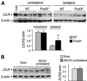
Interestingly, although hippocampus LDLR protein
lev-els were reduced in WT and Pcsk9
⫺ / ⫺mice on the lesioned
side of the brain after the ischemic stroke, the decrease in
LDLR levels was attenuated by 50% in Pcsk9
⫺ / ⫺mice ( Fig. 6A ).
Note that the LDLR protein levels in the nonlesioned
side of the hippocampus were similar to those of sham-
operated control mice ( Fig. 6B ). Altogether, our results
evidence that PCSK9 mRNA levels are upregulated
follow-ing a traumatic injury in vivo.
Because tMCAO impacts on behavior in a manner related
to the extent of lesion, we measured the lesion volume and
mouse behavior following tMCAO ( 20 ). The data showed
that mouse behavior (scale 2, corresponding to mild
consis-tent curling, with >50% attempts to curl to the contralateral
side, see Methods) and lesion volumes of
Pcsk9
⫺ / ⫺mice
(
ⵑ 70% compared with the nonlesioned side) did not differ
from those of WT mice ( Fig. 5A , B ).
Following tMCAO, PCSK9 was not expressed in the
in-farct and penumbra areas, suggesting that PCSK9 does not
play a signifi cant role in neuronal death. In contrast, it was
expressed after ischemic stroke in the dentate gyrus ( Fig. 4 ),
a brain area in which neurogenesis takes place ( 26 ).
Be-cause Pcsk9
⫺
/⫺
mice exhibited impaired hepatocyte
pro-liferation following partial hepatectomy ( 6 ), we tested the
impact of PCSK9 on neurogenesis in hippocampus
follow-ing ischemic stroke. Although cell proliferation, based on
BrdU incorporation, tends to increase on the stroke side, in
WT and Pcsk9
⫺
/⫺
mice, our data failed to reveal any signifi
-cant impact of PCSK9 on the de novo neurogenesis in the
Fig. 3. LDLR regulation by PCSK9 at adulthood. A: In situ hybridization of sagital adult mouse brain cryosections with a PCSK9 and LDLR antisense 35 S-labeled cRNA riboprobes. B: Representative immu-noblots showing LDLR and  -actin protein levels in the RE-OP and olfactory bulb (200 g protein load/ lane) and in adult liver (30 g of protein load/lane) of WT and Pcsk9 ⫺ / ⫺ mice. The specifi city of the LDLR antibody is emphasized by the absence of sig-nal in the liver of Ldlr ⫺ / ⫺ mice. Bar diagrams repre-sent the protein levels of LDLR normalized to those of  -actin in RE-OP and olfactory bulb (n = 9 for WT and Pcsk9 ⫺ / ⫺ mice). All error bars represent the SEM. C: Nissl staining showing the structure of the RE-OP in WT and Pcsk9 ⫺ / ⫺ mice. The enlarged squared regions are shown on the right. Bars = 400 m (left) and 100 m (right). D: Nissl staining re-vealing the structure of the olfactory bulb in WT and Pcsk9 ⫺ / ⫺ mice. Bar = 1 mm.
Fig. 5. Impact of ischemic stroke on Pcsk9 ⫺ / ⫺ mice. A: Behavior analysis after a tMCAO (1 h) ischemic stroke at different times of brain reperfusion (1, 24, 48, and 72 h) of WT and Pcsk9 ⫺ / ⫺ mice. B: Lesion volume at 72 h of brain reperfusion following tMCAO in WT and Pcsk9 ⫺ / ⫺ mice. C: BrdU was injected intraperitoneally for two days (twice a day at 8 h intervals) following tMCAO versus in sham-operated mice. WT and Pcsk9 ⫺ / ⫺ mice were euthanized at 72 h of brain reperfusion. BrdU positive cells were counted in ipsilateral (lesioned side) and contralateral (nonlesioned side) dentate gyrus. Bar diagrams represent the percent ratio of the number of BrdU positive cells in the ipsilateral side versus the contralateral one (n = 5 for WT and Pcsk9 ⫺ / ⫺ mice). All error bars represent the SEM.
Fig. 4. Time-dependent PCSK9 expression following an ischemic stroke. mRNA distribution pattern using PCSK9 antisense ribo-probe, seen as bright labeling under darkfi eld illumination in mouse coronal cryosections following transient (1 h) tMCAO at different times following brain reperfusion (6, 24, and 72 h and 1 week). DG, hippocampal dentate gyrus .
by guest, on November 9, 2019
www.jlr.org
Downloaded from
.html
the upregulation of LDLR in Pcsk9
⫺ / ⫺mice enhances
apoE degradation only during development, and not at
adulthood.
DISCUSSION
The strikingly lower levels of LDL-cholesterol (
⫺ 40% or
⫺ 85%) found in individuals carrying loss-of-function
mu-tations on one or both PCSK9 alleles generated a strong
interest from researchers and pharmaceutical industries
to develop a PCSK9 inhibitor/silencer for the treatment of
dyslipidemias (
8, 9
). The injection of PCSK9-specifi c
monoclonal antibodies ( 30 ) or antisense oligonucleotides
in mice ( 31, 32 ) and cynomolgus monkeys ( 33 ) provided
the proof of principle that PCSK9 inhibition/silencing is a
promising therapeutic approach for dyslipidemias. It was
thus critical to defi ne the clinical phenotypes of the lack of
PCSK9 to fully understand its physiological roles in various
organs. From this perspective, the impact of the lack of
PCSK9 on brain recovery following ischemic stroke was
also of interest for future patients who would be treated
with a PCSK9 inhibitor/silencer.
We herein demonstrated that, in mouse brain, PCSK9 is
co-expressed with the LDLR in the telencephalon at E12.5,
cerebellum at P7, and RE-OP at adulthood ( Figs. 1–3 ).
The low LDLR protein levels in the RE-OP and olfactory
bulb are unchanged in adult Pcsk9
⫺ / ⫺mice compared with
WT mice, suggesting that in adult mice PCSK9 does not
promote the degradation of the LDLR in these brain areas
( Fig. 3 ). Our data are consistent with those reported for
adult transgenic mice overexpressing PCSK9 in the liver
( 34 ), in which no LDLR regulation by ectopic PCSK9 in
the adult hippocampus and cortex was observed ( 1, 6 ).
However, in the present study we provide the fi rst
dem-onstration that endogenous PCSK9 regulates the levels of
LDLR during mouse brain development and following
ischemic stroke. Total LDLR protein levels were indeed
⭓ 2.5-fold higher in the telencephalon at E12.5 and in the
cerebellum at P7 of Pcsk9
⫺ / ⫺embryos or newborns ( Figs.
2, 3
). Furthermore, they decreased 2-fold less in the
Pcsk9
⫺ / ⫺-lesioned dentate gyrus compared with that in WT
mice ( Fig. 6A ). Consistently, cell surface LDLR levels
dur-ing brain development were increased, suggestdur-ing that, as
in liver, PCSK9 enhances LDLR internalization and
degra-dation in the analyzed developing or lesioned brain areas.
Although annexin A2 has been shown to be an inhibitor of
extra-hepatic PCSK9 ( 35 ), in Pcsk9
⫺ / ⫺brain during
devel-opment and at adulthood, the protein levels of annexin
A2 were unchanged compared with those of WT mice
(supplemental Fig. V). However, we cannot exclude that,
at adulthood, the lack of LDLR regulation by PCSK9 in
WT mice is due to a possible modulation of PCSK9
bind-ing to LDLR by annexin A2, especially because LDLR
lev-els are particularly low in the RE-OP.
The absence of V5 immunoreactivity in the CSF of
trans-genic mice overexpressing V5-tagged PCSK9 in liver
(sup-plemental Fig. VI) demonstrated that circulating PCSK9
does not cross the blood-brain barrier (BBB). This may
also explain the absence of brain LDLR regulation by
suggest that, even though LDLR levels were
downregu-lated following an ischemic stroke, PCSK9 still promotes
its degradation.
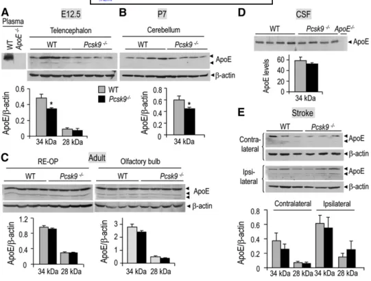
Downregulation of the protein levels of apoE in Pcsk9
ⴚ / ⴚmice during development
To determine the role of the regulation of brain LDLR
by PCSK9 and because apoE is the main apolipoprotein in
brain that can bind LDLR, we quantifi ed the protein levels
of apoE during development, at adulthood, and following
ischemic stroke. Western blot analyses showed that the
ⵑ 34 kDa untruncated apoE protein levels were ⵑ 25%
lower in Pcsk9
⫺ / ⫺mice in the telencephalon at E12.5 and
in the cerebellum at P7 ( Fig. 7A , B ), but not at adulthood
in the RE-OP, olfactory bulb, and cerebrospinal fl uid
(CSF) ( Fig. 7C, D ). Following ischemic stroke, the
ⵑ 34
kDa apoE protein levels increased
ⵑ 1.6-fold in the
le-sioned dentate gyrus compared with the nonlele-sioned side
( Fig. 7E ), consistent with the upregulation of apoE in
in-farcted cortex and striatum ( 28 ). However, no signifi cant
difference in the levels of upregulated apoE protein levels
between WT and Pcsk9
⫺ / ⫺mice was observed. Note that
the apoE protein levels of sham-operated mice were
simi-lar to those of nonlesioned dentate gyrus (data not shown).
In addition to the
ⵑ 34 kDa apoE form, another form
run-ning with an apparent molecular weight of
ⵑ 28 kDa is
pres-ent at all stages except at P7, as previously reported ( 29 ).
The protein levels of this apoE form are also not
modu-lated in Pcsk9
⫺ / ⫺mice. Altogether, these data suggest that
Fig. 6. LDLR regulation by PCSK9 following tMCAO. A: Repre-sentative immunoblots showing the protein levels of LDLR and  -actin in the hippocampus of WT and Pcsk9 ⫺ / ⫺ mice following
tMCAO (1 h) and 24 h of brain reperfusion (200 g protein load/ lane) and in adult liver (30 g protein load/lane). Ipsilateral and contralateral hippocampi were isolated and analyzed separately. Bar diagrams represent the quantitation of the protein levels of LDLR normalized to those of  -actin (n = 4 for WT and Pcsk9 ⫺ / ⫺ mice). * P < 0.05 by Dunnett test. All error bars represent the SEM. B: Representative immunoblots showing the protein levels of LDLR and  -actin in the contralateral hippocampus of WT mice follow-ing or not tMCAO (1 h) and 24 h of brain reperfusion (200 g protein load/lane). Bar diagrams represent the quantitation of the protein levels of LDLR normalized to those of  -actin (n = 4 for sham and n = 3 for tMCAO).
by guest, on November 9, 2019
www.jlr.org
Downloaded from
adulthood, apoE ( Fig. 7C, D ) and LDLR ( Fig. 3B ) protein
levels are not regulated in the brain of Pcsk9
⫺ / ⫺mice.
Fol-lowing transient ischemic stroke, in spite of a diminution
of LDLR by
ⵑ 2-fold less in Pcsk9
⫺ / ⫺mice compared with
WT mice, apoE protein levels similarly increased in WT
and Pcsk9
⫺ / ⫺lesioned mice compared with the nonlesioned
side ( Fig. 7E ), suggesting that the
ⵑ 2-fold higher levels of
LDLR in the lesioned hippocampus of Pcsk9
⫺ / ⫺mice is not
suffi cient to enhance the general degradation of apoE in
brain. However, during brain development, PCSK9 induces
a downregulation of the protein levels of the nontruncated
apoE form (
ⵑ 25%) ( Fig. 7A, B ), which may be due to the
increase of cell surface LDLR and consequently to an
in-crease in apoE uptake and degradation by lysosomes. This
is consistent with the upregulation of apoE protein levels
reported in CSF and cortex of Ldlr
⫺ / ⫺mice ( 46 ), and the
downregulation by 50-90% of apoE levels in the brain of
transgenic mice overexpressing LDLR ( 40 ).
The downregulation or degradation of apoE levels by
PCSK9, probably through LDLR upregulation, is not
enough to induce clear physiological consequences, since
during development and following transient ischemic
stroke we did not observe any brain morphology alteration
or modulation of markers of cell proliferation, cell
differ-entiation, synapses, or clusterin, a neuroprotective protein
for ischemic stroke (supplemental Figs. I–IV). Thus, the
physiological role of PCSK9 in the brain remains to be
de-fi ned. Further investigations may be warranted as we
can-not exclude from our study that the effect of PCSK9 on
PCSK9 at adulthood. Because the BBB is more permissive
at earlier developmental stages and the establishment of its
integrity during embryogenesis, or at postnatal stages, has
been controversial ( 36, 37 ), the potential contribution of
circulating PCSK9 in LDLR downregulation at E12.5 and
P7 remains to be elucidated. However, the excellent
colo-calizations of PCSK9 and LDLR expression in the frontal
cortex (E12.5) and external granular cell layer (P7)
sug-gest a major contribution of local PCSK9. Furthermore, we
showed by in situ hybridization that the expression level of
other members of the PC family relevant to cholesterol
regulation (Furin, PC5/6, and SKI-1/S1P) ( 38 ) do not
change upon loss of PCSK9 expression in the liver, where
the major expression of PCSK9 occurs (Ref. 1 and
supple-mental Fig. VII), suggesting that absence of PCSK9 does
not infl uence the mRNA expression of these proprotein
convertases (PCs). Consequently, the observed regulation
of LDLR levels in Pcsk9
⫺ / ⫺brain is likely a consequence of
the absence of PCSK9.
The physiological and pathological functions of LDLR
in the nervous system remain unclear. Ldlr
⫺ / ⫺mice have
normal brain morphology but exhibit impaired learning
and memory ( 39 ), and the impact of excess LDLR levels on
general brain functions has not yet been reported, except
in the case of Alzheimer’s disease progression ( 40 ).
How-ever, it has been shown that LDLR has the highest affi nity
for apoE in brain ( 41–44 ). ApoE, a
ⵑ 34 kDa glycoprotein,
is the major apolipoprotein in the brain ( 45 ), and it plays
an important role in brain cholesterol metabolism. At
Fig. 7. Regulation of apoE protein levels in Pcsk9 ⫺ / ⫺ mouse brain during development, at adulthood, and following tMCAO. Representative immunoblots (100 g protein load/lane) of WT and Pcsk9 ⫺ / ⫺ mice show-ing the protein levels of apoE and  -actin in the telencephalon at E12.5 (A), in the cerebellum at P7 (B), at adulthood in the REOP and olfactory bulb (C), in adult CSF (D), and in the adult contralateral and ipsilat-eral hippocampus following tMCAO (E). The specifi city of the apoE antibody is emphasized by the absence of signal in the plasma and CSF of apoE ⫺ / ⫺ mice (A, D). Bar diagrams represent the normalized protein levels of apoE to those of  -actin (n = 4). The error bars represent the SEM.
by guest, on November 9, 2019
www.jlr.org
Downloaded from
.html
3 . Kotowski , I. K. , A. Pertsemlidis , A. Luke , R. S. Cooper , G. L. Vega , J. C. Cohen , and H. H. Hobbs . 2006 . A spectrum of PCSK9 alleles contributes to plasma levels of low-density lipoprotein cholesterol. Am. J. Hum. Genet. 78 : 410 – 422 .
4 . Allard , D. , S. Amsellem , M. Abifadel , M. Trillard , M. Devillers , G. Luc , M. Krempf , Y. Reznik , J. P. Girardet , A. Fredenrich , et al . 2005 . Novel mutations of the PCSK9 gene cause variable pheno-type of autosomal dominant hypercholesterolemia. Hum. Mutat. 26 : 497 – 506 .
5 . Rashid , S. , D. E. Curtis , R. Garuti , N. N. Anderson , Y. Bashmakov , Y. K. Ho , R. E. Hammer , Y. A. Moon , and J. D. Horton . 2005 . Decreased plasma cholesterol and hypersensitivity to statins in mice lacking Pcsk9. Proc. Natl. Acad. Sci. USA . 102 : 5374 – 5379 . 6 . Zaid , A. , A. Roubtsova , R. Essalmani , J. Marcinkiewicz , A.
Chamberland , J. Hamelin , M. Tremblay , H. Jacques , W. Jin , J. Davignon , et al . 2008 . Proprotein convertase subtilisin/kexin type 9 (PCSK9): hepatocyte-specifi c low-density lipoprotein receptor deg-radation and critical role in mouse liver regeneration. Hepatology . 48 : 646 – 654 .
7 . Cohen , J. , A. Pertsemlidis , I. K. Kotowski , R. Graham , C. K. Garcia , and H. H. Hobbs . 2005 . Low LDL cholesterol in individuals of African descent resulting from frequent nonsense mutations in PCSK9. Nat. Genet. 37 : 161 – 165 .
8 . Seidah , N. G. 2009 . PCSK9 as a therapeutic target of dyslipidemia. Expert Opin. Ther. Targets . 13 : 19 – 28 .
9 . Horton , J. D. , J. C. Cohen , and H. H. Hobbs . 2009 . PCSK9: a con-vertase that coordinates LDL catabolism. J. Lipid Res. 50 ( Suppl ): S172 – S177 .
10 . Lagace , T. A. , D. E. Curtis , R. Garuti , M. C. McNutt , S. W. Park , H. B. Prather , N. N. Anderson , Y. K. Ho , R. E. Hammer , and J. D. Horton . 2006 . Secreted PCSK9 decreases the number of LDL re-ceptors in hepatocytes and inlivers of parabiotic mice. J. Clin. Invest. 116 : 2995 – 3005 .
11 . Grefhorst , A. , M. C. McNutt , T. A. Lagace , and J. D. Horton . 2008 . Plasma PCSK9 preferentially reduces liver LDL receptors in mice. J. Lipid Res. 49 : 1303 – 1311 .
12 . Schmidt , R. J. , T. P. Beyer , W. R. Bensch , Y. W. Qian , A. Lin , M. Kowala , W. E. Alborn , R. J. Konrad , and G. Cao . 2008 . Secreted proprotein convertase subtilisin/kexin type 9 reduces both hepatic and extrahepatic low-density lipoprotein receptors in vivo. Biochem. Biophys. Res. Commun. 370 : 634 – 640 .
13 . Roubtsova , A. , M. N. Munkonda , Z. Awan , J. Marcinkiewicz , A. Chamberland , C. Lazure , K. Cianfl one , N. G. Seidah , and A. Prat . 2011 . Circulating proprotein convertase subtilisin/kexin 9 (PCSK9) regulates VLDLR protein and triglyceride accumulation in visceral adipose tissue. Arterioscler. Thromb. Vasc. Biol. 31 : 785 – 791 . 14 . Salero , E. , and M. E. Hatten . 2007 . Differentiation of ES cells into
cerebellar neurons. Proc. Natl. Acad. Sci. USA . 104 : 2997 – 3002 . 15 . Poirier , S. , A. Prat , E. Marcinkiewicz , J. Paquin , B. P. Chitramuthu ,
D. Baranowski , B. Cadieux , H. P. Bennett , and N. G. Seidah . 2006 . Implication of the proprotein convertase NARC-1/PCSK9 in the development of the nervous system. J. Neurochem. 98 : 838 – 850 . 16 . Seidah , N. G. , G. Mayer , A. Zaid , E. Rousselet , N. Nassoury , S.
Poirier , R. Essalmani , and A. Prat . 2008 . The activation and physi-ological functions of the proprotein convertases. Int. J. Biochem. Cell Biol. 40 : 1111 – 1125 .
17 . Chiang , L. W. , J. M. Grenier , L. Ettwiller , L. P. Jenkins , D. Ficenec , J. Martin , F. Jin , P. S. DiStefano , and A. Wood . 2001 . An orches-trated gene expression component of neuronal programmed cell death revealed by cDNA array analysis. Proc. Natl. Acad. Sci. USA . 98 : 2814 – 2819 .
18 . Bingham , B. , R. Shen , S. Kotnis , C. F. Lo , B. A. Ozenberger , N. Ghosh , J. D. Kennedy , J. S. Jacobsen , J. M. Grenier , P. S. DiStefano , et al . 2006 . Proapoptotic effects of NARC 1 (= PCSK9), the gene encoding a novel serine proteinase. Cytometry A . 69 : 1123 – 1131 . 19 . Belayev , L. , R. Busto , W. Zhao , G. Fernandez , and M. D. Ginsberg .
1999 . Middle cerebral artery occlusion in the mouse by intralumi-nal suture coated with poly-L-lysine: neurological and histological validation. Brain Res. 833 : 181 – 190 .
20 . Beaulieu , J. M. , J. Kriz , and J. P. Julien . 2002 . Induction of periph-erin expression in subsets of brain neurons after lesion injury or cerebral ischemia. Brain Res. 946 : 153 – 161 .
21 . Jiang , S. X. , J. Lertvorachon , S. T. Hou , Y. Konishi , J. Webster , G. Mealing , E. Brunette , J. Tauskela , and E. Preston . 2005 . Chlortetracycline and demeclocycline inhibit calpains and protect mouse neurons against glutamate toxicity and cerebral ischemia. J. Biol. Chem. 280 : 33811 – 33818 .
mouse brain is more subtle, since cholesterol is involved in
nerve conduction velocity through the formation of
myeli-nation and synaptogenesis ( 47 ). It also plausible that local
hydroxy-cholesterol, rather than circulating cholesterol, is
critical for CNS development ( 48 ).
Injection of purifi ed PCSK9 or a recombinant
adenovi-rus overexpressing PCSK9 in mice reduced the protein
lev-els of the LDLR in liver, lung, kidney, and small intestine,
but not in the adrenal glands ( 11, 12 ). In addition, upon
overexpression in kidney, the secreted plasma PCSK9
pro-motes LDLR degradation mostly in the liver and raises
plasma LDL ( 49 ). Altogether, our data and those in the
lit-erature show that not all tissues respond equally to local or
circulating PCSK9 and that in adult brain LDLR does not
respond to circulating PCSK9. Because the gene ablation of
PCSK9 in mice, which completely abolishes PCSK9
expres-sion, does not affect brain organization or brain recovery
following ischemic stroke, the silencing of PCSK9, either
using monoclonal antibodies ( 30 ) or antisense therapies
( 33 ) that may partially inhibit PCSK9 expression, should
not affect brain PCSK9 levels. However, we cannot exclude
that low circulating LDL-cholesterol in treated adult
pa-tients with PCSK9-silencing approaches could affect brain
LDLR levels via regulation of PCSK9 in the central nervous
system ( 1 ), likely through the SREBP-2 ( 50 ) or HNF-1
␣
pathways ( 51 ). Furthermore, rare human subjects
exhibit-ing either PCSK9 loss-of-function mutations [e.g., R46L ( 3 )
and R434W ( 52 )] or gain-of-function mutations [e.g., and
S127R ( 2 ) and D374Y ( 53 )], may during development also
exhibit higher or lower levels of LDLR in the brain,
respec-tively, resulting in subtle effects later on in adults.
In conclusion, the present results suggest that PCSK9
inhibition should not interfere with brain development
and morphology or with brain recovery/damage after an
ischemic stroke. They are consistent with humans carrying
heterozygous loss-of-function mutations in PCSK9 who
ap-pear healthy and have normal a lifespan ( 54, 55 ). Thus,
PCSK9 inhibition is likely not to have overt deleterious
ef-fects on patients affected by a stroke event and would not
be expected to cause major side effects in patients treated
with a PCSK9 inhibitor or silencer for
hypercholester-olemia.
The authors thank Yuan Cheng Weng for his precious teaching of ischemia procedure and Claudia Toulouse for excellent animal care. Many thanks to all the members of the Seidah laboratory for helpful discussions and to Brigitte Mary for effi cacious editorial assistance.
REFERENCES
1 . Seidah , N. G. , S. Benjannet , L. Wickham , J. Marcinkiewicz , S. B. Jasmin , S. Stifani , A. Basak , A. Prat , and M. Chretien . 2003 . The secretory proprotein convertase neural apoptosis-regulated con-vertase 1 (NARC-1): liver regeneration and neuronal differentia-tion. Proc. Natl. Acad. Sci. USA . 100 : 928 – 933 .
2 . Abifadel , M. , M. Varret , J. P. Rabes , D. Allard , K. Ouguerram , M. Devillers , C. Cruaud , S. Benjannet , L. Wickham , D. Erlich , et al . 2003 . Mutations in PCSK9 cause autosomal dominant hypercholes-terolemia. Nat. Genet. 34 : 154 – 156 .
by guest, on November 9, 2019
www.jlr.org
Downloaded from
22 . Seidah , N. G. , R. Day , M. Marcinkiewicz , and M. Chretien . 1998 . Precursor convertases: an evolutionary ancient, cell-specifi c, com-binatorial mechanism yielding diverse bioactive peptides and pro-teins. Ann. N. Y. Acad. Sci. 839 : 9 – 24 .
23 . Seidah , N. G. , M. Mbikay , M. Marcinkiewicz , and M. Chretien . 1998 . The mammalian precursor convertases: paralogs of the subtilisin/ kexin family of calcium-dependent serine proteinases. In Proteolytic and Cellular Mechanisms in Prohormone and Neuropeptide Precursor Processing. V. Y. Hook, editor. R.G. Landes Company, Georgetown, TX. 49 – 76 .
24 . Gao , W. O. , N. Heintz , and M. E. Hatten . 1991 . Cerebellar gran-ule cell neurogenesis is regulated by cell-cell interactions in vitro. Neuron . 6 : 705 – 715 .
25 . Furley , A. J. , S. B. Morton , D. Manalo , D. Karagogeos , J. Dodd , and T. M. Jessell . 1990 . The axonal glycoprotein TAG-1 is an immuno-globulin superfamily member with neurite outgrowth-promoting activity. Cell . 61 : 157 – 170 .
26 . Kaplan , M. S. , and J. W. Hinds . 1977 . Neurogenesis in the adult rat: electron microscopic analysis of light radioautographs. Science . 197 : 1092 – 1094 .
27 . Imhof , A. , Y. Charnay , P. G. Vallet , B. Aronow , E. Kovari , L. E. French , C. Bouras , and P. Giannakopoulos . 2006 . Sustained astro-cytic clusterin expression improves remodeling after brain ische-mia. Neurobiol. Dis. 22 : 274 – 283 .
28 . Kitagawa , K. , M. Matsumoto , K. Kuwabara , T. Ohtsuki , and M. Hori . 2001 . Delayed, but marked, expression of apolipoprotein E is involved in tissue clearance after cerebral infarction. J. Cereb. Blood Flow Metab. 21 : 1199 – 1207 .
29 . Huang , Y. , X. Q. Liu , T. Wyss-Coray , W. J. Brecht , D. A. Sanan , and R. W. Mahley . 2001 . Apolipoprotein E fragments present in Alzheimer’s disease brains induce neurofi brillary tangle-like in-tracellular inclusions in neurons. Proc. Natl. Acad. Sci. USA . 98 : 8838 – 8843 .
30 . Chan , J. C. , D. E. Piper , Q. Cao , D. Liu , C. King , W. Wang , J. Tang , Q. Liu , J. Higbee , Z. Xia , et al . 2009 . A proprotein convertase subtil-isin/kexin type 9 neutralizing antibody reduces serum cholesterol in mice and nonhuman primates. Proc. Natl. Acad. Sci. USA . 106 : 9820 – 9825 .
31 . Graham , M. J. , K. M. Lemonidis , C. P. Whipple , A. Subramaniam , B. P. Monia , S. T. Crooke , and R. M. Crooke . 2007 . Antisense in-hibition of proprotein convertase subtilisin/kexin type 9 reduces serum LDL in hyperlipidemic mice. J. Lipid Res. 48 : 763 – 767 . 32 . Gupta , N. , N. Fisker , M. C. Asselin , M. Lindholm , C. Rosenbohm ,
H. Orum , J. Elmen , N. G. Seidah , and E. M. Straarup . 2010 . A locked nucleic acid antisense oligonucleotide (LNA) silences PCSK9 and enhances LDLR expression in vitro and in vivo. PLoS ONE . 5 : e10682 .
33 . Frank-Kamenetsky , M. , A. Grefhorst , N. N. Anderson , T. S. Racie , B. Bramlage , A. Akinc , D. Butler , K. Charisse , R. Dorkin , Y. Fan , et al . 2008 . Therapeutic RNAi targeting PCSK9 acutely lowers plasma cholesterol in rodents and LDL cholesterol in nonhuman primates. Proc. Natl. Acad. Sci. USA . 105 : 11915 – 11920 .
34 . Liu , M. , G. Wu , J. Baysarowich , M. Kavana , G. H. Addona , K. K. Bierilo , J. S. Mudgett , G. Pavlovic , A. Sitlani , J. J. Renger , et al . 2010 . PCSK9 is not involved in the degradation of LDL receptors and BACE1 in the adult mouse brain. J. Lipid Res. 51 : 2611 – 2618 . 35 . Mayer , G. , S. Poirier , and N. G. Seidah . 2008 . Annexin A2 is a
C-terminal PCSK9-binding protein that regulates endogenous low density lipoprotein receptor levels. J. Biol. Chem. 283 : 31791 – 31801 . 36 . Lossinsky , A. S. , and R. R. Shivers . 2004 . Structural pathways for
macro-molecular and cellular transport across the blood-brain barrier during infl ammatory conditions. Review. Histol. Histopathol. 19 : 535 – 564 . 37 . Daneman , R. , L. Zhou , A. A. Kebede , and B. A. Barres . 2010 .
Pericytes are required for blood-brain barrier integrity during embryogenesis. Nature . 468 : 562 – 566 .
38 . Seidah , N. G. , and A. Prat . 2007 . The proprotein convertases are potential targets in the treatment of dyslipidemia. J. Mol. Med. 85 : 685 – 696 .
39 . Mulder , M. , G. Koopmans , G. Wassink , M. G. Al , M. L. Simard , L. M. Havekes , J. Prickaerts , and A. Blokland . 2007 . LDL receptor defi ciency
results in decreased cell proliferation and presynaptic bouton density in the murine hippocampus. Neurosci. Res. 59 : 251 – 256 .
40 . Kim , J. , J. M. Castellano , H. Jiang , J. M. Basak , M. Parsadanian , V. Pham , S. M. Mason , S. M. Paul , and D. M. Holtzman . 2009 . Overexpression of low-density lipoprotein receptor in the brain markedly inhibits amyloid deposition and increases extracellular A beta clearance. Neuron . 64 : 632 – 644 .
41 . Mahley , R. W. 1988 . Apolipoprotein E: cholesterol transport pro-tein with expanding role in cell biology. Science . 240 : 622 – 630 . 42 . Mahley , R. W. , and S. C. Rall , Jr . 2000 . Apolipoprotein E: far more
than a lipid transport protein. Annu. Rev. Genomics Hum. Genet. 1 : 507 – 537 .
43 . Innerarity , T. L. , and R. W. Mahley . 1978 . Enhanced binding by cul-tured human fi broblasts of apo-E-containing lipoproteins as com-pared with low density lipoproteins. Biochemistry . 17 : 1440 – 1447 . 44 . Innerarity , T. L. , R. E. Pitas , and R. W. Mahley . 1979 . Binding of
arginine-rich (E) apoprotein after recombination with phospho-lipid vesicles to the low density lipoprotein receptors of fi broblasts. J. Biol. Chem. 254 : 4186 – 4190 .
45 . Elshourbagy , N. A. , W. S. Liao , R. W. Mahley , and J. M. Taylor . 1985 . Apolipoprotein E mRNA is abundant in the brain and adrenals, as well as in the liver, and is present in other peripheral tissues of rats and marmosets. Proc. Natl. Acad. Sci. USA . 82 : 203 – 207 .
46 . Fryer , J. D. , R. B. Demattos , L. M. McCormick , M. A. O’Dell , M. L. Spinner , K. R. Bales , S. M. Paul , P. M. Sullivan , M. Parsadanian , G. Bu , et al . 2005 . The low density lipoprotein receptor regulates the level of central nervous system human and murine apolipoprotein E but does not modify amyloid plaque pathology in PDAPP mice. J. Biol. Chem. 280 : 25754 – 25759 .
47 . Dietschy , J. M. , and S. D. Turley . 2004 . Thematic review series: brain lipids. Cholesterol metabolism in the central nervous system during early development and in the mature animal. J. Lipid Res. 45 : 1375 – 1397 .
48 . Bryleva , E. Y. , M. A. Rogers , C. C. Chang , F. Buen , B. T. Harris , E. Rousselet , N. G. Seidah , S. Oddo , F. M. LaFerla , T. A. Spencer , et al . 2010 . ACAT1 gene ablation increases 24(S)-hydroxycholesterol content in the brain and ameliorates amyloid pathology in mice with AD. Proc. Natl. Acad. Sci. USA . 107 : 3081 – 3086 .
49 . Luo , Y. , L. Warren , D. Xia , H. Jensen , T. Sand , S. Petras , W. Qin , K. S. Miller , and J. Hawkins . 2009 . Function and distribution of circulating human PCSK9 expressed extrahepatically in transgenic mice. J. Lipid Res. 50 : 1581 – 1588 .
50 . Dubuc , G. , A. Chamberland , H. Wassef , J. Davignon , N. G. Seidah , L. Bernier , and A. Prat . 2004 . Statins upregulate PCSK9, the gene encoding the proprotein convertase neural apoptosis-regu-lated convertase-1 implicated in familial hypercholesterolemia. Arterioscler. Thromb. Vasc. Biol. 24 : 1454 – 1459 .
51 . Dong , B. , M. Wu , H. Li , F. B. Kraemer , K. Adeli , N. G. Seidah , S. W. Park , and J. Liu . 2010 . Strong induction of PCSK9 gene expres-sion through HNF1 ␣ and SREBP2: mechanism for the resistance to LDL-cholesterol lowering effect of statins in dyslipidemic hamsters. J. Lipid Res. 51: 1486–1495.
52 . Davignon , J. , G. Dubuc , and N. G. Seidah . 2010 . The infl uence of PCSK9 polymorphisms on serum low-density lipoprotein choles-terol and risk of atherosclerosis. Curr. Atheroscler. Rep. 12 : 308 – 315 . 53 . Timms , K. M. , S. Wagner , M. E. Samuels , K. Forbey , H. Goldfi ne ,
S. Jammulapati , M. H. Skolnick , P. N. Hopkins , S. C. Hunt , and D. M. Shattuck . 2004 . A mutation in PCSK9 causing dominant hypercholesterolemia in a Utah pedigree. Hum. Genet. 114 : 349 – 353 .
54 . Zhao , Z. , Y. Tuakli-Wosornu , T. A. Lagace , L. Kinch , N. V. Grishin , J. D. Horton , J. C. Cohen , and H. H. Hobbs . 2006 . Molecular characterization of loss-of-function mutations in PCSK9 and iden-tifi cation of a compound heterozygote. Am. J. Hum. Genet. 79 : 514 – 523 .
55 . Fasano , T. , A. B. Cefalu , E. Di Leo , D. Noto , D. Pollaccia , L. Bocchi , V. Valenti , R. Bonardi , O. Guardamagna , M. Averna , et al . 2007 . A novel loss of function mutation of PCSK9 gene in white subjects with low-plasma low-density lipoprotein cholesterol. Arterioscler. Thromb. Vasc. Biol. 27 : 677 – 681 .
by guest, on November 9, 2019
www.jlr.org
Downloaded from
.html