HAL Id: hal-03243156
https://hal-amu.archives-ouvertes.fr/hal-03243156
Submitted on 1 Jun 2021
HAL is a multi-disciplinary open access
archive for the deposit and dissemination of sci-entific research documents, whether they are pub-lished or not. The documents may come from teaching and research institutions in France or abroad, or from public or private research centers.
L’archive ouverte pluridisciplinaire HAL, est destinée au dépôt et à la diffusion de documents scientifiques de niveau recherche, publiés ou non, émanant des établissements d’enseignement et de recherche français ou étrangers, des laboratoires publics ou privés.
infection in Greece
Stavroula Labropoulou, Ekatherina Charvalos, Stylianos Chatzipanagiotou,
Anastasios Ioannidis, Panagiotis Sylignakis, Styliani T aka, Ioulia
Karageorgou, Maria Linou, Giota Mpizta, Andreas Mentis, et al.
To cite this version:
Stavroula Labropoulou, Ekatherina Charvalos, Stylianos Chatzipanagiotou, Anastasios Ioannidis, Panagiotis Sylignakis, et al.. Sunbathing, a possible risk factor of murine typhus infection in Greece. PLoS Neglected Tropical Diseases, Public Library of Science, 2021, 15 (3), pp.e0009186. �10.1371/jour-nal.pntd.0009186�. �hal-03243156�
RESEARCH ARTICLE
Sunbathing, a possible risk factor of murine
typhus infection in Greece
Stavroula Labropoulou1, Ekatherina Charvalos2, Stylianos ChatzipanagiotouID3,
Anastasios IoannidisID4, Panagiotis Sylignakis5, StylianiΤakaID6, Ioulia Karageorgou1, Maria Linou1, Giota Mpizta1, Andreas Mentis1, Sophie Edouard7, Didier Raoult7,8,
Emmanouil AngelakisID1,8*
1 Laboratory of Medical Microbiology, Hellenic Pasteur Institute, Athens, Greece, 2 IASO Medical Research (IMR) department, IASO Gynecology Obstretrics and Pediatrics Hospital, Marousi, Greece, 3 Department of Biopathology and Clinical Microbiology, Aeginition Hospital, Medical School, National and Kapodistrian University of Athens, Athens, Greece, 4 Department of Nursing, Faculty of Health Sciences University of Peloponnese, Tripoli, Greece, 5 Pananio—Venizelio General Hospital of Heraklion, Heraklion, Greece, 6 Allergy Department, 2nd Pediatric Clinic, National and Kapodistrian University of Athens, Athens, Greece, 7 Aix Marseille Univ, IRD, IHU Me´diterrane´ e Infection, MEPHI, Marseille, France, 8 Aix Marseille Universite´, IRD, APHM, VITROME, IHU-Me´diterrane´e Infection, Marseille, France
Abstract
Background
There are few studies about the presence of murine typhus in Greece. Our objective was to conduct a large scale retrospective investigation to determine the clinical and epidemiologi-cal features of patients diagnosed with murine typhus in Greece.
Methodology/Principal findings
From 2012 to 2019 serum samples from hospitalized patients and outpatients throughout Greece suspected for murine typhus infection were tested by immunofluorescence assay for Rickettsia typhi. Immunofluorescence positive samples obtained since 2016 were also tested by qPCR targeting R. typhi. Clinical and epidemiological data were retrospectively collected for the patients with confirmed murine typhus. Overall, we tested 5,365 different patients and, in total, 174 patients from all geographic regions of Greece were diagnosed with murine typhus. The most frequently reported sign or symptom was fever (89%), fol-lowed by headache (84%) and rash (81%). The classical triad of fever, headache, and rash was present in 72% of patients during their illness. Severe infections with complications including acute renal failure or septic shock were not recorded. The majority of cases (81%) occurred during May–October and peaked in June and September. Most of patients (81%) infected in Athens, recalled that their only activity the last weeks before symptoms onset was swimming on the beach and 59% of them also reported an insect bite while sunbathing.
Conclusions/Significance
Our results may reflect the reemergence of murine typhus in Greece and we highlight the importance of awareness of this difficult-to-recognize undifferentiated febrile illness.
a1111111111 a1111111111 a1111111111 a1111111111 a1111111111 OPEN ACCESS
Citation: Labropoulou S, Charvalos E,
Chatzipanagiotou S, Ioannidis A, Sylignakis P,Τaka S, et al. (2021) Sunbathing, a possible risk factor of murine typhus infection in Greece. PLoS Negl Trop Dis 15(3): e0009186.https://doi.org/10.1371/ journal.pntd.0009186
Editor: Job E. Lopez, Baylor College of Medicine,
UNITED STATES
Received: May 7, 2020 Accepted: January 27, 2021 Published: March 12, 2021
Copyright:© 2021 Labropoulou et al. This is an open access article distributed under the terms of theCreative Commons Attribution License, which permits unrestricted use, distribution, and reproduction in any medium, provided the original author and source are credited.
Data Availability Statement: All relevant data are
within the manuscript.
Funding: The author(s) received no specific
funding for this work.
Competing interests: The authors have declared
Author summary
Murine typhus, a febrile disease caused byRickettsia typhi, is often misdiagnosed due to its non-specific presentation. Evidence suggests that murine typhus is more prevalent in Greece than originally believed. We retrospectively collected clinical and epidemiological data for all the patients with confirm and probable murine typhus diagnosed in our labo-ratory the last eight years and we found evidence of murine typhus throughout Greece. Comparing this evidence to other studies, we found that most of Greek patients presented rash and the typical triad probably due to the awareness of physicians, as during our inter-rogation, most of physicians mentioned that they considered murine typhus based on rash appearance. Finally, we found that most murine typhus cases in the city of Athens were associated with sunbathing on the beach and we raise the question of beach littering in murine typhus endemic areas.
Introduction
Murine typhus, an acute zoonotic infection caused byRickettsia typhi, is transmitted to humans byXenopsylla cheopis, a flea that infests rats [1]. The emergence of murine typhus has become apparent mostly in the field of travel medicine, as increasing numbers of individuals are being exposed [2,3]. Indeed, the 2.5% of international travelers reported to the GeoSentinel surveillance network from 1996 to 2008 were from patients with typhus group rickettsioses [4]. The disease occurs worldwide and is underdiagnosed and largely under-reported probably because of its non specifιc clinical manifestations and frequently mild course, a lack of active monitoring and the limited awareness among physicians. The clinical features of murine typhus are non-specific and include fever, rash, headache, myalgia and gastrointestinal symp-toms [5,6]. While the clinical course of murine typhus is typically uncomplicated, serious com-plications have been associated with acute infections, such as central nervous system
abnormalities. The death rate is generally low but can reach 4% without the use of antibacterial drugs [6,7].
Murine typhus was first described in Greece in 1932 and, in 1948, 1,420 cases of murine typhus and 17 deaths were reported [8]. Since then, the disease is considered endemic on the Greek islands of Crete [9] and Euboea [10] whereas sporadic cases have been reported in northern Greece [11] and in the island of Kasos [12]. Because of murine typhus nonspecific clinical presentation and the lack of awareness by physicians we raised the question of the under-diagnosis ofR. typhi infections in Greece. As a reference laboratory for the diagnosis of rickettsial infections in Greece, we routinely receive serum samples from patients suspected of having murine typhus. To gain a better understanding of murine typhus in Greece, we retro-spectively collected clinical and epidemiological data for all the patients diagnosed withR. typhi in our laboratory the last eight years.
Materials
Ethics statement
This study is based on routine diagnosis samples. All collected data were anonymized in stan-dardized forms according to the Ethic and Scientific Committee of the Hellenic Pasteur
Insti-Patients
We studied serum samples from patients with suspected murine typhus that were sent to our laboratory from January 2012 to December 2019. We received sera from hospitalized patients and outpatients with suspected rickettsioses throughout Greece. For each patient, an acute-phase serum sample was obtained within three weeks after the onset of symptoms and, when possible, a convalescent-phase serum sample (i.e., one collected >2 weeks after onset of symp-toms) was also obtained. Clinical data, medical history and complications during illness, habi-tat and environmental characteristics, presence of pets, livestock, or rodents, daily activities in the 30 days before symptom onset and travel during the previous one month were documented with structured conversations forR. typhi positive patients by physicians thanks to phone calls.
R. typhi antigen preparation
R. typhi (strain Wilmington) was grown in confluent HEL cell monolayers using 12-mm round coverslips seeded with 1 ml of medium containing 50,000 cells and incubated in a 5% CO2 incubator at 37˚C. The density ofR. typhi was assessed by microscopic observation of Gimenez stained smears. The supernatants of 15 flasks were pelleted by centrifugation at 5000g for 15 min and resuspended in 1 mL of phosphate-buffered saline (PBS) at pH 7.3 with 0.1% formaldehyde. Cells were fragmented by sonication, and cellular debris was removed by two successive centrifugations (100g, 10 min each). After the supernatants were centrifuged through 20 mL of PBS with 25% sucrose (6000g, 30 min), the resulting pellet was rinsed three times in PBS (6000g, 10 min) and, using spectrophotometry, was suspended in sterile distilled water at a concentration of 2 mg protein/mL prior to being frozen at− 20˚C.
Serology assays
Patients’ sera at a dilution of 1:50 and 1:100 were firstly screened againstR. typhi by immuno-fluorescence assay (IFA) using conjugate-anti-Human IgG/IgA/IgM-FITC as previously described [13,14]. Positive and negative controls were diluted at 1:25 and 1:50. Sera found pos-itive againstR. typhi at a 1:50 dilution were further entered into the quantification test and tested for IgG and IgM. For quantification of IgG and IgM, patients’ sera were initially diluted to 1:8 in PBS milk and subsequently diluted in twofold dilutions as previously described [13,14]. Finally, to exclude false-positive results due to cross-reactivity betweenR. typhi and spotted fever group (SFG) rickettsioses all murine typhus positive patients diagnosed from 2016 to 2019 were also tested by IFA for SFGR. conorii conorii antigens as previously described [13,14]. When cross-reactions were observed, a rickettsial antigen was considered to represent the infectious agent if cumulated titres of IgG and IgM antibodies against this antigen were at least twice as high as those of the others. All reactions were performed in dark room and at least two people performed each microscopical examination.
Molecular assays
Total genomic DNA was retrospectively extracted from samples obtained from 2016 to 2019 with a positiveR. typhi IFA using a QIAamp tissue kit (Qiagen, Hilden, Germany). Samples were handled under sterile conditions to avoid cross-contamination. Samples were screened by qPCR targeting a fragment of the Rpr 274P gene coding for a hypothetical protein, as described previously [15]. Positive results were confirmed by qPCR targeting the glycosyltrans-ferase gene using a previously described Rpr 331 system [15]. A maximum of 10 samples were tested along with negative controls (DNA from IFA negative sera and sterile water) and a posi-tive control (DNA fromR. typhi Wilmington (ATCC VR-144). The quality of DNA handling
and extraction of human samples was verified by qPCR for a housekeeping gene encoding beta-actin [10]
Case definition
A confirmed case of murine typhus was defined as a patient meeting the minimum presump-tive clinical criteria and having 1) a posipresump-tive molecular assay, 2) a single serum with antibody titers of �1:64 for IgM and �1:128 for IgG antibodies [13,14], acute and convalescent sera showing 3) a seroconversion or 4) a fourfold or greater increase in titers. A probable case of murine typhus was defined as a patient having a single serum with an indirect immunofluores-cence assay antibody titer of >1:128 and a clinically compatible illness.
Statistical analysis
EpiInfo version 6.0 software (Centers for Disease Control and Prevention, Atlanta, GA, USA) was used for significance variations in the number of positive patients between two consecu-tive months, nonconsecuconsecu-tive months and seasons. Seasons were defined as winter (January– March), spring (April–June), summer (July–September), and autumn (October–December). The Mantel-Haenszel test or the Fisher exact test was used to test for significance. Ap value <0.05 was considered significant.
Results
We tested 5,506 different sera obtained from 5,365 patients and we identified 172 patients pos-itive byIFA (Table 1). In addition, we realised 133 qPCR and a positive result was obtained for 12 (9%) serum samples. Indeed, 11 acute and 1 convalescent-phase sera were positive by qPCR. For two patients qPCR was positive although serology was negative (Table 2). As a result, in total we diagnosed 174 (98 confirmed and 76 probable) patients with murine typhus. The median age± interquartile range (IR) of patients was 53 ± 15 years, (range: 1–91 years) and 53% were males.
Geographical distribution of cases and epidemiological data
Overall, we found 171 autochthonous murine typhus patients whereas two patients reported a recent travel to Cyprus and one had recently returned from Uruguay. Totally, autochthonous cases were recorded to 33 (58%) out of the 54 prefectures of Greece (Fig 1A). Moreover, we found that murine typhus distribution varied widely among Greek regions and the highest percentage of patients was observed in Central Greece (n = 70, 40%) followed by the region of Peloponnese (n = 29, 17%) (Table 3). Epidemiological information was obtained for 118 patients and among the recorded cases 49 (42%) reported living in rural areas and 19 (16%) were farmers in contact with livestock animals. In total, 101 (86%) mentioned recent/constant outdoor activities, 58 (49%) recalled an exposure to insects or an insect bite, 66 (56%) a close contact with animals and 9 (7%) patients a contact with rodents. Finally, two patients reported a recent travel to Cyprus and one had recently returned from Uruguay.
Table 1.Rickettsia typhi results of serology and PCR assays.
Patients tested Number of realized qPCR (positives) Serology positive Total cases
Acute serum 5,224 115 (11) 166 168
Convalescent-phase serum 141 18 (1) 19 19
Seasonality
Positive murine typhus cases were plotted for each month to identify seasonal distributions from 2012 through 2019 (Fig 1B). Monthly cases of murine typhus cases were lowest from November through April, followed by significant increases during June (p = 0.002) and during September (p <0.001). During October—November, the number of cases decreased signifi-cantly (p = 0.005), then plateaued from November through April. Cases increased slightly in April, then increased significantly during May—October (p = 0.002). The number of murine typhus cases was significantly higher in summer (July—September) that in autumn (October— December) (p<0.0001) and in spring (April—June) than in winter (January—March)
(p<0.0001). The number of cases did not differ significantly from spring to summer (p = 0.07).
Murine typhus in Athens, Attica region
Totally, 53 (30%) murine typhus patients were diagnosed in the city of Athens. The seasonality of murine typhus cases slightly differed for Athens than for other regions, as all cases were
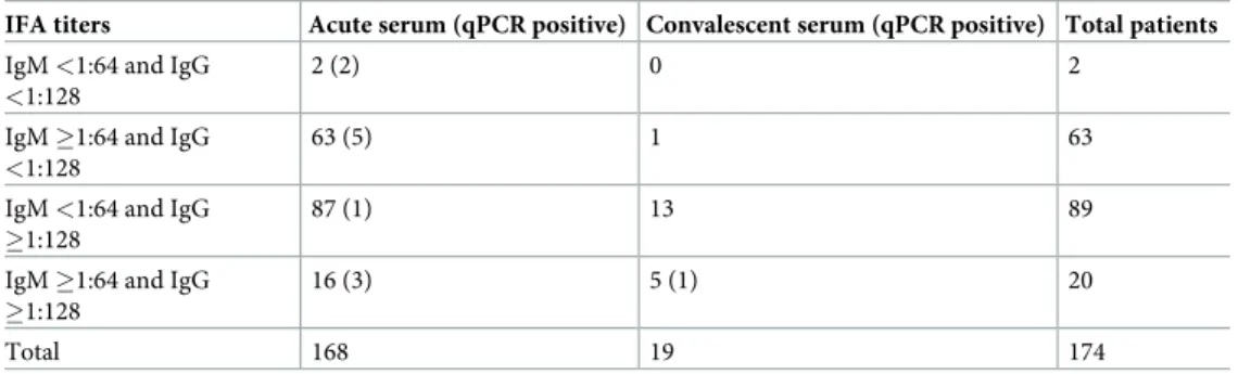
Table 2. IF assay antibody titers for the 174 confirmed murine typhus patients.
IFA titers Acute serum (qPCR positive) Convalescent serum (qPCR positive) Total patients
IgM <1:64 and IgG
<1:128
2 (2) 0 2
IgM �1:64 and IgG
<1:128
63 (5) 1 63
IgM <1:64 and IgG �1:128
87 (1) 13 89
IgM �1:64 and IgG �1:128
16 (3) 5 (1) 20
Total 168 19 174
https://doi.org/10.1371/journal.pntd.0009186.t002
Fig 1. A, Murine typhus patients throughout Greece; B, Seasonal distribution of patients diagnosed for murine
typhus, in Greece and in Athens, 2012–2019. Red stars: present murine typhus cases. Blue stars: previous murine typhus reported cases based on the researched of PubMed for peer-reviewed, articles published in English up to June 2020. The search terms were combinations of “murine typhus”, “Rickettsia typhi” and “Greece”.
https://doi.org/10.1371/journal.pntd.0009186.g001
observed from April through October (Fig 1B). Epidemiological information was obtained for 43 patients and eight reported a recent travel in Greece outside Athens whereas three reported a travel abroad. We considered that 32 patients were infected in/or close to Athens and among these only six (19%) reported outdoor activities. Surprisingly, all the rest of these patients (81%) mentioned that their only activity the last weeks before symptoms onset was swimming on the beach and they did not have any other murine typhus associated risk factors. Moreover, 12 (59%) of them also recalled an insect bite while sunbathing. None of these patients men-tioned a direct contact with rodents.
Clinical manifestations
Clinical information was obtained for 61 patients and for 31 (51%) of them a hospitalization was necessary. This high percentage of hospitalization is probably because we obtained clinical data mostly from patients who had previously presented to hospitals. The most frequently reported sign or symptom was fever (89%), followed by headache (84%) and rash (81%) (Table 4). The duration of fever ranged from 4 to 15 days (38 to 40˚C) whereas the rash was macular or maculopapular with a duration from three to 10 days. The classical triad of fever, headache, and rash was present in 72% of patients during their illness. Other less common manifestations were cough (67%), anorexia (45%) and arthralgia (35%). Gastrointestinal tract symptoms like nausea or vomiting was found in 38% of cases, and diarrhea in 26% of cases.
Table 3. Murine typhus patients among the nine greek regions.
Regions Number of patients (%)
Aegean Islands 16 (9%) Central Greece 70 (40%) Crete 16 (9%) Epirus 14 (8%) Macedonia 4 (2%) Ionian Islands 4 (2%) Peloponnese 29 (17%) Thessaly 18 (10%) Thrace 3 (2%) https://doi.org/10.1371/journal.pntd.0009186.t003
Table 4. Clinical characteristics of murine typhus patients.
Clinical signs and symptoms Number of patients (%)
Fever 54 (89%)
Headache 51 (84%)
Rash 49 (81%)
The classic triad� 44 (72%)
Cough 41 (67%)
Pneumonia or pulmonary infiltrates 37 (61%)
Anorexia 27 (45%) Nausea/vomiting 23 (38%) Myalgia/arthralgia 21 (35%) Diarrhea 17 (26%) Confusion 4 (7%) Photophobia 2 (4%) Hepatosplenomegaly 2 (4%)
Neurological manifestations were also reported, including confusion in 7% and photophobia in 4%. Liver or spleen enlargement was observed in 4% of patients. All hospitalized patients presented pulmonary manifestations including pneumonia or pulmonary infiltrates. Finally, severe infections with complications including acute renal failure or septic shock were not recorded.
Discussion
We report a large series ofR. typhi infections and we found evidence of murine typhus throughout Greece. Our immunofluorescence and molecular assays were sensitive and versa-tile and have been routinely used for the detection and diagnosis of murine typhus [13–15]. Furthermore, we routinely included large numbers of negative controls in our assays that were processed identically to the test samples. Serological tests are the easiest methods for the diag-nosis ofR. typhi infection but seroconversion is usually detected 7–15 days after disease onset [2]. We detected two samples positive by qPCR but negative by IFA and a limitation of our study was that samples were retrospectively tested using molecular assays as we have managed to test only immunofluorescence positive samples since 2016. Indeed, it is very possible that this number could be higher if sera were routinely tested by qPCR on the basis of both clinical and epidemiology data also. Finally, we cannot exclude that some patients were false-positive diagnosed with murine typhus before 2016 as only samples after 2016 were also tested for SFG rickettsioses.
Previously in Greece,R. typhi infections were considered endemic in some Greek regions [9,10] whereas for Europe, exposure toR. typhi has been recorded in Spain, Canary Islands [16], Cyprus [17], Italy [18], and Croatia [19]. Although autochthonous cases of murine typhus have not been described in the countries of North Europe, sporadic cases are commonly iden-tified in travelers who visited endemic areas [7,15,20]. In Cyprus although to date many cases of murine typhus have been described, the first confirmed case was reported in a Swedish woman who developed fever, severe headache, myalgia and rash, three weeks after a visit to Cyprus [21].
The diagnosis of murine typhus has been characterized as a challenge because many physi-cians are unfamiliar with the nonspecific symptoms during the early stages of illness. Rash is considered to be the most typical finding of murine typhus but its prevalence varies from 28– 82% depending on the geographic region. Indeed, rash was found to 20% of patients from Thailand, 38% of patients from Laos, 49% from Texas, 63% from Spain and 80% from Greece [2,6]. Similarly, in previous studies the typical triad for murine typhus occurred in less than half of the patients [6,7,22]. In our study, most of patients presented rash and the typical triad. We believe that this higher rate of rash as a symptom is due to the awareness of physicians, as during our interrogation, most of physicians mentioned that they considered murine typhus based on rash appearance.
Our findings show that the number of murine cases in Greece varies by season. In studies where seasonal variation was recorded, at least half of cases were diagnosed during summer and early autumn [6,15]. A similar seasonality was also previously described in Greece where most cases occurred during summer with a peak on the late summer [10]. In other studies, the highest incidence was observed in May and June [23,24] suggesting that climatic conditions are the main factors contributing to different seasonal distributions of human cases in different geographical areas. Human infection with murine typhus has been associated with the pres-ence of rats and their fleas. This probably explains the two seasonal peaks during June and Sep-tember that we observed due to the higher abundance ofX. cheopis in our country during these months possibly because rats are reproducing these periods.
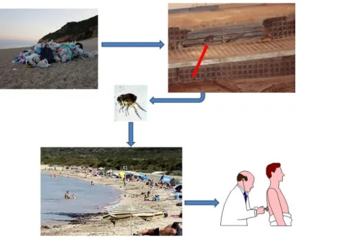
Surprisingly we observed that most cases of murine typhus in Athens were associated with sunbathing on the beach. As the most of the reported human cases of murine typhus have been associated in sites with large rat populations we believe that during summer rats infest beach environments probably because of littering and humans are infected by the rat fleas while sunbathing (Fig 2). Although adultX. cheopis spend most of their time on their host, they can survive for relative long time in off-host environments [25]. Indeed unfed adultX. cheopis survived for 38 days after emergence while those given blood meals lived for 100 days [26]. Survival ofX. cheopis depends also on climatic conditions and their population decreased quickly during hot, dry weather [25].
In conclusion, we provide evidence thatR. typhi is widespread in Greece. Our data affect local practice as we found that physicians consider murine typhus mostly when rash is pre-sented. Murine typhus cases are frequently described to travelers visiting Greece [4,7] and although further work needs to be done to confirm our hypothesis, we believe that some of the travelers are possibly infected on the beach environments considering that tourists spend most of their time on the beach side. Finally, we raise the question of beach littering in murine typhus endemic areas as it is associated with increased rodent populations and as a conse-quence increased risk of infection byR. typhi.
Author Contributions
Data curation: Ekatherina Charvalos, Stylianos Chatzipanagiotou, Anastasios Ioannidis,
Panagiotis Sylignakis, StylianiΤaka.
Investigation: Stavroula Labropoulou, Ioulia Karageorgou, Maria Linou, Giota Mpizta,
Andreas Mentis.
Supervision: Emmanouil Angelakis.
Writing – original draft: Ekatherina Charvalos, Sophie Edouard, Didier Raoult. Writing – review & editing: Emmanouil Angelakis.
Fig 2. Sunbathing on the beach and murine typhus. https://doi.org/10.1371/journal.pntd.0009186.g002
References
1. Balleydier E, Camuset G, Socolovschi C, Moiton MP, Kuli B, Foucher A, et al. Murine typhus, Reunion, France, 2011–2013. Emerg Infect Dis 2015; 21:316–319.https://doi.org/10.3201/eid2102.140850
PMID:25625653
2. Angelakis E, Botelho E, Socolovschi C, Sobas CR, Piketty C, Parola P, et al. Murine typhus as a cause of Fever in travelers from Tunisia and mediterranean areas. J Travel Med 2010; 17:310–315.https:// doi.org/10.1111/j.1708-8305.2010.00435.xPMID:20920051
3. Delord M, Socolovschi C, Parola P. Rickettsioses and Q fever in travelers (2004–2013). Travel Med Infect Dis 2014; 12:443–458.https://doi.org/10.1016/j.tmaid.2014.08.006PMID:25262433
4. Jensenius M, Davis X, von SF, Schwartz E, Keystone JS, Leder K, et al. Multicenter GeoSentinel analy-sis of rickettsial diseases in international travelers, 1996–2008. Emerg Infect Dis 2009; 15:1791–1798.
https://doi.org/10.3201/eid1511.090677PMID:19891867
5. Blanton LS, Walker DH. Flea-Borne Rickettsioses and Rickettsiae. Am J Trop Med Hyg 2017; 96:53– 56.https://doi.org/10.4269/ajtmh.16-0537PMID:27799640
6. Tsioutis C, Zafeiri M, Avramopoulos A, Prousali E, Miligkos M, Karageorgos SA. Clinical and laboratory characteristics, epidemiology, and outcomes of murine typhus: A systematic review. Acta Trop 2017; 166:16–24.https://doi.org/10.1016/j.actatropica.2016.10.018PMID:27983969
7. Rauch J, Eisermann P, Noack B, Mehlhoop U, Muntau B, Schafer J, et al. Typhus Group Rickettsiosis, Germany, 2010-2017(1). Emerg Infect Dis 2018; 24:1213–1220.https://doi.org/10.3201/eid2407. 180093PMID:29912688
8. Dimissas KA. Epidemic diseases trasmitted by instects; 1948, pp 517–560.
9. Tsioutis C, Chaliotis G, Kokkini S, Doukakis S, Tselentis Y, Psaroulaki A, et al. Murine typhus in elderly patients: a prospective study of 49 patients. Scand J Infect Dis 2014; 46:779–782.https://doi.org/10. 3109/00365548.2014.943283PMID:25119441
10. Chaliotis G, Kritsotakis EI, Psaroulaki A, Tselentis Y, Gikas A. Murine typhus in central Greece: epide-miological, clinical, laboratory, and therapeutic-response features of 90 cases. Int J Infect Dis 2012; 16: e591–e596.https://doi.org/10.1016/j.ijid.2012.03.010PMID:22658872
11. Sarikloglou E, Goula A, Sidiropoulos C, Tsolia M, Papa A. Murine Typhus with Marked Thrombocytope-nia in a Child in Northern Greece and Literature Review. Jpn J Infect Dis 2018; 71:368–369.https://doi. org/10.7883/yoken.JJID.2018.091PMID:29848847
12. Spernovasilis N, Tsioutis C, Zafeiri M, Hamilos G, Gikas A. Severe Murine Typhus Presenting with Acal-culous Cholecystitis: A Case Report and Literature Review. Case Rep Med 2017; 2017:3769074.
https://doi.org/10.1155/2017/3769074PMID:28473857
13. Angelakis E, Richet H, Rolain JM, La Scola B, Raoult D. Comparison of real-time quantitative PCR and culture for the diagnosis of emerging Rickettsioses. PLoS Negl Trop Dis 2012; 6:e1540.https://doi.org/ 10.1371/journal.pntd.0001540PMID:22413026
14. Angelakis E, Munasinghe A, Yaddehige I, Liyanapathirana V, Thevanesam V, Bregliano A, et al. Detec-tion of rickettsioses and Q fever in Sri Lanka. Am J Trop Med Hyg 2012; 86:711–712.https://doi.org/10. 4269/ajtmh.2012.11-0424PMID:22492158
15. Walter G, Botelho-Nevers E, Socolovschi C, Raoult D, Parola P. Murine typhus in returned travelers: a report of thirty-two cases. Am J Trop Med Hyg 2012; 86:1049–1053.https://doi.org/10.4269/ajtmh. 2012.11-0794PMID:22665617
16. Bernabeu-Wittel M, Pachon J, Alarcon A, Lopez-Cortes LF, Viciana P, Jimenez-Mejias ME, et al. Murine typhus as a common cause of fever of intermediate duration: a 17-year study in the south of Spain. Arch Intern Med 1999; 159:872–876.https://doi.org/10.1001/archinte.159.8.872PMID:
10219934
17. Psaroulaki A, Christou C, Chochlakis D, Tsiligianni I, Sandalakis V, Georgalis L, et al. Murine typhus in Cyprus: a 9-year survey. Trans R Soc Trop Med Hyg 2012; 106:489–495.https://doi.org/10.1016/j. trstmh.2012.02.010PMID:22537566
18. Ciceroni L, Pinto A, Ciarrocchi S, Ciervo A. Current knowledge of rickettsial diseass in Italy. Ann N Y Acad Sci 2006; 1078:143–149.https://doi.org/10.1196/annals.1374.024PMID:17114696
19. Punda-polic V, Luksic B, Capkun V. Epidemiological features of Mediterranean spotted fever, murine typhus, and Q fever in Split-Dalmatia County (Croatia), 1982–2002. Epidemiol Infect 2008; 136:972– 979.https://doi.org/10.1017/S0950268807009491PMID:17850690
20. Morand A, Angelakis E, Ben CM, Parola P, Raoult D, Gautret P. Seek and Find! PCR analyses of skin infections in West-European travelers returning from abroad with an eschar. Travel Med Infect Dis 2018; 26:32–36.https://doi.org/10.1016/j.tmaid.2018.02.009PMID:29501703
21. Strand O, Stromberg A. Ciprofloxacin treatment of murine Typhus. Scand J Infect Dis 1990; 22:503– 504.https://doi.org/10.3109/00365549009027084PMID:2218412
22. Grouteau G, Lancelot O, Bertolotti A, Poubeau P, Manaquin R, Foucher A, et al. Emergence of murine typhus in La Reunion, France, 2012–2017. Med Mal Infect 2020; 50:22–27.https://doi.org/10.1016/j. medmal.2019.06.003PMID:31387814
23. Erickson T, da Silva J, Nolan MS, Marquez L, Munoz FM, Murray KO. Newly Recognized Pediatric Cases of Typhus Group Rickettsiosis, Houston, Texas, USA. Emerg Infect Dis 2017; 23:2068–2071.
https://doi.org/10.3201/eid2312.170631PMID:29148369
24. Cherry CC, Binder AM. Trends in clinical diagnoses of typhus group rickettsioses among a large U.S. insurance claims database. Zoonoses Public Health 2020.https://doi.org/10.1111/zph.12687PMID:
31984654
25. Kreppel KS, Telfer S, Rajerison M, Morse A, Baylis M. Effect of temperature and relative humidity on the development times and survival of Synopsyllus fonquerniei and Xenopsylla cheopis, the flea vectors of plague in Madagascar. Parasit Vectors 2016; 9:82.https://doi.org/10.1186/s13071-016-1366-z
PMID:26864070
26. Burroughs AL. Sylvatic plague studies. X. Survival of rodent fleas in the laboratory. Parasitology 1953; 43:35–48.https://doi.org/10.1017/s0031182000018321PMID:13046890