HAL Id: hal-03065957
https://hal.archives-ouvertes.fr/hal-03065957
Submitted on 29 Dec 2020
HAL is a multi-disciplinary open access
archive for the deposit and dissemination of
sci-entific research documents, whether they are
pub-lished or not. The documents may come from
teaching and research institutions in France or
abroad, or from public or private research centers.
L’archive ouverte pluridisciplinaire HAL, est
destinée au dépôt et à la diffusion de documents
scientifiques de niveau recherche, publiés ou non,
émanant des établissements d’enseignement et de
recherche français ou étrangers, des laboratoires
publics ou privés.
The enigmatic mechanisms by which Plasmodium vivax
infects Duffy-negative individuals
Jean Popovici, Camille Roesch, Virginie Rougeron
To cite this version:
Jean Popovici, Camille Roesch, Virginie Rougeron. The enigmatic mechanisms by which Plasmodium
vivax infects Duffy-negative individuals. PLoS Pathogens, Public Library of Science, 2020, 16 (2),
pp.e1008258. �10.1371/journal.ppat.1008258�. �hal-03065957�
REVIEW
The enigmatic mechanisms by which
Plasmodium vivax infects Duffy-negative
individuals
Jean Popovici
ID1,2*, Camille Roesch
ID1,2, Virginie Rougeron
ID31 Malaria Molecular Epidemiology Unit, Institut Pasteur du Cambodge, Phnom Penh Cambodia, 2 Malaria Translational Research Unit, Institut Pasteur, Paris & Institut Pasteur du Cambodge, Phnom Penh, Cambodia, 3 Laboratoire MIVEGEC (Universite´ de Montpellier-CNRS-IRD), Montpellier, France
*jean.o.popovici@gmail.com
Abstract
The absence of the Duffy protein at the surface of erythrocytes was considered for decades
to confer full protection against Plasmodium vivax as this blood group is the receptor for the
key parasite ligand P. vivax Duffy binding protein (PvDBP). However, it is now clear that the
parasite is able to break through this protection and induce clinical malaria in Duffy-negative
people, although the underlying mechanisms are still not understood. Here, we briefly
review the evidence of Duffy-negative infections by P. vivax and summarize the current
hypothesis at the basis of this invasion process. We discuss those in the perspective of
malaria-elimination challenges, notably in African countries.
Author summary
Plasmodium vivax is the most widespread of the parasite species causing human malaria.
It was considered for decades to be almost absent from sub-Saharan African countries
because the majority of people in these areas lack a red blood cell receptor, the Duffy
pro-tein, which is the critical gateway for the parasite’s entry into erythrocytes. In the last
years, this dogma has been challenged by an increasing number of observations of
P.
vivax clinical infections into Duffy-negative people (individuals lacking the Duffy
recep-tor) in many African countries as well as in South America where a significant number of
people of African descent are also Duffy-negative. However, we still do not know how
P.
vivax is able to infect those individuals. Understanding how this invasion takes place will
be critical for the implementation of adapted control solutions targeting this parasite
spe-cies in order to achieve malaria elimination, especially in African countries where
tremen-dous decline in the prevalence of the other major human malaria species,
P. falciparum,
has been achieved in the past decade.
Introduction
Plasmodium vivax is one of the five Plasmodium species causing human malaria and although
considered as benign during prior decades, it is now recognized as a significant cause of
a1111111111
a1111111111
a1111111111
a1111111111
a1111111111
OPEN ACCESSCitation: Popovici J, Roesch C, Rougeron V (2020) The enigmatic mechanisms by which Plasmodium
vivax infects Duffy-negative individuals. PLoS
Pathog 16(2): e1008258.https://doi.org/10.1371/ journal.ppat.1008258
Editor: Chetan E. Chitnis, Institut Pasteur, FRANCE Published: February 20, 2020
Copyright:© 2020 Popovici et al. This is an open access article distributed under the terms of the
Creative Commons Attribution License, which permits unrestricted use, distribution, and reproduction in any medium, provided the original author and source are credited.
Funding: Agence Nationale de la Recherche Tremplin-ERC (TERC3) 2017
http://www.agence-nationale-recherche.fr/Project-ANR-17-ERC3-0002 (grant number EVAD: Evolutionary history and genetic adaptation of Plasmodium vivax). Received by VR. The funder had no role in study design, data collection and analysis, decision to publish, or preparation of the manuscript.
Competing interests: The authors have declared that no competing interests exist.
morbidity and mortality in endemic populations [
1
–
4
]. Its geographic distribution is the
wid-est, and more than three billion people live within the
P. vivax transmission limits [
5
]. For
decades, this parasite was considered to be almost absent from the African continent with the
exception of the Horn of Africa [
6
,
7
]. Indeed, during the era of malariotherapy, it was noted
that individuals of African ancestry were often resistant to
P. vivax infection [
8
–
10
]. Later on,
it was shown that individuals lacking the Duffy antigen, a red blood cell membrane protein
known to be absent in several populations of African origin, were protected from
P. vivax
infections, establishing the evidence that the receptor of
P. vivax at the erythrocyte surface was
the Duffy protein [
11
–
13
]. It’s not until the 2000s that the dogma was challenged by reports of
Duffy-negative people infected by
P. vivax [
14
,
15
]. Here we review the Duffy nomenclature,
the different polymorphisms affecting
P. vivax invasion, the evidence describing infections of
Duffy-negative individuals by
P. vivax as well as the hypotheses currently held to explain the
molecular basis of Duffy-negative
P. vivax infections. We discuss those in the perspective of
malaria-elimination challenges notably in African countries.
How Duffy affects P. vivax invasion
The Duffy protein is a receptor for chemokines (Duffy antigen receptor for chemokines
[DARC]), officially called the atypical chemokine receptor 1 (ACKR1) [
16
,
17
]. This protein is
the receptor for
P. vivax Duffy binding protein (PvDBP), allowing entry of the parasite into
the red blood cells and more precisely, into the reticulocytes, the target cells of
P. vivax
inva-sion [
18
]. Duffy is polymorphic, and many variants have been described, with nomenclature
often changing over time. In this Review, we briefly describe the main variants of relevance for
interaction with
P. vivax, using the nomenclature commonly used in the field (see [
19
] for
thorough up to date description of the Duffy polymorphism). There are two major alleles
known for Duffy: FY
�A (coding for the Fy
aantigen) [
20
] and the ancestral allele FY
�B (coding
for the Fy
bantigen) [
21
–
26
] (
Table 1
). Three major genotypes exist (FY
�A/FY
�A, FY
�B/FY
�B,
and FY
�A/FY
�B), resulting in three possible phenotypes (corresponding to the associated
blood groups): Fy(a+b-), b+), and Fy(a+b+), collectively called Duffy-positive. The
Fy(a-b-) phenotype is referred to as the Duffy-negative phenotype [
11
]. One single point mutation
in the GATA-1 transcription factor binding site of the promoter of the gene at position
−67
changing a T nucleotide to a C underlies the Duffy-negative phenotype [
19
,
24
]. This genotype
is referred to as erythrocyte silent (ES) because individuals homozygous for this mutation lack
the Duffy protein on erythrocytes, while heterozygous individuals display approximately 50%
of the Duffy protein [
27
–
30
]. Duffy-negative people of African ancestry are homozygous for
this mutation that is at near fixation in sub-Saharan Africa [
31
]. In those individuals, the
muta-tion is located upstream of the FY
�B allele, leading to the homozygous FY
�B
ES/FY
�B
ESgeno-type responsible for the refractoriness of Duffy-negative people to
P. vivax infections.
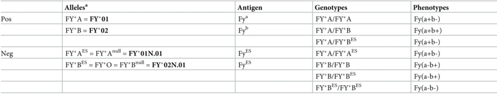
Table 1. Major Duffy alleles, genotypes and phenotypes described in human populations and alternate nomenclature.
Allelesa Antigen Genotypes Phenotypes
Pos FY�A = FY�01 Fya FY�A/FY�A Fy(a+b-)
FY�B = FY�02 Fyb
FY�A/FY�B Fy(a+b+)
FY�A/FY�BES Fy(a+b-)
Neg FY�AES= FY�Anull= FY�01N.01 FyES FY�A/FY�AES Fy(a+b-)
FY�BES = FY�O = FY�Bnull = FY�02N.01 FyES FY�B/FY�B Fy(a-b+) FY�B/FY�BES Fy(a-b+) FY�BES/FY�BES Fy(a-b-) a
The official allele nomenclature is indicated in bold. Neg, negative, Pos, positive
Discrepant results have been reported concerning the impact on
P. vivax infection of
hav-ing only one ES allele. It was initially shown in Papua New Guinea that FY
�A/FY
�A
EShetero-zygous individuals were at lower risk of
P. vivax infection compared to FY
�A/FY
�A people (in
Papua, the
−67 T to C mutation has arisen upstream of the FY
�A allele, independently of the
FY
�B
ESfound in Africa) [
30
,
32
]. Similarly, in the Brazilian Amazon, heterozygous FY
�A/
FY
�B
ESindividuals were at lower risk of
P. vivax malaria compared to FY
�A/FY
�B people [
33
].
However, in the same Brazilian study, heterozygous FY
�B/FY
�B
ESindividuals were at
increased risk of malaria infection compared to homozygous FY
�A/FY
�B people [
33
]. These
apparently conflicting observations might simply reflect differences between the numbers of
individuals analyzed for each genotype, resulting in underpowered analysis. More
epidemio-logical studies in different endemic settings are needed to get a clearer understanding of the
associations between the Duffy genotypes and infection outcomes.
In addition, independently of the ES genotype, there seems to be a protective effect of the
FY
�A allele compared to the FY
�B one against
P. vivax infections. Indeed, in vitro studies have
shown that the PvDBP binding to the FY
�A allele was reduced compared to binding to the
FY
�B allele [
34
]. It was also observed that FY
�A was associated with clinical protection, while
FY
�B was associated with increased infection risk by
P. vivax [
33
,
34
].
Altogether, those studies suggest that the link between Duffy genotypes and susceptibility
to
P. vivax is more complex than just the association between Duffy-negative and full
protection.
Evidence of P. vivax infections in Duffy-negative people
Travelers infected by
P. vivax coming back from sub-Saharan African
regions
The first indirect evidence of
P. vivax infection in Duffy-negative individuals comes from the
reports of travelers presenting a
P. vivax infection after returning from African areas where
Duffy-negative is at near fixation [
35
,
36
]. However, those observations did not question the prevailing
dogma of Duffy-negative refractoriness for several reasons. First,
P. vivax is morphologically
simi-lar to
P. ovale endemic in most Africa, which could lead to microscopic misinterpretation. The
advent of molecular techniques to diagnose parasites have ruled out this confounding factor and
have allowed to unequivocally identify
P. vivax [
37
–
39
]. Second, because of the occurrence of
P.
vivax hypnozoites hiding in the liver for months or even years before triggering a relapse infection,
the origin of the parasite is difficult to determine. Third, it was suggested that a small proportion
of Duffy-positive individuals is enough to sustain the transmission cycle of
P. vivax independently
of Duffy-negative infections [
40
]. Similarly, the detection of
P. vivax in Anopheles mosquitoes
col-lected in areas where the majority of people are Duffy-negative cannot conclusively demonstrate
Duffy-negative infections [
14
]. Therefore, additional evidence is clearly needed to demonstrate the
ability of
P. vivax to infect Duffy-negative individuals.
P. vivax infections in Duffy-negative people from sub-Saharan Africa
In the last decade, reports of
P. vivax infections in Duffy-negative individuals have increased
steadily. There is no doubt that the advent of molecular techniques unambiguously detecting
P. vivax at high sensitivity has greatly contributed to those observations. However, those
obser-vations come with caveats that can only be resolved by microscopic obserobser-vations. Indeed,
detection of
P. vivax DNA by PCR in a Duffy-negative individual’s blood sample can arise from
pre-erythrocytic stages of the parasites even in the absence of actual red blood cell invasion [
41
].
Similarly, seropositivity to
P. vivax antigens can result from the presence of pre-erythrocytic
parasite forms independently of blood-stage infection (not to mention the possible
crossreactiv-ity of the serological analysis with other
Plasmodium species) [
42
]. Therefore, microscopic
observation of
P. vivax within a Duffy-negative erythrocyte, along with genotyping
confirma-tion (especially to exclude a
P. ovale infection), is necessary to prove Duffy-negative infection.
The first mention of a microscopic observation of
P. vivax in a Duffy-negative erythrocyte was
made in 2006 [
14
] (Culleton and colleagues [
40
] mentioned a report by Van Ros in 1985 but we
could not retrieve the original publication). However, when the slides were double read by two
expert microscopists for confirmation,
P. vivax could not be confirmed [
14
]. It is in 2010 that
the definitive evidence of a
P. vivax infection within Duffy-negative erythrocyte from patients
living in Madagascar was reported [
15
]. It was hypothesized that it is the admixture of
Duffy-positive and Duffy-negative people occurring in Madagascar that has allowed the parasites to
become adapted to Duffy-negative individuals. Of note, when Bray assessed the susceptibility of
30 Nigerian people to
P. vivax through experimental mosquito bite infections, he did observe P.
vivax blood-stage infection in one of the subjects [
43
]. It can be speculated that this Nigerian
individual was Duffy-negative, though this cannot be confirmed. Interestingly, the strain used
by Bray was the “Madagascar strain” of
P. vivax, and it is tempting to speculate that its Malagasy
genetic background allowed the parasite to infect a Duffy-negative individual. However, the
exact origin of this strain is controversial, and it cannot be ascertained that it indeed comes
from Madagascar [
2
,
44
–
46
]. Since then, further unequivocal
P. vivax Duffy-negative infections
were reported from Ethiopia where Duffy-negative and Duffy-positive people coexist [
47
,
48
]
and also from countries where Duffy negativity is at near fixation, such as Cameroon [
49
] and
Mali [
50
] (though in some of those studies the morphological features of erythrocytic
P. vivax
were not well resolved). Additionally, the detection of
P. vivax in Duffy-negative individuals
either by molecular diagnostic or serological analysis only has been made in a large number of
sub-Saharan countries (for extensive review, see [
36
]). The description of Duffy-negative
patients harboring microscopically-confirmed
P. vivax erythrocyte infections coupled to
molec-ular diagnostic confirmation in genotyped Duffy-negative individuals have undoubtedly
chal-lenged the established paradigm that prevailed for decades, opening new research questions
regarding this neglected malaria parasite.
P. vivax infections of Duffy-negative people from the American continent
Current American populations are characterized as an admixture of individuals of different
ancestries, due to the historical human migrations over the last centuries (European, African,
Native American, Asian, etc.). Depending on the areas, some variable numbers of people are
Duffy-negative due to their African ancestry and are distributed throughout South America up to
southern United States of America [
6
]. In South and Central America, where
P. vivax is endemic,
the paradigm of Duffy-negative protecting against
P. vivax malaria was quite well established
with a number of studies from diverse locations [
51
–
56
]. However, similarly to the situation in
Africa, several studies have suggested through serological, molecular, and microscopic detection
analysis that Duffy negativity is not completely protective against
P. vivax infection [
33
,
57
–
59
].
All those studies clearly show that the protection of the Duffy negativity does not constitute
an absolute barrier against
P. vivax infections.
Towards understanding the mechanisms of Duffy-negative invasion by
P.
vivax
How does
P. vivax invade Duffy-negative red blood cells? Has P. vivax recently evolved
strate-gies to infect Duffy-negative erythrocytes, or are alternative invasion pathways ubiquitous in
strategies to prevent the emergence of Duffy-negative infections. No clear answer has been
made yet, mainly because of the inherent difficulty in working on
P. vivax as there is still no in
vitro continuous culture available for this species [
60
,
61
]; however, some hypothesis can be
raised on the parasite and/or human sides.
Parasite side: PvDBP duplication and/or other parasite ligands?
The first mechanistic hypothesis that has been made following the observation of
Duffy-nega-tive,
P. vivax-infected patients in Madagascar came through whole genome sequencing (WGS)
of field isolates. This analysis showed a duplication in the PvDBP gene in Malagasy isolates,
and further molecular epidemiology surveys revealed that this duplication was observed in
parasites isolated from a wide range of geographic locations (South America, East Africa,
Mad-agascar, Asia-Pacific) [
62
]. Noteworthy, the highest frequency of isolates with
pvdbp
duplica-tion (nearly 53%) was detected in Madagascar, in comparison to other areas (12.5% in Sudan,
9% in Cambodia). It was thus speculated that this duplication might have been selected in
Madagascar to respond to the Duffy negativity barrier perhaps by increasing the amount of
PvDBP protein on the merozoite’s surface to bind to an unknown low-affinity receptor [
62
].
Interestingly, in two different studies,
P. vivax-infecting, Duffy-negative individuals from
Ethi-opia (where the prevalence of
pvdbp amplification is between 55% to 80% [
63
,
64
]) carried
multiple copies of
pvdbp, although the number of individuals were too low to draw definitive
conclusions (only two in each report) [
48
,
64
]. However, PvDBP itself has been shown to not
bind to Duffy-negative erythrocytes, questioning how it could be involved in Duffy-negative
invasion [
18
,
48
]. Furthermore, the analysis by WGS of more than 200
P. vivax isolates from
around the world indicated that 33% of Cambodian parasites carried the
pvdbp duplication,
much higher than the previously reported 9% [
65
]. In fact the initial PCR-based surveys have
missed isolates carrying the duplication, because of variation in the boundaries of the
duplica-tion among parasites [
66
]. Using a quantitative PCR assay that enables the gene copy number
assessment independently of the duplication boundaries, no difference in the
pvdbp copy
number between Cambodian parasites (where there is virtually no Duffy-negative individuals)
and Malagasy ones (where an admixture of Duffy-negative and Duffy-positive individuals
coexist) was observed [
67
]. Those results suggest that
pvdbp amplification is not selected in
response to the Duffy negativity barrier [
67
]. In a recent molecular epidemiology study
con-ducted on Ethiopian
P. vivax isolates, it was observed that the proportion of parasites with
pvdbp amplification was higher in individuals carrying the FY
�A allele compared to
individu-als carrying the FY
�B one [
64
]. As mentioned above, because the binding of PvDBP to FY
�A is
lower than to FY
�B, it can be speculated that
pvdbp amplification could have been selected to
increase the affinity to erythrocytes expressing the FY
�A allele by supposedly increasing the
amount of PvDBP protein at the surface of the merozoites. In the same study, the proportion
of parasites with multiple
pvdbp copies was significantly higher in symptomatic patients
com-pared to asymptomatic individuals, raising the possibility that
pvdbp amplification is involved
in immune evasion of the parasites. Further investigations are clearly needed to understand
the role of
pvdbp amplification.
In addition to copy number variation, there is a number of evidence pointing to the absence
of a specific sequence polymorphism in PvDBP in relation to Duffy-negative invasion.
Recently, PvDBP sequence analysis of parasites infecting Duffy-negative and Duffy-positive
people from Sudan showed that most alleles were shared between parasites, and those alleles
were found globally [
68
]. Also, recombinant PvDBP alleles isolated from two Duffy-negative
individuals infected by
P. vivax in Ethiopia were expressed in vitro, and none could bind to
There are other parasite ligands described, such as
P. vivax reticulocyte binding proteins
(PvRBPs), although they have received much less attention than PvDBP and their role in
eryth-rocyte invasion (whether Duffy-positive or Duffy-negative) for most of them is yet to be
deter-mined [
69
,
70
]. PvRBPs are a family of ligands shown to bind to erythrocytes, but conflicting
results were obtained for some of them regarding specific binding to reticulocytes [
70
,
71
].
Among the different PvRBPs, the binding to Duffy-negative erythrocytes was evaluated only
for PvRBP1a and PvRBP2c, and both were found to bind to Duffy-negative reticulocytes,
indi-cating they could be involved in Duffy-independent invasion pathways [
69
].
Naturally-acquired antibodies against some RBPs inhibiting binding to erythrocytes or being associated
with clinical protection have been described; however, the involvement of those ligands in
actual parasite invasion was not assessed, except for PvRBP1a and PvRBP2b [
72
–
77
]. While
PvRBP1a does not seem to be critical for
P. vivax invasion in Duffy-positive cells [
75
],
PvRBP2b was recently shown to be a key ligand involved in reticulocyte recognition and
inva-sion through the transferrin receptor 1 (TfR1) [
76
]. However, the roles of PvRBP1a and of
PvRBP2b–TfR1 have not been evaluated specifically in the invasion of Duffy-negative red
blood cells. Recently,
P. vivax merozoite surface protein-1 paralog (PvMSP1P) and P. vivax
glycosylphosphatidylinositol-anchored micronemal antigen (PvGAMA) were shown to bind
to both Duffy-positive and Duffy-negative red blood cells, and antibodies against PvMSP1P
blocked the related
P. knowlesi invasion in Duffy-positive reticulocytes [
78
–
80
]. Those results
suggest that PvGAMA and PvMSP1P are ligands that could be involved in Duffy-independent
reticulocyte invasion pathways, although functional demonstration is still lacking. Finally,
P.
vivax erythrocyte-binding protein (PvEBP, also known as PvDBP2) has also been shown to
moderately bind to Duffy-negative reticulocytes [
48
,
81
,
82
]. Of interest, it was recently shown
that this gene can be amplified and a higher proportion of parasites from Madagascar (where
Duffy-negative and Duffy-positive people coexist) carry multiple copies of PvEBP compared
to isolates from Cambodia (where only Duffy-positive people are present) [
67
]. Also, while
only duplication was observed in Cambodia, up to five copies of PvEBP were detected in
Mala-gasy parasites [
67
]. Altogether, those results suggest that PvEBP might be also involved in
Duffy-negative invasion mechanisms, but again, functional demonstration is still lacking.
Functional studies need to be developed in the future to figure out the role of each of the
men-tioned parasite ligands in the invasion of Duffy-positive and Duffy-negative reticulocytes as
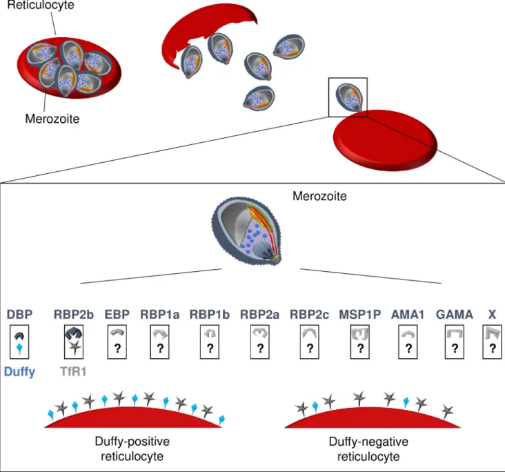
well as to identify possible unknown ligands involved in alternative invasion pathways (
Fig 1
).
Human side: Alternate receptor or Duffy involvement?
On the human erythrocyte side, the main question is: Through what receptor is
P. vivax able
to enter Duffy-negative red blood cells? While for
P. falciparum many receptors have been
described to ensure the parasite entry through multiple pathways, for
P. vivax (until recently)
only the Duffy protein was known. As mentioned above, TfR1 has recently been identified as
the receptor to PvRBP2b for reticulocyte recognition and invasion [
76
]. TfR1 is one of the
many membrane proteins lost during red blood cell maturation and is thus absent from
nor-mocytes [
83
,
84
]. It is present on both Duffy-positive and Duffy-negative reticulocytes;
how-ever, it is believed that the critical interaction between PvRBP2b and TfR1 occurs upstream of
the PvDBP–Duffy one, not independently of it [
76
]. Anyway, the role of this interaction in
Duffy-negative has not been assessed, and an alternate human red blood cell receptor involved
in Duffy-independent invasion pathways has yet to be discovered.
Could the invasion of Duffy-negative people require a particular Duffy protein not
recog-nized by serological tools and therefore falsely assigned as negative? Probably not, as
sequenc-ing of this gene has consistently shown that the basis for Duffy-negative is a ssequenc-ingle mutation in
the promoter of the gene upstream of the FY
�B allele in African populations [
24
]. More
con-clusively, the full coding sequence of the Duffy gene, including its promoter, of 14
Duffy-Fig 1. Schematic representation of receptor-ligands involved inP. vivax invasion process of reticulocytes. At the end of the erythrocytic cycle, theschizonts burst and release merozoites in the blood stream, enabling the invasion of uninfected reticulocytes. The recognition of reticulocytes and the invasion process require interactions between parasite ligands and reticulocyte receptors. For Duffy-positive reticulocyte invasion, PvRBP2b binds first to the TfR1 present on reticulocytes, and subsequently, PvDBP engages with the Duffy protein allowing the entry of the merozoite in the cell. Other ligands such as PvEBP, PvRBPs, PvMSP1P, PvAMA1, or PvGAMA are currently being investigated for their involvement in this invasion process, and their putative receptors are unknown. PvRBP2b probably also recognizes TfR1 of Duffy-negative reticulocytes; however, the subsequent invasion steps are still unknown. Are there a few Duffy molecules present on the surface of the erythrocyte enabling parasites with multiple PvDBP gene copies to invade the cell? Conversely, is the invasion process of Duffy-negative reticulocytes occurring through alternate, yet-to-identity receptors of, perhaps, ligands such as PvEBP, PvMSP1P, or PvGAMA? Finally, the invasion process might occur through complete unknown pathways with both unidentified ligands (noted with X) and receptors. AMA1, anchored micronemal antigen 1; DBP, Duffy binding protein; EBP, erythrocyte-binding protein; GAMA,
glycosylphosphatidylinositol-anchored micronemal antigen; MSP1P, merozoite surface protein-1 paralog; Pv,Plasmodium vivax; RBP, reticulocyte
binding proteins; TfR1, transferrin receptor 1.
negative individuals actually infected by
P. vivax, have been sequenced, and all were
homozy-gous with the expected
−67 T to C mutation upstream of the FY
�B alleles, indicating that no
cryptic allele was at the origin of the infection [
15
].
However, what has not been definitely ruled out and is perhaps the most parsimonious
explanation for the mechanisms of Duffy-negative invasion is that the Duffy-negative
pheno-type is not Duffy null [
85
]. Indeed, it can be speculated that although the
−67 T to C mutation
reduces the binding of the GATA-1 transcription factor and therefore transcription of the
gene, this reduction might not be complete and RNA transcription could still be occurring at
very low levels, leading to an amount of Duffy protein below the limit of detection of current
analytical tools. Some recent data suggest that Duffy protein is notably detectable in erythroid
precursor cells typically found in the bone marrow where
P. vivax invasion is believed to occur
to some extent [
85
–
87
]. It is worth mentioning here that Duffy-negative people do express the
Duffy protein in other cell types such as in endothelial cells, indicating that the expression of
Duffy is not intrinsically abolished by the
−67 T to C mutation [
23
,
88
]. This hypothesis would
explain the very low parasitemia usually observed in Duffy-negative infected patients and also
imply that
P. vivax does not necessarily require an alternate pathway but could instead go
through the classical PvDBP−Duffy invasion process. Such a hypothesis is so far only a
specu-lation, and the involvement of an unknown receptor cannot be excluded (
Fig 1
). More
investi-gations are needed to determine how the parasite is able to infect Duffy-negative red blood
cells.
Future directions and conclusive remarks
With such evidence of
P. vivax infections in Duffy-negative people, a better understanding of
the epidemiology of this parasite is necessary and will involve the implementation of specific
diagnostics in the African continent, where apart from the Horn of Africa and Madagascar,
P.
vivax is usually not looked for. A characteristic of P. vivax infections in Duffy-negative
individ-uals is the usually low parasitemia easily missed by microscopy or conventional rapid
diagnos-tics tests (RDTs) and leading to asymptomatic infections often detected through
community-based, cross-sectional surveys [
50
,
89
]. As such, sensitive molecular detection tools such as
PCR should be implemented for
P. vivax diagnostics.
To provide better insights in the mechanisms underlying Duffy-negative infections,
next-generation sequencing technologies will be extremely useful both at the genomic and
tran-scriptomic level. By identifying genomic loci under selection in
P. vivax isolates infecting
Duffy-negative individuals compared to Duffy-positive ones or originating from areas where
Duffy negativity is at near fixation compared to areas where Duffy-positive are predominant,
candidate genes involved in the adaptation to Duffy-negative reticulocytes might be detected.
Similarly, by comparing the gene expression profiles of parasites infecting Duffy-negative or
Duffy-positive people, the signature of genes specifically regulated to respond to the Duffy
neg-ativity barrier might be identified. Such an approach could also make use of the
Saimiri and
Aotus nonhuman primate model—as both species can be infected by P. vivax, while PvDBP
can bind only to
Aotus erythrocytes and not to Saimiri ones, indicating an alternative invasion
pathway reminiscent of what is observed for Duffy-negative human erythrocytes [
90
].
Alterna-tive models to decipher erythrocyte invasion mechanisms could also rely on the genetically
tractable
P. knowlesi that shares many biological similarities with P. vivax, including the
requirement of the Duffy receptor for erythrocyte invasion [
91
] or even on
P. cynomolgi,
closely related to
P. vivax and recently adapted to in vitro culture [
92
].
Providing definitive evidence of the involvement of any candidate ligand involved in
Duffy-negative reticulocytes’ invasion will anyway require functional demonstration in a
P.
vivax human reticulocyte model. However, as mentioned before, there is still no continuous in
vitro culture for this parasite species, and consequently no genetic transformation is possible.
Only tedious, short-term cultures conducted in a handful of laboratories around the world can
be performed [
76
,
93
–
95
]. Nevertheless, using those assays in order to evaluate the capacity of
parasites with different genotypes (i.e., single or multicopy
pvdbp parasites) to invade
Duffy-negative reticulocytes in presence or absence of monoclonal antibodies specific for a candidate
ligand or receptor will allow one to determine the pathways used by
P. vivax to infect
Duffy-negative erythrocytes. In addition, on the erythrocyte side, elegant technologies have been
developed to manipulate the repertoire of proteins expressed on erythropoietic cells using an
immortalized erythroid progenitor cell line that can provide additional evidence on the
involvement of specific receptors in
P. vivax invasion [
96
]. All those approaches, combined
with structural biology techniques, have the potential to identify receptor–ligand interactions
involved in
P. vivax invasion as performed for the recent discovery of PvRBP2b–TfR1
involve-ment [
76
].
In Africa, malaria burden is largely due to the more virulent and severe
P. falciparum that
is, of course, currently the primary target of elimination strategies. However, it can be expected
that in the future while the elimination of falciparum malaria hopefully continues as seen for
the last decade [
97
], more cases of
P. vivax will be reported, as often the proportion of P. vivax
cases increases while elimination progresses [
5
]. In those countries and elsewhere, in order to
achieve elimination of all malaria species it will be necessary to implement strategies targeting
P. vivax [
98
]. Among those, a PvDBP-based vaccine is currently under clinical development
[
99
,
100
], and deciphering the molecular pathways by which
P. vivax is able to infect
Duffy-negative people will be critical for ensuring the success of such a strategy.
References
1. Barber BE, William T, Grigg MJ, Parameswaran U, Piera KA, Price RN, et al. Parasite Biomass-Related Inflammation, Endothelial Activation, Microvascular Dysfunction and Disease Severity in Vivax Malaria. PLoS Pathog. 2015; 11(1):e1004558.https://doi.org/10.1371/journal.ppat.1004558
PMID:25569250
2. Baird JK. Evidence and Implications of Mortality Associated with Acute Plasmodium vivax Malaria. Clinical Microbiology Reviews. 2013; 26(1):36–57.https://doi.org/10.1128/CMR.00074-12PMID:
23297258
3. Douglas N, Pontororing G, Lampah D, Yeo T, Kenangalem E, Poespoprodjo J, et al. Mortality attribut-able to Plasmodium vivax malaria: a clinical audit from Papua, Indonesia. BMC Medicine. 2014; 12 (1):217.https://doi.org/10.1186/s12916-014-0217-zPMID:25406857
4. Siqueira A, Lacerda M, Magalhaes B, Mourao M, Melo G, Alexandre M, et al. Characterization of Plas-modium vivax-associated admissions to reference hospitals in Brazil and India. BMC Medicine. 2015; 13(1):57.https://doi.org/10.1186/s12916-015-0302-yPMID:25889040
5. Battle KE, Lucas TCD, Nguyen M, Howes RE, Nandi AK, Twohig KA, et al. Mapping the global endemicity and clinical burden of Plasmodium vivax, 2000–17: a spatial and temporal modelling study. The Lancet. 2019.https://doi.org/10.1016/S0140-6736(19)31096-7.
6. Howes RE, Patil AP, Piel FB, Nyangiri OA, Kabaria CW, Gething PW, et al. The global distribution of the Duffy blood group. Nat Commun. 2011; 2:266.https://doi.org/10.1038/ncomms1265PMID:
21468018
7. Rosenberg R. Plasmodium vivax in Africa: hidden in plain sight? Trends Parasitol. 2007; 23(5):193–6.
https://doi.org/10.1016/j.pt.2007.02.009PMID:17360237
8. Mayne B. NOTE ON EXPERIMENTAL INFECTION OF ANOPHELES PUNCTIPENNIS WITH QUAR-TAN MALARIA. Public Health Reports 1932; 47(35):1771–811.https://doi.org/10.2307/4580870
9. Boyd MF, Stratman-Thomas WK. STUDIES ON BENIGN TERTIAN MALARIA: 4. ON THE REFRAC-TORINESS OF NEGROES TO INOCULATION WITH PLASMODIUM VIVAX. American Journal of Epidemiology. 1933; 18(2):485–9.
10. Young MD, Don EE, Burgess RW, Jeffery GM. Experimental Testing of the Immunity of Negroes to Plasmodium vivax. The Journal of Parasitology. 1955; 41(3):315–8.https://doi.org/10.2307/3274214
11. Sanger R, Race RR, Jack J. The Duffy Blood Groups of New York Negroes: The Phenotype Fy (a−b −). British Journal of Haematology. 1955; 1(4):370–4.https://doi.org/10.1111/j.1365-2141.1955. tb05523.xPMID:13269673
12. Miller L, Mason S, Dvorak J, McGinniss M, Rothman I. Erythrocyte receptors for (Plasmodium know-lesi) malaria: Duffy blood group determinants. Science. 1975; 189(4202):561–3.https://doi.org/10. 1126/science.1145213PMID:1145213
13. Miller LH, Mason SJ, Clyde DF, McGinniss MH. The Resistance Factor to Plasmodium vivax in Blacks. N Engl J Med. 1976; 295(6):302–4.https://doi.org/10.1056/NEJM197608052950602PMID:778616. 14. Ryan JR, Stoute JA, Amon J, Dunton RF, Mtalib R, Koros J, et al. Evidence for transmission of
Plas-modium vivax among a duffy antigen negative population in Western Kenya. The American journal of tropical medicine and hygiene. 2006; 75(4):575–81. PMID:17038676
15. Me´nard D, Barnadas C, Bouchier C, Henry-Halldin C, Gray LR, Ratsimbasoa A, et al. Plasmodium vivax clinical malaria is commonly observed in Duffy-negative Malagasy people. Proceedings of the National Academy of Sciences. 2010; 107(13):5967–71.https://doi.org/10.1073/pnas.0912496107
PMID:20231434
16. Bachelerie F, Ben-Baruch A, Burkhardt AM, Combadiere C, Farber JM, Graham GJ, et al. Interna-tional Union of Basic and Clinical Pharmacology. [corrected]. LXXXIX. Update on the extended family of chemokine receptors and introducing a new nomenclature for atypical chemokine receptors. Phar-macol Rev. 2014; 66(1):1–79.https://doi.org/10.1124/pr.113.007724PMID:24218476.
17. Horuk R, Chitnis CE, Darbonne WC, Colby TJ, Rybicki A, Hadley TJ, et al. A receptor for the malarial parasite Plasmodium vivax: the erythrocyte chemokine receptor. Science. 1993; 261(5125):1182.
https://doi.org/10.1126/science.7689250PMID:7689250
18. Chitnis C, Miller LH. Identification of the erythrocyte binding domains ofPlasmodium vivax and Plasmo-dium knowlesi proteins involved in erythrocyte invasion. Journal of Experimental Medicine. 1994; 180:497–506.https://doi.org/10.1084/jem.180.2.497PMID:8046329
19. Ho¨her G, Fiegenbaum M, Almeida S. Molecular basis of the Duffy blood group system. Blood Trans-fus. 2018; 16(1):93–100. Epub 01/30.https://doi.org/10.2450/2017.0119-16PMID:28151395. 20. Cutbush M, Mollison PL. The Duffy blood group system. Heredity (Edinb). 1950; 4(3):383–9. Epub
1950/12/01.https://doi.org/10.1038/hdy.1950.31PMID:14802995.
21. Ikin EW, Mourant AE, Pettenkofer HJ, Blumenthal G. Discovery of the expected haemagglutinin, anti-Fyb. Nature. 1951; 168(4288):1077–8. Epub 1951/12/22.https://doi.org/10.1038/1681077b0PMID:
14910641.
22. Seixas S, Ferrand N, Rocha J. Microsatellite Variation and Evolution of the Human Duffy Blood Group Polymorphism. Mol Biol Evol. 2002; 19(10):1802–6.https://doi.org/10.1093/oxfordjournals.molbev. a004003PMID:12270907
23. Chaudhuri A, Polyakova J, Zbrzezna V, Pogo AO. The coding sequence of Duffy blood group gene in humans and simians: restriction fragment length polymorphism, antibody and malarial parasite speci-ficities, and expression in nonerythroid tissues in Duffy- negative individuals. Blood. 1995; 85(3):615. PMID:7833466
24. Tournamille C, Colin Y, Cartron JP, Le Van Kim C. Disruption of a GATA motif in the Duffy gene pro-moter abolishes erythroid gene expression in Duffy-negative individuals. Nat Genet. 1995; 10(2):224– 8.https://doi.org/10.1038/ng0695-224PMID:7663520
25. Tournamille C, Blancher A, Le Van Kim C, Gane P, Apoil PA, Nakamoto W, et al. Sequence, evolution and ligand binding properties of mammalian Duffy antigen/receptor for chemokines. Immunogenetics. 2004; 55(10):682–94.https://doi.org/10.1007/s00251-003-0633-2PMID:14712331
26. Li J, Iwamoto S, Sugimoto N, Okuda H, Kajii E. Dinucleotide repeat in the 30flanking region provides a
clue to the molecular evolution of the Duffy gene. Hum Genet. 1997; 99(5):573–7.https://doi.org/10. 1007/s004390050408PMID:9150720
27. Michon P, Woolley I, Wood EM, Kastens W, Zimmerman PA, Adams JH. Duffy-null promoter hetero-zygosity reduces DARC expression and abrogates adhesion of the P. vivax ligand required for blood-stage infection. FEBS Letters. 2001; 495(1–2):111–4. https://doi.org/10.1016/s0014-5793(01)02370-5PMID:11322957
28. Yazdanbakhsh K, Rios M, Storry JR, Kosower N, Parasol N, Chaudhuri A, et al. Molecular mecha-nisms that lead to reduced expression of Duffy antigens. Transfusion. 2000; 40(3):310–20.https://doi. org/10.1046/j.1537-2995.2000.40030310.xPMID:10738032
29. Woolley IJ, Hotmire KA, Sramkoski RM, Zimmerman PA, Kazura JW. Differential expression of the Duffy antigen receptor for chemokines according to RBC age and FY genotype. Transfusion. 2000; 40 (8):949–53.https://doi.org/10.1046/j.1537-2995.2000.40080949.xPMID:10960522
30. Zimmerman PA, Woolley I, Masinde GL, Miller SM, McNamara DT, Hazlett F, et al. Emergence of FY*Anull in a Plasmodium vivax-endemic region of Papua New Guinea. Proceedings of the National Academy of Sciences. 1999; 96(24):13973–7.https://doi.org/10.1073/pnas.96.24.13973PMID:
10570183
31. Hamblin MT, Di Rienzo A. Detection of the signature of natural selection in humans: evidence from the Duffy blood group locus. Am J Hum Genet. 2000; 66(5):1669–79. Epub 04/12.https://doi.org/10.1086/ 302879PMID:10762551.
32. Kasehagen LJ, Mueller I, Kiniboro B, Bockarie MJ, Reeder JC, Kazura JW, et al. Reduced Plasmo-dium vivax erythrocyte infection in PNG Duffy-negative heterozygotes. PLoS ONE. 2007; 2(3):e336– e.https://doi.org/10.1371/journal.pone.0000336PMID:17389925.
33. Kano FS, de Souza AM, de Menezes Torres L, Costa MA, Souza-Silva FA, Sanchez BAM, et al. Sus-ceptibility to Plasmodium vivax malaria associated with DARC (Duffy antigen) polymorphisms is influ-enced by the time of exposure to malaria. Scientific reports. 2018; 8(1):13851–.https://doi.org/10. 1038/s41598-018-32254-zPMID:30218021.
34. King CL, Adams JH, Xianli J, Grimberg BT, McHenry AM, Greenberg LJ, et al. Fy(a)/Fy(b) antigen polymorphism in human erythrocyte Duffy antigen affects susceptibility to Plasmodium vivax malaria. Proc Natl Acad Sci U S A. 2011; 108(50):20113–8.https://doi.org/10.1073/pnas.1109621108
PMC3250126. PMID:22123959
35. Howes RE, Reiner RC Jr., Battle KE, Longbottom J, Mappin B, Ordanovich D, et al. Plasmodium vivax Transmission in Africa. PLoS Negl Trop Dis. 2015; 9(11):e0004222–e.https://doi.org/10.1371/journal. pntd.0004222PMID:26587988.
36. Twohig KA, Pfeffer DA, Baird JK, Price RN, Zimmerman PA, Hay SI, et al. Growing evidence of Plas-modium vivax across malaria-endemic Africa. PLoS Negl Trop Dis. 2019; 13(1):e0007140–e.https:// doi.org/10.1371/journal.pntd.0007140PMID:30703083.
37. Rubio JM, Benito A, Roche J, Berzosa PJ, Garcı´a ML, Mico´ M, et al. Semi-nested, multiplex polymer-ase chain reaction for detection of human malaria parasites and evidence of Plasmodium vivax infec-tion in Equatorial Guinea. The American Journal of Tropical Medicine and Hygiene. 1999; 60(2):183– 7.https://doi.org/10.4269/ajtmh.1999.60.183PMID:10072133
38. Gautret P, Legros F, Koulmann P, Rodier MH, Jacquemin JL. Imported Plasmodium vivax malaria in France: geographical origin and report of an atypical case acquired in Central or Western Africa. Acta Trop. 2001; 78(2):177–81.https://doi.org/10.1016/s0001-706x(00)00181-9PMID:11230828
39. BLOSSOM DB, KING CH, ARMITAGE KB. OCCULT PLASMODIUM VIVAX INFECTION DIAG-NOSED BY A POLYMERASE CHAIN REACTION–BASED DETECTION SYSTEM: A CASE REPORT. The American Journal of Tropical Medicine and Hygiene. 2005; 73(1):188–90.https://doi. org/10.4269/ajtmh.2005.73.188. PMID:16014856
40. Culleton RL, Mita T, Ndounga M, Unger H, Cravo PVL, Paganotti GM, et al. Failure to detect Plasmo-dium vivax in West and Central Africa by PCR species typing. Malaria Journal. 2008; 7(1):174.https:// doi.org/10.1186/1475-2875-7-174PMID:18783630
41. Abkallo HM, Liu W, Hokama S, Ferreira PE, Nakazawa S, Maeno Y, et al. DNA from pre-erythrocytic stage malaria parasites is detectable by PCR in the faeces and blood of hosts. Int J Parasit. 2014; 44 (7):467–73.https://doi.org/10.1016/j.ijpara.2014.03.002.
42. Culleton R, Ndounga M, Zeyrek FY, Coban C, Casimiro PN, Takeo S, et al. Evidence for the Transmis-sion of Plasmodium vivax in the Republic of the Congo, West Central Africa. J Infect Dis. 2009; 200 (9):1465–9.https://doi.org/10.1086/644510PMID:19803728
43. Bray RS. The Susceptibility of Liberians to the Madagascar Strain of Plasmodium vivax. The Journal of Parasitology. 1958; 44(4):371–3.https://doi.org/10.2307/3274317
44. Battle K, Karhunen M, Bhatt S, Gething P, Howes R, Golding N, et al. Geographical variation in Plas-modium vivax relapse. Malaria Journal. 2014; 13(1):144.https://doi.org/10.1186/1475-2875-13-144
PMID:24731298
45. Garnham PC, Bray RS, Bruce-Chwatt LJ, Draper CC, Killick-Kendrick R, Sergiev PG, et al. A strain of Plasmodium vivax characterized by prolonged incubation: morphological and biological characteris-tics. Bulletin of the World Health Organization. 1975; 52(1):21–32. PMID:764993.
46. Lover AA. Note on the origin of the Madagascar strain of Plasmodium vivax. The American journal of tropical medicine and hygiene. 2014; 91(6):1283–.https://doi.org/10.4269/ajtmh.14-0507PMID:
25473067.
47. Woldearegai TG, Kremsner PG, Kun JF, Mordmu¨ ller B. Plasmodium vivax malaria in Duffy-negative individuals from Ethiopia. Transactions of The Royal Society of Tropical Medicine and Hygiene. 2013; 107(5):328–31.https://doi.org/10.1093/trstmh/trt016PMID:23584375
48. Gunalan K, Lo E, Hostetler JB, Yewhalaw D, Mu J, Neafsey DE, et al. Role of Plasmodium vivax Duffy-binding protein 1 in invasion of Duffy-null Africans. Proc Natl Acad Sci U S A. 2016; 113
(22):6271–6. Epub 2016/05/18.https://doi.org/10.1073/pnas.1606113113PMID:27190089; PubMed Central PMCID: PMC4896682.
49. Fru-Cho J, Bumah V, Safeukui I, Nkuo-Akenji T, Titanji V, Haldar K. Molecular typing reveals substan-tial Plasmodium vivax infection in asymptomatic adults in a rural area of Cameroon. Malaria Journal. 2014; 13(1):170.https://doi.org/10.1186/1475-2875-13-170PMID:24886496
50. Niangaly A, Karthigayan G, Amed O, Coulibaly D, Sa´ JM, Adams M, et al. Plasmodium vivax Infections over 3 Years in Duffy Blood Group Negative Malians in Bandiagara, Mali. The American journal of trop-ical medicine and hygiene. 2017; 97(3):744–52. Epub 07/24.https://doi.org/10.4269/ajtmh.17-0254
PMID:28749772.
51. Spencer HC, Miller LH, Collins WE, Knud-Hansen C, McGinnis MH, Shiroishi T, et al. The Duffy Blood Group and Resistance to Plasmodium Vivax in Honduras. The American Journal of Tropical Medicine and Hygiene. 1978; 27(4):664–70.https://doi.org/10.4269/ajtmh.1978.27.664PMID:356634
52. Weppelmann TA, Carter TE, Chen Z, von Fricken ME, Victor YS, Existe A, et al. High frequency of the erythroid silent Duffy antigen genotype and lack of Plasmodium vivax infections in Haiti. Malaria Jour-nal. 2013; 12(1):30.https://doi.org/10.1186/1475-2875-12-30PMID:23347639
53. Cavasini CE, Pereira FJT, Ribeiro WL, Wunderlich G, Ferreira MU. Duffy blood group genotypes among malaria patients in Rondoˆ nia, Western Brazilian Amazon. Revista da Sociedade Brasileira de Medicina Tropical. 2001; 34:591–5.https://doi.org/10.1590/s0037-86822001000600016PMID:
11813069
54. Sousa TN, Sanchez BAM, Cera´volo IP, Carvalho LH, Brito CFA. Real-time multiplex allele-specific polymerase chain reaction for genotyping of the Duffy antigen, the Plasmodium vivax invasion recep-tor. Vox Sanguinis. 2007; 92(4):373–80.https://doi.org/10.1111/j.1423-0410.2007.00902.xPMID:
17456162
55. Ginouves M, Veron V, Musset L, Legrand E, Stefani A, Prevot G, et al. Frequency and distribution of mixed Plasmodium falciparum-vivax infections in French Guiana between 2000 and 2008. Malaria Journal. 2015; 14(1):446.https://doi.org/10.1186/s12936-015-0971-1PMID:26555553
56. Vallejo AF, Chaparro PE, Benavides Y, A´ lvarez A´, Quintero JP, Padilla J, et al. High prevalence of sub-microscopic infections in Colombia. Malaria Journal. 2015; 14(1):201.https://doi.org/10.1186/ s12936-015-0711-6PMID:25971594
57. HERRERA S, GO´ MEZ A, VERA O, VERGARA J, VALDERRAMA-AGUIRRE A, MAESTRE A, et al. ANTIBODY RESPONSE TO PLASMODIUM VIVAX ANTIGENS IN FY-NEGATIVE INDIVIDUALS FROM THE COLOMBIAN PACIFIC COAST. The American Journal of Tropical Medicine and Hygiene. 2005; 73(5_suppl):44–9.https://doi.org/10.4269/ajtmh.2005.73.44.
58. Cavasini CE, Mattos LCd, Couto A´ ADA, Bonini-Domingos CR, Valencia SH, Neiras WCdS, et al. Plas-modium vivax infection among Duffy antigen-negative individuals from the Brazilian Amazon region: an exception? Transactions of The Royal Society of Tropical Medicine and Hygiene. 2007; 101 (10):1042–4.https://doi.org/10.1016/j.trstmh.2007.04.011PMID:17604067
59. Carvalho TA, Queiroz MG, Cardoso GL, Diniz IG, Silva AN, Pinto AY, et al. Plasmodium vivax infection in Anajas, State of Para: no differential resistance profile among Duffy-negative and Duffy-positive individuals. Malaria journal. 2012; 11(1):430.
60. Bermu´dez M, Moreno-Pe´ rez DA, Are´valo-Pinzo´n G, Curtidor H, Patarroyo MA. Plasmodium vivax in vitro continuous culture: the spoke in the wheel. Malaria journal. 2018; 17(1):301–.https://doi.org/10. 1186/s12936-018-2456-5PMID:30126427.
61. Noulin F, Borlon C, Van Den Abbeele J, D’Alessandro U, Erhart A. 1912–2012: a century of research on Plasmodium vivax in vitro culture. Trends Parasitol. 2013; 29(6):286–94.https://doi.org/10.1016/j. pt.2013.03.012PMID:23623759
62. Menard D, Chan ER, Benedet C, Ratsimbasoa A, Kim S, Chim P, et al. Whole Genome Sequencing of Field Isolates Reveals a Common Duplication of the Duffy Binding Protein Gene in Malagasy Plasmo-dium vivax Strains. PLoS Negl Trop Dis. 2013; 7(11):e2489.https://doi.org/10.1371/journal.pntd. 0002489PMID:24278487
63. Auburn S, Getachew S, Pearson R, Amato R, Miotto O, Trimarsanto H, et al. Genomic analysis of plasmodium vivax in southern Ethiopia reveals selective pressures in multiple parasite mechanisms. The Journal of Infectious Diseases. 2019.
64. Lo E, Hostetler JB, Yewhalaw D, Pearson RD, Hamid MMA, Gunalan K, et al. Frequent expansion of Plasmodium vivax Duffy Binding Protein in Ethiopia and its epidemiological significance. PLoS Negl Trop Dis. 2019; 13(9):e0007222.https://doi.org/10.1371/journal.pntd.0007222PMID:31509523
65. Pearson RD, Amato R, Auburn S, Miotto O, Almagro-Garcia J, Amaratunga C, et al. Genomic analysis of local variation and recent evolution in Plasmodium vivax. Nature Genetics. 2016; 48:959.https:// doi.org/10.1038/ng.3599PMID:27348299
66. Hostetler JB, Lo E, Kanjee U, Amaratunga C, Suon S, Sreng S, et al. Independent Origin and Global Distribution of Distinct Plasmodium vivax Duffy Binding Protein Gene Duplications. PLoS Negl Trop Dis. 2016; 10(10):e0005091.https://doi.org/10.1371/journal.pntd.0005091PMID:27798646
67. Roesch C, Popovici J, Bin S, Run V, Kim S, Ramboarina S, et al. Genetic diversity in two Plasmodium vivax protein ligands for reticulocyte invasion. PLoS Negl Trop Dis. 2018; 12(10):e0006555–e.https:// doi.org/10.1371/journal.pntd.0006555PMID:30346980.
68. Hoque MR, Elfaki MMA, Ahmed MA, Lee S-K, Muh F, Ali Albsheer MM, et al. Diversity pattern of Duffy binding protein sequence among Duffy-negatives and Duffy-positives in Sudan. Malaria Journal. 2018; 17(1):297.https://doi.org/10.1186/s12936-018-2425-zPMID:30119671
69. Galinski MR, Medina CC, Ingravallo P, Barnwell JW. A reticulocyte-binding protein complex of plasmo-dium vivax merozoites. Cell. 1992; 69(7):1213–26.https://doi.org/10.1016/0092-8674(92)90642-p
PMID:1617731
70. Chan L-J, Dietrich MH, Nguitragool W, Tham W-H. Plasmodium vivax Reticulocyte Binding Proteins for invasion into reticulocytes. Cell Microbiol. 0(0):e13110.https://doi.org/10.1111/cmi.13110PMID:
31469946
71. Gunalan K, Niangaly A, Thera MA, Doumbo OK, Miller LH. Plasmodium vivax Infections of Duffy-Neg-ative Erythrocytes: Historically Undetected or a Recent Adaptation? Trends Parasitol. 2018; 34 (5):420–9.https://doi.org/10.1016/j.pt.2018.02.006PMID:29530446
72. Franc¸a CT, White MT, He W-Q, Hostetler JB, Brewster J, Frato G, et al. Identification of highly-protec-tive combinations of Plasmodium vivax recombinant proteins for vaccine development. eLife. 2017; 6: e28673.https://doi.org/10.7554/eLife.28673PMID:28949293
73. Gupta ED, Anand G, Singh H, Chaddha K, Bharti PK, Singh N, et al. Naturally Acquired Human Anti-bodies Against Reticulocyte-Binding Domains of Plasmodium vivax Proteins, PvRBP2c and PvRBP1a, Exhibit Binding-Inhibitory Activity. The Journal of Infectious Diseases. 2017; 215 (10):1558–68.https://doi.org/10.1093/infdis/jix170PMID:28379500
74. Franc¸a CT, He W-Q, Gruszczyk J, Lim NTY, Lin E, Kiniboro B, et al. Plasmodium vivax Reticulocyte Binding Proteins Are Key Targets of Naturally Acquired Immunity in Young Papua New Guinean Chil-dren. PLoS Negl Trop Dis. 2016; 10(9):e0005014.https://doi.org/10.1371/journal.pntd.0005014
PMID:27677183
75. Gupta S, Singh S, Popovici J, Roesch C, Shakri AR, Guillotte-Blisnick M, et al. Targeting a Reticulo-cyte Binding Protein and Duffy Binding Protein to Inhibit ReticuloReticulo-cyte Invasion by Plasmodium vivax. Scientific reports. 2018; 8(1):10511–.https://doi.org/10.1038/s41598-018-28757-4PMID:30002416. 76. Gruszczyk J, Kanjee U, Chan L-J, Menant S, Malleret B, Lim NTY, et al. Transferrin receptor 1 is a
reticulocyte-specific receptor for Plasmodium vivax. Science (New York, NY). 2018; 359(6371):48–55.
https://doi.org/10.1126/science.aan1078PMID:29302006.
77. He W-Q, Karl S, White MT, Nguitragool W, Monteiro W, Kuehn A, et al. Antibodies to Plasmodium vivax reticulocyte binding protein 2b are associated with protection against P. vivax malaria in popula-tions living in low malaria transmission regions of Brazil and Thailand. PLoS Negl Trop Dis. 2019; 13 (8):e0007596–e.https://doi.org/10.1371/journal.pntd.0007596PMID:31425514.
78. Han J-H, Cheng Y, Muh F, Ahmed MA, Cho J-S, Nyunt MH, et al. Inhibition of parasite invasion by monoclonal antibody against epidermal growth factor-like domain of Plasmodium vivax merozoite sur-face protein 1 paralog. Scientific Reports. 2019; 9(1):3906. https://doi.org/10.1038/s41598-019-40321-2PMID:30846737
79. Han J-H, Cho J-S, Cheng Y, Muh F, Yoo WG, Russell B, et al. Plasmodium vivax Merozoite Surface Protein 1 Paralog as a Mediator of Parasite Adherence to Reticulocytes. Infect Immun. 2018; 86(9): e00239–18.https://doi.org/10.1128/IAI.00239-18PMID:29967091
80. Cheng Y, Lu F, Wang B, Li J, Han J-H, Ito D, et al. Plasmodium vivax GPI-anchored micronemal anti-gen (PvGAMA) binds human erythrocytes independent of Duffy antianti-gen status. Scientific reports. 2016; 6:35581–.https://doi.org/10.1038/srep35581PMID:27759110.
81. Hester J, Chan ER, Menard D, Mercereau-Puijalon O, Barnwell J, Zimmerman PA, et al. De Novo Assembly of a Field Isolate Genome Reveals Novel Plasmodium vivax Erythrocyte Invasion Genes. PLoS Negl Trop Dis. 2013; 7(12):e2569.https://doi.org/10.1371/journal.pntd.0002569PMID:
24340114
82. Ntumngia FB, Thomson-Luque R, Torres LdM, Gunalan K, Carvalho LH, Adams JH. A Novel Erythro-cyte Binding Protein of Plasmodium vivax Suggests an Alternate Invasion Pathway into Duffy-Positive Reticulocytes. mBio. 2016; 7(4):e01261–16.https://doi.org/10.1128/mBio.01261-16PMID:27555313. 83. Pan B-T, Johnstone RM. Fate of the transferrin receptor during maturation of sheep reticulocytes in
vitro: Selective externalization of the receptor. Cell. 1983; 33(3):967–78. https://doi.org/10.1016/0092-8674(83)90040-5PMID:6307529
84. Malleret B, Xu F, Mohandas N, Suwanarusk R, Chu C, Leite JA, et al. Significant Biochemical, Bio-physical and Metabolic Diversity in Circulating Human Cord Blood Reticulocytes. PLoS ONE. 2013; 8 (10):e76062.https://doi.org/10.1371/journal.pone.0076062PMID:24116088
85. Dechavanne C, Dechavanne S, Metral S, Roeper B, Krishnan S, Fong R, et al. Duffy Antigen Expres-sion in Erythroid Bone Marrow Precursor Cells of Genotypically Duffy Negative Individuals. bioRxiv. 2018:508481.https://doi.org/10.1101/508481
86. Malleret B, Li A, Zhang R, Tan KSW, Suwanarusk R, Claser C, et al. Plasmodium vivax: restricted tro-pism and rapid remodeling of CD71-positive reticulocytes. Blood. 2015; 125(8):1314–24. Epub 11/20.
https://doi.org/10.1182/blood-2014-08-596015PMID:25414440.
87. Obaldia N 3rd, Meibalan E, Sa JM, Ma S, Clark MA, Mejia P, et al. Bone Marrow Is a Major Parasite Reservoir in Plasmodium vivax Infection. mBio. 2018; 9(3):e00625–18.https://doi.org/10.1128/mBio. 00625-18PMID:29739900.
88. Peiper SC, Wang ZX, Neote K, Martin AW, Showell HJ, Conklyn MJ, et al. The Duffy antigen/receptor for chemokines (DARC) is expressed in endothelial cells of Duffy negative individuals who lack the erythrocyte receptor. The Journal of experimental medicine. 1995; 181(4):1311–7.https://doi.org/10. 1084/jem.181.4.1311PMID:7699323.
89. Lo E, Yewhalaw D, Zhong D, Zemene E, Degefa T, Tushune K, et al. Molecular epidemiology of Plas-modium vivax and PlasPlas-modium falciparum malaria among Duffy-positive and Duffy-negative popula-tions in Ethiopia. Malaria journal. 2015; 14:84–.https://doi.org/10.1186/s12936-015-0596-4PMID:
25884875.
90. Gunalan K, Sa´ JM, Moraes Barros RR, Anzick SL, Caleon RL, Mershon JP, et al. Transcriptome profil-ing of Plasmodium vivax in Saimiri monkeys identifies potential ligands for invasion. Proceedprofil-ings of the National Academy of Sciences. 2019; 116(14):7053.https://doi.org/10.1073/pnas.1818485116
PMID:30872477
91. Mohring F, Hart MN, Rawlinson TA, Henrici R, Charleston JA, Diez Benavente E, et al. Rapid and iter-ative genome editing in the malaria parasite Plasmodium knowlesi provides new tools for P. vivax research. eLife. 2019; 8:e45829.https://doi.org/10.7554/eLife.45829PMID:31205002.
92. Chua ACY, Ong JJY, Malleret B, Suwanarusk R, Kosaisavee V, Zeeman A-M, et al. Robust continu-ous in vitro culture of the Plasmodium cynomolgi erythrocytic stages. Nature communications. 2019; 10(1):3635–.https://doi.org/10.1038/s41467-019-11332-4PMID:31406175.
93. Russell B, Suwanarusk R, Borlon C, Costa FTM, Chu CS, Rijken MJ, et al. A reliable ex vivo invasion assay of human reticulocytes by Plasmodium vivax. Blood. 2011; 118(13):e74–e81.https://doi.org/10. 1182/blood-2011-04-348748PMID:21768300
94. Urusova D, Carias L, Huang Y, Nicolete VC, Popovici J, Roesch C, et al. Structural basis for neutrali-zation of Plasmodium vivax by naturally acquired human antibodies that target DBP. Nature Microbiol-ogy. 2019; 4(9):1486–96.https://doi.org/10.1038/s41564-019-0461-2PMID:31133752
95. Prajapati SK, Borlon C, Rovira-Vallbona E, Gruszczyk J, Menant S, Tham W-H, et al. Complement Receptor 1 availability on red blood cell surface modulates Plasmodium vivax invasion of human retic-ulocytes. Scientific reports. 2019; 9(1):8943–.https://doi.org/10.1038/s41598-019-45228-6PMID:
31221984.
96. Scully EJ, Shabani E, Rangel GW, Gruring C, Kanjee U, Clark MA, et al. Generation of an immortal-ized erythroid progenitor cell line from peripheral blood: A model system for the functional analysis of Plasmodium spp. invasion. Am J Hematol. 2019. Epub 2019/05/31.https://doi.org/10.1002/ajh.25543
PMID:31148215.
97. Weiss DJ, Lucas TCD, Nguyen M, Nandi AK, Bisanzio D, Battle KE, et al. Mapping the global preva-lence, incidence, and mortality of Plasmodium falciparum, 2000–17: a spatial and temporal modelling study. The Lancet. 2019.https://doi.org/10.1016/S0140-6736(19)31097-9.
98. Lover AA, Baird JK, Gosling R, Price RN. Malaria Elimination: Time to Target All Species. The Ameri-can Journal of Tropical Medicine and Hygiene. 2018; 99(1):17–23. https://doi.org/10.4269/ajtmh.17-0869PMID:29761762
99. Singh K, Mukherjee P, Shakri AR, Singh A, Pandey G, Bakshi M, et al. Malaria vaccine candidate based on Duffy-binding protein elicits strain transcending functional antibodies in a Phase I trial. NPJ vaccines. 2018;3:48-.https://doi.org/10.1038/s41541-017-0043-3PMID:30302285.
100. Payne RO, Silk SE, Elias SC, Milne KH, Rawlinson TA, Llewellyn D, et al. Human vaccination against Plasmodium vivax Duffy-binding protein induces strain-transcending antibodies. JCI Insight. 2017; 2 (12):e93683.https://doi.org/10.1172/jci.insight.93683PMID:28614791.