HAL Id: hal-03001011
https://hal.inrae.fr/hal-03001011
Submitted on 5 Jan 2021
HAL is a multi-disciplinary open access
archive for the deposit and dissemination of sci-entific research documents, whether they are pub-lished or not. The documents may come from teaching and research institutions in France or abroad, or from public or private research centers.
L’archive ouverte pluridisciplinaire HAL, est destinée au dépôt et à la diffusion de documents scientifiques de niveau recherche, publiés ou non, émanant des établissements d’enseignement et de recherche français ou étrangers, des laboratoires publics ou privés.
homeostasis: a role in ectomycorrhizal symbiosis
between Hebeloma cylindrosporum and Pinus pinaster
Kevin Garcia, Maria del Carmen Guerrero-Galan, Hannah E.R. Frank,
Muhammad Zulqurnain Haider, Amandine Delteil, Geneviève Conejero,
Raphaël Lambilliotte, Cécile Fizames, Hervé Sentenac, Sabine Dagmar
Zimmermann
To cite this version:
Kevin Garcia, Maria del Carmen Guerrero-Galan, Hannah E.R. Frank, Muhammad Zulqurnain Haider, Amandine Delteil, et al.. Fungal Shaker-like channels beyond cellular K+ homeostasis: a role in ectomycorrhizal symbiosis between Hebeloma cylindrosporum and Pinus pinaster. PLoS ONE, Public Library of Science, 2020, 15, �10.1371/journal.pone.0242739�. �hal-03001011�
RESEARCH ARTICLE
Fungal Shaker-like channels beyond cellular K
+
homeostasis: A role in ectomycorrhizal
symbiosis between Hebeloma cylindrosporum
and Pinus pinaster
Kevin GarciaID1*, Carmen Guerrero-Gala´nID2, Hannah E. R. Frank1, Muhammad
Zulqurnain Haider2¤, Amandine Delteil2, Geneviève Cone´je´ro2,3, Raphae¨l Lambilliotte2, Ce´cile Fizames2, Herve´ Sentenac2, Sabine D. ZimmermannID2*
1 Department of Crop and Soil Sciences, North Carolina State University, Raleigh, California, United States
of America, 2 BPMP, Universite´ de Montpellier, CNRS, INRAE, Institut Agro, Montpellier, France,
3 Plateforme Histocytologie et Imagerie Cellulaire Ve´ge´tale, INRA-CIRAD Montpellier, France ¤ Current address: Department of Botany, Government College University, Faisalabad, Pakistan
*kgarcia2@ncsu.edu(KG);sabine.zimmermann@cnrs.fr(SDZ)
Abstract
Potassium (K+) acquisition, translocation and cellular homeostasis are mediated by various membrane transport systems in all organisms. We identified and described an ion channel in the ectomycorrhizal fungus Hebeloma cylindrosporum (HcSKC) that harbors features of animal voltage-dependent Shaker-like K+channels, and investigated its role in both free-liv-ing hyphae and symbiotic conditions. RNAi lines affected in the expression of HcSKC were produced and used for in vitro mycorrhizal assays with the maritime pine as host plant, under standard or low K+conditions. The adaptation of H. cylindrosporum to the downregu-lation of HcSKC was analyzed by qRT-PCR analyses for other K+-related transport proteins: the transporters HcTrk1, HcTrk2, and HcHAK, and the ion channels HcTOK1, HcTOK2.1, and HcTOK2.2. Downregulated HcSKC transformants displayed greater K+contents at standard K+only. In such conditions, plants inoculated with these transgenic lines were impaired in K+nutrition. Taken together, these results support the hypothesis that the reduced expression of HcSKC modifies the pool of fungal K+available for the plant and/or affects its symbiotic transfer to the roots. Our study reveals that the maintenance of K+ trans-port in H. cylindrosporum, through the regulation of HcSKC expression, is required for the K+nutrition of the host plant.
Introduction
Potassium (K+) is an essential cation involved in many biological processes in plants, such as growth, photosynthesis and stress tolerance [1,2]. However, most soil K+cations are com-plexed with minerals such as feldspars or micas, resulting in low mobility and availability. As a consequence, productivity of agricultural and agroforestry ecosystems is strongly dependent on large and regular addition of chemical fertilizers [3]. Plants developed various strategies to a1111111111 a1111111111 a1111111111 a1111111111 a1111111111 OPEN ACCESS
Citation: Garcia K, Guerrero-Gala´n C, Frank HER,
Haider MZ, Delteil A, Cone´je´ro G, et al. (2020) Fungal Shaker-like channels beyond cellular K+
homeostasis: A role in ectomycorrhizal symbiosis between Hebeloma cylindrosporum and Pinus
pinaster. PLoS ONE 15(11): e0242739.https://doi. org/10.1371/journal.pone.0242739
Editor: Erika Kothe, Friedrich Schiller University,
GERMANY
Received: May 20, 2020 Accepted: November 7, 2020 Published: November 20, 2020
Copyright:© 2020 Garcia et al. This is an open access article distributed under the terms of the
Creative Commons Attribution License, which permits unrestricted use, distribution, and reproduction in any medium, provided the original author and source are credited.
Data Availability Statement: All relevant data are
within the manuscript and itsSupporting Informationfiles.
Funding: RL and CGG were financially supported
by grants from the French Minister of Research and Technology, AD by funding of the ANR project "TRANSMUT" 2010 BLAN 1604 03. KG
acknowledges support of the North Carolina Agriculture Research Service (NCARS) and the North Carolina Soybean Producers Association
overcome this lack of K+availability and to improve its uptake, such as expression of high-affinity transport systems, exudation of organic acids by the root system, and symbiotic associ-ations with soil-living microbes (reviewed in [4–7]).
Ectomycorrhizal (ECM) symbiosis is an intimate relationship between the roots of woody plants and the mycelium of soil-borne fungi, and constitutes a major component of boreal and temperate forest ecosystems [8]. The main benefit of this association for the host plants is the improvement of macro- and micronutrient acquisition [9]. In ECM structures, nutrients are transferred from the soil-exploring extraradical mycelium to the hyphae developingin planta,
through a fungal sheath surrounding the roots. The intra-radical colonization by the fungus forms a specific structure called the Hartig net. This net develops between the host root epider-mis and cortex, where various transport proteins are expressed to drive nutrient exchanges between the two partners [9,10]. While most reports have focused on nitrogen and phosphorus nutrition in ECM plants, some studies have reported a significant transfer of fungal K+towards the host, particularly in K+-limited and stressful conditions [11,12]. One hypothesis is that the ectomycorrhiza-mediated K+nutrition could also be a strategy for the host plant to alleviate salt stress responses [13]. The transfer of K+through the mycorrhizal pathway requires the expression and organization of fungal transport proteins specific to the soil-fungus or plant-fungus interfaces, allowing K+movements from the soil into colonized roots. The fine tuning of this putative fungal hyphae polarization seems to be required for the distribution of K+from the soil to plant cortical apoplasm [4].
Several proteins from ECM fungi involved in plant nutrient allocation have been identified and characterized [9,14–16], and some of these might be responsible for the maintenance of ECM symbiosis, in addition to exclusive involvement in trophic exchanges [17]. To our knowl-edge, only a handful of transport proteins involved in K+transport has been reported in ECM fungi thus far. Among them,HcTrk1 from Hebeloma cylindrosporum [18] is specifically local-ized in extra-radical hyphae ofH. cylindrosporum-Pinus pinaster ectomycorrhizas, suggesting
a role in K+uptake from the soil [12]. Genome sequencing ofH. cylindrosporum [19] has allowed the identification of other putative transporters possibly involved in K+acquisition from the soil,HcTrk2 and HcHAK [4], but their roles remain to be elucidated. Recently, we also described for the first time three fungus-specific tandem-pore outwardly rectifying K+ (TOK) channels fromH. cylindrosporum [20,21]. Excitingly, we were able to use an overex-pression approach to demonstrate that one of them,HcTOK2.2, participates in the release of
K+towards colonizedP. pinaster roots.
In plants and animals, the efflux of K+from cells is a crucial biological process involved in the electrical polarization and energization of the plasma membrane, as well as in adaptation to biotic and abiotic stresses. Potassium channels and non-selective cation channels are involved in such mechanisms [22]. The large family of tetrameric voltage-dependent K+ chan-nels (KV), often calledShaker-like channels, constitutes a key component of the plasma
mem-brane conductance. Many members are major contributors to the voltage-dependent influx or efflux of K+in plants and animals [23–25]. To our knowledge, although someShaker-like
channels were identifiedin silico in ECM fungi, none has been studied so far [4]. Previous evi-dence in plants and animals establishes the relevance of this type of channels in the regulation of cellular K+homeostasis, and suggests fungalShaker-like channels are likely to have a similar
function. Moreover, they are possible candidates for K+allocation from ECM fungi towards the host plant at the symbiotic interface and/or K+storage into the vacuole.
In this work, we provide the first description of the fungalShaker-like KVchannel family by
in silico and phylogenetic analyses, and investigate the physiological role of one of them, HcSKC from H. cylindrosporum. To assess the role of this channel in the allocation of fungal
K+to the host plantP. pinaster, we generated transgenic fungal lines downregulated in HcSKC (2019-1656). SDZ is supported by the French ANR
project “MYCOTRANS”. The funders had no role in study design, data collection and analysis, decision to publish, or preparation of the manuscript.
Competing interests: The authors have declared
expression. We demonstrate thatHcSKC silencing affects the fungal K+homeostasis, alters the expression of other fungal K+transport systems, and attenuates the K+acquisition of ECMP. pinaster.
Materials and methods
Wild-type and transgenic fungal strains
The homokaryotic strain h7 of the ECM BasidiomycotaH. cylindrosporum Romagnesi [26] was grown in the dark at 26˚C in YMG medium (Yeast extract, Malt extract, Glucose) [27] either on agar-solidified Petri dishes or in liquid cultures.
TheAgrobacterium tumefaciens-mediated genetic transformation process and production
of the empty vector fungal strain (pPZP-133 control) have been detailed previously [12]. Using the same protocol and the bacterial strain LBA1126, carboxin-resistant RNAi-SKC fungal lines were produced.
Construction of RNAi-SKC plasmids
A 320 bpHcSKC-specific region was amplified on cDNA using RNAi-specific primers (S1 Table) for the construction of the silencing vector. The sense (S) and antisense (AS) fragments obtained were inserted into the two MCS of pSILBAγ vector used for gene silencing in
Lac-caria bicolor [28]. The silencing cassette was digested usingXbaI enzyme and inserted in
pPZP-133 plasmid in order to get the pPZP-RNAi-SKC vector.
Ectomycorrhiza production
Maritime pine seeds (P. pinaster Soland in Ait. from Medoc, Landes-Sore-VG source, France)
were sterilized with 37% H2O2[29] and sown on Petri dishes containing agarose (Eurobio
Molecular Biology Grade) and 0.2% glucose. The co-culture method in glass tubes as well as the composition of standard K+(SK, 1 mM K+) and low K+(LK, 0.05 mM K+) liquid N1 media were described previously [12]. Culture conditions were 16 h-photoperiod (210μmol. m-2.s-1), 20˚C and 60% relative humidity. SK medium was used forin situ hybridization
exper-iments, and both SK and LK media for assays analyzing the ECM phenotype of RNAi-SKC fungal lines. Six 2-month-old plants per condition were collected for each experiment.
In situ hybridization
Two successive PCR amplifications ofHcSKC cDNA led to the synthesis of sense and antisense
probes (300 bp). The first amplification was performed with ISHSKC-T7-S-F/ISHSKC-S-R or ISHSKC-AS-F/ISHSKC-T7-AS-R primers (S1 Table). A 1:100 dilution of the first PCR product was used for the second amplification with ISHT7-Prom and specific SKC-F or -R primers. Ectomycorrhizas were embedded in paraffin and 8μm sections were obtained using a micro-tome. Sample preparation and hybridization were performed as previously described [20,30]. Slides were observed with a Leica DM6000 wide-field microscope (Montpelier RIO Imaging platform,www.mri.cnrs.fr) and pictures were analyzed by Volocity Acquisition 5.1.0 software (Perkin Elmer,www.perkinelmer.com).
Potassium shortage in
Hebeloma cylindrosporum pure culture
Empty vector and RNAi-SKC fungal strains were cultivated in N6 liquid medium [31] contain-ing 10 mM K+(6 mM KNO3, 4 mM KCl). Fresh N6 was supplied on days 7 and 12. On day 14,
thalli were washed five times with N6 medium without added K+and cultivated in these media up to 48 h. In medium without K+, NO3was added in the form of Ca(NO3)2at the same
concentration as in the standard K+medium. Fungi were collected 0, 12, 24 and 48 h after K+ deprivation and washed twice in CaSO4(0.2 mM)–glucose (5 gl-1) solution. Half of each
sam-ple was flash-frozen in liquid nitrogen for qRT-PCR analyses and the rest was dried for one week at 60˚C, weighed and used for intracellular K+content determination.
Additionally, four media with high or low concentrations of K+(1 or 0.05 mM, respectively) and sodium (Na+; 1 or 0.2 mM, respectively) were used to assess fungal biomass of transgenic strains (S2 Table). These media were named: +K/-Na, +K/+Na, -K/-Na, and -K/+Na. Trans-genic fungal strains were cultured on a solid version of these media for 1 week at 26˚C, sub-cultured into 50 ml of their corresponding liquid media and placed at 26˚C for 28 days. Thalli were collected, dried at 70˚C, and dry weights were determined.
Quantification of potassium contents
Plant tissue and mycelia were collected and weighed to determine the dry biomass (DW). Acid extraction of tissue ion contents and assays of K+content by flame atomic absorption spectro-photometry were performed as previously described [12].
qRT-PCR analyses
H. cylindrosporum RNA extraction, cDNA synthesis and qRT-PCR protocol were described
[30]. Expression levels of the transport systemsHcSKC (protein ID 79961;https://mycocosm. jgi.doe.gov/Hebcy2/Hebcy2.home.html),HcTOK1 (31571), HcTOK2.1 (129509), HcTOK2.2
(127201),HcTrk1 (445173), HcTrk2 (176376), and HcHAK (435192) were determined
rela-tively to the internal controlα-tubulin (24108) on mycelium samples (for primers seeS1 Table).
In silico analysis of HcSKC
Hydrophobicity profiles ofHcSKC (H. cylindrosporum), LbSKC (L. bicolor), and XlKV2.1
(Xenopus laevis) subunits were obtained using Kyte-Doolittle algorithm with a window size of
11 amino acids [32] (http://gcat.davidson.edu/DGPB/kd/kyte-doolittle.htm).
Amino acid alignment ofHcSKC, LbSKC, and XlKV2.1 subunits was performed using
Clus-tal Omega program (http://www.ebi.ac.uk/Tools/msa/) and formatted with BoxShade program (http://www.ch.embnet.org/software/BOX_form.html).
Homology structure of theHcSKC subunit was modeled by Swiss-Model server using the
template structure ofRattus norvegicus RnKV2.1 subunit [33] (http://swissmodel.expasy.org/).
Amino acid alignment between these two subunits was obtained using the same program.
Phylogenetic tree construction
The protein sequences of SKC subunits of 185 fungi, 4 animals and 2 plants were collected from JGI [34], NCBI (NCBI Resource Coordinator 2015), UniProt (The UniProt Consortium 2015) or TAIR (www.arabidopsis.org) databases by BLASTP analysis based onHcSKC of H. cylindrosporum (S2 Table). All fungal databases used here are publicly available. All sequences without a predicted G[YF]G[DE] pore motif were removed manually. Protein sequences were aligned with MUSCLE algorithm v3.8.31 [35] and the alignment was cured by Gblocks pro-gram [36]. Phylogenetic analysis was performed by PhyML [37] using the maximum likelihood method (1000 bootstraps) and the tree was visualized by Dendroscope software v3.2.10 [38].
Construction of the
HcSKC-EGFP fusion and expression in Saccharomyces
cerevisiae
TheHcSKC-EGFP C-terminal cassette was created with a PCR fusion approach, amplifying
separatelyHcSKC from H. cylindrosporum cDNA and EGFP from a preexisting construct, with
primers overlapping the fusion region containing bothHcSKC and EGFP sequences (S1 Table). A third PCR with the flanking primers was required to amplify the whole construct, which was later digested with restriction enzymes and inserted in the corresponding vectors. Subcellular localization of theHcSKC-EGFP construct in the yeast strain ply232 was attempted
with two approaches, first a galactose-induced expression in the vector pYES2 (ThermoFisher Scientific) and later under the constitutive promoter PGK in the pFL61 vector [39].
Statistical analyses
Data normality was checked using the Wilk-Shapiro test. Differences between averages were analyzed by one-way ANOVA followed by Tukey HSD post-hoc tests, or by Student’s test, depending on the experiment. Mean comparisons were carried out using Dunnett’s test. Dun-nett’s test was also used to search for significant differences in pairwise comparisons. All statis-tical analyses were performed with R software at the 5% level of statisstatis-tical significance.
Results
Identification of a
Shaker-like K
Vchannel in
Hebeloma cylindrosporum
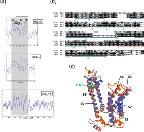
We previously identified a putative K+channel (STC no.: 009A07R1.0A1.1) in EST resources obtained fromH. cylindrosporum pure cultures [40]. BLASTP analysis of 009A07R1.0A1.1 protein sequence against theL. bicolor S238N genomic database allowed the identification of aunique orthologue (protein ID 297800;https://mycocosm.jgi.doe.gov/Lacbi2/Lacbi2.home. html). The hydrophobicity profile of these two proteins generated by the Kyte-Doolittle algo-rithm revealed the presence of 7 hydrophobic domains, similar to theXlKV2.1Shaker channel
fromX. laevis (Fig 1A). The amino acid alignment of these fungalShaker-like proteins with XlKV2.1 confirmed the presence of 6 conserved transmembrane domains and one pore
domain characterized by the GYGE motif ensuring K+selectivity (Fig 1B). Consequently, we named these proteins SKC (Shaker-like K+-Channel). The S4 domain (constituting the voltage sensor inShaker channels) of HcSKC and LbSKC harbored several arginine residues (R). Such
positively charged residues have been shown in other KVchannels to contribute to the channel
voltage sensitivity and gating charge [41]. When compared withXlKV2.1,HcSKC and LbSKC
have a shorter C-terminal region, suggesting reduced "built-in" regulation mechanisms (Fig 1A). Based on thesein silico analyses, we used the R. norvegicus RnKV2.1 channel
(NP_037318.1;S3 Table) as a template to propose a structural model for the fungalShaker-like
proteins (Fig 1C,S1 Fig).
Distribution of
Shaker-like channels in the fungal kingdom
UsingHcSKC as a reference, we performed BLASTP analysis on publicly available sequenced
fungi found on NCBI and JGI fungal portals (http://genome.jgi-psf.org/programs/fungi/index. jsf). In addition, a cure based on the presence of a putative G[YF]G[DE] pore motif was achieved. In the Basidiomycota phylum, the presence of putative SKC proteins was detected only in the Agaricomycotina subphylum, as is defined in the JGI phylogenetic tree (Fig 2). In addition, putative SKC proteins were found inWallemia melicola, which belongs to
Wallemio-mycetes, a sister group to Agaricomycotina [42]. Two putativeShaker-like channels were also
(Glomeromycotina subphylum) and other basal fungi belonging to Cryptomycota, Chytridio-mycota, BlastocladioChytridio-mycota, Zoopagomycota and Mucoromycota phyla (Fig 2). Surprisingly, no SKC was found in the Pucciniomycotina and Ustilaginomycotina subphyla of Basidiomy-cota, and in the Ascomycota phylum. Moreover, proteins similar to KVchannels, but without
the G[YF]G[DE] motif, were not found in these fungi, indicating the complete loss of
Shaker-like genes in Pucciniomycotina, Ustilaginomycotina and Ascomycota species, with the excep-tion in the Ascomycota phylum ofSaitoella complicata, which presents one putative SKC
channel (Fig 2).
The functional properties of
HcSKC could not be revealed in various
heterologous systems
Functional characterization is an important step for the analysis of K+channel activity to pre-dict transport features, such as selectivity and direction of rectification. Many strategies were applied to characterizeHcSKC, using multiple heterologous systems (X. laevis oocytes and
yeast) and co-injection of putative regulatory proteins (protein kinase K and putative channel β-subunits from H. cylindrosporum: HcKCNAB1 and HcKCNAB2) (S4 Table). No significant result was obtained from any of these experiments. Moreover, the expression ofHcSKC
Fig 1.In silico prediction of a Shaker-like channel in the fungus Hebeloma cylindrosporum. The hydrophobicity
profiles of theShaker subunits from H. cylindrosporum (HcSKC), L. bicolor (LbSKC), and X. laevis (XlKV2.1) (a) and their partial amino acid sequence alignment (b) predicted six transmembrane domains (S1 to S6). One pore domain in
HcSKC and LbSKC subunits containing the conserved G[YF]G[DE] motif of the Shaker channel selectivity filter was
also detected. The S4 transmembrane domain corresponds to the predicted voltage-sensor domain. (c) The plausible 3-D model of the fungalHcSKC subunit showing the transmembrane (S1 to S6) and pore (P) domains was generated
by homology with the 3-D structure ofRnKV2.1 K+channel from
R. norvegicus (NP_037318.1;S2 Table) as a template using Swiss-Model server [33] (http://swissmodel.expasy.org/). The GYGE signature motif of the pore region was highlighted in green. The sequence alignment ofHcSKC and RnKV2.1 polypeptides is displayed inS1 Fig.
triggers endogenous currents inX. laevis oocytes, making the observation of specific
exoge-nous currents tricky (S4 Table). In addition, the previously identifiedHcSKC channel in the H. cylindrosporum h1 strain [40] has been extensively tested by voltage-clamp experiments inX. laevis oocytes, without any conclusive result (S4 Table). As an alternative, a functional charac-terization of the homologous channel inL. bicolor LbSKC was also attempted in X. laevis
oocytes, but unsuccessfully (S4 Table). Finally, attempts to perform subcellular localizations in yeast usingHcSKC-EGFP fused constructs driven by a constitutive or a galactose-inducible
promoter failed too (S2 Fig). None of the experiments yielded a clear localization pattern of
HcSKC, probably due to misprocessing of the protein product, reinforcing the issues in
expressingHcSKC in the tested heterologous systems.
HcSKC transcripts are localized in all types of ectomycorrhizal hyphae
To elucidate the role ofHcSKC channels in the symbiotic association with P. pinaster, the
localization of corresponding transcripts was analyzed in 2-month-old ectomycorrhizas throughin situ hybridization experiments. Cross-section and probe hybridization processes
resulted in the detection ofHcSKC transcripts in extra-radical hyphae, the hyphal mantle, and
Fig 2. Phylogenetic tree of KVchannels in fungi, and representative animal and plant species. TheShaker-like K +
channel tree of publicly available sequenced fungi from the Basidiomycota phylum and from basal fungi (Cryptomycota, Chytridiomycota, Blastocladiomycota, Zoopagomycota and Mucoromycota) was calculated by the maximum likelihood method (1000 bootstraps).Shaker-like proteins were found only
in one putative Ascomycota species (Saitoella complicata) and not in the Pucciniomycotina and Ustilaginomycotina subphyla of Basidiomycota.
Ectomycorrhizal (ECM), arbuscular mycorrhizal (AM), and orchid mycorrhizal (OM) fungi are indicated by yellow, green, and blue dots, respectively. KVchannels from representative land plant and animal species were included in the tree. All species displayed in this tree are listed in
theS2 Table.Shaker-like proteins from H. cylindrosporum (HcSKC) and L. bicolor (LbSKC) used in this study are indicated by black arrows. https://doi.org/10.1371/journal.pone.0242739.g002
Fig 3.HcSKC transcript localization in Hebeloma cylindrosporum—Pinus pinaster ectomycorrhizas. In situ
hybridization in ectomycorrhizas from 2-month-old co-cultures ofP pinaster–H. cylindrosporum with HcSKC-specific
the Hartig net, indicating no specific expression of this channel in ectomycorrhizas (Fig 3). Only the antisense probe provided signals indicating the presence ofHcSKC transcripts (Fig 3B). Since the sense probe sequence is not complementary to theHcSKC mRNA sequence, it
cannot hybridize, and, therefore, serves as a negative control. Consequently, it did not give rise to any signal (Fig 3A). A construct harboring theEGFP gene marker fused to the HcSKC
pro-moter was cloned to show thatHcSKC was well expressed in free-living conditions (S3 Fig).
Silencing of
HcSKC affects Hebeloma cylindrosporum potassium
homeostasis
Decoding the role ofHcSKC in axenic conditions was attempted by RNAi downregulation of
the corresponding gene.HcSKC-silencing lines of H. cylindrosporum were produced by Agro-bacterium tumefaciens–mediated transformation. The expression level of HcSKC was
deter-mined by qRT-PCR in fungal mycelia growing on selection medium. Two isolates displaying a five-fold decrease inHcSKC expression, named RNAi-SKC-5 and RNAi-SKC-7, were selected
for ECM assays (S4 Fig). We then investigated the K+accumulation and expression of K+ transport-related genes in RNAi-SKC-5 and RNAi-SKC-7 lines. In fungi grown in standard medium (i.e., in presence of a relatively high external K+, 1 mM), internal K+content was sig-nificantly higher in RNAi-SKC transformants than in the control line, indicating an increase in K+acquisition and/or a decrease in K+efflux (Fig 4). Once these fungal isolates were trans-ferred into the K+-free medium, no difference in K+content between RNAi-SKC and control lines could be detected at 12, 24 and 48 h after the transfer (Fig 4). In addition, we assessed the effect of high and low K+(1 and 0.05 mM, respectively) and Na+(1 or 0.2 mM, respectively)
detected in extra-radical hyphae, the fungal sheath and the Hartig net of ectomycorrhizas. Bars, 50μm. c.c., cortical cell; exh, extraradical hyphae; Hn, Hartig net; sh, fungal sheath.
https://doi.org/10.1371/journal.pone.0242739.g003
Fig 4. Potassium content inHebeloma cylindrosporum HcSKC downregulated lines. K+content was measured in fungal lines transformed with the empty (Ctrl) orHcSKC-silencing vectors (RNAi-SKC-5 and RNAi-SKC-7) using an
atomic absorption flame spectrophotometer. Statistical differences were evaluated using the Student’s test with respect to the Ctrl strain (�, P < 0.05;��, P < 0.01). n = 6.
availability on fungal growth in all transgenic strains (S5 Fig). All media were prepared on the basis of the media used for the subsequent ECM assay (S4 Table). After 28 days of culture in axenic condition, dry weights were recorded. Although the biomass of both RNAi-SKC trans-genic lines differed in all conditions, they did not significantly differ from the control line, except for RNAi-SKC-5 that showed a slightly reduced biomass in +K/-Na condition com-pared to the control line (S5 Fig).
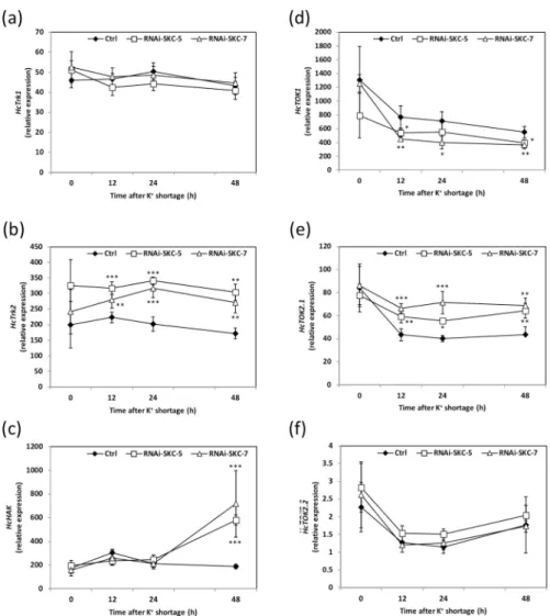
BesidesHcSKC, six other putative K+transport-related genes can be identified in theH. cylindrosporum genomic database [4,19]. To figure out whether their expression levels were affected by the downregulation ofHcSKC, the same fungal samples as those used for K+ con-tent measurements were analyzed by qRT-PCR. When grown in K+standard pure culture con-ditions (SK medium), none of the six targeted K+transport system genes was significantly differentially expressed in the RNAi-SKC lines compared to the control (Fig 5). In contrast, 12, 24 and 48 h after the transfer into the K+-free medium,HcTrk2 and HcTOK2.1 were
upregu-lated andHcTOK1 was downregulated in the two RNAi-SKC lines (Fig 5B, 5D and 5E). Inter-estingly,HcHAK was upregulated in both RNAi lines only 48 h after the transfer into the K+ -free medium (Fig 5C). The expression in the transgenic lines of the other two transport sys-tems,HcTrk1 and HcTOK2.2, was not significantly affected by the transfer into the K+-free medium (Fig 5A–5F). Taken together, these data indicate that the downregulation ofHcSKC is
sufficient to modify the K+homeostasis inH. cylindrosporum at standard K+. However, in con-ditions of K+shortage, the modification of the expression of three K+transport-related genes compensated for the effect ofHcSKC downregulation and restored a wild-type K+content in the fungus.
Downregulation of
HcSKC affects K
+transfer to the host plant at standard
K
+conditions
To investigate the role ofHcSKC in symbiotic conditions, P. pinaster seedlings were inoculated
with control, RNAi-SKC-5, or RNAi-SKC-7 lines, or kept non-inoculated, and grown in SK and LK conditions (Fig 6). Mycorrhization with the control fungal strain (transformed with the empty plasmid) resulted in statistically significant higher K+contents in the plant roots and shoots when grown in LK, but not in SK, conditions. In comparison with these control plants, mycorrhizal association with theSKC-silencing fungal lines resulted in a significant
decrease of K+contents in shoots and roots in SK conditions, indicating a less cooperative behavior ofH. cylindrosporum that affected K+transfer toP. pinaster (Fig 6A). Surprisingly, this negative effect ofHcSKC silencing on K+contents in the host plant was not observed in LK conditions (Fig 6B). Hence, in low K+conditions, the mycorrhizal pathway for plant K+ nutrition was not affected by the reduced expression ofHcSKC. This result suggests that, in
this LK condition, other underlying mechanisms are at work to achieve an efficient mycor-rhizal K+transport towards the host plant. In other words,HcSKC seems to play a significant
role in K+delivery toward the plant in SK conditions (1 mM), and not in conditions of K+ shortage (0.05 mM).
Discussion
As has recently been revealed, K+acquisition in higher fungi seems to be mediated by mem-brane transport proteins belonging to the Trk/Ktr/HKT and KT/KUP/HAK families [4,43]. In addition, ACU ATPases have been reported as being involved in K+and Na+uptake in Usti-lago maydis, and more generally envisioned as key players of K+transport in fungi [7,44]. PAT ATPases could emerge as mechanisms for absorption of these cations as well, but they have not been studied thoroughly [45,46]. Regarding outward K+transport systems, yeast and
filamentous fungi developed TOK andShaker-like K+channels, from which only some mem-bers have been investigated thus far [20,21]. To our knowledge, this work reports the first description of aShaker-like KVchannel in higher fungi and highlights its role in both K+
homeostasis and mycorrhizal symbiosis.
In silico analysis of HcSKC revealed features from animal Shaker K
+channels
The initial identification of theHcSKC Shaker-like channel was performed from an EST library
ofH. cylindrosporum produced in free-living conditions [40]. In this study, one putative
Shaker-like K+channel was found to be highly expressed (STC no.: 009A07R1.0A1.1), Fig 5. Relative expression of putative K+channels and transporters in
Hebeloma cylindrosporum control and HcSKC-silencing lines. Relative expressions of the K+
transportersHcTrk1 (a), HcTrk2 (b), HcHAK (c) and the K+
channelsHcTOK1 (d), HcTOK2.1 (e) and HcTOK2.2 (f) were analyzed in dependence on K+shortage in control and RNAi fungal lines. The relative expressions of the K+transporter
HcTrk2 (b) and the K+channels
HcTOK1 (d) and HcTOK2.1 (e) were altered in RNAi-SKC-5 and RNAi-SKC-7 lines in comparison to the transgenic control line (Ctrl)
under K+limiting conditions. The relative expressions ofHcTrk1 (a) and HcHAK (c) K+
transporters, and of the K+ channelHcTOK2.2 (f) were not affected by K+shortage in RNAi-SKC-5 and RNAi-SKC-7 lines in comparison to the transgenic control line (Ctrl). Mean values are provided with the standard error (n = 6). Statistical differences were determined using a one-way ANOVA followed by Dunnett’s test relative to Ctrl strain for each data point P < 0.05 (�), P < 0.01 (��) and P < 0.001 (���).
suggesting an important role of this transport system in the fungus. Thanks to the release ofH. cylindrosporum h7 genomic data [19,47,48],in silico analyses confirmed the homology of this
fungal candidate to animal and plant KVchannels (Fig 1). Moreover, phylogenetic analyses
provided additional evidence on the proximity of fungalShaker channels to animal ones (Fig 2). The major difference betweenHcSKC/LbSKC and animal channels is the length of their
C-terminal regions. Several studies have evidenced a crucial role of this C-C-terminal region in the regulation of plant and animalShaker channel activity [49,50]. In plants, it was reported that Fig 6. Potassium content in 2-month-oldPinus pinaster plants growing alone or in co-culture with Hebeloma cylindrosporum under K+-sufficient or -deficient conditions. Root and shoot contents were measured in
non-mycorrhizal plants (NM) and plants inoculated with fungi transformed with the empty (Ctrl) orHcSKC-silencing
vectors (RNAi-SKC-5 and RNAi-SKC-7), and growing at standard K+(SK medium, 1 mM K+, a) or low K+(LK medium, 0.05 mM K+, b). Mean values are provided with the standard error (n = 5–6). Different letters indicate significant differences between treatments according to one-way ANOVA followed by Tukey HSD post-hoc tests (P < 0.05).
the C-terminal regions are involved in intracellular K+sensing, heteromerization or voltage-dependent gating mechanisms [51–53]. In animalShaker-type channels, the C-terminus can
play multiple rolese.g. in regulation by β-subunits [54], by voltage [55], or in localization [56]. The fact that the C-terminal domains ofH. cylindrosporum and L. bicolor are significantly
shorter than those of the animalShaker channels (Fig 1) suggests differences in channel activ-ity regulation. Moreover,LbSKC seems to diverge by an additional sequence in the region of
the S4 domain (Fig 1B) with unknown function.
Shaker-like channels were lost in Pucciniomycotina, Ustilaginomycotina
and Ascomycota phyla
Although well-described in plants and animals, the presence of KVchannels in the fungal
king-dom has been hypothetical until now. Indeed, noShaker channel gene could be detected in the Saccharomyces cerevisiae genome sequence. Our phylogenetic analysis based on many fungal
genomes belonging to Basidiomycota and Ascomycota phyla, as well as basal fungi (Crypto-mycota, Chytridio(Crypto-mycota, Blastocladio(Crypto-mycota, Zoopagomycota and Mucoromycota phyla) revealed the absence of putative SKC proteins in Pucciniomycotina and Ustilaginomycotina, which are sub-phyla of Basidiomycota, and the entire Ascomycota phylum. Interestingly, one SKC protein was found inS. complicata, which is related to the Ascomycota phylum. It is,
however, worth noting that the affiliation ofS. complicata to Ascomycota remains unclear
[57,58], and its SKC protein sequence clusters with those of basal fungi, in agreement with the idea that this species might belong to a more basal phylum than previously thought. Given that
Shaker-like channels were found in basal fungi and in the most recent subphylum of
Badisio-mycota (Agaricomycotina), their absence in Pucciniomycotina, Ustilaginomycotina, and Ascomycota strongly suggests that they have been lost in the ancestors of these phyla. This indicates that other transport systems can fill the roles ofShaker channels and play a central
role in the maintenance of K+homeostasis in these organisms. The survey and analysis of other K+channels found in fungi, such as the TOK channels [4], have been integral in describ-ing other key regulators of K+transport and homeostasis in fungi (e.g. [20,21]).
HcSKC cannot be functionally characterized in heterologous systems
Although several attempts using different setups, strategies and heterologous systems were performed, the functional characterization ofH. cylindrosporum and L. bicolor Shaker-like
channels is still missing (S4 Table). Similarly, no conclusive data were obtained after express-ing aHcSKC-EGFP construct driven by a constitutive or a galactose-inducible promoter in S. cerevisiae (S2 Fig). This might result from the fact that conditions or proteins required for the expression, function, localization, and regulation of fungal SKC channels were missing in the tested heterologous systems. Recently, the similar failed expression of a zinc transporter from the ECM fungusSuillus luteus was reported, showing the risky nature of these experiments
[59]. It is well-known that animal and plantShaker channels can physically interact with
mod-ulatoryβ-subunits [60]. For example, rat KV1Shaker channels are able to form stable
com-plexes with cytosolicβ-subunits (KVβ), which modify the channel activity [61,62]. In the
model plantArabidopsis thaliana, KAB1 is a protein related to animal β-subunits that interacts
with the K+channel KAT1 [63]. We identified ten putativeβ-subunits in the genomic database ofH. cylindrosporum and two of them, HcKCNAB1 and HcKCNAB2, were co-expressed with HcSKC in X. laevis oocytes, resulting in no exogenous current detection. At this stage we
can-not conclude whether fungalShaker channels require β-subunits or other types of peptides for
their activation. It is known that some compounds can alter the activity of voltage-dependent K+channels, such as human hormones that inhibit KV1.3 and KV1.5 [64]. Thus, the presence
inX. laevis oocytes of undefined compounds that would inhibit fungal SKC currents might be
possible. Another possibility would also be a regulation by other membrane proteins. For instance, in the nematodeCaenorhabditis elegans, the two-pore K+channel SUP-9 forms a complex with two proteins, UNC-93 (Major Facilitator Superfamily) and SUP-10 (The Potas-sium Channel Regulatory Protein Sup-10 Family), which coordinate muscle contraction [65]. InA. thaliana, the homolog of UNC-93 regulates K+translocation from the root to the aerial parts of the plant. Disruption of this gene leads to a phenotype similar to that of knockout mutants of theAtSKOR Shaker-like channel, indicating that they may function together,
although no physical interactions have yet been observed [66,67]. There is one UNC-93 gene in the genome ofH. cylindrosporum which is induced in ectomycorrhizas, compared to
free-living mycelium, and could be a promising candidate to test as a regulator of the activity of
HcSKC.
Calcium signaling pathways are well-known regulators of K+transport systems in plants and animals. In plants, calcium mediates signals during biotic interactions and environmental stress, and regulates a series of proteins that can regulate the activity of K+channels through phosphorylation, dephosphorylation, structural modification, and physical interaction [68]. In animals these pathways are different, but there is also evidence of the modulation of K+ chan-nel transport by calcium-dependent phosphorylation and dephosphorylation [69]. In filamen-tous fungi, there are few links between the regulation of K+transport and calcium signaling, probably because most of the studies have been carried on the model Ascomycota yeastS. cere-visiae [70]. However, the first description ofShaker-like K+channels in fungi may lead to the discovery of new pathways. Supporting this hypothesis, a rapid survey of the genome ofH. cylindrosporum yields many putative calcium-regulated genes: three calmodulins (Protein ID
385514, 443591 and 443596), one calcineurin B (444455) and a wide range of Ca2+ /calmodu-lin-dependent and serine/threonine protein kinases. It is also tempting to imagine that the most promising heterologous system for the characterization of fungal transport systems would beS. cerevisiae, which is phylogenetically closer to higher fungi than animal cells. In
general, the use of yeast seems more adapted for the characterization of inward transport sys-tems than of outward syssys-tems, but so far it has not provided encouraging results in our uptake experiments (S4 Table). Direct transport measurements by the electrophysiological patch-clamp method might provide missing insights in fungal SKC functional properties.
Potassium homeostasis mediated by
HcSKC is a prerequisite for K
+allocation to pine
In order to investigatein vivo the role of the KVchannel SKC in both free-living and ECM
con-ditions, we decided to generate RNAi fungal lines affected inHcSKC expression. Analysis of
the phenotype of these transgenic fungal lines revealed a close relationship betweenHcSKC
expression, K+accumulation in the fungus, and improvement of K+nutrition of the colonized plants. Indeed,HcSKC silencing resulted in contrasting phenotypes of both the fungus and the
mycorrhizal plant, depending on external K+availability. At 1 mM K+, the downregulation of
HcSKC expression altered K+accumulation in the free-living mycelia and the ECM plants. It should be noted that, in our previous report [12],H. cylindrosporum had a significant effect on
K+allocation toP. pinaster only in K+limiting conditions and not in sufficient conditions. Such a phenotype is further confirmed in the present report by the comparison between plants inoculated by the control transformed fungus harboring the empty vector and the control non-mycorrhizal plants (Fig 4). However, this does not mean that K+transfer from the fungus to the plant did not occur in standard (SK) conditions. Indeed, the direct uptake of K+from the medium by the root system may be reduced upon mycorrhizal association, and this
reduction could be compensated by fungal K+allocation to the plants. Parts of the ECM root system are somehow isolated from the external medium due to a loss of root hairs and to the presence of the fungal mantle surrounding the roots, and thus are dependent on the ECM symbiont to acquire nutrients. Given thatHcSKC RNAi transformants accumulated more K+
than the control strain at standard external K+, it may be assumed that, when such transfor-mants were grown in association withP. pinaster, their reduced expression of HcSKC either
modified the pool of fungal K+available for the plant and/or affected its symbiotic transfer to host roots. This would result in impaired K+nutrition of the host. It is worth noting that vary-ing external concentrations of K+and Na+did not drastically impact the growth of the RNAi-SKC transgenic fungal lines (S5 Fig). This indicates that the alteration of K+nutrition observed in plants colonized by the RNAi-SKC lines at standard K+cannot be explained by a modifica-tion of fungal growth and sensitivity to external condimodifica-tions, but by the actual downregulamodifica-tion ofHcSKC. Interestingly, the over-accumulation of K+in the RNAi-SKC transformants was not found to involve the upregulation of putative inward K+transporters, and thus, might result from a decrease in K+efflux from the fungus. In contrast, in low K+conditions, three other putative K+transport systems were up- or downregulated in the RNAi lines. We postulate that the altered expression ofHcTrk2, HcHAK, HcTOK1 and HcTOK2.1 restored the overall fungal
K+transport activities and homeostasis, as suggested by the recovery of the fungal K+content observed in free-living conditions (Fig 4). This could explain the restoration of the K+contents displayed by the mycorrhizal plants, suggesting that efficient ectomycorrhiza-mediated K+ nutrition occurred again. Another hypothesis, thatHcSKC is involved in K+movements from/ to the vacuole, cannot be completely excluded, even though only plasma membrane tion of this type of ion channel has been described in any organism so far. Subcellular localiza-tion at either the plasma membrane or at the tonoplast should allow us to fully appreciate the role ofHcSKC, regarding its manifest importance in H. cylindrosporum biology under axenic
and symbiotic conditions.
To summarize, homology ofHcSKC with animal outward KVchannels and the observed
K+nutrition-related mycorrhizal and fungal phenotypes lead to the simplest hypothesis: that the fungalShaker-like channel HcSKC may be involved in K+efflux from fungal cells, allowing K+translocation towards the host plant. Thus, the role played byHcSKC in K+
homeostasis in
H. cylindrosporum seems crucial for fungal K+availability and/or release to the host plant.
Supporting information
S1 Fig. Alignment ofHcSKC subunit of Hebeloma cylindrosporum with Rattus norvegicus RnKV2.1. Structure-based sequence alignment and sequence conservation betweenHcSKC
andRnKV2.1 K+channel fromRattus norvegicus. The GYGE motif of the pore domain was
highlighted in green. (TIF)
S2 Fig. Attempts to localizeHcSKC with a C-terminal EGFP fusion in Saccharomyces cere-visiae. The HcSKC-EGFP construct was inserted into the pYES2 vector, with
galactose-induced expression (a,b), or into the pFL61 vector, under the PGK constitutive promoter (c,d). None of these attempts yielded a clear localization pattern ofHcSKC, probably due to
mispro-cessing of the protein product. (TIF)
S3 Fig. Analysis of EGFP expression under control of theHcSKC promoter in Hebeloma cylindrosporum. EGFP expression was observed in fungal lines harboring the EGFP cassette
YMG medium for 2–3 weeks before analysis. (a) bright field image (b) EGFP image (GFP filter 505–530 nm). Scale bar: 100μm.
(TIF)
S4 Fig. Determination ofHebeloma cylindrosporum silencing HcSKC lines. The relative
expression ofHcSKC was determined in empty vector (Ctrl), RNAi-SKC-5 and RNAi-SKC-7
fungal lines by qRT-PCR. Mean values are provided with the standard deviation (n = 6). Statis-tical differences were evaluated using the Student’s test with respect to E.V. strain (��,
P < 0.01). (TIF)
S5 Fig. Biomass ofHebeloma cylindrosporum HcSKC-silencing lines under high and low
potassium and sodium regimes. Dry weights of empty vector (Ctrl), SKC-5 and RNAi-SKC-7 fungal lines were determined after 28 days of culture in media containing high (+K and +Na) or low concentration (-K and -Na) of potassium (K+) and sodium (Na+), respectively. +K corresponds to 1 mM of K+, -K to 0.05 mM of K+, +Na to 1 mM of Na+, and -Na to 0.2 mM of Na+. Full recipes are provided inS1 Table. Mean values are provided with the standard deviation (n = 4–6). Different letters indicate significant differences between treatments according to one-way ANOVA followed by Tukey HSD post-hoc tests (P < 0.05). (TIF)
S1 Table. Primer list. Restriction enzyme sites indicated in primer name, were underlined in the corresponding 5’-3’ sequence.
(TIF)
S2 Table. Protocol of media used to assess the impact of high and low potassium and sodium conditions on the biomass of transgenic fungal strains Ctrl, RNAi-SKC-5 and RNAi-SKC7. Final concentrations of components constituting the +K/-Na, +K/+Na, -K/-Na, and -K/+Na media used inS5 Figwere presented in this table. pH was adjusted at 5.5 with Ca (OH)2 before autoclave.
(TIF)
S3 Table. Sequences used inin silico and phylogenetic analyses.
(XLS)
S4 Table. Attempts for functional characterization of theShaker-like channels HcSKC
(strain h7), HcSKC1&2 (strain h1) fromHebeloma cylindrosporum, and LbSKC from Lac-caria bicolor, respectively. Whole-cell current recordings in Xenopus oocytes expressing HcSKC alone or with two β-subunits or a kinase as well as expression of LbSKC did not give
evidence for K+-dependent currents. Growth of a triple yeast mutant PLY246 (Δtrk1Δtrk2Δtok1; Bertl et al., 2003) was not complemented by expression of HcSKC. (TIF)
Acknowledgments
We would like to thank Dr. A.G. Pardo and Dr. M.J. Kemppainen (Laboratorio de Micologı´a Molecular; Universidad Nacional de Quilmes, Argentina) for kindly providing the vector pSILBAγ. We also thank Dr. A. Kafle for assistance with some statistical analyses.
Author Contributions
Formal analysis: Kevin Garcia.
Funding acquisition: Kevin Garcia, Herve´ Sentenac, Sabine D. Zimmermann.
Investigation: Kevin Garcia, Hannah E. R. Frank, Amandine Delteil, Geneviève Cone´je´ro, Raphae¨l Lambilliotte, Ce´cile Fizames.
Methodology: Kevin Garcia, Carmen Guerrero-Gala´n, Muhammad Zulqurnain Haider, Amandine Delteil, Geneviève Cone´je´ro, Ce´cile Fizames, Sabine D. Zimmermann. Resources: Herve´ Sentenac.
Supervision: Sabine D. Zimmermann.
Writing – original draft: Kevin Garcia, Sabine D. Zimmermann.
Writing – review & editing: Kevin Garcia, Carmen Guerrero-Gala´n, Hannah E. R. Frank, Herve´ Sentenac, Sabine D. Zimmermann.
References
1. Zimmermann SD, Che´rel I. Potassium. In: Broadley MR, White PJ, editors. Plant Nutritional Genomics. Blackwell. Oxford, UK; 2005. pp. 26–65.
2. Anschu¨tz U, Becker D, Shabala S. Going beyond nutrition: Regulation of potassium homoeostasis as a common denominator of plant adaptive responses to environment. J Plant Physiol. 2014; 171(9): 670– 687.https://doi.org/10.1016/j.jplph.2014.01.009PMID:24635902
3. Zo¨rb C, Senbayram M, Peiter E. Potassium in agriculture—Status and perspectives. J Plant Physiol. 2014; 171(9): 656–669.https://doi.org/10.1016/j.jplph.2013.08.008PMID:24140002
4. Garcia K, Zimmermann SD. The role of mycorrhizal associations in plant potassium nutrition. Front Plant Sci. 2014; 5(337): 1–9.https://doi.org/10.3389/fpls.2014.00337PMID:25101097
5. Meena VS, Maurya BR, Verma JP. Does a rhizospheric microorganism enhance K+availability in
agri-cultural soils? Microbiological Research. 2014; 169: 337–347.https://doi.org/10.1016/j.micres.2013.09. 003PMID:24315210
6. Shin R. Strategies for improving potassium use efficiency in plants. Mol Cells. 2014; 37(8): 575–584.
https://doi.org/10.14348/molcells.2014.0141PMID:24938230
7. Haro R, Benito B. The role of soil fungi in K+plant nutrition. Int J Mol Sci. 2019; 20(13): 3169.
8. Smith SE, Read D. Mycorrhizal Symbiosis. 3rd ed. Smith SE, Read D, editors. London: Academic Press; 2008.
9. Becquer A, Guerrero-Gala´n C, Eibensteiner JL, Houdinet G, Bu¨cking H, Zimmermann SD, et al. The ectomycorrhizal contribution to tree nutrition. In: Advances in Botanical Research 89. Academic Press Inc.; 2019. pp. 77–126.
10. Guerrero-Gala´n C, Houdinet G, Calvo-Polanco M, Bonaldi KE, Garcia K, Zimmermann SD. The role of plant transporters in mycorrhizal symbioses. In: Advances in Botanical Research 87. Academic Press Inc.; 2018. pp. 303–342.
11. Jentschke G, Brandes B, Kuhn AJ, Schro¨der WH, Godbold DL. Interdependence of phosphorus, nitro-gen, potassium and magnesium translocation by the ectomycorrhizal fungus Paxillus involutus. New Phytol. 2001; 149(2): 327–337.
12. Garcia K, Delteil A, Cone´je´ro G, Becquer A, Plassard C, Sentenac H, et al. Potassium nutrition of ecto-mycorrhizal Pinus pinaster: Overexpression of the Hebeloma cylindrosporum HcTrk1 transporter affects the translocation of both K+and phosphorus in the host plant. New Phytol. 2014; 201: 951–960.
https://doi.org/10.1111/nph.12603PMID:24279702
13. Guerrero-Gala´n C, Calvo-Polanco M, Zimmermann SD. Ectomycorrhizal symbiosis helps plants to challenge salt stress conditions. Mycorrhiza. 2019; 29: 291–301. https://doi.org/10.1007/s00572-019-00894-2PMID:31011805
14. Casieri L, Ait Lahmidi N, Doidy J, Veneault-Fourrey C, Migeon A, Bonneau L, et al. Biotrophic transpor-tome in mutualistic plant-fungal interactions. Mycorrhiza. 2013; 23: 597–625.https://doi.org/10.1007/ s00572-013-0496-9PMID:23572325
15. Courty PE, Doidy J, Garcia K, Wipf D, Zimmermann SD. The transportome of mycorrhizal systems. In: Molecular Mycorrhizal Symbiosis. Wiley Blackwell; 2016. pp. 239–256.
16. Garcia K, Doidy J, Zimmermann SD, Wipf D, Courty PE. Take a trip through the plant and fungal trans-portome of mycorrhiza. Trends Plant Sci. 2016; 21(11): 937–950.https://doi.org/10.1016/j.tplants.2016. 07.010PMID:27514454
17. Garcia K, Delaux PM, Cope KR, Ane´ JM. Molecular signals required for the establishment and mainte-nance of ectomycorrhizal symbioses. New Phytol. 2015; 208(1): 79–87.https://doi.org/10.1111/nph. 13423PMID:25982949
18. Corratge´ C, Zimmermann S, Lambilliotte R, Plassard C, Marmeisse R, Thibaud JB, et al. Molecular and functional characterization of a Na+-K+transporter from the Trk family in the ectomycorrhizal fungus
Hebeloma cylindrosporum. J Biol Chem. 2007; 282: 26057–26066.https://doi.org/10.1074/jbc. M611613200PMID:17626012
19. Kohler A, Kuo A, Nagy LG, Morin E, Barry KW, Buscot F, et al. Convergent losses of decay mecha-nisms and rapid turnover of symbiosis genes in mycorrhizal mutualists. Nat Genet. 2015; 47(4): 410– 415.https://doi.org/10.1038/ng.3223PMID:25706625
20. Guerrero-Gala´n C, Delteil A, Garcia K, Houdinet G, Cone´ je´ro G, Gaillard I, et al. Plant potassium nutri-tion in ectomycorrhizal symbiosis: properties and roles of the three fungal TOK potassium channels in
Hebeloma cylindrosporum. Environ Microbiol. 2018; 20(5): 1873–1887. https://doi.org/10.1111/1462-2920.14122PMID:29614209
21. Guerrero-Gala´n C, Garcia K, Houdinet G, Zimmermann SD. HcTOK1 participates in the maintenance of K+homeostasis in the ectomycorrhizal fungus Hebeloma cylindrosporum, which is essential for the
symbiotic K+nutrition of Pinus pinaster. Plant Signal Behav. 2018; 13(6): e1480845.https://doi.org/10.
1080/15592324.2018.1480845PMID:29939816
22. Gambale F, Uozumi N. Properties of shaker-type potassium channels in higher plants. J Membr Biol. 2006; 210(1): 1–19.https://doi.org/10.1007/s00232-006-0856-xPMID:16794778
23. Zimmermann S, Sentenac H. Plant ion channels: from molecular structures to physiological functions. Curr Opin Plant Biol. 1999; 2(6): 477–482.https://doi.org/10.1016/s1369-5266(99)00020-5PMID:
10607654
24. Hedrich R. Ion channels in plants. Physiol Rev. 2012; 92(4): 1777–1811.https://doi.org/10.1152/ physrev.00038.2011PMID:23073631
25. Jegla T, Busey G, Assmann SM. Evolution and structural characteristics of plant voltage-gated K+ chan-nels. Plant Cell. 2018; 30(12): 2898–2909.https://doi.org/10.1105/tpc.18.00523PMID:30389753 26. Debaud JC, Gay G. In vitro fruiting under controlled conditions of the ectomycorrhizal fungus Hebeloma
cylindrosporum associated with Pinus pinaster. New Phytol. 1987; 105: 429–435.
27. Rao PS, Niederpruem DJ. Carbohydrate metabolism during morphogenesis of Coprinus lagopus (sensu Buller). J Bacteriol. 1969; 100(3): 1222–1228.https://doi.org/10.1128/JB.100.3.1222-1228.1969
PMID:5391229
28. Kemppainen MJ, Pardo AG. pHg/pSILBAγvector system for efficient gene silencing in homobasidiomy-cetes: optimization of ihpRNA–triggering in the mycorrhizal fungus Laccaria bicolor. Microb Biotechnol. 2010; 3(2): 178–200.https://doi.org/10.1111/j.1751-7915.2009.00122.xPMID:21255319
29. Ali MA, Louche J, Legname E, Duchemin M, Plassard C. Pinus pinaster seedlings and their fungal sym-bionts show high plasticity in phosphorus acquisition in acidic soils. Tree Physiol. 2009; 29(12): 1587– 1597.https://doi.org/10.1093/treephys/tpp088PMID:19840995
30. Garcia K, Haider MZ, Delteil A, Corratge´ -Faillie C, Cone´jero G, Tatry M-V, et al. Promoter-dependent expression of the fungal transporter HcPT1.1 under Pi shortage and its spatial localization in ectomycor-rhiza. Fungal Genet Biol. 2013;58–59: 53–61.https://doi.org/10.1016/j.fgb.2013.06.007PMID:
23850603
31. Louche J, Ali MA, Cloutier-Hurteau B, Sauvage FX, Quiquampoix H, Plassard C. Efficiency of acid phosphatases secreted from the ectomycorrhizal fungus Hebeloma cylindrosporum to hydrolyse organic phosphorus in podzols. FEMS Microbiol Ecol. 2010; 73(2): 323–335.https://doi.org/10.1111/j. 1574-6941.2010.00899.xPMID:20533944
32. Kyte J, Doolittle RF. A simple method for displaying the hydropathic character of a protein. J Mol Biol. 1982; 157(1): 105–132.https://doi.org/10.1016/0022-2836(82)90515-0PMID:7108955
33. Biasini M, Bienert S, Waterhouse A, Arnold K, Studer G, Schmidt T, et al. SWISS-MODEL: modelling protein tertiary and quaternary structure using evolutionary information. Nucleic Acids Res. 2014; 42(1): 252–258.https://doi.org/10.1093/nar/gku340PMID:24782522
34. Grigoriev IV, Cullen D, Goodwin SB, Hibbett D, Jeffries TW, Kubicek CP, et al. Fueling the future with fungal genomics. Mycology. 2011; 2(3): 192–209.
35. Edgar RC. MUSCLE: multiple sequence alignment with high accuracy and high throughput. Nucleic Acids Res. 2004; 32(5): 1792–1797.https://doi.org/10.1093/nar/gkh340PMID:15034147
36. Castresana J. Selection of conserved blocks from multiple alignments for their use in phylogenetic anal-ysis. Mol Biol Evol. 2000; 17(4): 540–552.https://doi.org/10.1093/oxfordjournals.molbev.a026334
PMID:10742046
37. Guindon S, Dufayard JF, Lefort V, Anisimova M, Hordijk W, Gascuel O. New algorithms and methods to estimate maximum-likelihood phylogenies: assessing the performance of PhyML 3.0. Syst Biol. 2010; 59: 307–321.https://doi.org/10.1093/sysbio/syq010PMID:20525638
38. Huson DH, Richter DC, Rausch C, Dezulian T, Franz M, Rupp R. Dendroscope: An interactive viewer for large phylogenetic trees. BMC Bioinformatics. 2007; 8(1): 460. https://doi.org/10.1186/1471-2105-8-460PMID:18034891
39. Minet M, Dufour M-E, Lacroute F. Complementation of Saccharomyces cerevisiae auxotrophic mutants by Arabidopsis thaliana cDNAs. Plant Journal. 1992; 2: 417–422.https://doi.org/10.1111/j.1365-313x. 1992.00417.xPMID:1303803
40. Lambilliotte R, Cooke R, Samson D, Fizames C, Gaymard F, Plassard C, et al. Large-scale identifica-tion of genes in the fungus Hebeloma cylindrosporum paves the way to molecular analyses of ectomy-corrhizal symbiosis. New Phytol. 2004; 164: 505–513.
41. Seoh SA, Sigg D, Papazian DM, Bezanilla F. Voltage-sensing residues in the S2 and S4 segments of the Shaker K+channel. 1996; 16(6): 1159–1167.https://doi.org/10.1016/s0896-6273(00)80142-7
PMID:8663992
42. Matheny PB, Gossmann JA, Zalar P, Kumar TKA, Hibbett DS. Resolving the phylogenetic position of the Wallemiomycetes: An enigmatic major lineage of Basidiomycota. Can J Bot. 2006; 84(12): 1794– 1805.
43. Corratge´ -Faillie C, Jabnoune M, Zimmermann S, Ve´ry AA, Fizames C, Sentenac H. Potassium and sodium transport in non-animal cells: The Trk/Ktr/HKT transporter family. Cell Mol Life Sci. 2010; 67 (15): 2511–2532.https://doi.org/10.1007/s00018-010-0317-7PMID:20333436
44. Benito B, Garciadebla´s B, Schreier P, Rodrı´guez-Navarro A. Novel P-type ATPases mediate high-affin-ity potassium or sodium uptake in fungi. Eukaryot Cell. 2004; 3(2): 359–368.https://doi.org/10.1128/ec. 3.2.359-368.2004PMID:15075266
45. Fietto LG, Pugliese L, Gomes SL. Characterization and expression of two genes encoding isoforms of a putative Na, K-ATPase in the Chytridiomycete Blastocladiella emersonii. Biochim Biophys Acta. 2002; 1576(1–2): 59–69.https://doi.org/10.1016/s0167-4781(02)00297-xPMID:12031485
46. Benito B, Garciadebla´s B, Fraile-Escanciano A, Rodrı´guez-Navarro A. Potassium and sodium uptake systems in fungi. The transporter diversity of Magnaporthe oryzae. Fungal Genet Biol. 2011; 48(8): 812–822.https://doi.org/10.1016/j.fgb.2011.03.002PMID:21406243
47. Dore´ J, Perraud M, Dieryckx C, Kohler A, Morin E, Henrissat B, et al. Comparative genomics, proteo-mics and transcriptoproteo-mics give new insight into the exoproteome of the basidiomycete Hebeloma
cylin-drosporum and its involvement in ectomycorrhizal symbiosis. New Phytol. 2015; 208: 1169–1187. https://doi.org/10.1111/nph.13546PMID:26171947
48. Dore´ J, Kohler A, Dubost A, Hundley H, Singan V, Peng Y, et al. The ectomycorrhizal basidiomycete
Hebeloma cylindrosporum undergoes early waves of transcriptional reprogramming prior to symbiotic
structures differentiation. Environ Microbiol. 2017; 19: 1338–1354.https://doi.org/10.1111/1462-2920. 13670PMID:28076886
49. Che´rel I. Regulation of K+channel activities in plants: from physiological to molecular aspects. J Exp Bot. 2004; 55(396): 337–351.https://doi.org/10.1093/jxb/erh028PMID:14739260
50. Li Y, Um SY, Mcdonald TV. Voltage-gated potassium channels: regulation by accessory subunits. Neu-rosci. 2006; 12(3): 199–210.
51. Dreyer I, Pore´e F, Schneider A, Mittelsta¨dt J, Bertl A, Sentenac H, et al. Assembly of plant Shaker-like Koutchannels requires two distinct sites of the channelα-subunit. Biophys J. 2004; 87(2): 858–872.
https://doi.org/10.1529/biophysj.103.037671PMID:15298894
52. Liu K, Li L, Luan S. Intracellular K+sensing of SKOR, a Shaker-type K+channel from Arabidopsis. Plant
J. 2006; 46(2): 260–268.https://doi.org/10.1111/j.1365-313X.2006.02689.xPMID:16623888 53. Naso A, Dreyer I, Pedemonte L, Testa I, Gomez-Porras JL, Usai C, et al. The role of the C-terminus for
functional heteromerization of the plant channel KDC1. Biophys J. 2009; 96(10): 4063–4074.https:// doi.org/10.1016/j.bpj.2009.02.055PMID:19450478
54. Sokolova O, Accardi A, Gutierrez D, Lau A, Rigney M, Grigorieff N. Conformational changes in the C terminus of Shaker K+channel bound to the rat Kvβ2-subunit. Proc Natl Acad Sci. 2003; 100(22):
12607–12612.https://doi.org/10.1073/pnas.2235650100PMID:14569011
55. Zandany N, Marciano S, Magidovich E, Frimerman T, Yehezkel R, Shem-Ad T, et al. Alternative splicing modulates Kv channel clustering through a molecular ball and chain mechanism. Nat Commun. 2015; 6 (1): 6488.https://doi.org/10.1038/ncomms7488PMID:25813388
56. Ruiz-Cañada C, Koh YH, Budnik V, Tejedor FJ. DLG differentially localizes Shaker K+-channels in the
central nervous system and retina of Drosophila. J Neurochem. 2002; 82(6): 1490–1501.https://doi. org/10.1046/j.1471-4159.2002.01092.xPMID:12354297
57. Goto S, Sugiyama J, Hamamoto M, Komagata K. Saitoella, a new anamorph genus in the Cryptococca-ceae to accommodate two himalayan yeast isolates formerly identified as Rhodotorula glutinis. J Gen Appl Microbiol. 1987; 33(1): 75–85.
58. Nishida H, Hamamoto M, Sugiyama J. Draft genome sequencing of the enigmatic yeast Saitoella
com-plicata. J Gen Appl Microbiol. 2011; 57(4): 243–246.https://doi.org/10.2323/jgam.57.243PMID:
21914972
59. Ruytinx J, Coninx L, Nguyen H, Smisdom N, Morin E, Kohler A, et al. Identification, evolution and func-tional characterization of two Zn CDF-family transporters of the ectomycorrhizal fungus Suillus luteus. Environ Microbiol Rep. 2017; 9(4): 419–427.https://doi.org/10.1111/1758-2229.12551PMID:
28557335
60. Torres YP, Morera FJ, Carvacho I, Latorre R. A marriage of convenience:β-subunits and voltage-dependent K+channels. J Biol Chem. 2007; 282(34): 24485–24489.https://doi.org/10.1074/jbc. R700022200PMID:17606609
61. Rettig J, Heinemann SH, Wunder F, Lorra C, Parcej DN, Oliver Dolly J, et al. Inactivation properties of voltage-gated K+channels altered by presence ofβ-subunit. Nature. 1994; 369(6478): 289–294.
https://doi.org/10.1038/369289a0PMID:8183366
62. Pan Y, Weng J, Cao Y, Bhosle RC, Zhou M. Functional coupling between the Kv1.1 channel and aldo-ketoreductase Kvbeta1. J Biol Chem. 2008; 283(13): 8634–8642.https://doi.org/10.1074/jbc. M709304200PMID:18222921
63. Tang H, Vasconcelos AC, Berkowitz GA. Physical association of KAB1 with plant K+channel alpha sub-units. Plant Cell. 1996; 8(9): 1545–1553.https://doi.org/10.1105/tpc.8.9.1545PMID:8837508
64. Yu J, Park M-H, Jo S-H. Inhibitory effects of cortisone and hydrocortisone on human Kv1.5 channel cur-rents. Eur J Pharmacol. 2015; 746: 158–166.https://doi.org/10.1016/j.ejphar.2014.11.007PMID:
25449034
65. de la Cruz IP, Levin JZ, Cummins C, Anderson P, Horvitz HR. sup-9, sup-10, and unc-93 may encode components of a two-pore K+channel that coordinates muscle contraction in Caenorhabditis elegans. J Neurosci. 2003; 23(27): 9133–9145. PMID:14534247
66. Xiang J, Zhou X, Zhang X, Liu A, Xiang Y, Yan M, et al. The Arabidopsis AtUNC-93 acts as a positive regulator of abiotic stress tolerance and plant growth via modulation of ABA signaling and K+ homeosta-sis. Front Plant Sci. 2018; 9: 718.https://doi.org/10.3389/fpls.2018.00718PMID:29899751
67. Gaymard F, Pilot G, Lacombe B, Bouchez D, Bruneau D, Boucherez J, et al. Identification and disrup-tion of a plant Shaker-like outward channel involved in K+release into the xylem sap. Cell. 1998; 94(5):
647–655.https://doi.org/10.1016/s0092-8674(00)81606-2PMID:9741629
68. Che´rel I, Gaillard I. The complex fine-tuning of K+fluxes in plants in relation to osmotic and ionic abiotic
stresses. Int J Mol Sci. 2019; 20(3): 715.https://doi.org/10.3390/ijms20030715PMID:30736441 69. Roeper J, Lorra C, Pongs O. Frequency-dependent inactivation of mammalian A-type K+channel
KV1.4 regulated by Ca2+/Calmodulin-dependent protein kinase. J Neurosci. 1997; 17(10): 3379–3391.
PMID:9133364
70. Casado C, Yenush L, Melero C, del Carmen Ruiz M, Serrano R, Pe´rez-Valle J, et al. Regulation of Trk-dependent potassium transport by the calcineurin pathway involves the Hal5 kinase. FEBS Lett. 2010; 584(11): 2415–2420.https://doi.org/10.1016/j.febslet.2010.04.042PMID:20412803