Annals of Oncology 9: 95-100. 1998.
© 1998 Kluwer Academic Publishers. Printed in the Netherlands.
Clinical case
Management of an isolated thymic mass after primary therapy for lymphoma
S. Anchisi, R. Abele, M. Guetty-Alberto & P. Alberto
Division of Oncology, Department of Medicine, University Hospital, Geneva, SwitzerlandKey words: CT scan, galliumscintigraphy, Hodgkin's disease, lymphoma, residual mass, thymus hyperplasia
Introduction
The appearance of an anterior mediastinal mass during the follow-up of patients successfully treated for lym-phoma is worrisome, and may suggest treatment failure. However, benign 'rebound' thymic hyperplasia (TH), a well documented phenomenon in children [1-3] and in young adults treated for malignant testicular teratoma [4], is an important differential diagnosis even in the adult population [5].
Case reports
Between July 1990 and December 1994, we observed two cases of TH among 58 adults treated consecutively at our institution. Thirty of the 58 had de novo Hodgkin's disease (HD) and 28 had aggressive non-Hodgkin's lymphoma (NHL). During the same period, another case was diagnosed by one of us in private practice.
Casel
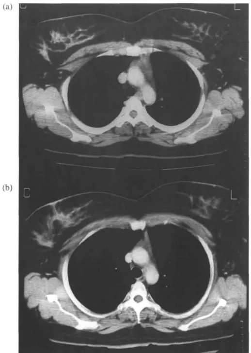
A 32-year-old man presented with a diffuse centroblastic B-cell NHL of the nasopharynx, stage IIA. Thymic and mediastinal lymph node enlargements were documented by thoracic computerized tomography (CT, Figure la). He obtained a complete response (CR) after three courses of BCDVP chemotherapy (bleomycin, cyclo-phosphamide, doxorubicin, vincristine and prednisone) with intrathecal methotrexate CNS prophylaxis. A total of six courses was given, followed by radiation therapy (36 Gy) to his Waldeyer's ring and cervical lymph nodes. Eight months later, left-sided lower cervical and axillary adenopathies were noted. A thoracic CT revealed an enlarged thymic mass in comparison with the initial study, but without abnormal mediastinal lymph nodes (Figure lb). A surgical biopsy of the axillary adenopathy disclosed a benign lymphoid hyperplasia. Four months later the persisting thymic mass, measuring 6 x 4.5 x 1 cm, was surgically excised. Histology with immuno-staining showed a benign thymic hyperplasia, with
adi-pose tissue constituting less than 40% of the total mass. The patient is still in CR 51 months after the end of treatment.
Case 2
A 26-year-old woman presented with a stage IVB nodular sclerosing HD, with liver and spleen infiltration and a bulky mediastinum.
Magnetic resonance imaging (MRI) disclosed a dif-fuse infiltration of the thoracic and lumbar vertebral bodies. The result of a bone-marrow biopsy was normal. Five courses of VEMP chemotherapy (vincristine, eto-poside, mitoxantrone, prednisone) produced a partial response greater than 75%. The treatment was intensi-fied with a BEAM chemotherapy (carmustine, etoposide, cytosine arabinoside, melphalan), followed by an autol-ogous bone marrow transplant (BMT). Four months later, the planned CT revealed that the residual mass in the antero-superior mediastinum was enlarging, meas-uring 4 x 1.5 x 3 cm. There was no other sign of lymphoma progression, and the mass was surgically excised. Histology showed TH and fibrotic lymph nodes. The patient is still in CR 30 months after completion of treatment.
Case 3
A 30-year-old female presented with a stage IIA diffuse centroblastic B-cell NHL of the nasopharynx. She en-tered CR after three courses of BCDVP chemotherapy with intrathecal methotrexate CNS prophylaxis. A total of six courses of chemotherapy were given and radio-therapy (34 Gy) was delivered to her Waldeyer's ring and cervical lymph nodes. Two months after the end of treat-ment, a thoracic CT showed an isolated thymic mass measuring 4 x 2 x 3 cm (Figure 2a), somewhat larger than prior to treatment. A new work-up was performed and CR was confirmed. A presumptive diagnosis of TH was made, and remained unchanged for three months. Thirteen months later CT showed an important regres-sion, with the thymus measuring 2 x 1.2 x 1.5 cm
triangular, partly bi-lobed mass in the anterior mediastinum (see 'o'), measuring 4 x 2 x 4.5 cm., without enlarged mediastinal lymph nodes.
(Figure 2b). The patient is currently in persistent CR 28 months after cessation of treatment.
Discussion
Due to the potential for lymphomatous involvement of the thymus, disclosure of a new or growing anterior mediastinal mass after successful treatment of lym-phoma is ominous. During the first six months post-therapy (occasionally up to one year) the relevant diag-noses are essentially relapse, TH, and residual 'fibrotic' mass if the anterior mediastinum was already involved at presentation. We will confine our discussion to relapse
versus TH.
Does any specific radiological sign differentiate between lymphoma recurrence and TH?
CTandMRI
On CT the appearance of normal thymus in the anterior mediastinal space is described as a triangular or bi-lobed structure, with soft-tissue density, whose size varies according to age [6-8].
Thymic enlargement is usually suspected if the thick-ness of at least one lobe is greater than two standard deviations above those of age-related controls [2, 6]. Mackall et al. [9] defined TH as a two-fold or greater increase in thymic volume during the first year after chemotherapy over the thymic volume at presentation.
Lucker and Siegel [2] studied 23 children who were followed with thoracic CT after treatment of HD with mediastinal involvement. They reported TH in four (17%) children who were in CR. TH was defined as an isolated enlargement of the thymus, observed on the first follow-up scan.
Heron et al. [6] studied 100 pathological thoracic CT in 84 patients (aged 15-33 years) with HD. One case of thymic cyst without nodal enlargement was observed out of 50 CT performed at the time of suspected relapse. In contrast, seven cases of thymic enlargement with proven HD relapse were associated with enlarged mediastinal lymph nodes. The authors report five cases of TH, not included in the above series, which occurred 4 to 10 months after cessation of treatment.
On MRI the signal intensity of the normal thymus is less than that of surrounding fat on Tl-weighted image, and approaches that of fat on T2-weighted scans. TH is described as homogeneous and uniform, as opposed to that of lymphoma [8].
In most if not all cases of TH, the enlarging mass retained the characteristic shape of normal thymus [2,4, 6-8].
Is Gallium-67 scan helpful?
Reported cases of TH on thoracic CT are mostly asso-ciated with positive [1, 9-15] Ga-67 scans. Negative scans are described [1], particularly in instances of thymic cyst or hemorrhage [5, 12].
9?
(a)
(b)
Figure 2 Thoracic CTscan of case no. 2. (a) Thymic enlargment ( 4 x 2 x 3 cm) diagnosed two months after completion of treatment, (b) Control
CTscan 13 months later, showing significant regression of the thymic mass (2 x 1.2 x 1.5 cm).
survivors followed with Ga-67 scans after treatment of NHL. Ten (16%) TH, defined as an isolated antero-superior mediastinal uptake, were observed one to eight months after completion of chemotherapy. In this study, only four children had both Ga-67 scans and thoracic CT. The CT revealed a thymic mass in one child with negative scintigraphy and in one of three with positive scintigraphy.
Since Ga-67 scan cannot distinguish between lym-phoma relapse and TH, its main utility would be to disclose other positive sites suggestive of relapse. Yet, in a study of 101 HD patients, post-treatment Ga-67 scan identified only 4 of 20 patients who eventually failed, and Ga-67 scan was the sole predictor of failure for only two patients [16].
Could other radiological techniques contribute to the diagnosis of TH?
To our knowledge no prospective studies have been per-formed to assess the accuracy and practical usefulness of other radiological techniques for the diagnosis of TH [17] such as PET-scan, thallium-201 scintigraphy [10] or octreo-scan [15]. There is no proof that they can really differentiate between proliferating 'normal reactive' and 'lymphomatous' lymphocytes.
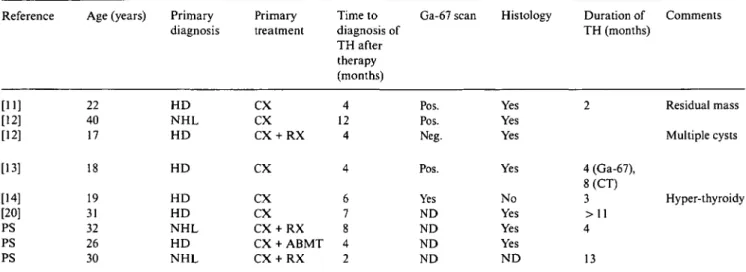
Table I. Characteristics of some recently reported cases of TH disclosed on CT after primary therapy for lymphoma in adults. Reference [11] [12] [12] [13] [14] [20] PS PS PS Age (years) 22 40 17 18 19 31 32 26 30 Primary diagnosis H D N H L H D HD H D H D N H L HD N H L Primary treatment CX CX CX + RX CX CX CX CX + RX CX + ABMT CX + RX Time to diagnosis of TH after therapy (months) 4 12 4 4 6 7 8 4 2 Ga-67 scan Pos. Pos. Neg. Pos. Yes ND ND ND ND Histology Yes Yes Yes Yes No Yes Yes Yes ND Duration of TH (months) 2 4 (Ga-67), 8 (CT) 3 > 1 1 4 13 Comments Residual mass Multiple cysts Hyper-thyroidy
Abbreviations: CX - chemotherapy; RX - radiotherapy; Pos. - positive; Neg. - negative; ND - not done; PS - present series.
Radiology, summary
As pointed out by these studies [1, 2, 6,7], the absence of concurrent lymph node enlargement or other signs of relapsing lymphoma, and the timing of thymic enlarge-ment or uptake, are the major clues favoring the diag-nosis of TH, which retains the characteristic shape of normal thymus.
Rebound thymic hyperplasia
Histology
Rebound TH correspond to a true hyperplasia involving both the cortex and the medulla [18]. It is to be distin-guished from 'thymic lymphoid follicular hyperplasia' of the medulla or 'germinal center hyperplasia', which can be associated with myastenia gravis [18]. In 30%-50% of the latter CTscan shows a normal-sized thymus [7].
Pathophysiology of TH
As illustrated by Mackall et al. [9], the regenerative capacity of thymopoiesis decreased with age, which could represent a decline in the intrinsic capacity of lymphopoietic stem cells and/or changes in the thymic microenvironment. This group showed an inverse rela-tion between the patients' ages and the recovery of CD4+ T-lymphocytes six months after chemotherapy. This re-covery was correlated quantitatively with the appear-ance of CD45RA+-naive T-lymphocytes in the periph-eral blood and with thymic enlargement (P - 0.015). At three months post-therapy, there was no relationship among age, TH, and CD8-positive lymphocytes which had returned to baseline [19].
Clinical manifestation ofTH, and associated situation
TH is asymptomatic and usually regresses within a few months, but long-standing cases are reported [1, 20]. 'Rebound' TH has been associated with prolonged sur-vival and may be a good prognostic sign [4, 5].
Rebound TH has been described following hyper-thyroidy or resolution of important stress in children (burns, surgery, corticotherapy) [18], and treatment of Cushing's syndromes [21].
TH following chemotherapy and/or radiotherapy is not restricted to patients treated for lymphoma. Kissin et al. [4] reported that 11.6% of (14 of 120) patients developed TH 3 to 14 months after cessation of chemo-therapy for malignant testicular teratoma, and that one TH occurred among 80 patients treated by surgery only. Three cases of TH described by Mackall et al. [9] oc-curred among children treated for sarcoma.
Is TH after primary treatment for lymphoma a plausible diagnosis at any age?
'Rebound' TH has essentially been described in children and adolescents, in whom frequencies of 16%: 10 of 62 (1); 17%: 4 of 23 (2) and 12%: 6 of 51 (3) have been found. The 10 patients reported by Peylan-Ramu et al. [1] were younger than 15 years.
Though infrequent, TH has been observed in adults with lymphoma. In a review of the literature, Langer et al. [5] found 21 cases following treatment for HD, with a median age of 23 years (range 8-40). More recently, further cases of TH following treatment of HD or NHL have been described in adults [11-14, 20] and are listed in Table 1.
99
Particular aspects of TH illustrated by our cases Cases no. 1 and no. 3 showed an enlarged thymus before any treatment, which was not considered to be infiltrated. This anomaly persisted as a residual growing mass after completion of treatment and achievement of a CR. TH could already have been present at the time of presenta-tion [6], as shown by two cases reported by Langer et al. [5], and by the study of Moul et al. [22], which disclosed at presentation four TH in 221 patients with germ cell testicular tumors.
Case no. 2 presented a growing residual mediastinal mass after autologous BMT for HD. Diagnosis of TH was made by surgical biopsy. Thymic hyperplasia occur-ring after allogeneic BMT has been described in a nine-year-old child suffering from an acute lymphoblastic leukemia [23]. In our case, with bulky mediastinum at presentation, thymus was considered to be infiltrated by HD. It demonstrates that TH must be considered in the differential diagnosis of a residual mass.
Management ofTH
We acknowledge, with Peylan-Ramu et al. [1], that a careful clinical and radiological follow-up is frequently sufficient. There is no need for specific treatment. Although regression of the mass with steroid challenge 60 mg/m2 daily for 7-10 days [24] could favor TH if the initial diagnosis was not steroid-sensitive leukemia or lymphoma.
An invasive diagnostic procedure can usually be avoided, but should be considered if a new oncological treatment is planned, in order to obtain histological confirmation of lymphoma relapse.
Conclusion
Thymic hyperplasia following chemotherapy and/or radiotherapy for lymphoma is probably not rare in the adult population up to the age of 40. As illustrated by our three cases, TH can be present before any treatment and/or recognized as a growing anterior mediastinal mass during the first months after therapy (up to one year). It should be included in the differential diagnosis of a residual mass. Using strict diagnostic criteria (iso-lated radiological mass retaining the characteristic shape and density of normal thymus, without mediastinal aden-opathy and without symptoms or signs of lymphoma relapse), a careful clinical and radiological follow-up is generally sufficient. An invasive diagnostic procedure can usually be avoided, but should be considered before administering a new oncological treatment, in order to ascertain lymphoma relapse and rule out TH.
References
1. Peylan-Ramu N, Haddy TB, Jones E et al. High frequency of benign mediastinal uptake of gallium-67 after completion of che-motherapy in children with high-grade non-Hodgkin's lympho-ma. J Clin Oncol 1989; 7: 180O-6.
2. Luker GD, Siegel MJ. Mediastinal Hodgkin disease in children: Response to therapy. Radiology 1993; 189: 737-40.
3. Mohamed Y, Leonidas J, Mehta H et al. Thymic hyperplasia (TH) after treatment of childhood Hodgkin's disease (HD). Proc Am Soc Clin Oncol 1997; 527a (Abstr 1899).
4. Kissin CM, Husband JE, Nicholas D et al. Benign thymic enlargement in adults after chemotherapy: CT demonstration. Radiology 1987; 163: 67-70.
5. Langer CJ, Keller SM, Erner SM. Thymic hyperplasia with hemorrhage simulating recurrent Hodgkin disease after chemo-therapy-induced complete remission. Cancer 1992; 70: 2082-6. 6. Heron CW, Husband JE, Williams MP. Hodgkin disease: CT of
the thymus. Radiology 1988; 167. 647-51.
7. Gamsu G. The mediastinum. In Moss AA, Gamsu G, Genant HK (eds): Computed Tomography of the Body with Magnetic Resonance Imaging. Second edition, Vol 1. Philadelphia: WB Saunders 1992; 43-118.
8. Zerhouni EA, Herold CJ, Hahn D Mediastinum and lung. In Stark DD, Bradley WG (eds): Magnetic Resonance Imaging. Second edition, Vol 2. St. Louis: Mosby Year Book 1992; 1429-89. 9. Mackall CL, Fleisher TA, Brown MR et al. Age, thymopoiesis, and CD4+ T-lymphocyte regeneration after intensive chemother-apy. N Engl J Med 1995; 332: 143-9.
10. Harris EW, Rakow JI, Weiner M et al. Thallium-201 scintigraphy for assessment of a gallium-67-avid mediastinal mass following therapy for Hodgkin's disease. J Nucl Med 1993; 34: 1326-30. 11. Small EJ, Venook AP, Damon LE. Gallium-avid thymic
hyper-plasia in an adult after chemotherapy for Hodgkin disease. Can-cer 1993; 72: 905-8.
12. Abdulnour E, Beauregard P. Anterior mediastinal masses after cancer therapy: Recurrences or benign lesions? JCC 1993; 36: 504-36.
13. Burns DE, Schiffman FJ. Beguiled by the gallium. Thymic re-bound in an adult after chemotherapy for Hodgkin's disease. Chest 1993; 104: 1916-9.
14. Pendlebury SC, Boyages S, Koutts J et al. Thymic hyperplasia associated with Hodgkin disease and thyrotoxicosis. Cancer 1992; 70: 1985-7.
15. Rettenbacher L, Galvan G. Differentiation between residual can-cer and thymic hyperplasia in malignant non-hodgkin's lym-phoma with somatostatin receptor scintigraphy. Clin Nucl Med
1994; 19: 64-5.
16. Salloum E, Schwab Brandt D, Caride VJ et al. Gallium scans in the management of patients with Hodgkin's disease: A study of 101 patients. J Clin Oncol 1997; 15: 518-27.
17. Israel O, Front D. Benign mediastinal and parahilar uptake of gallium-67 in treated lymphoma: Do we have all the answers? J Nucl Med 1993; 34: 1330-2.
18. Kornstein MJ, deBlois GG. Non-neoplastic pathology of the thymus. In Kornstein MJ (ed): Pathology of the Thymus and Mediastinum. First edition. Philadelphia: WB Saunders 1995; 34-66.
19. Mackall CL, Fleisher TA, Brown MR et al. Distinctions between CD8+ and CD4+ T-cell regenerative pathways result in prolonged T-cell subset imbalance after intensive chemotherapy. Blood 1997; 89: 3700-7.
20. Hermann R, Greminger P, Dommann-Scherrer C et al. Diffuse Thymushyperplasie nach Chemotherapie eines nodular-sklero-sierenden Hodgkin-Lymphoms. Schweiz Med Wochenschr 1994;
124: 1666-71.
21. Tabarin A, Catargi B, Chanson P et al. Pseudo-tumours of the thymus after correction of hypercortisolism in patients with ectopic ACTH syndrome: A report of five cases. Clin Endocrinol 1995; 42: 207-13.
22. Moul JW, Fernandez EB, Bryan MG et al. Thymic hyperplasia in newly diagnosed testicular germ cell tumors. J Urol 1994; 152: 1480-3.
23. Miniero R, Busca A, Leonardo E et al. Rebound thymic hyper-plasia following high dose chemotherapy and allogeneic BMT. Bone Marrow Tranplant 1993; 11: 67-70.
24. Ford EG, Lockhart SK, Sullivan MP et al. Mediastinal mass following chemotherapeutic treatment of Hodgkin's disease: Re-current tumor or thymic hyperplasia? J Pediatr Surg 1987; 22: 1155-9.
Received 19 June 1997; accepted 1 October 1997.
Correspondence to: Dr. Sandro Anchisi Division d'oncologie Hopital cantonal Rue Micheli-du-Crest 24 CH-1211 Geneve 14 Switzerland