CORRESPONDENCE
Multiple Endocrine Neoplasia
1 Gene Alterations in
MEN1-Associated and
Sporadic Lipomas
Multiple endocrine neoplasia 1 (MEN1) syndrome is characterized by the development of parathyroid adeno-mas, pituitary adenoadeno-mas, and duodenal and/or pancreatic neuroendocrine tu-mors. The vast majority, and possibly all, of these tumors are characterized by two genetic hits—germline mutation of the MEN1 tumor suppressor gene com-bined with allelic deletion of the corre-sponding wild-type allele (1). Alter-ations of the MEN1 gene are not only found in MEN1-associated tumors but also, with decreasing frequency, in spo-radic neuroendocrine tumors of the fore-gut (2,3), parathyroid tumors (4), and pi-tuitary adenomas (5).
Lipomatous tumors are known to oc-cur in a relatively high proportion of pa-tients with MEN1 disease. In this study, we attempted to determine the role of the MEN1 gene in the development of MEN1-associated and sporadic lipomas. For genetic tissue analysis of MEN1-associated lipomas, we obtained prepa-rations by touching tumor tissue to a glass slide to transfer a limited number of cells; we then performed fluorescence in situ hybridization (FISH) on such touch preparations to detect deletions of the MEN1 wild-type allele in two tu-mors that were excised from two pa-tients with known MEN1 gene germline mutations (case 1—a 61-year-old male with an abdominal wall lipoma, muta-tion status L22R; case 2—a 69-year-old male with a lipoma of the right thigh, mutation status W436R). FISH was per-formed using as a probe cosmid clone c10B11 (size, 40 kilobases), which con-tains the MEN1 gene (6). For analysis of sporadic lipomas, we performed poly-merase chain reaction (PCR)-based single-strand conformation polymor-phism and sequence analysis using for-malin-fixed, paraffin-embedded tissue.
FISH analysis of the two lipomas, which were excised from patients with known MEN1 disease, revealed loss of one MEN1 allele in 53% of the cells examined from case 1 (Fig. 1, left panel) and in 63% of the cells examined from case 2. It appears from this finding that the lipoma cells are affected by genetic deletion, whereas both MEN1 gene cop-ies were visualized in normal cellular constituents. Furthermore, in both cases, two copies of the chromosome 11 alpha satellite, located in the centromere, were present in all of the cells, as shown by a control probe. In conjunction with a re-cent PCR-based deletion analysis of three lipomas (7), our results confirm the hypothesis that mutation of the MEN1 gene and subsequent loss of the wild-type allele are associated with or causative for the development of lipo-matous tumors in patients with MEN1 disease.
To investigate the role of the MEN1 gene in sporadic lipomas, we analyzed six sporadic tumors (female : male 4 2:4; mean age4 33 ± 16 years). Lipo-matous and normal control tissue was scraped from sections of paraffin-embedded tissue after deparaffinization in xylene and alcohol. The removed tis-sue was placed in proteinase K buffer for DNA extraction. The DNA samples
were PCR amplified with 13 pairs of markers amplifying the coding regions of the MEN1 gene, including exons 2– 10, as previously described (2). The am-plification products were visualized af-ter polyacrylamide gel electrophoresis. Due to poor tissue quality, amplification products were only obtained for exons 2, 3, 8, and 9. In one case, single-strand conformation polymorphism analysis and subsequent sequence analysis re-vealed a four-nucleotide deletion in exon 2 (Fig. 1, right panel). This dele-tion was present only in the tumor tissue but not in the normal tissue control from the same patient. From this finding, we conclude that MEN1 gene mutation may play a role not only in the development of MEN1-associated lipomas but also in sporadic lipomas. ALEXANDERO. VORTMEYER ROLANDBO¨NI EVGENIAPAK SVETLANAPACK ZHENGPING ZHUANG
References
(1) Chandrasekharappa SC, Guru SC, Manickam
P, Olufemi SE, Collins FS, Emmert-Buck MR, et al. Positional cloning for the gene for mul-tiple endocrine neoplasia-type 1. Science 1997;276:404–7.
(2) Zhuang Z, Vortmeyer AO, Pack S, Huang S,
Fig. 1. Detection of genetic alterations in multiple endocrine neoplasia 1 (MEN1)-associated and sporadic
lipomas. Left panel) fluorescence in situ hybridization analysis of a preparation, obtained by touching tumor tissue to a glass slide to transfer a limited number of cells, from lipomatous tissue (case 1) using cosmid clone c10B11, which contains the MEN1 gene as a probe; red signals4 the presence of the MEN1 gene on chromosome 11q13, green signals4 the centromeric region of chromosome 11; four cells exhibit loss of one MEN1 allele; one cell in the right upper corner shows the presence of both MEN1 gene copies, and therefore is interpreted as a non-neoplastic stromal cell. Right panel) Sequence analysis of a sporadic lipoma (case 1) reveals a TGTC deletion in the tumor cells when compared with normal control tissue from the same patient.
Pham TA, Wang C, et al. Somatic mutations of the MEN1 tumor suppressor gene in spo-radic gastrinomas and insulinomas. Cancer Res 1997;57:4682–6.
(3) Toliat M, Berger W, Ropers HH, Neuhaus P,
Wiedenmann B. Mutations in the MEN I gene in sporadic neuroendocrine tumours of gastroen-teropancreatic system. Lancet 1997;350:1223.
(4) Heppner C, Kester MB, Agarwal SK,
De-belenko LV, Emmert-Buck MR, Guru SC, et al. Somatic mutation of the MEN1 gene in parathy-roid tumours. Nat Genet 1997;16:375–8.
(5) Zhuang Z, Ezzat SZ, Vortmeyer AO, Weil R,
Oldfield EH, Park WS, et al. Mutations of the MEN1 tumor suppressor gene in pituitary tu-mors. Cancer Res 1997;57:5446–51.
(6) Guru S, Olufemi SE, Manickam P, Cummings
C, Gieser LM, Pike BL, et al. A 2.8-Mb clone contig of the multiple endocrine neoplasia type 1 (MEN1) region at 11q13. Genomics 1997;42:436–45.
(7) Dong Q, Debelenko LV, Chandrasekharappa
SC, Emmert-Buck MR, Zhuang Z, Guru SC, et al. Loss of heterozygosity at 11q13: analysis of pituitary tumors, lung carcinoids, lipomas, and other uncommon tumors in subjects with familial multiple endocrine neoplasia type 1. J Clin Endocrinol Metab 1997;82:1416–20.
Notes
Affiliations of authors: A. Vortmeyer, E. Pak, S.
Pack, Laboratory of Pathology, Division of Clini-cal Sciences, National Cancer Institute, Bethesda, MD; R. Bo¨ni, Department of Dermatology, Uni-versity Hospital, Zurich, Switzerland.
Correspondence to: Zhengping Zhuang, M.D.,
Ph.D., National Institutes of Health, Bldg. 10, Rm. 2N212, Bethesda, MD 20892.
Re: Prevalence of Cancer
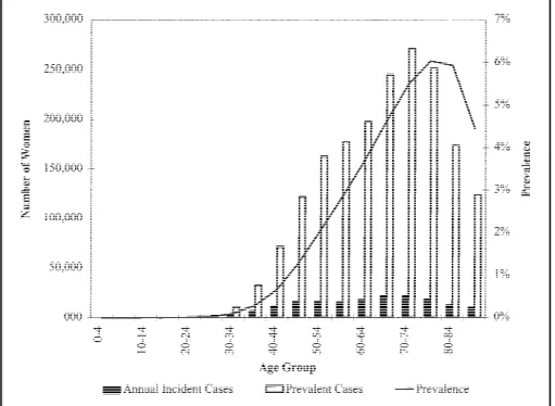
A recent Stat Bite (1) showed the prevalent cases of eight cancer types in the U.S. population. Practitioners of public health often ignore prevalence as a measure of disease frequency, prob-ably because differences in prevalence between groups can arise from differ-ences in incidence or from differdiffer-ences in the average survival (2). While this am-biguity may cloud etiologic interpreta-tion of differences in prevalence, the mixture of occurrence and survival makes prevalence an important measure of the distribution of disease.Fig. 1 shows our estimates of the prevalence, prevalent cases, and annual incident cases of breast cancer by age among U.S. women in 1997. We esti-mated the prevalence by applying the method of Alho (3) to data reported by the National Cancer Institute (NCI) (4), the National Center for Health Statistics,
and the Census Bureau. A further de-scription is available from the authors.
The age-specific prevalence provides insight into the distribution of breast cancer among U.S. women. This insight differs from what one would learn by examining incidence rates alone. For ex-ample, while about 22% of incident cases of breast cancer occur in women younger than age 50 years, only about 12% of the prevalent cases exist in that group. Similarly, 51% of new cases oc-cur in women younger than age 65 years, but only about 42% of the preva-lent cases exist in that group. The ratio of the number of prevalent cases to an-nual incident cases increases in older age groups. Among women 40–44 years old, the ratio of prevalent cases to an-nual incident cases is about 6 to 1. Among women 70–74 years old, the ra-tio is about 12 to 1.
This description of prevalent breast cancer emphasizes the importance of in-cluding the elderly population in studies of cancer survivors. There are about as many prevalent cases of breast cancer among women ages 80–84 years as there are among women ages 55–59 years. Women may weigh aspects of cancer treatment and survival differently depending on their age. For example, younger women are often concerned
about the effect of treatment on their ability to meet their obligations, such as caring for family members (5). About 20% of the women less than 75 years old at diagnosis said this was a very impor-tant consideration in making decisions about treatment for breast cancer. Among women age 75 years and older, 7% said this was a very important con-sideration and 83% said it was not im-portant at all. Younger women may weight most heavily the expected dura-tion of survival, whereas older women may weight most heavily the quality of their expected survival, particularly their ability to live independently. The elderly have often been excluded from studies of treatment efficacy, and the frequency of age-related exclusion has increased in re-cent years (6), at least in the case of acute myocardial infarction. As the NCI em-barks on a formal program to study cancer survivors (7), the program must take care to avoid a reprise of this ageist history.
TIMOTHYL. LASH
REBECCAA. SILLIMAN
References
(1) Stat bite: Prevalence of cancer [news]. J Natl
Cancer Inst 1997;89:1093.
(2) Rothman KJ. Modem epidemiology Boston:
Little, Brown, 1986.
(3) Alho, JM. On prevalence, incidence, and
du-Fig. 1. Estimated prevalence, number of prevalent cases, and annual number of incident cases of breast
cancer among U.S. women in 1997.