Invasive pulmonary aspergillosis:
effects of early resection in a neutropenic rat model
J.M. Habicht
a,*, M. Preiss
a, J. Passweg
b, P. Dalquen
c, P. Matt
a, H. Adler
d,
R. Frei
d, H.-R. Zerkowski
aaDivision of Cardio-Thoracic Surgery, University Hospital, Basel, Switzerland bDivision of Hematology, University Hospital, Basel, Switzerland
cDivision of Pathology, University Hospital, Basel, Switzerland dBacteriology Laboratory, University Hospital, Basel, Switzerland
Received 7 April 2002; received in revised form 26 June 2002; accepted 8 July 2002
Abstract
Objective: Invasive pulmonary aspergillosis is frequent in neutropenic patients. Usually localized in the beginning, the disease spreads and mortality is high despite antifungal treatment. The role of early adjuvant surgery is not clear. Surgery may help to confirm fungal disease, may control fungal disease locally and may prevent systemic spreading. This study examines effects of early resection on survival and dissemination in a rat model of localized invasive pulmonary aspergillosis. Methods: Forty persistently neutropenic male albino rats were challenged with standardized conidial aspergillus inoculum injected into peripheral lung tissue of the right upper lobe under direct vision. Animals were divided into four groups. Twenty animals were treated with amphotericin B at 1 mg/kg per day beginning 48 h after inoculation, 20 animals were left untreated. In each group half the animals underwent early resection of localized invasive aspergillosis by lobectomy. Animals were checked daily and mortality was recorded up to 28 days after which surviving animals were sacrificed. Results: Significantly higher survival was observed in resected animals in the non-Am B groups (survival: 10 ^ 19% without early resection and 50 ^ 32% with early resection; P ¼ 0:044). However, early resection did not lead to improved survival in animals treated with amphotericin B (survival 70 ^ 29% without early resection and 50 ^ 32% with early resection; P ¼ 0:316). Conclusions: In this rat model of localized invasive pulmonary aspergillosis effects of early resection on survival could be demonstrated only in animals not receiving amphotericin B treatment. q 2002 Elsevier Science B.V. All rights reserved.
Keywords: Fungal infection; Invasive pulmonary aspergillosis; Neutropenia; Surgery; Animal
1. Introduction
Mortality of invasive pulmonary aspergillosis (IPA) is reported to be 40–60% despite adequate medical treatment [1]. Descriptive studies [2–9] and a recent retrospective comparative cohort study [10] of surgically versus medi-cally treated patients have reported lower case fatality rates of 30% or less in surgically treated patients. Although these results are promising, the role of early surgery in the treatment of IPA in neutropenic hematological patients has not been generally established given the lack of prospective randomized studies.
In the present study a rat model of resection of localized IPA is presented. The aim of this study is to follow an
established model of diffuse IPA in persistently neutropenic rats initially developed for comparisons of antifungal drugs [11,12] and to modify the model to observe the effects of early resection of localized IPA on mortality and disease dissemination.
2. Materials and methods 2.1. Animals
Forty male albino rats (Sprague–Dawley, specified patho-gen free, weight 180–250 g; RCC Ltd., Fullinsdorf, Switzer-land). The animals received humane care in compliance with the European Convention on Animal Care and the study was approved by the local institutional ethics commit-tee (Supervisory Board for Animal Experiments, Kanto-nales Veterinaeramt Basel; protocol #1772).
1010-7940/02/$ - see front matter q 2002 Elsevier Science B.V. All rights reserved. PII: S 1 0 1 0 - 7 9 4 0 ( 0 2 ) 0 0 4 6 7 - 0
www.elsevier.com/locate/ejcts
* Corresponding author. Herzzentrum, Hirslanden Klinik Im Schachen, Ziegelrain 23, CH-5000 Aarau, Switzerland. Tel.: 141-62-823-0704; fax: 141-62-823-2315.
2.2. Materials
Amphotericin B (Fungizone, Bristol-Myers Squibb); cyclophosphamide (Endoxan, Asta Medica); amoxicillin (Clamoxyl, SmithKline Beecham); gentamicin (Garamycin, Essex Chemie); isoflurane (Forene, Abott), Sabouraud agar (Biomerieux, Marcy l’Etoile, France for production of coni-dia and Becton Dickinson, Meylan Cedex, France for controls).
2.3. Anesthesia
All intraperitoneal (i.p.) and intramuscular (i.m.) injec-tions and all surgical procedures were performed under spontaneous ventilation with an isoflurane/oxygen gas mixture (evaporation system: Medical Supplies & Services Int. Ltd., UK) in an induction box and/or on an operation table with a mask and waste-air suction system (Vet-Anest System, Provet Ltd., Lyssach, Switzerland).
2.4. Fungal strain
A strain of Aspergillus fumigatus was isolated from a fatal case of leukemia with disseminated IPA. MIC of amphoter-icin B for this strain was 0.5 mg/l. The fungus was cultured at 37 8C on Sabouraud agar and conidia were harvested in sterile saline. Preparation of a microscopically confirmed hyphal-free conidial suspension was performed as described
by Leenders et al. [11]. A suspension of 5 £ 105conidia/ml
was produced freshly a few hours before inoculation. Each
inoculum consisted of 0.02 ml of suspension (1 £ 104
coni-dia) in separate syringes (Micro Fine 0.5 ml, Becton Dick-inson, Meylan Cedex, France). Viability of conidia in inoculum was tested by simultaneously culturing a sample of inoculum on Sabouraud agar.
2.5. Experimental local lung infection, early resection and histological stain
All rats received inoculum under direct vision through a small thoracotomy into the peripheral tissue of right upper lobe 5 days after beginning of immunosuppression. In resec-tion groups lobectomy was performed on day 13 after inoculation. The thoracotomy was reopened, the lobe clipped at the base (Premium Surgiclip M 11.5 Auto Suture, Tyco Healthcare Ltd., Wollerau, Switzerland) and removed aseptically. A smear of the lobe was taken on Sabouraud and the complete lobe sent for histological examination. Hema-toxylin–eosin, PAS and Grocott-stains were performed. In non-resected animals the complete right lung was removed at autopsy together with the other organs as described below.
2.6. Immunosuppression, supportive care and experimental setting
Persistent neutropenia was induced according to the method described by Leenders et al. [11] and Van Etten et
al. [12]. Cyclophosphamide 90 mg/kg per day was injected i.p. 5 days before fungal inoculation, followed by repeated doses of cyclophosphamide 60 mg/kg per day at 4-day inter-vals on days 21, 13, 17, 111, etc. By obtaining blood samples through orbital puncture under light anesthesia on a regular basis this immunosuppressive regime has been demonstrated to lead to profound neutropenia beginning 5 days after first dose of cyclophosphamide [11]. In the present study leukocyte counts were determined from blood samples at the time of death or sacrifice to confirm aplasia (automatic blood cell counter: Advia 120, Bayer Diagnostics, Mu¨nchen, Germany).
Animals were kept under strict hygienic conditions and allowed free rodent laboratory fodder (Kliba Mu¨hlen Ltd., Kaiseraugst, Switzerland) and water. To prevent bacterial superinfection amoxicillin i.m. (40 mg/kg per dose) was added daily beginning on day 21 and gentamycin i.m. (6 mg/kg per dose) was given on days 21 and 0. In amphoter-icin B (Am B) treatment groups Am B i.p. (1 mg/kg per day) was begun 48 h after inoculation. Mycelial growth is known to be firmly established after 30 h in neutropenic rats [12]. Animals were checked daily and mortality was recorded up to 28 days after which surviving rats were killed (in rats surviving for longer time periods the influence on survival by other factors such as anemia and bacterial infections become a major concern). From all animals both lungs, liver, spleen and kidneys were removed, and examined macroscopically and histologically. At the time of death or sacrifice residual fungal disease was classified as either absent, local disease or disseminated disease. Ipsilateral histologically verified fungal manifestations in the inocu-lated lung, pleura or thoracic wall were defined as local disease. Disseminated disease was defined as histologically verified invasive aspergillosis in one or more of the follow-ing organs: contralateral lung, mediastinum, liver, spleen, kidneys and retroperitoneal space.
2.7. Statistical analysis
To compare categorical variables among groups the chi-squared or the Fisher’s exact test were used where appro-priate. Survival of rats was analyzed using the Kaplan– Meier estimator and compared among groups using the log-rank test. Rats surviving to 28 days and killed at that time were considered censored observations.
3. Results
Median white blood cell count was 0:53 £ 109=l and
median neutrophil count 0:11 £ 109=l.
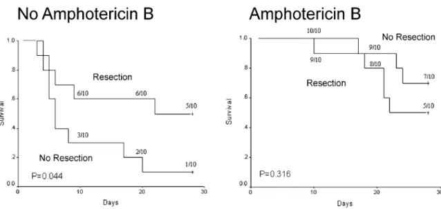
Survival probabilities at 28 days were 30 ^ 21% (95% confidence interval) in the group not receiving Am B, and
60 ^ 22% in the group receiving Am B (P ¼ 0:0085).
Survival was 40 ^ 22% in animals not undergoing early
resection, and 50 ^ 22% (P ¼ 0:412) in animals with
Early resection did not lead to improved survival in animals treated with Am B, (survival 70 ^ 29% without early resection and 50 ^ 32% with early resection;
P ¼ 0:316), but significantly higher survival was observed
in resected animals in the non-Am B group (survival: 10 ^ 19% without early resection and 50 ^ 32% with
early resection; P ¼ 0:044) (Fig. 1).
IPA was observed in all resected lobes histologically. When dissemination occurred (43%), the liver was most often involved (77%), followed by spleen (35%), contralat-eral lung (18%), mediastinum (6%) and retroperitoneum (6%). No IPA manifestations in kidneys and brain were observed in this series. At death or autopsy local disease was seen in 25% of animals undergoing early resection
and in 65% of animals without early resection
(P ¼ 0:011). Dissemination of disease was observed in
50% of animals undergoing early resection and in 35% of
animals without early resection (P ¼ 0:337) In the subgroup
of animals not receiving Am B, dissemination of disease was observed in 50% of animals undergoing early resection
and in 60% of animals without early resection (P ¼ 0:653).
In the same subgroup local disease was seen in 30% of animals undergoing early resection and in 90% of animals
without early resection (P ¼ 0:021). Absence of disease at
death or sacrifice was observed in 50% of animals treated
with Am B and 20% of animals not treated (P ¼ 0:047).
4. Discussion
The incidence of IPA is reported to vary from 2 to 14% in hematologic patients in the literature [10,13–16]. Inhalation of conidia by immunocompetent individuals rarely has any adverse effect, since conidia are eliminated efficiently by immune mechanisms [17]. Conidia enter the lungs through the airways and in neutropenic patients the mycelium then
infiltrates through the bronchial wall into adjacent pulmon-ary arteries causing thrombosis and infarction [18], which facilitates further mycelial growth and hampers induction of
effective antifungal drug concentrations. Seemingly,
outcome of IPA is more dependent on marrow regeneration and termination of immunosuppressive state than on any therapeutic strategy, but antifungal drugs such as amphoter-icin B or azole derivatives are the standard of care. Although general contamination of the bronchial tree with conidia must be assumed, a diffuse fungal pneumonia is rarely seen in humans at the time of diagnosis. Initially the disease is typically localized, offering the possibility of early adju-vant resection.
Animal models for IPA have been established in a variety of species and there is no consensus about the best model. The models most often employed are those used in rabbits [19–21], rats [11,12,22] and mice [23–25] and were devel-oped to study the efficacy of antifungal drugs or to evaluate diagnostic methods. Challenge protocols vary considerably in terms of concentration of conidia as well as route of injection (intranasal, intratracheal or intravenous). Of note, all comparable published models use intratracheal or intrabronchial inoculation with aspergillus conidia, thus leading to widespread lung involvement. We chose an established rat model of neutropenia and diffuse IPA [11,12] and with a direct injection into peripheral lung parenchyma aimed at simulating the clinical situation with a localized focus. Thoracotomy and injection under direct vision was chosen in contrast to blind intercostal injection in order to avoid intrapleural contamination and intravascular spread into major pulmonary vessels. Although great care was given to avoid any undesirable propagation of conidia by the above mentioned mechanisms, there is no tool avail-able to exclude this possibility. However, no case of diffuse unilateral intrapleural aspergillosis nor any animal dying exceptionally early of diffuse multiple organ spread was
Fig. 1. Effects of resection of a focus of localized invasive pulmonary aspergillosis (IPA) in animals with and without amphotericin B treatment (1 mg/kg per day). Neutropenic rats were inoculated in right upper lobe with 2 £ 104Aspergillus fumigatus conidia at time zero (Kaplan–Meier plot).
observed, supporting the probability that no intravascular dissemination occurred during injection.
Our results show resection of localized IPA to improve survival in animals not treated by Am B. Early resection is effective through local control of IPA rather than through prevention of disseminated disease. Dissemination was found in 50–60% in resected and non-resected animals. This supports the hypothesis that resection will not cure fungal disease in most instances but might be useful to gain time for hematological reconstitution of IPA-patients so treated. The slightly higher incidence of disease dissemi-nation observed in resected animals could point to a danger of disseminating aspergillosis through surgery. The differ-ence of dissemination between resected animals and animals not undergoing resection was, however, not signif-icant and this would have to be studied in a larger experi-mental series and preferentially also in the human setting. Resection may be undertaken more extensively in humans as opposed to small experimental animals which may mini-mize the danger of iatrogenic dissemination, but in our formerly published clinical comparative series [10] there was no evidence of a larger rate of disseminated disease in patients undergoing surgery as opposed to patients treated conservatively. It is probable that surviving non-Am B animals showing distant organ involvement would have eventually died of IPA later on if they had not been killed at end of observation period. However, resection enabled more animals to reach cut-off point at 28 days. In the clinical situation the time gained may in part explain the good results reported in surgical series in patients with reversible aplasia. The fact that at autopsy a substantial number of non-resected animals treated with Am B showed absence of any fungal manifestation, together with the observation that IPA could be demonstrated in all early resection specimens, supports the interpretation that Am B-treated neutropenic animals may spontaneously clear an IPA-manifestation even when caused by a major dose of conidia, as long as it originates and stays confined to a single small focus. In animals not treated with Am B such an observation could not be made, inasmuch as none of the non-resected animals were disease-free, even if surviving up to 28 days.
Results shown here may not be transferred easily to IPA encountered in neutropenic patients. In contrast to the clin-ical situation, this experiment precisely defines onset of neutropenia and onset of fungal infection. Therefore the restriction of the beneficial effect of early resection to animals not treated with Am B and the limitation of its effect to local control may not be valid in the human situation. Experimentally induced aspergillosis is a hyperacute infec-tion obtained with a high concentrainfec-tion of conidia given at a single point in time. In humans IPA is often a more indolent disease caused by continuous or repeated exposure to small numbers of conidia, and time of initiation of antifungal treatment varies considerably.
The value of early resection in the human situation is not clear. So far only observational studies are available
suggesting a beneficial effect. Localized invasive aspergil-losis is often operated on even though there is little evidence for these interventions. The current experimental study shows that resection can be life-saving in animals not receiving early antifungal treatment. In the clinical situation where antifungal treatment is often initiated only at a time when invasive aspergillosis is well established, resection strategies could prove equally beneficial. It is only through a large randomized study comparing resection strategies to more conservative treatment that such a benefit could be proven.
We conclude that in the presented rat model of localized invasive pulmonary aspergillosis early resection of a single fungal lesion improved survival in a subgroup of animals. Early resection was effective for local control of the disease but did not necessarily prevent systemic spread. A beneficial effect of resection could be observed if animals were not simultaneously treated with amphotericin B.
References
[1] Denning DW, Stevens DA. Antifungal and surgical treatment of inva-sive aspergillosis: review of 2121 published cases. Rev Infect Dis 1990;12:1147–1201.
[2] Young VK, Maghur HA, Luke DA, McGovern EM. Operation for cavitating invasive pulmonary aspergillosis in immunocompromised patients. Ann Thorac Surg 1992;53:621–624.
[3] Wong K, Waters CM, Walesby RK. Surgical management of invasive pulmonary aspergillosis in immunocompromised patients. Eur J Cardiothorac Surg 1992;6:138–143.
[4] Moreau P, Zahar JR, Milpied N, Baron O, Mahe B, Wu D, Germaud P, Despins P, Delajartre AY, Haroussea JL. Localized invasive pulmonary aspergillosis in patients with neutropenia. Effectiveness of surgical resection. Cancer 1993;72:3223–3226.
[5] Robinson LA, Reed EC, Galbraith TA, Alonso A, Moulton AL, Flem-ing WH. Pulmonary resection for invasive aspergillus infections in immunocompromised patients. J Thorac Cardiovasc Surg 1995;109:1182–1197.
[6] Caillot D, Casasnovas O, Bernard A, Couaillier JF, Durand C, Cuisi-nier B, Solary E, Piard F, Petrella T, Bonnin A, Couillault G, Dumas M, Guy H. Improved management of invasive pulmonary aspergillo-sis in neutropenic patients using early thoracic computed tomographic scan and surgery. J Clin Oncol 1997;15:139–247.
[7] Baron O, Guillaume´ B, Moreau P, Germaud P, Despins P, De Lajartre AY, Michaud JL. Aggressive surgical management in localized pulmonary mycotic and nonmycotic infections for neutropenic patients with acute leukemia: report of eighteen cases. J Thorac Cardi-ovasc Surg 1998;115:63–69.
[8] Salerno CT, Ouyang DW, Pederson TS, Larson DM, Shake JP, John-son EM, Maddaus MA. Surgical therapy for pulmonary aspergillosis in immunocompromised patients. Ann Thorac Surg 1998;65:1415– 1419.
[9] Reichenberger F, Habicht JM, Kaim P, Dalquen P, Bernet F, Schlap-fer R, Stulz P, Perruchoud AP, Tichelli A, Gratwohl A, Tamm M. Lung resection for invasive pulmonary aspergillosis in neutropenic patients with hematologic diseases. Am J Respir Crit Care Med 1998;158:885–890.
[10] Habicht JM, Matt P, Passweg JR, Reichenberger F, Gratwohl A, Zerkowski H-R, Tamm M. Invasive pulmonary fungal infection in hematologic patients: is resection effective? Hematol J 2001;2:250– 256.
[11] Leenders ACAP, De Marie S, Ten Kate MT, Bakker-Woudenberg IAJM, Verbrugh HA. Liposomal amphotericin B (Am Bisome) reduces dissemination of infection as compared with amphotericin B deoxycholate (Fungizone) in a rat model of pulmonary aspergillo-sis. J Antimicrob Chemother 1996;38:215–225.
[12] Van Etten EWM, Stearne-Cullen LET, Ten Kate MT, Bakker-Woudenberg IAJM. Efficacy of liposomal Amphotericin B with prolonged circulation in blood in treatment of severe pulmonary aspergillosis in leukopenic rats. Antimicrob Agents Chemother 2000;44:540–545.
[13] McWhinney PHM, Kibbler CC, Hamon MD, Smith OP, Gandhi L, Berger LA, Walesby RK, Hoffbrand AV, Prentice HG. Progress in the diagnosis and management of aspergillosis in bone marrow transplan-tation: 13 years’ experience. Clin Infect Dis 1993;17:397–404. [14] Morrison VA, Haake RJ, Weisdorf DJ. Non-Candida fungal
infec-tions after bone marrow transplantation: risk factors and outcome. Am J Med 1994;96:497–503.
[15] Barnes AJ, Oppenheim BA, Chang J, Morgenstern GR, Scarffe JH. Early investigation and initiation of therapy for invasive pulmonary aspergillosis in leukaemic and bone marrow transplant patients. Mycoses 1999;42:403–408.
[16] Kami M, Tanaka Y, Kanda Y, Ogawa S, Masumoto T, Ohtomo K, Matsumura T, Saito T, Machida U, Kashima T, Hirai H. Computed tomographic scan of the chest, latex agglutination and plasma (1AE3)-beta-d-glucan assay in early diagnosis of invasive pulmonary aspergillosis: airways study of 215 patients. Haematologica 2000;85:745–752.
[17] Latge´ JP. Aspergillus fumigatus and aspergillosis. Clin Microbiol Rev 1999;12:310–350.
[18] Sobonya RE. Fungal diseases, including allergic bronchopulmonary
aspergillosis. In: Thurlbeck WM, editor. Pathology of the lung, New York: Thieme Medical, 1995. p. 307.
[19] Spreadbury CL, Krausz T, Pervez S, Cohen J. Invasive aspergillosis: clinical and pathological features of a new animal model. J Med Vet Mycol 1989;27:5–15.
[20] Berenguer J, Ali NM, Allende MC, Lee J, Garrett K, Battaglia S, Piscitelli SC, Rinaldi MG, Pizzo PA, Walsh TJ. Itraconazole for experimental pulmonary aspergillosis: comparison with amphotericin B, interaction with cyclosporin A and correlation between therapeutic response and itraconazole concentrations in plasma. Antimicrob Agents Chemother 1994;38:1303–1308.
[21] Berenguer J, Allende MC, Lee JW, Garrett K, Lyman C, Ali NM, Bacher J, Pizzo PA, Walsh TJ. Pathogenesis of pulmonary aspergil-losis: granulocytopenia versus cyclosporine and methylprednisolone-induced immunosuppression. Am J Respir Crit Care Med 1995;152:1079–1086.
[22] Murphy M, Bernard EM, Ishimaru T, Armstrong D. Activity of Vora-conazole (UK-109,496) against clinical isolates of Aspergillus species and its effectiveness in an experimental model of invasive pulmonary aspergillosis. Antimicrob Agents Chemother 1997;41: 696–698.
[23] Dixon DM, Polak A, Walsh TJ. Fungus dose-dependent primary pulmonary aspergillosis in immunosuppressed mice. Infect Immun 1989;57:1452–1456.
[24] Cenci E, Perito S, Enssle KH, Mosci P, Latge´ JP, Romani L, Bistoni F. Th1 and Th2 cytokines in mice with invasive aspergillosis. Infect Immun 1997;65:564–570.
[25] Cenci E, Mencacci A, d’Ostiani CF, Montagnoli C, Bacci A, Sero GD, Perito S, Bistomni F, Romani L. Cytokine- and T-helper-depen-dent immunity in murine aspergillosis. Res Immunol. 1998;149:445– 453.