Exp Brain Res (2003) 153: 591–604 DOI 10.1007/s00221-003-1616-0
R E S E A R C H A RT I C L E
Michela Adriani . Philippe Maeder . Reto Meuli . Anne Bellmann Thiran . Rolf Frischknecht . Jean-Guy Villemure . James Mayer .
Jean-Marie Annoni . Julien Bogousslavsky . Eleonora Fornari . Jean-Philippe Thiran . Stephanie Clarke
Sound recognition and localization in man: specialized cortical
networks and effects of acute circumscribed lesions
Received: 14 November 2002 / Accepted: 4 July 2003 / Published online: 18 September 2003 # Springer-Verlag 2003
Abstract Functional imaging studies have shown that information relevant to sound recognition and sound localization are processed in anatomically distinct cortical networks. We have investigated the functional organiza-tion of these specialized networks by evaluating acute effects of circumscribed hemispheric lesions. Thirty patients with a primary unilateral hemispheric lesion, 15 with right-hemispheric damage (RHD) and 15 with left-hemispheric damage (LHD), were evaluated for their capacity to recognise environmental sounds, to localize sounds in space and to perceive sound motion. One patient with RHD and 2 with LHD had a selective deficit in sound recognition; 3 with RHD a selective deficit in sound localization; 2 with LHD a selective deficit in sound
motion perception; 4 with RHD and 3 with LHD a combined deficit of sound localization and motion perception; 2 with RHD and 1 with LHD a combined deficit of sound recognition and motion perception; and 1 with LHD a combined deficit of sound recognition, localization and motion perception. Five patients with RHD and 6 with LHD had normal performance in all three domains. Deficient performance in sound recognition, sound localization and/or sound motion perception was always associated with a lesion that involved the shared auditory structures and the specialized What and/or Where networks, while normal performance was associated with lesions within or outside these territories. Thus, damage to regions known to be involved in auditory processing in normal subjects is necessary, but not sufficient for a deficit to occur. Lesions of a specialized network was not always associated with the corresponding deficit. Conversely, specific deficits tended not be associated predominantly with lesions of the corresponding network; e.g. deficits in auditory spatial tasks were observed in patients whose lesions involved to a larger extent the shared auditory structures and the specialized What network than the specialized Where network, and deficits in sound recog-nition in patients whose lesions involved mostly the shared auditory structures and to a varying degree the specialized What network. The human auditory cortex consists of functionally defined auditory areas, whose intrinsic orga-nization is currently not understood. In particular, areas involved in the What and Where pathways can be conceived as: (1) specialized regions, in which lesions cause dysfunction limited to the damaged part; observed deficits should be then related to the specialization of the damaged region and their magnitude to the extent of the damage; or (2) specialized networks, in which lesions cause dysfunction that may spread over the two specia-lized networks; observed deficits may then not be related to the damaged region and their magnitude not propor-tional to the extent of the damage. Our results support strongly the network hypothesis.
M. Adriani . A. B. Thiran . S. Clarke (*) Division de Neuropsychologie, CHUV, 1011 Lausanne, Switzerland
e-mail: Stephanie.Clarke@chuv.hospvd.ch Tel.: +41-21-3141309
Fax: +41-21-3141319
P. Maeder . R. Meuli . E. Fornari
Service de Radiodiagnostic et Radiologie Interventionnelle, CHUV,
Lausanne, Switzerland R. Frischknecht
Service de Rhumatologie et Médecine Réhabilitative, CHUV, Lausanne, Switzerland
J. Villemure
Service de Neurochirurgie, CHUV, Lausanne, Switzerland
J. Mayer . J. Annoni Service de Neurologie, HCU, Geneva, Switzerland J. Bogousslavsky
Service de Neurologie, CHUV, Lausanne, Switzerland J. Thiran
Institut de Traitement des Signaux, EPFL, Lausanne, Switzerland
Keywords Auditory . Cortex . Parallel processing . Diaschisis . Plasticity
Introduction
Evidence from human (Clarke et al. 1998; Alain et al. 2001; Anourova et al. 2001; Maeder et al. 2001) and non-human primate (Rauschecker et al. 1995; Rauschecker 1998; Kaas et al. 1999; Recanzone 2000; Rauschecker and Tian 2000; Tian et al. 2001), psychophysical, electro-physiological and activation studies suggests that auditory information relevant to sound recognition and that relevant to sound localization are processed in parallel, anatomi-cally distinct cortical networks, often referred to as the What and Where processing streams. In man sound stimuli conveying information about sound-source identity are selectively processed within a network which involves bilaterally the temporal convexity as well as a part of the left inferior frontal gyrus, while those conveying informa-tion about sound locainforma-tion within a bilateral network comprise the posterior parietal and prefrontal cortices (Fig. 1a; Maeder et al. 2001).
Human lesion data support further evidence for anatomically distinct What and Where processing streams. Selective deficits in sound recognition without accom-panying deficits in sound localization have been described in some cases after right or bilateral lesions (Spreen et al. 1965; Jerger et al. 1972; Fujii et al. 1990) and in one case of selective impairment of auditory motion perception after a right-hemispheric lesion that included the insular and parietal convexity (Griffiths et al. 1996, 1997). Double dissociations, i.e. selective deficits in sound recognition and selective deficits in sound localization without accompanying deficits in the other domain, have been described following right- or left-hemispheric lesions (Clarke et al. 2000, 2002). Anatomo-clinical correlations have shown that these selective deficits tended to be associated with lesions centred on the corresponding specialized network (Clarke et al. 2000, 2002). Two typical cases, with right unilateral hemispheric lesions, are illustrated in Fig. 1b; one patient who presented a deficit in sound recognition without an accompanying deficit in sound localization had a lesion of the lateral and anterior part of the temporal lobe, while another patient who presented a deficit in sound localization without an accompanying deficit in sound recognition had a parieto-prefrontal lesion. It has to be noted that in previous studies lesions were relatively large and the psychophysical testing of the patients was done in the chronic stage, often several months after the lesion occurred.
The intrinsic organization of the sound recognition and sound localization networks is currently unknown. Two distinct organizational principles may apply. First, the regions that are activated in normal subjects specifically by sound recognition or sound localization may constitute functionally specialized areas. If so, lesions involving selectively one of the networks, but not the other, will cause deficits in the corresponding function. Furthermore,
the magnitude of the deficit will be proportional to the extent of the damage to the corresponding network. Second, the regions that are activated in normal subjects by sound recognition and sound localization, respectively, may constitute two specialized but interconnected net-works which share a certain degree of suppliance. If so, lesions involving selectively one of the networks, but not the other, will not necessarily cause deficits that are associated with the pre-lesion specialization of the damaged area. Also, the magnitude of the deficit will not necessarily be proportional to the extent of the damage to the corresponding network (Fig. 1c).
Previous investigators have been unable to decide between these two possibilities, because the studies were based on results obtained in the chronic stage. Growing evidence from activation studies suggests that a fair degree of functional reorganization occurs during the weeks following brain damage, including within the auditory cortex (Musso et al. 1999). However, there are limits to the functional organization that may occur and be accom-panied by recovery. This may explain why relatively large lesions may be associated, in the chronic stage, with selective deficits corresponding to the network that is rather selectively damaged (Clarke et al. 2000, 2002). We report here on the effects that circumscribed hemispheric lesions cause in the acute stage, i.e. before major reorganization occurs.
Materials and methods
Thirty patients with a primary unilateral hemispheric lesion were included in this study (Table 1). Patients were recruited from in-patients of the Departments of Neurosurgery, Neurology or Neurorehabilitation in the University Hospital in Lausanne and from the Department of Neurology of the University Hospital in Geneva. Fifteen were male and 15 female; the mean age was 51.6 years (SD 16.3 years) for the whole patient population, 51.6 years (SD 17.0 years) for the female and 51.6 years (SD 16.1 years) for the male patients. Fifteen patients had sustained right-hemispheric damage (RHD; 8 men and 7 women; mean age 49.8 years, SD 15.2 years) and 15 a left-hemispheric damage (LHD; 7 men and 8 women; mean age 53.4 years, SD 17.6 years). The auditory testing reported here was administered on average 10.4 days (SD 6.9 days) after the lesion occurred. There was no significant difference in this delay between the patients who were deficient on at least one of the three auditory tests described below (mean delay 10.9 days, SD 7.2 days) and those who were normal on all three (mean delay 9.4 days, SD 6.5 days).
It has to be stressed that patients in this study were tested at a stage when the penumbra was dissolved (Hossmann 1994) and the dysfunction that was observed was likely to result from long-distance effects of focal lesions (for review, see Witte et al. 2000). The most likely interpretation of our observations in that a disorganization occurs within functionally linked networks.
Recognition of environmental sounds
The test consisted of 50 samples of environmental sounds from different semantic categories (tools, objects, music instruments, human sounds, animal cries), each of which lasted 7 s and was accompanied with a multiple-choice display of five drawings and the corresponding words (in French): the target and four distracters
which were acoustically and semantically related to the sound; only semantically related; only acoustically related; or neither acousti-cally nor semantiacousti-cally related. The subject had to indicate the correct sound source. Detailed description of the test and normative data on
60 control subjects have been published previously (Clarke et al. 1996). The mean number of correct replies among the normal subjects was 46.88 (SD 2.45). The limit of normal performance was set 2 SD below the mean, which corresponded to a score of 42. Fig. 1a–c Cortical networks
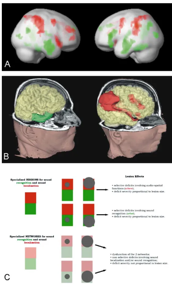
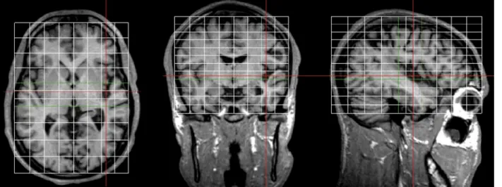
involved in sound recognition and sound localization. a Mean activation of 18 normal subjects in a functional MRI (fMRI) study on sound recognition and sound localization; green de-notes regions activated more by sound recognition than sound localization and red those acti-vated more by sound localiza-tion than sound recognilocaliza-tion. Note the presence of distinct cortical networks for sound rec-ognition (mainly temporal con-vexity bilaterally and left pre-frontal convexity) and for sound localization (mainly parietal and frontal convexities bilaterally). (Adapted from Maeder et al. 2001.) b Lesions associated, in the chronic stage, with selective deficit in sound recognition (left) or selective deficit in sound localization (right); both patients sustained rather large lesions, centred on the specia-lized auditory networks, and were examined in the chronic stage. A lesion of the temporal convexity, involving the region selectively activated, in normal subjects, by sound recognition, was associated with sound-rec-ognition deficit and normal sound localization. A lesion of the parieto-frontal convexity, involving regions selectively activated, in normal subjects, by sound localization, was asso-ciated with sound-localization deficit and normal sound recog-nition. c Schematic representa-tion of the auditory What and Where pathways (green What, red Where) as specialized areas (top) or specialized networks (bottom). Hatched circles re-present lesions, which involve one or the other pathway and are of different sizes. Predictions are made as to the associated per-formance. (Adapted from Clarke et al. 2002)
T able 1 Patients participating in this study and their performance in environmental sound recognition, sound localization and sound motion perception, indicated by z-scores. (CC corpus callosum, CN caudate nucleus, Cs centrum semiovale, FG fusiform gyrus, FOP frontal operculum, GB globus pallidus, IFG inferior frontal gyrus, IOG inferior occipital gyrus, IPlo inferior parietal lobule, ITG inferior temporal gyrus, LG lingual gyrus, MOG middle occipital gyrus, MTG middle temporal gyrus, PG parahippocampal gyrus, PoCG postcentral gyrus, Pr eCG precentral gyrus, PUT putamen, SMG supramar ginal gyrus, STG superior temporal gyrus) Case Age (years) Sex
Socio- professional level
Handedness Brain imaging Site of lesion Etiology of lesion Sound recognition Sound l ocalization
Sound motion perception
RIGHT 1 3 7 M 1 Right CT ITG, MTG, insula Haematoma –4.850 a –0.641 0.738 2 7 2 M 2 Left, forced right CT PUT , G P, external capsula, supracapsular region, thalamus Stroke –5.667 a –0.084 –3.520 a 3 6 8 F 1 Right MRI Capsulothalamic region Stroke –3.218 a 0.474 –3.296 a 4 5 0 M 1 Right MRI Thalamus, external capsula Stroke –1.585 –5.661 a –1.503 5 5 5 F 1 Right MRI Posterior STG, insula, CS Stroke 0.456 –2.872 a –1.055 6 4 8 M 2 Right MRI STG, MTG, insula T umor resection –1.993 –1.757 a,b –0.496 7 4 5 F 2 Right MRI STG, MTG, insula, CN, PUT , G P, IFG, IPlo Stroke –1.585 –3.430 a –3.072 a 8 5 4 M 1 Left, forced right MRI IOG, MOG, FG, LG, PG, ITG, thalamus, CN Stroke –0.360 –3.430 a –2.399 9 1 8 M 2 Right MRI Posterior STG, insula T umor resection 0.456 –2.872 a –2.51 1 a 10 42 F 1 Right MRI Anterior STG and MTG, insula Stroke –0.767 –2.872 a 0.289 a,b 1 1 53 F 2 Right MRI Insula, IPlo, PoCG, parietal subcortical region Stroke 0.048 –0.084 –0.271 12 61 M 2 Right MRI Subcortical lesion, insula, thalamus, external capsula, PUT , G P Stroke 0.048 –0.641 –0.607 13 68 F 2 Right CT Anterior and inferior thalamus Stroke –1.585 1.032 0.738 14 25 F 3 Right MRI Pallidum, external capsula Stroke 1.272 0.474 0.047 15 51 M 2 Left MRI PUT , G P, supracapsular region, external capsula, MTG, insula Stroke 0.456 1.032 0.625 LEFT 16 47 M 2 Right MRI ITG, MTG, STG, IFG, insula, PUT , G P, CN, anterior cc Stroke –4.850 a –0.641 –0.943 17 74 M 1 Right MRI FOP , STG, MTG Stroke –4.034 a 0.474 –1.279 18 39 M 2 Right MRI STG, MTG, insula, IFG, PreCG, thalamus, PUT , G P Stroke –6.891 a –1.757 –3.520 a 19 63 M 3 Right MRI IOG, MOG, FG, LG, ITG, posterior MTG Stroke 0.456 –3.430 a –2.175 a 20 30 M 3 Right MRI STG, IFG, MFG, PreCG, PoCG, insula Stroke 0.864 –2.315 a –1.055 a,b 21 68 F 1 Right MRI Thalamus, internal capsula, PUT , G P Haematoma 0.456 –1.199 a,b –0.719 a,b 22 49 F 1 Right MRI PUT , G P, insula, internal capsula Stroke –0.769 –0.641 –2.287 a 23 38 F 1 Right MRI STG, insula, PoCg, IPlo Stroke –0.769 0.474 –0.607 a,b 24 73 M 3 Right CT PoCG, IPlo, SMG Stroke –3.218 a –2.315 a –3.520 a 25 70 F 2 Right MRI Cuneus, MOG Haemorrhage –1.177 –0.084 –1.279 26 71 F 1 Right MRI External PUT Stroke –1.177 –0.084 0.065
Sound localization
Detailed description of this test and normative data on 60 control subjects have been reported elsewhere (Clarke et al. 2000; Bellman 2001; Bellmann et al. 2001). Sound lateralization was simulated with differences in interaural time. The stimulus was a 2-s broadband bumblebee sound, shaped with 100-ms rise and fall times, and presented through earphones. One central and four lateral positions, two in each hemispace, were simulated. The lateral positions were created by delaying the left or right channel by 0.3 ms or 1 ms. Sixty items, 12 in each position, were presented in pseudorandom order. The patients were asked to indicate the perceived position on their head with their ipsilesional hand (same procedure as Altman et al. 1979; Bisiach et al. 1984). A graduated half-circle fixed on the headphones was used to determine the angular value of the position (from 0° at the vertex to 90° at each ear). As a measure of overall performance (maximum 59), the relative positions attributed to two consecutive stimuli were compared; a response was counted as correct when a stimulus was correctly placed to the left or the right of the previous stimulus in correspondence with the difference in interaural temporal discrep-ancy or within ±15° of the previous location for identical interaural temporal values. Alloacuses (perception of stimuli as shifted to the other side of the mediosagittal plane) were also recorded. The patients individual scores were converted into z-scores relative to the mean and standard deviation of the control population (mean 57.15, SD 1.79); the limit of normal performance was set 2 SD below the mean (z<−2).
Sound motion perception
Auditory motion recognition was tested by creating an illusion of sound motion in the azimuthal plane by changing the interaural time difference (ITD) progressively. Six different motions were simu-lated: extreme left to extreme right and the reverse; extreme left to midsagittal plane and the reverse; and extreme right to midsagittal plane and the reverse. Motion from an extreme lateral position was created by using an initial ITD of 1 ms and progressive changes by 100 ms for each 50 ms of stimulus duration until both channels were in phase (for motion stopping at the midsagittal plane) or until the reverse balance was reached (for motion between the extreme lateral positions). A detailed description of this test and normative data on 60 control subjects have been reported elsewhere (Clarke et al. 2000).
Anatomical evaluation
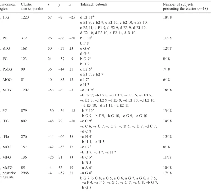
The normalized coordinate system of Talairach and Tournoux (1988) was adopted for the comparison of sites of lesions with anatomical data from normal postmortem material. Lesions were delineated on MRI or CT for all patients and were displayed and analysed using OrthoViewer, an image-visualization and -processing software, developed in our laboratory, able to display orthogonal views of a 3D image (Fig. 2). The OrthoViewer is implemented to superimpose the Talairach grid on the images based on a manual selection of the anterior and posterior commissures and of the anterior, posterior, top, bottom and lateral limits of the brain.
The lesions were analysed in relation to the degree of damage to the shared auditory structures (Talairach and Tournoux 1988; Maeder et al. 2001), the auditory callosal pathway (Alexander and Warren 1988) and/or the specialized auditory networks (Maeder et al. 2001). The shared auditory structures included the auditory thalamus (a E2 8,−a E3 7, −a E3 8, −a F 8, a E3 7, a E3 8, a F 8; Engelien et al. 1995; Frith and Friston 1996; Zatorre et al. 1999; Thivard et al. 2000), the acoustic radiation and Heschls gyrus (c E2 7, c E2 8, c E3 7, c E3 8, c F 7, c F 8,−c E2 7, −c E2 8, −c E3 7, −c E3 8,−c F 7, −c F 8; Engelien et al. 1995; Belin et al. 1998; Griffiths and Green 1999; Weeks et al. 1999; Thivard et al. 2000; Pavani et al. 2002; Zatorre et al. 2002), the auditory callosal pathways (a D 6, a
Case
Age (years)
Sex
Socio- professional level
Handedness Brain imaging Site of lesion Etiology of lesion Sound recognition Sound l ocalization
Sound motion perception
27 57 M 3 Right MRI External capsula, CG, internal capsula, anterior CC, insula, frontal and parietal subcortical regions Stroke –1.177 1.032 0.313 28 63 F 2 Right MRI FOP Stroke 0.864 –0.084 0.177 29 16 F 1 Right MRI Anterior thalamus, pulvinar , FG, hippocampus Stroke 0.456 0.474 0.513 30 43 F 1 Right MRI ITG, MTG, STG, insula Stroke –0.360 1.032 0.850 a Deficient performance defined by z<2 b Deficient performance defined by the presence of numerous alloacusis and/or errors in motion direction detection T able 1 (continued)
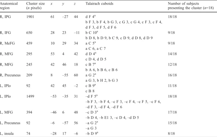
E1 6, a E2 6, a F 6, b D 6, b E1 6, b E2 6, b E3 6, b F 6,−a D 6, −a E1 6,−a E2 6, −a F 6, −b D 6, −b E1 6, −b E2 6, −b E3 6, −b F 6) and areas that were equally activated by the sound recognition and sound localization tasks (Maeder et al. 2001) on the supratemporal plane (c F 8, b C 9, b E3 8, b F 7, c C 9, c E3 8, c E3 9, c F 9, c C 7, c D 5, c D 6, c E2 8, c E2 9, c F 7, c C 6, c D 9, c C 8, c E1 9, c D 8, c E3 7, d C 6, d D 6, d D 9, d D 7, d E1 9, d E2 7, d E2 8, d E2 9, d E3 7, d E3 8, d E3 9, d F 7, d F 8, d F 9, d G 7, d G 8,−c F 7, −b C 9,−b F 7, −b F 8, −c E3 7, −c E3 8, −c E3 9, −c C 9, −c D 9, −c F 8, −c D 10, −c E1 10, −c E2 10, −c E2 9, −c E2 8, −c F 6, −c F 9, −d D 9,−d E2 8, −d E2 9, −d E3 7, −d E3 8, −d E3 9, −d F 6, −d F 7, −d F 8,−d E1 8, −d E2 7) or on the middle prefrontal gyrus (−a C 5, a C 5, −c D 6, −d D 6). The sound recognition network (the What network) and the sound localization network (the Where network) included clusters identified in our previous study (Maeder et al. 2001) and summarized here in Tables 2 and 3. The shared auditory structures include 51 cuboids in the right hemisphere and 45 in the left; the What network, 29 cuboids in the right hemisphere and 44 in the left; and the Where network, 34 cuboids in the right hemisphere and 18 in the left.
Results
Performance on sound recognition, sound localization and sound motion perception
Seven patients (cases 1–3, 16–18, 24) presented deficits in
sound recognition (Table 1); their correct replies were between 30 and 39 of 50 possible ones (cut-off 42). Their errors corresponded partially to those made by normal controls, but included also errors never done by controls, such as choosing a neither acoustically nor semantically related distracter as target. For example, case 17 chose the drawing of a frog when he heard the sound of a toothbrush and case 18 chose a cricket instead of an axe.
Eleven patients (cases 4–10, 19–21, 24) presented
deficits in sound localization. Their performance was characterized by disturbed positioning of consecutive Fig. 3a–d Performance in
sound localization. The x-axis indicates the 5 positions and the y-axis the possible azimuthal positions (degrees) of the right (positive values) and the left (negative values) auditory hemifields. The means (circles) and the standard deviations (bars) of positions chosen by the subjects for each ITD are re-presented. a Normal subjects performance; b patients unable to discriminate between the 2 positions of the same hemispace and attributing the central posi-tion systematically to one hemispace; c as in profile b, but in this case patients attribute the central position to either hemi-space; d the central position is correct, whereas the localization of the lateralized positions is very imprecise
Fig. 2 Analysis of brain lesions in Talairach coordinates. MRI scans were acquired with a 1.5-T Siemens Vision and were displayed and analysed using OrthoViewer. The Talairach grid
was superimposed (white lines and green lines). The lesion is here indicated by in the intersection of red lines (in the right supratem-poral plane)
stimuli and/or by the occurrence of alloacusis (Table 1). All patients were unable to discriminate positions within either hemispace; 4 of them attributed the central position systematically to the periphery of one hemispace (cases 4, 6, 9, 24; Fig. 3b), 2 to one or the other space (cases 8, 19; Fig. 3c), and 3 to the central part of the auditory field (cases 5, 10, 21). Two patients presented alloacusis (cases 7, 20; Fig. 3d).
Thirteen patients (cases 2–3, 7–10, 18–24) presented
deficits in sound motion perception (Table 1). Their performance was characterized by incorrect appreciation of the trajectory, and in particular the starting and finishing
positions, and/or the presence of incorrect appreciations of motion direction.
Selective versus combined deficits
One patient with RHD (case 1) and two patients with LHD (cases 15, 16) had a selective deficit of sound recognition, i.e. without an associated deficit in sound localization or sound motion perception (Table 1). Three patients with
RHD (cases 4–6) had a selective deficit in sound
localization, and two with LHD (cases 22, 23) a selective deficit in sound motion perception. Four patients with Table 2 Regions selectively activated by sound recognition (the
What network) as demonstrated by a functional MRI (fMRI) paradigm in normal subjects (Maeder et al. 2001). Anatomical location of the clusters, clusters size, gravity centre coordinates and
number of subjects presenting each cluster have been published previously. The Talairach cuboids which contain the activation clusters are listed in column 6 (unpublished data). Same conventions as in Table 1(MeFG medial frontal gyrus)
Anatomical region
Cluster size (n pixels)
x y z Talairach cuboids Number of subjects
presenting the cluster (n=18)
R, ITG 1220 57 –7 –25 d E1 11a 18/18 c E1 9, c E2 9, c E1 10, c E2 10, c E3 10, c E2 11, d E1 9, d E2 9, d E3 9, d E1 10, d E2 10, d E3 10, d E2 11, d D 10 R, PG 312 26 –36 –20 b F 10a 11/18 b F 9 R, STG 168 50 –57 23 c G 6a 12/18 d G 6 R, FG 123 24 –57 –9 b G 9a 8/18 b H 9 R, PoCG 99 36 –14 21 c E2 6a 7/18 c E1 7, c E2 7 R, MOG 81 40 –83 12 c I 7a 6/18 c H 7 L, MTG 1202 –53 –6 –3 –d E1 9a 18/18 –b E2 7, –b E2 8, –b E3 7, –c E3 6, –c E3 7, –c E2 8, –d E2 9 –d E3 9, –d E1 10, –d E2 10, –d E3 10, –d E1 11, –d E2 11 L, PG 879 –30 –34 –18 –b F 10a 13/18 –b G 9, –b F 9, –b G 10, –c G 9, –c G 10 L, IFG 802 –48 29 –10 –c C 9a 14/18 –c C 6, –c C 7, –c C 8, –c D 6, –c D 7, –d C 7, –d C 8 L, IPlo 276 –44 –66 38 –c H 4a 15/18 –b H 4, –c H 5 L, MOG 157 –42 –83 12 –c I 7a 8/18 –b H 7, –b I 7, –c H 7 L, MFG 136 –26 31 33 –b C 5a 11/18 –b B 5 L, MeFG 85 –4 53 19 –a A 6a 10/18 L, posterior cingulate 2968 –4 –57 21 –a G 6a 17/18 b G 7, b G 8, a G 5, a G 6, a G 7, a G 8, a F 5, –a F 4, –a F 5, –a G 5, –a G 7, –a G 8, –b G 7, –b G 8
a
RHD (cases 7–10) and three with LHD (cases 19–21) had a combined deficit of sound localization and sound motion perception; and two with RHD (cases 2, 3) and one with LHD (case 18) a combined deficit of sound recognition and sound motion perception. One patient with LHD (case 24) was deficient in all three tests. Five patients with RHD
(cases 11–15) and six with LHD (cases 25–30) performed
normally in all three domains.
Effects of lesions of specialized networks
The performance of patients was analysed in relation to the site of lesion and in particular to the degree of damage to the specialized What and Where networks, as well as to auditory structures shared by both networks (Fig. 4, Tables 2, 3).
Deficient performance in sound recognition, sound localization and/or sound motion perception, observed in
ten patients with RHD (cases 1–10) and in nine patients
with LHD (cases 16–24), was always associated with a
lesion that involved, at least partially, the specialized What network, the specialized Where network and/or the shared auditory structures. Lesions that did not involve the two networks or the shared auditory structures, found in one
patient with RHD (case 14) and 1 patient with LHD (case 26), were associated with normal performance in sound recognition, sound localization and sound motion percep-tion. Thus, damage to regions known to be involved in auditory processing in normal subjects is necessary for a deficit to occur. However, it is not sufficient to cause a
deficit. Four patients with RHD (cases 11–13, 15) and five
with LHD (cases 25, 27–30) had normal performance in
sound recognition, sound localization and sound motion perception, despite the fact that their lesions involved parts of the specialized What network, the specialized Where network and/or the shared auditory structures.
Lesions of a specialized network were not always associated with the corresponding deficit (Fig. 4). A partial lesion of the What network was associated with normal performance in recognition (case 25), while a combined
" Fig. 4a–g Distribution of lesions associated with deficits in sound localization (rows 2 and 3), sound motion perception (rows 4 and 5) or sound recognition (rows 6 and 7). The position of the frontal (red), parietal (mauve), temporal (blue) and occipital (yellow) lobes as well as of the insula and the lenticular nucleus (green) is shown in the top row. Three parasagittal levels are shown (d–b; from left to right). The number of cases that sustained a lesion in a given cuboid are indicated with a colour (for code, see top left; LHD left hemisphere damage, RHD right hemisphere damage)
Table 3 Regions selectively activated by sound localization (the Where network) as demonstrated by an fMRI paradigm in normal subjects (Maeder et al. 2001). Anatomical location of the clusters,
clusters size, gravity centre coordinates, corresponding Talairach cubes and number of subjects presenting each cluster. Same conventions as in Tables 1 and 2
Anatomical region
Cluster size (n pixels)
x y z Talairach cuboids Number of subjects
presenting the cluster (n=18)
R, IPG 1901 61 –27 44 d F 4a 18/18 b F 3, b F 4, b G 3, c G 3, c G 4, c F 3, c F 4, d F 3, d F 5, d F 6 R, IFG 650 28 23 –11 b C 10a 9/18 b D 8, b D 9, b C 9, c D 9, d D 8, d D 9 R, MeFG 459 10 29 34 a C 5a 9/18 a C 6, a C 7 R, MFG 295 53 4 42 d D 4a 14/18 c D 4, d D 5 R, MFG 245 42 46 18 c B 7a 12/18 b A 6, b B 6, c B 6 R, Precuneus 209 8 –55 60 a G 2a 16/18 a G 3, b H 2, b G 3 L, IPlo 92 42 45 –2 c B 9a 11/18 c B 8 L, IPlo 1499 –53 –35 31 –d F 5a 18/18 –b F 3, –b F 4, –c F 3, –c F 4, –c F 5, –c F 6, –d F 3, –d F 4, –d F 6 L, MFG 394 –46 6 48 –c D 3a 17/18 –b D 4, –b E1 3, –c D 4, –d D 5 L, Precuneus 92 –6 –57 56 –a G 2a 15/18 –a G 3 L, insula 74 –28 17 –6 –b D 9a 8/18 a
lesion of the What network and the shared auditory structures was associated with selective deficit in sound recognition (case 1), selective deficit in sound localization (case 6), selective deficit in sound motion perception (case 22), combined deficit in sound localization and sound motion perception (cases 8, 19, 21), or with normal performance in all three domains (cases 29, 30). A combined but partial lesion of the Where network and the shared auditory structures was associated with combined deficit in sound recognition and sound motion perception (case 2), combined deficit in sound recognition, sound localization and sound motion perception (case 24), or normal performance (case 27). A combined but partial lesion of the specialized What and Where networks and of the shared auditory structures was associated with selec-tive deficit in sound recognition (case 16); selecselec-tive deficit in sound localization (case 5); selective deficit in sound motion perception (case 23); combined deficit in sound localization and sound motion perception (cases 7, 20); combined deficit in sound recognition and sound local-ization (case 18); or with normal performance in all three domains (cases 11, 12, 15).
Deficits in auditory spatial tasks were observed as often in patients whose lesions involved the shared auditory
structures (cases 4, 9–10; Fig. 5) or the shared auditory
structures and the specialized What network (cases 6, 8,
19–22) as in patient with lesions of the specialized Where
network in combination with the shared auditory structures (cases 2, 24) or the shared auditory structures and the What network (cases 5, 7, 18, 20, 23). Deficits in sound recognition were observed as often in patients whose lesions involved the shared auditory structures alone (cases 3, 17) or in combination with the Where network (cases 2, 24) as in patients with lesions of the What network plus the shared auditory structures (case 1) or of the What and Where networks plus the shared auditory structures (cases 16, 18). Normal performance in sound recognition, sound localization and sound motion percep-tion was associated with lesions involving varying degrees the shared auditory structures and the specialized What and Where networks.
Performance of patients with RHD and LHD in sound recognition, sound localization and sound motion percep-tion was analysed using analysis of variance (ANOVA) with repeated measures with side of lesion as a between-subject factor and performance in the three tests as a within-subject factor. No main effects (performance in the three tests: F=0.59, P=0.5577; side of lesion: F=0.11, P=0.7371) nor interaction (F=0.43, P=0.6518) were significant.
Discussion
The present study investigated acute effects of circum-scribed hemispheric lesions affecting the auditory What and Where processing streams. Three standardized psy-chophysical tests have been used to assess sound recog-nition, sound localization and sound motion perception
(Clarke et al. 2000, 2002). Damage to shared auditory structures and the What and Where streams have been evaluated in individual patients. In addition, lesions associated with a particular deficit have been analysed by the superposition method (Fig. 4); this method confirmed the role of cortical networks, but visualized also the involvement of subcortical white and grey matter (see rightmost column in Fig. 4). The deep parts of lesions are likely to have caused disconnections that are currently difficult to interpret.
What and Where processing streams in audition
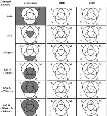
Neurophysiological and anatomical connectivity studies in non-human primates suggest the auditory information is processed in a parallel and hierarchical fashion within the What and Where streams (Rauschecker 1998; Kaas et al. 1999). The most compelling evidence comes from electrophysiological recordings in nonprimary auditory areas of the lateral belt, which tend to be selective for complex sounds (Rauschecker et al. 1995). These areas were shown to have specific selectivity along the monkey calls versus spatial location axis: the antero-lateral belt area for monkey calls, and the caudo-lateral area for spatial location (Rauschecker and Tian 2000; Tian et al. 2001). Fig. 5 Relationship between performance in sound recognition, sound localization and sound motion perception and the damage of auditory regions as predicted by the model of strictly specialized regions for sound recognition and sound localization and as observed in case of right (RHD) and left-hemispheric damage (LHD). Numerals in circles indicate cases with deficient perfor-mance in the corresponding domain. (R sound recognition, L sound localization, M sound motion perception, SAS shared auditory structures)
Several observations support a similar organization in man: (1) the presence of multiple auditory areas in man (Rivier and Clarke 1997; Tardif and Clarke 2000; Wallace et al. 2002; Chiry et al. 2003); (2) specific involvement of the posterior part of the planum temporale in spatial processing (Baumgart et al. 1999; Weeks et al. 2000; Warren et al. 2002), of the lateral part in sound pattern analysis (Griffiths and Warren 2002) and of the more anterior part of the superior temporal gyrus (on the left side) in speech processing (Binder et al. 2000); (3) hierarchical processing of complex sounds (Wessinger et al. 2001) and of auditory spatial information (Zatorre et al. 2002) within the nonprimary auditory areas; (4) anatom-ical segregation of specialized sounds recognition and sound localization networks on the temporal and parieto-frontal convexities respectively (Alain et al. 2001; Maeder et al. 2001); and (5) the association of lesions that comprise large parts of regions known to be involved in sound recognition or sound localization with the corre-sponding deficits (Clarke et al. 2000, 2002; Bellmann et al. 2001; Bellmann Thiran and Clarke 2003).
The functional organization of the dorsal pathway remains to be elucidated. It has been proposed to play a major role in the perception of the temporal evolution of sounds, or spectral motion, and to contribute thus to speech and melody analysis (Belin and Zatorre 2000). A recent activation study demonstrated that posterior audi-tory cortex responded to sound that varied in their spatial distribution, but only when multiple complex stimuli were presented simultaneously, suggesting a role in the segre-gation of sound objects, while inferior parietal cortex was found to be specifically involved in localization tasks (Zatorre et al. 2002). Relative independence of the use of spatial cues for sound localization and sound object segregation is further supported by a case of spatial deafness with spared use of spatial cues for sound object segregation and for spatial attention (Bellmann Thiran and Clarke 2003). Furthermore, the dorsal pathway is likely to be organized in a modular fashion; evoked potential (Ducommun et al. 2001), fMRI (Baumgart et al. 1999) and lesion (here) studies suggest that at least partially distinct neural populations are involved in sound localization and sound motion perception.
Effects of acute lesions on auditory What and Where processing streams
In the present study we investigated effects of acute lesions of specialized cortical networks for sound recog-nition and sound localization as anatomically demonstra-ted in normal subjects (Maeder et al. 2001). Our results revealed three major features. First, selective deficits in sound recognition, sound localization or sound motion perception occurred in the acute stage. The type of deficits observed here in the acute stage was similar of those reported in the chronic stage (Clarke et al. 2000, 2002; Bellmann 2001). This speaks in favour of separate neural mechanisms for these three functions even in acutely
damaged networks. Second, deficient performance in sound recognition, sound localization and/or sound motion perception was always associated with a lesion that involved the shared auditory structures and the specialized What and/or Where networks, while normal performance was associated with lesions within or outside these territories. Thus, damage to regions known to be involved in auditory processing in normal subjects is necessary, but not sufficient for a deficit to occur. This allows us to exclude non-specific effects of hemispheric lesions, but suggests that the precise site of lesion and/or its extent are critical. Third, in the acute stage, lesions of a specialized network were not always associated with the correspond-ing deficit. Conversely, specific deficits tended not to be associated predominantly with lesions of the correspond-ing network. For example, deficits in auditory spatial tasks were observed in patients whose lesions involved to a larger extent the shared auditory structures and the specialized What network than the specialized Where network, and sound recognition in patients whose lesions involved mostly the shared auditory structures and to a varying degree the specialized Where network. This finding was very different from our previous data collected from patients with focal lesions in the chronic stage (Clarke et al. 2000, 2002; Bellmann 2001; Bellmann et al. 2001). In these previous studies, sound-recognition deficits were associated with lesions centred on the temporal convexity and sound-localization deficits with lesions centred on the parieto-frontal convexity (see Fig. 1).
The discrepancy in effects of acute versus chronic lesions can be interpreted in three ways: as a case against the auditory What and Where processing streams; as a shift from bilateral to unilateral processing; or as a feature of the intrinsic organization of specialized auditory networks.
Early lesion results: A case against the auditory What and Where processing streams?
The effects of early lesions are partially similar to those of chronic lesions. Nine cases reported in this study speak clearly in favour of specialized What and Where networks, insofar as there is agreement between the damage to specialized networks and the type of deficit (Fig. 5). Case 1, with damage to shared auditory structures and to the What network, had a selective deficit in sound recognition. Cases 2 and 24, with damage to shared auditory structures and to the Where network, had combined deficit of sound recognition plus sound motion perception and of sound localization plus sound motion perception, respectively. Cases 5, 7, 16, 18, 20 and 23, with damage to shared auditory structures and to the What and Where networks, had deficits in at least one of the auditory functions.
Analysis of lesions associated with sound-recognition deficits stressed the key role of the temporal convexity in the acute stage (Fig. 4, bottom two rows), in a similar fashion to the chronic stage (Clarke et al. 2000, 2002).
The main discrepancy between the effects of acute and chronic lesions concerns the unexpected association between lesions of the What network and the occurrence of auditory spatial deficits. Cases 6, 8, 19, 21 and 22, with damage to shared auditory structures and to the What network, had deficits in one or two auditory spatial functions, but not in sound recognition. The analysis of lesions associated with sound-localization deficits in the acute stage showed a striking involvement of the temporal convexity in the right hemisphere (Fig. 4, rows 2 and 3), while the parieto-frontal convexity was involved in the chronic stage (Clarke et al. 2000, 2002; Bellmann et al. 2001; Bellmann Thiran and Clarke 2003). This frequent association of auditory spatial deficits with lesions of the What network, observed in the acute stage, suggests interactions between the What and the Where networks, such as those proposed to play a role in sound object segregation and spatial attention (Zatorre et al. 2002; Bellmann Thiran and Clarke 2003; Clarke and Bellmann Thiran 2003).
Bilateral What and Where streams
The What and Where processing streams are each present in both hemispheres. In the chronic stage, relatively large unilateral lesions were found to cause deficits in sound recognition, sound localization and/or sound motion perception (Clarke et al. 2000, 2002; Bellmann et al. 2001; Bellmann Thiran and Clarke 2003). This observa-tion can be interpreted in two ways. First, the two hemispheres may be specialized for different aspects of auditory processing, as proposed for sound recognition where the right hemisphere is believed to contribute to perceptual-discriminative and the left hemisphere to semantic aspects (Faglioni et al. 1969; Vignolo 1982). Second, the processing within the intact hemisphere may be disturbed by the contralateral lesion. Our preliminary results suggest that this may be the case (Clarke et al. 2002).
In the acute stage, unilateral right or left lesions have been associated with deficits in sound recognition, sound localization and/or sound motion perception, suggesting that similar interhemispheric interactions occur as in the chronic stage. However, we have also observed a relatively high number of cases who had normal performance although auditory structures were damaged: either the shared auditory structures alone (cases 13 and 28); the What network (case 25); the shared auditory structures and the What network (cases 29 and 30); the shared auditory structures and the Where network (case 27); or the shared auditory structures and the What and Where networks (case 11, 12 and 15). This observation suggests lesser hemispheric specialization or lesser distur-bance of the processing in the intact contralateral hemi-sphere in the acute than in the chronic stage.
Intrinsic organization of specialized auditory networks The What and Where auditory processing streams, which were demonstrated in activation studies (Maeder et al. 2001), can be conceived of in two different ways. First, they can correspond to specialized regions or centres that contain anatomically defined modules. Partial lesions of these centres would cause dysfunction of specific modules. Observed deficits should be then related to the specialization of the damaged region and their magnitude to the extent of the damage. Second, they can correspond to specialized networks, which are interconnected via the shared auditory structures and more directly with each other. Partial lesions of one of these networks would cause widespread dysfunction within the network and may influence processing within the shared auditory structures and possibly the other network. Observed deficits may thus not be related to the damaged region and their magnitude not proportional to the extent of the damage. Effects of focal lesions that we observed in the acute stage strongly support the second hypothesis.
Disorganization of networks beyond the functional subunits, but within connected networks, has been dem-onstrated for speech processing. Lesions to Brocas area were shown to be associated with abnormal activation patterns to a reading task within the left posterior inferior temporal cortex (Price et al. 2001). Regions which showed these long-distance effects are known to be part of a network which has been demonstrated functionally (Price et al. 2001) and anatomically in man (Di Virgilio and Clarke 1997). Furthermore, electrophysiological evidence suggests that functional changes occur within specific networks after stroke, as demonstrated by reduced intracortical inhibition in the anatomically intact motor cortex of the affected hemisphere (Liepert et al. 2000).
Conclusions
The identity of a sound source and its location are processed independently along two distinct cortical path-ways, which have be visualized previously using fMRI. Two models may account for the functional organization of these pathways, which can be then interpreted as: (1) specialized regions, in which lesions cause dysfunction that is limited to the damaged part; (2) specialized networks, in which lesions cause dysfunction that may spread over the two specialized networks and that may be associated with considerable and rapid functional substi-tution by the non-damaged network. The present results support strongly the network model. In cases of unilateral hemispheric lesions we have demonstrated a relative independence between a small damage to a network and the type of deficit that occurs. These findings are of importance to our understanding of recovery and rehabil-itation in cases of brain damage.
Acknowledgements This study was supported by Swiss National Science Foundation grants 3138-51173.97 and 3238-064604.01 and by the Lausanne Medical Faculty RATP grant to S.C.
References
Alain C, Arnott SR, Hevenor S, Graham S, Grady CL (2001) What and Where in the human auditory system. Proc Natl Acad Sci USA 98:12301–12306
Alexander MP, Warren RL (1988) Localization of callosal auditory pathways: a CT case study. Neurology 38:802–804
Altman J, Balanov L, Delgin V (1979) Effects of unilateral disorder of the brain hemispheric functions in man on directional hearing. Neuropsychologia 17:295–301
Anourova I, Nikouline VV, Ilmoniemi RJ, Hotta J, Aronen HJ, Carlson S (2001) Evidence for dissociation of spatial and nonspatial auditory information processing. Neuroimage 14:1268–1277
Baumgart F, Gaschler-Markefski B, Woldorff MG, Heinze H-J, Scheich H (1999) A movement-sensitive area in auditory cortex. Nature 400:724–726
Belin P, Zatorre RJ (2000) What, where and how in auditory cortex. Nat Neurosci 3(10):965–966
Belin P, Zilbovicius M, Crozier S, Thivard L, Fontaine A, Masure M-C, Samson Y (1998) Lateralization of speech and auditory temporal processing. J Cogn Neurosci 10:536–540
Bellmann A (2001) Le traitement des données spatiales en audition: une approche neuropsychologique. Thèse de Doctorat en Psychologie, University of Geneva, Switzerland
Bellmann Thiran A, Clarke S (2003) Preserved use of spatial cues for sound segregation in a case of spatial deafness. Neuropsy-chologia 41:1254–1261
Bellmann A, Meuli R, Clarke S (2001) Two types of auditory neglect. Brain 124:676–687
Binder JR, Frost JA, Hammeke TA, Bellgowan PSF, Springer JA, Kaufman JN, Possing ET (2000) Human temporal lobe activation by speech and nonspeech sounds. Cereb Cortex 10:512–528
Bisiach E, Cornacchia L, Sterzi R, Vallar G (1984) Disorders of perceived auditory lateralization after lesions of the right hemisphere. Brain 107:37–52
Chiry O, Tardif E, Magistretti P, Clarke S (2003) Patterns of calcium binding proteins support parallel and hierarchical organization of human auditory areas. Eur J Neurosci 17(2):397–410 Clarke S, Bellmann Thiran A (2003) Auditory neglect: What and
Where in auditory space. Cortex (In press)
Clarke S, Bellmann A, Ribaupierre F de, Assal G (1996) Non-verbal auditory recognition in normal subjects and brain-damaged patients: evidence for parallel processing. Neuropsychologia 34:587–603
Clarke S, Adriani M, Bellmann A (1998) Distinct short-term memory systems for sound content and sound localization. Neuroreport 9:3433–3437
Clarke S, Bellmann A, Meuli RA, Assal G, Steck AJ (2000) Auditory agnosia and auditory spatial deficits following left-hemispheric lesions: evidence for distinct processing pathways. Neuropsychologia 38:797–807
Clarke S, Bellmann Thiran A, Maeder P, Adriani M, Vernet O, Regli L, Cuisenaire O, Thiran J-Ph (2002) What and Where in human audition: selective deficits following focal hemispheric lesions. Exp Brain Res 147:8–15 DOI: 10.1007/s00221–002–1203–9 Di Virgilio G, Clarke S (1997) Direct interhemispheric visual input
to human speech areas. Hum Brain Mapp 5:347–354 Ducommun C, Murray MM, Thut G, Bellmann A, Viaud-Delmon I,
Clarke S, Michel CM (2002) Segregated processing of auditory motion and auditory location: an ERP mapping study. Neuro-image 16:76–88
Engelien A, Silbersweig D, Stern E, Huber W, Döring W, Frith C, Frackowiak RSJ (1995) The functional anatomy of recovery from auditory agnosia. A PET study of sound categorization in a neurological patient and normal controls. Brain 111:1395– 1409
Faglioni P, Spinnler H, Vignolo LA (1969) Contrasting behavior of right and left hemisphere-damaged patients on a discriminative and a semantic task of auditory recognition. Cortex 5:366–389 Frith CD, Friston KJ (1996) The role of the thalamus in Top Down
modulation of attention to sound. Neuroimage 4:210–215 Fujii T, Fukatsu R, Watabe S, Ohnuma A, Teramura K, Kimura I,
Saso S, Kogure K (1990) Auditory sound agnosia without aphasia following a right lobe lesion. Cortex 26:263–268 Griffiths TD, Green GGR (1999) Cortical activation during
perception of rotating wide-field acoustic stimulus. Neuroimage 10:84–90
Griffiths TD, Warren JD (2002) The planum temporale as a computational hub. Trends Neurosci 25:348–353
Griffiths TD, Rees A, Witton C, Shakir RA, Henning GB, Green GGR (1996) Evidence for a sound movement area in the human cerebral cortex. Nature 383:425–427
Griffiths TD, Rees A, Witton C, Cross PM, Shakir RA, Green GG (1997) Spatial and temporal auditory processing deficits following right hemisphere infarction. A psychophysical study. Brain 120:785–794
Hossmann KA (1994) Viability thresholds and the penumbra of focal ischemia. Ann Neurol 36:557–65
Jerger J, Loverling L, Wertz M (1972) Auditory disorder following bilateral temporal lobe insult: report of a case. J Speech Hear Disord 37:523–535
Kaas JH, Hackett TA, Tramo MJ (1999) Auditory processing in primate cerebral cortex. Curr Opin Neurobiol 9:164–170 Liepert J, Storch P, Fritsch A, Weiller C (2000) Motor cortex
disinhibition in acute stroke. Clin Neurophysiol 111:671–676 Maeder P, Meuli R, Adriani M, Bellmann A, Fornari E, Thiran J-Ph,
Pittet A, Clarke S (2001) Distinct pathways involved in sound recognition and localization: a human fMRI study. Neuroimage 14:802–816
Musso M, Weiller C, Kiebel S Muller SP, Bulau P, Rijntjes M (1999) Training-induced brain plasticity in aphasia. Brain 122:1781– 1790
Pavani F, Macaluso E, Warren JD, Driver J, Griffiths TD (2002) A common cortical substrate activated horizontal and vertical sound movement in the human brain. Curr Biol 12:1584–1590 Price CJ, Warburton EA, Moore CJ, Frackowiak RS, Friston KJ (2001) Dynamic diaschisis: anatomically remote and context-sensitive human brain lesions. J Cogn Neurosci 13:419–29 Rauschecker JP (1998) Parallel processing in the auditory cortex of
primates. Audiol Neurootol 3:86–103
Rauschecker JP, Tian B (2000) Mechanisms and streams for processing of what and where in auditory cortex. Proc Natl Acad Sci USA 97:11800–11806
Rauschecker JP, Tian B, Hauser M (1995) Processing of complex sounds in the macaque non primary auditory cortex. Science 268:111–114
Recanzone GH (2000) Spatial processing in the auditory cortex of the macaque monkey. Proc Natl Acad Sci USA 97:11829– 11835
Rivier F, Clarke S (1997) Cytochrome oxidase, acetylcholinesterase, and NADPH-diaphorase staining in human supratemporal and insular cortex: evidence for multiple auditory areas. Neuro-image 6:288–304
Spreen O, Benton AL, Fincham RW (1965) Auditory agnosia without aphasia. Arch Neurol 13:84–92
Talairach J, Tournoux P (1988) Co-planar stereotaxic atlas of the human brain. Thieme Medical, New York
Tardif E, Clarke S (2000) Intrinsic connectivity of human auditory areas: a tracing study with Dil. Eur J Neurosci 13:1045–1050 Thivard L, Belin P, Zilbovicius M, Poline JB, Samson Y (2000) A
cortical region sensitive to auditory spectral motion. Neurore-port 11:2969–2972
Tian B, Reser D, Durham A, Kustov A, Rauschecker JP (2001) Functional specialization in rhesus monkey auditory cortex. Science 292:290–293
Vignolo LA (1982) Auditory agnosia. Philos Trans R Soc Lond B Biol 298:49–57
Wallace MN, Johnston PW, Palmer AR (2002) Histochemical identification of cortical areas in the auditory region of the human brain. Exp Brain Res 143:499–508
Warren JD, Zielinski BA, Green GGR, Rauschecker JP, Griffiths TD (2002) Perception of sound-source motion by the human brain. Neuron 34:139–148
Weeks RA, Aziz-Sultan A, Bushara KO, Tian B, Wessinger CM, Dang N, Rauschecker JP, Hallet M (1999) A PET study of human auditory spatial processing. Neurosci Lett 262:155–158
Weeks R, Horwitz B, Aziz-Sultan A, Tian B, Wessinger CM, Cohen LG, Hallet M, Rauschecker JP (2000) A positron emission tomography study of auditory localization in congenitally blind. J Neurosci 20:2664–2672
Wessinger CM, VanMeter J, Tian B, Van Lare J, Pekar J, Rauschecker JP (2001) Hierarchical organization of the human auditory cortex revealed by functional magnetic reso-nance imaging. J Cogn Neurosci 13:1–7
Witte OW, Bidmon HJ, Schiene K, Redecker C, Hagemann G (2000) Functional differentiation of multiple perilesional zones after focal cerebral ischemia. J Cereb Blood Flow Metab 20:1149–65
Zatorre RJ, Mondor TA, Evans AC (1999) Auditory attention to space and frequency activates similar cerebral systems. Neuro-image 10:544–554
Zatorre RJ, Bouffard M, Ahad P, Belin P (2002) Where is where in the human auditory cortex? Nat Neurosci 5(9):905–909