100
Antibiotic Treatment of Experimental Endocarditis Due to Methicillin-Resistant
Staphylococcus epidermidis
Jose M. Entenza, Ursula Fluckiger, Michel P. Glauser,
and Philippe Moreillon
Division of Infectious Diseases. Department of Internal Medicine. Centre Hospitalier Universitaire Vaudois. Lausanne. Switzerland
The natural history and treatment of experimental endocarditis due to heterogeneous and homogeneous methicillin-resistant Staphylococcus epidermidis was investigated. Amoxicillin/ clavulanate or vancomycin were administered for 3 days via a computerized pump to mimic human drug kinetics in animals. After challenge with the minimum inoculum producing 90% of infections (ID9o) ,bacteria in the vegetations grew logarithmically for 16 h. Then, bacterial densi-ties stabilized (at -108cfu/g) and growth rates sharply declined. Both regimens cured~60%of endocarditis (due to heterogeneous or homogeneous bacteria) when started 12-16 h after infec-tion, although the bacterial densities in the vegetations had increased by 20 times in between. In contrast, treatment started after 24 h failed in most animals, while bacterial densities had not increased any more. Thus, while both regimens were equivalent, the therapeutic outcome was best predicted by growth rates in the vegetations, not by bacterial densities. These observations highlight the importance of phenotypic tolerance developing in vivo.
In the early 1940s, the introduction of penicillin in medi-cine was rapidly followed by the emergence of penicillinase-producing penicillin-resistant staphylococci, posing the first major challenge to the then-nascent era ofantimicrobial che-motherapy [I]. Twenty years later this scenario was partially repeated when it appeared that antibiotic pressure with the newly available penicillinase-stable methicillin selected for a new, so-called intrinsic type of resistance to ,8-lactams [2]. Intrinsic resistance to methicillin was not due to penicillin-ase production but appeared to require the production of a new, unique, penicillin-binding protein (PBP) with low ,8-lactam affinity (called PBP 2A or 2') in addition to the nor-mal set of staphylococcal PBPs [3,4]. PBP 2A is also consid-ered to be responsible for cross-resistance to most other ,8-lactam antibiotics (for review see [5]).
Today, methicillin resistance is genuinely ubiquitous in hospital isolates of staphylococci. In Switzerland, for in-stance, 40%-60% of hospital isolates of Staphylococcus epi-dermidisare resistant to methicillin [6] (unpublished data). Although less common, the frequency of methicillin-resis-tant Staphylococcus aureus varies from 3%in our hospital [7] to 20% in other hospitals of the country (Auckenthaler R, personal communication). In the United States, up to 40%of S. aureus isolates are resistant to methicillin in certain
insti-Received 8 November 1993; revised 23 February 1994.
Presented in part: 32nd Interscience Conference on Antimicrobial Agents and Chemotherapy. October 1992. Anaheim. California (abstract 1458).
Grant support: Swiss National Foundation for Research (32-2-7777). Reprints or correspondence: Prof. Michel P. Glauser. Div. ofInfectious Diseases. Dept. oflnternal Medicine. Centre Hospitalier Universitaire Vau-dois. CH-I 0 IJ.Lausanne. Switzerland
The Journal of Infectious Diseases 1994;170:100-9
© 1994 by The University of Chicago. All rights reserved. 0022-1899/94/7001-0015$01.00
tutions [8]. In addition, most of these organisms are resistant to a number of other drugs [5, 9], posing a serious challenge to antimicrobial therapy.
The only nonexperimental antibiotic to which methicillin-resistant staphylococci are still considered uniformly sensi-tive is vancomycin, which is potentially toxic and can be given only parenterally (see [5]). However, staphylococci may be on the verge of becoming totally resistant to any available antibiotics, since it has been shown in the labora-tory that vancomycin resistance could be transferred to S. aureus from vancomycin-resistant enterococci [10]. Transfer of vancomycin resistance into methicillin-resistant staphylo-cocci would have major consequences for public health. To decrease the risk of this occurrence, it is necessary both to interfere with the spread of methicillin-resistant staphylo-cocci and to develop alternative, nonglycopeptide antimicro-bial agents to prevent selection of glycopeptide resistance in such organisms.
The purpose of the present study was to investigate the efficacy of the nonglycopeptide combination amoxicillin/ clavulanate in the treatment of experimental endocarditis due to methicillin-resistant S. epidermidis(MRSE). The ratio-nale of the experiments was based on the dual observations that amoxicillin/clavulanate could cure experimental endo-carditis due to methicillin-resistant S. aureus (MRSA)
[II.
12] (presumably because of the relatively good affinity of amoxicillin for PBP 2A [12]) and that a similar PBP 2A medi-ates high level of methicillin resistance in both MRSA and MRSE [13-15]. In addition, since the course of experimen-tal endocarditis due to S. epidermidis appeared to be more indolent and resulted in less mortality than with S. aureus, we investigated the therapeutic outcome of treatment started either early or relatively late after the induction of infection in animals.1ID 1994; 170 (July) Treatment of MRSE Endocarditis 101
Table1. Principal characteristics of the methicillin-resistantS.epidermidis (MRSE) isolates.
Antibiotic susceptibility
MRSE isolate Source Penicillinase Slime Gentamicin Clindamycin Erythromycin Cotrimoxazole
1386 Wound infection + + R R R S
4890 Peritonitis + +++ S R R R
1468 Wound infection + S S S S
I 386-Hom* Laboratory derivative + + R R R S
NOTE. Penicillinase and slime production were measured by semiquantitative methods [17, 18]. S, susceptible; R, resistant as determined by standard disk diffusion method [19].
* Homogeneous.
Materials and Methods
Microbiologic Methods
Microorganisms and growth conditions. The principal char-acteristics ofthe four strains ofMRSE used in these experiments are summarized in table I. Strains MRSE 1386,4890, and 1468 were clinical isolates recovered from distinct geographic areas and typically expressing so-called heterogeneous resistance to methicillin. Strain MRSE 1386-Hom was a homogeneously methicillin-resistant derivative of strain MRSE 1386, which had , been selected by serial passage of the parent strain on methicil-lin-containing medium as described [16]. Strain I 386-Hom was stable, as it appeared to retain its homogeneously resistant phe-notype for up to 60 generations in the absence of antibiotic pressure in vitro (see [16
D.
All 4 strains produced penicillinase as assessed both by a positive nitrocefin test and by inactivation ofam oxic illin using a described method [17]. The production of slime was assessed by a semiquantitative method [18] and antibi-otic susceptibilities were determined by standard disk diffusion test [19].Unless otherwise stated. the bacteria were grown at 35°C in Mueller-Hinton broth (MHB; Difco, Detroit) supplemented with 2% NaCl or on Columbia agar (C agar; Becton Dickinson, Cockeysville, MD) supplemented with 4% NaCl. Stocks were kept at -70°C in MHB supplemented with 10%glycerol.
Antibiotics. Amoxicillin and clavulanate (either individu-ally or in a 5: I [wt/wt] ratio) and flucloxacillin were obtained from Beecham Laboratories (Brockham Park, UK) and vanco-mycin from Eli Lilly (Indianapolis).
Antibiotic susceptibility. MlCs of several antibiotics were de-termined by a standard macrobroth method [20] in MHB supple-mented with calcium and 2%NaCl (as recommended for suscep-tibility testing of MRSA [21
D
using 105cfu/rnl, as an inoculum. The MlC was defined as the lowest drug concentration inhibit-ing visible bacterial growth after 24 h of incubation at 35°C. For MlC determinations of the combination of arnoxicillin/ clavulanate, the drugs were used in a ratio of 2: I(wt/wt).Population analysis profile. The phenotypic expression of methicillin resistance in vitro was determined as described [12]. In brief, serial dilutions of MRSE cultures (in MHB supple-mented with 2% NaCI) in the late exponential phase of growth were inoculated onto C agar plates supplemented with 4% NaCl and containing increasing concentrations of either methicillin or amoxicillin/clavulanate. Clavulanate was used at a fixed
con-centration of 5 mg/L. Large(>108cfu) and smaller (106 , 104, and 102cfu) bacterial numbers were plated. The numbers of colonies growing on the antibiotic-containing plates were deter-mined after 48-72 h of incubation at 35°C. Curves of the popu-lation analysis profile were generated by plotting the numbers of colonies growing on the plates against the concentrations ofanti-biotic in the plates.
In vitro time-kill curves. Series of flasks containing 20 mL of fresh MHB (prewarmed at 35°C) supplemented with 2% NaCI were inoculated with 1/100(vol/vol) of an overnight culture of bacteria (grown in the same medium) and incubated with aera-tion in a shaking incubator (at 120 rpm). Antibiotics (either amoxicillin/clavulanate or vancomycin) were added at different times during the logarithmic or the stationary phase of growth, and bacterial survival was followed by plating dilutions of the cultures onto C agar plates supplemented with 2000 units of penicillinase/ml, (Bacto-Penase concentrate; Difco). Final con-centrations of antibiotics were chosen to mimic the peak serum levels obtained in humans (or rats) during intravenous (iv) ther-apy: 90 and 20 mg/L for amoxicillin and clavulanate, respec-tively, and 40 mg/L for vancomycin. All measurements were made in triplicate.
Animal Experiments
Production ofendocarditis and installation ofan infusion pump device. Sterile aortic vegetations were produced in female Wis-tar rats (180-200 g) as described [22]. In brief, the animals were anesthetized and a polyethylene catheter (Guerbet Biomedical. Louvres, France) was inserted via the right carotid artery across the aortic valve. The catheter was secured with a silk ligature and left in place for the remainder of the experiment. At the same time, an iv line consisting of a sterile Silastic catheter (in-side and out(in-side diameters, 0.02 and 0.037 mm, respectively; Dow Corning, Midland, MI) was inserted via the jugular vein into the superior vena cava for the delivery of the antibiotics. The distal portion of the catheter was brought to the skin of the interscapular region and connected to a programmable infusion pump device (Pump 44; Harvard Apparatus, South Natick, MA) through a swivel that permitted the animals to move in an unre-strained fashion in their cage. Bacterial endocarditis was in-duced 24 h after catheterization by iv challenge of the animals with various inocula.
102 Entenza et al. JID 1994; 170 (July)
To determine the minimum size of bacterial inoculum produc-ing endocarditis in 90% of the rats(1090),groups of rats (8-10 animals) were challenged through a tail vein with 0.5 mL of saline containing 103-106cfu of each test organism. The ani-mals were killed 24 h after inoculation. One milliliter of blood was drawn and plated for quantitative blood cultures. The aortic vegetations were sterilely dissected, weighed, homogenized in I mL of saline, and serially diluted before plating for quantitative cultures. The technique permitted the detection of?:210gJOcfu/ g of tissue. The spleens were also processed for quantitative cul-tures as described. The numbers of colonies growing on the plates were determined after incubation at 35°C for 48 h.
To determine the kinetics of bacterial growth in the animals, groups ofrats challenged with either the1090or 10 X1090were killed at various times after iv bacterial challenge and the blood, vegetations, and spleens were cultured as described.
Treatment ofexperimental endocarditis. Groups of rats chal-lenged with either the 1090or lOX1090of various strains of MRSE were treated with either flucloxacillin, amoxicillin/ clavulanate, or vancomycin (flucloxacillin was used instead of methicillin because the latter antibiotic is no longer used in the clinic). To simulate in animals the human serum kinetics during standard iv treatment (i.e., 1.2 g four times daily for am oxicillin/ clavulanate and 1.0 g twice daily for vancomycin), these drugs were delivered to the animals at changing flow rates using a programmable pump device. This required the injection of 164.4 mg of amoxicillin/clavulanate (5: I [wt/wt] ratio )/kg every 6 h (beginning with a bolus of80.0 mg/kg followed by the progressively decreasing infusion of the remaining 84.4 mg/kg) and 23.2 mg of vancomycin/kg delivered at changing flow rates over a l2-h period. Flucloxacillin (200 mg/kg every 5 h) was administered subcutaneously (sc).
Therapy was started at various times after bacterial inocula-tion (see Results) and lasted for 3 days. The rats were sacrificed 12 h after the last antibiotic dose (when drug was no longer detected in the blood), and endocarditis was evaluated quantita-tively as described. Each therapeutic experiment was matched against a control group of rats killed at the initiation of therapy.
Determination ofserum antibiotic concentrations. Serum lev-els of antibiotics were determined in both uninfected and in-fected rats (groups of 3-6 animals) at various time points during and after antibiotic administration. Antibiotic concentrations were measured by an agar diffusion bioassay usingBacillus
subti-lis ATeC 6633 for amoxicillin, flucloxacillin, and vancomycin andKlebsiella pneumoniae ATCC 29665 plus 60 mg ofpenicil-lin/L for clavulanate [23]. For standard curves, antibiotics were diluted in pooled rat serum.
Expression of antibiotic resistance in vivo. To evaluate the number of highly methicillin-resistant organisms present on the valves at the onset of therapy, quantitative cultures of vegeta-tions from control animals were plated on agar containing 25 mg of methicillin/L in parallel with cultures on plain medium. In addition, the vegetations of animals treated with amoxicillin/ clavulanate were plated simultaneously on plain and antibiotic-containing agar plates (antibiotic-containing 5 mg of clavulanate/L plus either 25 or 100 mg ofamoxiciUin) to detect the possible emer-gence of antibiotic-resistant organisms in vivo. This screening was not done in vancomycin-treated animals.
Statistical analysis. The
x
2test with Yates's correction was used to compare the incidence of valvular and spleen infections.
Results
Antibiotic Determinations
Antibiotic susceptibility. Table 2 shows the MICs of sev-eral antibiotics for the 4 isolates of MRSE plus I clinical isolate of methicillin-sensitive S. epidermidis used as a con-trol. Since most methicillin-resistant staphylococci express a so-called heterogeneous type of resistance to ~-lactams, we also determined the population analysis profile of each of the test organisms on~-lactam-containingplates (figure 1). All three clinical isolates (MRSE 1386, 4890, and 1468) typi-cally expressed heterogeneous resistance to methicillin (as exemplified for strain MRSE 1386 in figure lA) and ·tained subpopulations of bacteria able to grow on plates con-taining 500-1000 mg of rnethicillin/L, Importantly, the pro-file of methicillin resistance was not altered when clavulanate (5 mg/L) was added to the medium. In contrast, no bacteria could grow on plates containing >25 mg of amoxicillin/L in the presence of clavulanate (5 rngjL). Fig-ure 1B shows that serial passage of the parent strain MRSE 1386 on methicillin resulted in a derivative (MRSE 1386-Hom) that was homogeneously resistant to methicillin, that
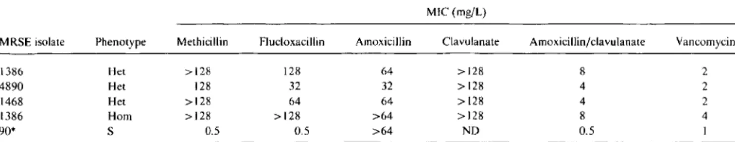
Table2. Phenotype of methicillin resistance and MICs of methicillin-resistant S. epidermidis(MRSE) test organisms to various antibi-otics.
MIC (mg/L)
MRSE isolate Phenotype Methicillin Flucloxacillin Amoxicillin Clavulanate Amoxicillinjclavulanate Vancomycin
1386 Het >128 128 64 >128 8 2
4890 Het 128 32 32 >128 4 2
1468 Het >128 64 64 >128 4 2
1386 Hom >128 >128 >64 >128 8 4
90* S 0.5 0.5 >64 NO 0.5 1
NOTE. Het, heterogeneously resistant; Hom, homogeneously resistant to methicillin; NO, not determined.
JID 1994; 170 (July) Treatment of MRSE Endocarditis 103 10
A
1000B
100 10Antibiotic concentration (m{JtI) .1
D---D----C~i~======~=========li
\
'
"\
0",,-'\,
\
2 8 10 ::l U.o
6 Cl o ..JFigure 1. Population analysis profile of heterogeneously methicillin-resistant S. epidermidis isolate 1386 (A) and its homogeneously resistant derivative 1386-Hom (B). Bacteria (»108 cfu) were plated on Columbia agar supple-mented with 4%NaCl and twofold serial dilutions of either methicillin (0), amoxicillin(0),methicillin/clavulanate (.), or arnoxicillin/clavulanate (.). Clavulanate was used at fixed concen-tration of 5 mg/L. Heterogeneous pro-file of I 386-Hom on amoxicillin alone was most likely due to residual penicil-linase secretion by bacteria.
.1 10 100 1000
Antibiotic concentration (mg/I)
is, all colony-forming units in the bacterial population ex-pressed high-level resistance to the drug. The homogeneous derivative was also homogeneously susceptible to amoxicil-lin in the presence of clavulanate. In contrast, when plated on amoxicillin alone, some heterogenicity was observed. This was most likely because of production of penicillinase by the bacteria, which resulted in degradation of amoxicillin in situ.
Antibiotic serum levels. After one injection of'flucloxacil-lin (200 mg/kg sc), both the peak serum levels (124.3mg/L)
and the half-life of the drug (0.5 h) were comparable to those observed during iv therapy in humans (125.2 mg/L and 0.5 h, respectively [24]). After continuous infusion, serum levels of amoxicillin, clavulanate, and vancomycin closely
fol-lowed the human serum kinetics produced by iv injections of I g of amoxicillin, 0.2 g of clavulanate, and I g of van co my-cin [25, 26]. Serum levels in rats at 30 min and 2,4, and 6 h were as follows: arnoxicillin, 89.4± 23, 13.9 ± 1.4, 3.5 ±
2.0, and 1.2±0.9 mg/L: clavulanate, 13.4± 5.0, 2.1± 0.5, 1.1±0.4, and 0.3 ±0.1 mg/L; and vancomycin, 40.5±6.0, 30.0
±
4.9, 21.5±
8.9, and 15.4±
2.0 mg/L (and 3.7±
2.8 mg/L at 12 h).Animal Studies
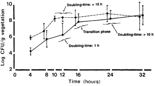
Natural history ofexperimental MRSE endocarditis. The minimum bacterial inoculum producing endocarditis in 90% of the rats (1090 )was 10
5
104 En tenza et al. JID 1994;170 (July)
Figure 2. Natural course of experi-mental endocarditis due to methicillin-resistant S. epidermidis (MRSE) isolate 1386 in rats challenged with either 105
cfu (ID90 ,solid line) or 106cfu (lOX
ID9o , dashed line). Each point repre-sents mean±SD of7-25 animals. Dou-bling times were derived from the growth curves on the graph. After chal-lenge with lOX ID9o ,bacterial growth in vegetations stopped --6 h earlier than after ID9o •
32
24
~
Doubling-time: 1 h ; Doubling-time:> . . - -1
,.t---r---
---
± ---- ...,
~
~\
Transition phase Doubling-time: > 10 h8 10 12
16
Time (hours)
4
~
1°1
J
8
1
~
6
1
~
4
1
2 - - - -
-
-o
with such an inoculum (solid line, figure 2), bacteria in the vegetations first entered a phase of rapid division (generation time, --1 h) that lasted until bacterial densities on the valves had reached '"'"-' 108cfu/g of vegetation, at
r -16 h after
bacte-rial challenge. At that time, the generation time progressively decreased (by about I0 times) and bacterial densities reached a plateau. The proportion of animals with positive blood cultures increased with the severity of valvular infec-tion (from 36% to 53% and 72% at 12, 16, and 24 h, respec-tively, for all 3 clinical isolates). In contrast, all the spleen cultures were positive from the beginning, but total bacterial numbers and densities in this organ remained relatively stable over the course of the disease (log., cfu/g of spleen, 2.8-3.4).
After challenge with 106cfu (i.e., 10 X ID
90 ;dashed line,
figure 2), the course of experimental endocarditis was roughly parallel to that following injection of the ID90 but
the growth curve was globally shifted to the left.
Expression ofmethicillin resistance in vivo. Expression of methicillin resistance in the vegetations of untreated rats did not vary over time. For the 3 heterogeneously resistant clini-cal isolates, the frequency of bacterial subpopulations able to grow on plates containing 25 mg of methicillin/L was 2-6 X 10⁻⁵ during the whole course of infection. Similar control experiments with the derivative MRSE 1386-Hom showed that the homogenous phenotype also persisted in vivo, as 100% of the bacteria recovered from the vegetations of un-treated animals grew on plates containing 100 mg of methi-cillin/L.
Therapeutic experiments. The rather indolent course of MRSE endocarditis allowed us to investigate the efficacy of antibiotherapy started either early ( 12 or 16 h after bacterial challenge) or relatively late (24 h after inoculation). This type oflate treatment was not possible in former experiments done with MRSA isolates [II, 12] because the more aggres-sive course of S. aureus endocarditis resulted in the prema-ture deaths of many of the animals.
Figure 3 depicts the therapeutic efficacy of various antibi-otic regimens against experimental endocarditis after chal-lenge with the ID90ofMRSE 1386, MRSE 4890, and MRSE
1468. According to the experimental design, antibiotic treat-ment was started either 12, 16, or 24 h after bacterial chal-lenge and lasted for 3 days. Animals were always sacrificed 12 h after the last antibiotic dose. As expected, treatment with flucloxacillin failed against all three organisms. In con-trast, both amoxicillin/clavulanate and vancomycin success-fully treated most of the infected rats when started 12 or 16 h after bacterial challenge. However, the therapeutic efficacy of both regimens tended to decrease with delayed onset of treatment and clearly failed (at least for MRSE 1386 and MRSE 4890) when started 24 h after bacterial challenge.
Analysis of the vegetations showed that this drop in effec-tiveness did not correlate well with the bacterial densities in the valvular lesions but did correlate with the rate ofbacte-rial growth in situ (figure 2, 3). Indeed, treatment started at either 12 or 16 h resulted in practically similar cure rates despite the fact that bacterial densities in the vegetations had increased by 20 times between those time points. During this time, bacterial growth was relatively fast (figures 2, 3). In contrast, the cure rates decreased between 16 and 24 h, while the rate of bacterial growth in situ had come to a virtual halt (doubling time, ~10 h) and bacterial densities in the vegeta-tions did not increase any more. Treatment failures were not due to organisms resistant to amoxicillin/clavulanate, as bac-teria recovered from the infected valves and directly plated on antibiotic-containing agar had retained the sensitivity of the original strain (data not shown). Thus, in the present experiments, the therapeutic efficacy of amoxicillin/ clavulanate and vancomycin was best predicted by the rate of bacterial growth in situ, not by the bacterial densities in the vegetations.
To investigate how therapy might be affected by larger inocula, groups of rats were inoculated with lOX ID90 (I06
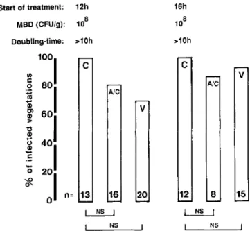
lID 1994; 170 (July) Treatment of MRSE Endocarditis 105
Start of treatment: 12h MBD (CFU/g): 1-5x10⁶
Doubling-time: 1h Transition phase >10h
v
17 28 AlC NSc
26 P< 0.005DO
,pc0.001, ...' _ _---:; __ P< 0.001c
25~c
fl
~bJ
F 11c
n: 15v
13 11 Ale NS 8c
P< 0.05DO
,pc0.05 I ...' ;..-. ...J P< 0.05 5c
V 9 A/C[;]
7 Fc
n= 10 Figure 3. Antibiotic treatment ofex-perimental endocarditis in rats infected with 105cfu (ID
90 ) of
methicillin-resis-tantS.epidermidisisolates 1386 (top), 4890 (center), and 1468 (bottom). MBD, median bacterial densities. Vege-tations were considered sterile when no growth occurred from cultures of undi-luted homogenates. Numbers of ani-mals in each group are indicated at bottom of columns. C control; F, f1u-cloxacillin; A/C amoxicillin/clavula-nate; V, vancomycin; NS, not signifi-cant.
c
n= 15 F 8 AlC[ill
o
c
13fl
h!J
o
c
v
11 NS NS IP< 0.0011...1 ...1 P< 0.005 IIL. --:.:..:..- ...J106 Entenza et al.
nn
1994;170 (July)Figure 4. Antibiotic treatment of experimental endocarditis in rats infected with 106cfu (lOX ID
90 ) of methicillin-resistantS.
epidermidisisolate 1386. See figure 3 legend for details.
shown in figure 2 (dashed line), after challenge with this larger inoculum, the bacterial densities in the vegetations attained the plateau (-., 108cfu/g) sooner than after
inocula-tion with the 1090(presumably because the valves had been
originally colonized with more bacteria after this larger inocu-lum, as in experimental streptococcal endocarditis [27]) and bacterial growth in situ already came to a halt 10 h after bacterial challenge (instead of> 16 h after challenge with the
1090 ) ,Therefore, 3 days of treatment with either amoxicillin/
clavulanate or vancomycin failed, even when started as soon as 12 h after infection (figure 4).
Experiments with the homogeneously resistant derivative MRSE 1386-Hom. Since only 2-6 X 10- 5 of the heteroge-neously resistant bacteria expressed high-level methicillin re-sistance at the onset of therapy, the question arose as to whether the efficacy of amoxicillin/clavulanate was exagger-ated as a result of relatively few highly resistant bacteria in the vegetations at the onset of therapy. Therefore, the experi-ments described above were repeated in rats challenged with 105 cfu (1090) of the homogeneously resistant derivative
MRSE I 386-Hom. As mentioned in Materials and Methods, the homogeneous character of this organism was stable in vitro. This also appeared to be true in vivo, as the whole bacterial population in the vegetations expressed high-level methicillin resistance at the beginning of treatment. Oespite this, amoxicillin/clavulanate started either 12 or 16 h after infection successfully treated 84% and 78% of the animals, respectively (P
<
10-4 and 10-3 compared with controls,respectively). Consequently, it appeared that the efficacy of amoxicillin/clavulanate had not been overestimated by the heterogeneous nature of the clinical isolates.
Survival ofbacteria in the spleens. Spleen cultures tended
>10h
NOTE. NO, not determined.
*Arnoxicillin/clavulanate vs. vancomycin. NS. not significant. Inoculum. time of Amoxicillin/
treatment onset Flucloxacillin clavulanate Vancomycin p*
No. spleens culture-positive/total(%)
1090 (I05cfu) 12 h 20/20 (100) 1/42 (2) 10/29 (34) <.001 16 h NO 5/44 (II) 11/29 (38) <.05 24 h NO 13/33 (39) 15/30 (50) NS 10X 1090(10 6 cfu) 12 h NO 0/9 7/12 (58) <.05 16 h NO 1/5 (20) 5/8 (62) NS
to be more frequently sterile in rats treated with amoxicillinj clavulanate than in rats treated with vancomycin. The trend was observed at each treatment time and for both inoculum sizes (table 3). However, the differences between groups treated with amoxicillinjclavulanate and vancomycin were statistically significant only when therapy was started at ei-ther 12 or 16 h after challenge with the1090or at 12 h after challenge with 10 X1090(table 3).
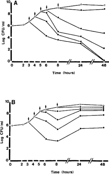
In vitro time-kill curves. Since cardiac vegetations are con-sidered devoid of host defense mechanisms [28, 29], the ther-apeutic results described above are likely to result almost exclusively from antibiotic-induced bacterial killing in the vegetations. Thus, it must be possible to mirror the results obtained in vivo in time-kill curves determined in vitro. The results of such an experiment (using strain MRSE 1386) are shown in figure 5.During the logarithmic phase of growth, both amoxicillinjclavulanate and vancomycin (used at con-centrations mimicking the peak serum levels in vivo) were highly bactericidal over a large range of bacterial densities, which encompassed several orders of magnitude. In contrast, as bacterial growth came to a halt, antibiotic-induced killing abruptly decreased while bacterial densities in the medium remained stable (between 108and 109cfu/rnl.).Thus, as was the case in vivo, antibiotic-induced killing in vitro depended essentially on the rate of bacterial growth, not on bacterial densities in the medium. Similar results were also observed with the 2 other clinical isolates ofMRSE. Note that the late regrowth of bacteria in vancomycin-treated cultures (figure 5B)was not due to the emergence of resistant organisms but to the progressive loss of efficiency of the drug, as deter-mined using an agar diffusion bioassay. Indeed, after 48 h of incubation, the antibiotic activity of vancomycin in the cul-ture supernatants had decreased by 10 times, whereas that of amoxicillinjclavulanate had decreased only by 2 times.
Discussion
In the present experiments, amoxicillinjclavulanate was as effective as vancomycin against experimental endocarditis
Table 3. Number of rats with positive spleen cultures in antibi-otic-treated animals.
v
15 8 Ale ~ NSc
12v
20 Ale 16 ~ NS Doubling-time: >10h Start of treatment: 12h MBD (CFUlg): 108JID 1994; 170 (July) Treatment of MRSE Endocarditis 107
Time (hours)
Time (hours)
Figure 5. Time-kill curves showing bactericidal acuvity of amoxicillin/clavulanate (A) and vancomycin (B) against methicil-lin-resistantS. epidermidisisolate 1386. Cultures were treated at different time points (arrows) with antibiotic concentrations re-flecting peak serum level. Similar results were obtained with iso-lates 4890 and 1468.0, untreated control cultures; ., antibiotic-treated cultures. Late regrowth of bacteria in (B) was due not to emergence of resistant organisms but to progressive loss of effi-ciency of drug (after 48 h of incubation, antibiotic activity of van-comycin culture supernatants, as determined by bioassay, had de-creased by 10 times, whereas that of amoxicillin/clavulanate had decreased by only 2 times).
due to both heterogeneously and homogeneously resistant strains of MRSE. These results are reminiscent of those ob-tained in experimental endocarditis due to MRSA, where amoxicillin/clavulanate was also equivalent to vancomycin [II, 12]. In addition, in the present experiments the spleens of animals treated with amoxicillin/clavulanate were sterile more frequently than were those of animals receiving vanco-mycin. While the reason(s) for this difference is speculative (e.g., do amoxicillin/clavulanate and vancomycin interfere differently with bacterial killing in the spleen"), the results
underline the efficacy of the ,B-lactam combination in this setting.
The finding is of interest because the clustering ofmultiple antibiotic resistance genes in methicillin-resistant staphylo-cocci has brought these organisms to the verge of becoming totally resistant to any available drugs. Therefore, it is essen-tial to broaden our therapeutic armamentarium so we can treat infections due to these pathogens without selecting resis-tance against one of the last nonexperimental antibiotics that is still effective, that is, vancomycin.
On the basis of these results, it might be envisioned to test amoxicillin/clavulanate in patients infected with methicillin-resistant staphylococci. Ideally, such a study should first be done in patients with MRSE infections, because these organ-isms produce much less severe diseases than do MRSA while sharing the mechanism(s) of resistance to methicillin. Sev-eral arguments suggest that such a trial might be feasible. First, in the present setting, amoxicillin/clavulanate was as effective as vancomycin in experiments in which both drugs were administered to the animals in doses that strictly mim-icked the human pharmacokinetics of the antibiotics. Sec-ond, failures after experimental treatment with amoxicillin/ clavulanate did not select for more resistant bacteria in animals, thus limiting the risk of such occurrence in humans. Third, coagulase-negative staphylococci tend to produce smaller amounts of penicillinase than does S. aureus [30], which may be important because it has been shown that pro-duction of large quantities of penicillinase by MRSA might decrease the efficacy ofamoxicillin/clavulanate in the model ofexperimental endocarditis [12]. Finally, S. epiderrnidistyp-ically produces relatively indolent infections (in contrast to S. aureus), ensuring a relatively safe time in which to evalu-ate and readjust therapy in patients with potential treatment failures. Therefore, it might be possible to safely assess the efficacy of amoxicillin/clavulanate against MRSE infections in humans.
A second observation in the present study is the abrupt loss of efficacy of both amoxicillin/clavulanate and vanco-mycin as bacterial growth came to a halt in the vegetations. Decreased killing ofslow-growing bacteria by cell-wall inhibi-tors (as well as by other antibiotics) is well known in vitro and has been referred to as phenotypic tolerance [31]. Pheno-typic tolerance undoubtedly also occurs in vivo [31, 32] but is difficult to demonstrate in the complex biologic environ-ment of animal models of infections. Poorly controlled fac-tors such as host defense mechanisms, progressive alterations in the nature of the infective lesions, or alterations in the diffusion or stability of antibiotics in situ may influence the results either positively or negatively.
In this regard, experimental endocarditis is an advan-tageous model because cardiac vegetations are virtually de-void of cellular host defenses [28, 29]. In addition, in the present experiments, the failure of delayed therapy (started at 24 h) was not the result of major changes in the vegetation or alteration of local drug diffusion for two reasons. The
108 Entenza et al. 110 1994;170 (July)
mass of the valvular lesions did not increase between 16 and 24 h after infection (data not shown) and the correlation between the abrupt switch off of antibiotic-induced bacterial killing and the slowdown of bacterial growth at high bacterial densities was fully reproducible in liquid cultures in vitro, where antibiotic diffusion is not a major issue. Thus, the present experiments clearly illustrate the phenomenon of phenotypic tolerance in vivo. The results also provide a plau-sible explanation for previously reported treatment failures associated with high bacterial densities in the vegetations [33, 34]. Such treatment failure (sometimes attributed to an ill-defined inoculum effect) may largely result from slow growth (i.e., a state of phenotypic tolerance) of bacteria in situ rather than from high bacterial densities per se. In the present experiments, a genuine inoculum effect was illus-trated in animals challenged with large bacterial inocula ( 10 X ID9o) :Halt in growth due to high bacterial densities in situ was attained sooner than after injection of smaller inocula, resulting in phenotypic tolerance and decreased therapeutic efficacy even when treatment was started as early as 12 h after infection. Therefore, it is crucial to precisely define both the size of bacterial inoculum and the timing of therapy onset in order to obtain reproducible results in experimental models of infection.
In conclusion, the good efficacy of arnoxicillin/clavula-nate treatment against experimental endocarditis due to MRSE, associated with its previously reported success against MRSA infections [11, 12], suggests that this fj-lactam combination might be used in clinical trials to treat patients with MRSE infections. In addition, our observations high-light the importance of phenotypic tolerance in vivo. The rapid slowdown of in situ bacterial growth rates observed between 107 and 108 cfu/g of tissue makes it difficult to merely rely on bacterial densities in the vegetations to ensure reproducible results.Itis crucial to precisely define the exper-imental conditions in animal models of infections, as slight variations in the size of bacterial inoculum or in the timing of therapy may dramatically alter the nature of the experimen-tal results, without influencing the most commonly tested parameter in experimental endocarditis, the bacterial densi-ties in the cardiac vegetations.
Acknowledgments
We thank Marlyse Giddey and Jacques Vouillamoz for excel-lent technical assistance.
References
I. Abraham EP. Introduction. In: Morin RB, Gorman M, eds. Chemistry and biology of beta-lac tam antibiotics. Vol I. New York: Academic Press 1982:21-38.
2. Hewitt JH, Coe WA, Parker MT. The detection of methicillin resis-tance inStaphvlococcus aureus.J Med Microbiol 1969;2:443-56.
3. Brown OFJ, Reynolds PE. Intrinsic resistance to beta-Iactarn antibi-otics inStaphylococcus aureus.FEBS Lett 1980;122:275-8. 4. Hartman BJ, Tomasz A. Low-affinity penicillin-binding protein
asso-ciated with f1-lactam resistance inStaphylococcus aureus.J Bacteriol 1984; I 58:513-6.
5. Brumfitt W, Hamilton-Miller J. Methicillin-resistant Staphylococcus aureus.N Engl J Med 1989;320: 1188-96.
6. Izard NC, HachlerH,Grehn M, Kayser FH. Ribotyping of coagulase-negative staphylococci with special emphasis on intraspecific typing ofStaphylococcus epidermidis.J Clin Microbiol 1992;30:817-23. 7. Lugeon C, Blanc D, Wenger A, Francioli P. Molecular epidemiology of
methicillin-resistantStaphylococcus aureus(MRSA) over a 4-year pe-riod in a tertiary university hospital [abstract 1073]. In: Program and abstracts of the 6th European Congress of Clinical Microbiology and Infectious Diseases, Seville, Spain, 1993.
8. Panlilio AL, Culver DH, Gaynes RP, et al. Methicillin-resistant Staphy-lococcus aureusin U.S. hospitals, 1975-1991. Infect Control Hosp Epidemiol 1992; 13:582-6.
9. Martin MA, Pfaller MA, Wenzel RP. Coagulase-negative staphylococ-cal bacteremia. Ann Intern Med 1989; 110:9-16.
10. Noble WC, Virani Z, Cree RGA. Co-transfer of vancomycin and other resistance genes fromEnterococcus faecalisNCTC 1220 I to Staphylo-coccus aureus.FEMS Microbiol Lett 1992;93: 195-8.
II. CantoniL,Wenger A, Glauser MP, Bille J. Comparative efficacy of amoxicillin-clavulanate, cloxacillin, and vancomycin against methi-cillin-sensitive and methicillin-resistantStaphylococcus aureus.J In-fect Dis 1989; 159:989-93.
12. Franciolli M, Bille J, Glauser MP, Moreillon P. f1-lactam resistance mechanisms of methicillin-resistantStaphylococcus aureus.J Infect Dis 1991;163:514-23.
13. Chambers HF. Coagulase-negative staphylococci resistant to f1-lactam antibiotics in vivo produce penicillin-binding protein 2a. Antimicrob Agents Chemother 1987;31: 1919-24.
14. Pierre JR, Williamson R, Bornet M, Gutmann L. Presence of an addi-tional penicillin-binding protein in methicillin-resistant Staphylococ-cus epidermidis.S.haemolyticus,S.hominis.and S.siniulanswith low affinity for methicillin, cephalothin, and cefamandole. Antimicrob Agents Chemother 1990;34: 1691-4.
15. Olsson-Liljequist B, Larsson P. Ringertz S, Lofdahl S. Use of a DNA hybridization method to verify results of screening for methicillin resistance in staphylococci. Eur J Clin Microbiol Infect Dis 1993;12:527-33.
16. Tomasz A, Nachmann S, LeafH. Stable classes of phenotypic expres-sion in methicillin-resistant clinical isolates ofstaphylococci. Antimi-crob Agents Chemother 1991;35:124-9.
17. Goldman PL. PetersdorfRG. Importance off1-lactamaseinactivation in the treatment of experimental endocarditis caused byStaphylococcus aureus.J Infect Dis 1980;141:331-7.
18. Praller M. Davenport D, Bale M, Barrett M, Koontz F, Massanari RM. Development of the quantitative micro-test for slime production by coagulase-negative staphylococci. Eur J Clin Microbiol Infect Dis 1988;7:30-3.
19. National Committee for Clinical Laboratory Standards. Approved stan-dard M2-A4. Performance stanstan-dards for antimicrobial disk suscepti-bility test. 4th ed. Villanova, PA: NCCLS, 1990.
20. Sahm DF, Washington JA. Antibacterial susceptibility test: dilution methods. In: Balows A, Hausler WJ, Herrmann KL, Isemberg HD, Shadomy HJ, eds. Manual of clinical microbiology. 5th ed. Washing-ton. DC: American Society for Microbiology, 1991: 1109-16. 21. Thornsberry C, McDougal LK, Gavan TL. Successful use of
microdilu-tion in susceptibility tests for methicillin-resistant (heteroresistant) staphylococci. J Gen Microbiol 1983; 18:1084-91.
22. Heraief E, Glauser MP. Freedman LR. Natural history of aortic-valve endocarditis in rats. Infect Immun 1982;37:127-31.
hu-JID 1994; 170 (July) Treatment of MRSE Endocarditis 109
man body fluids: techniques and significance. In: Lorian V, ed. Anti-biotics in laboratory medicine. 3rd ed. Baltimore: Williams& Wil-kins. 1991:295-366.
24. Frank U. Schmidt-Eisenlorh E, Schlosser V. Spillner G. Schindler M, Daschner FD. Concentrations of flucloxacillin in heart valves and subcutaneous and muscle tissues of patients undergoing open-heart surgery. Antimicrob Agents Chemother 1988;32:930-1.
25. Staniforth DR Jackson D, Norton R. Davies B. Parenteral augmentin: pharmacokinetics. Int J Clin Pharmacol Ther Toxicol 1984;22:430-4.
26. Blouin RA. Bauer LA. Miller DD. Record KE, Griffen WOo Vancomy-cin pharmacokinetics in normal and morbidly obese subjects. Anti-microb Agents Chemother 1982;21 :575-80.
27. Moreillon P. Francioli P. Overholser D. Meylan P, Glauser MP. Mecha-nisms of successful amoxicillin prophylaxis of experimental endocar-ditis due to Streptococcus intermedius. J Infect Dis 1986;5:801-7. 28. Durack DT, Beeson PB. Experimental bacterial endocarditis.I.
Coloni-zation of a sterile vegetation. Br J Exp Pathol 1972;53:44-9. 29. Berney P, Francioli P. Successful prophylaxis of experimental
strepto-coccal endocarditis with single-dose amoxicillin administered after bacterial challenge. J Infect Dis 1990; 161:281-5.
30. Rosdahl VT, Jarlev JO. Knudsen AM. Beta-Iactamase production in coagulase-negative micrococcaceae. Acta Pathol Microbiol Immunol Scand [B] 1986;94:423-7.
31. Tuomanen E. Phenotypic tolerance: the search for ,s-Iactam antibiotics that kill nongrowing bacteria. Rev Infect Dis 1986;8(suppl 8):279-91.
32. Wood WB, Smith MR. An experimental analysis of the curative action of penicillin in acute bacterial infections.I.The relationship ofbacte-rial growth rates to the antimicrobial effect of penicillin. J Exp Med 1956; 103:487-97.
33. Cantoni L. Glauser MP. Bille J. Comparative efficacy of daptomycin, vancomycin, and cloxacillin for the treatment of Staphylococcus
aur-eus endocarditis in rats and role of test conditions in this
determina-tion. Antimicrob Agents Chemother 1990;34:2348-53.
34. Catheral EJ, Irwing R, Mizen LW. Efficacy of arnoxicillin/clavulanic acid in experimental Staphylococcus aureus endocarditis in rats. J Antimicrob Chemother 1991;27: 117-26.