REVIEW
The blood
–brain and the blood–cerebrospinal fluid
barriers: function and dysfunction
Britta Engelhardt&Lydia SorokinReceived: 8 July 2009 / Accepted: 13 August 2009 / Published online: 25 September 2009
# Springer-Verlag 2009
Abstract The central nervous system (CNS) is tightly sealed from the changeable milieu of blood by the blood– brain barrier (BBB) and the blood–cerebrospinal fluid (CSF) barrier (BCSFB). While the BBB is considered to be localized at the level of the endothelial cells within CNS microvessels, the BCSFB is established by choroid plexus epithelial cells. The BBB inhibits the free paracellular diffusion of water-soluble molecules by an elaborate network of complex tight junctions (TJs) that interconnects the endothelial cells. Combined with the absence of fenestrae and an extremely low pinocytotic activity, which inhibit transcellular passage of molecules across the barrier, these morphological peculiarities establish the physical permeability barrier of the BBB. In addition, a functional BBB is manifested by a number of permanently active transport mechanisms, specifically expressed by brain capillary endothelial cells that ensure the transport of nutrients into the CNS and exclusion of blood-borne molecules that could be detrimental to the milieu required for neural transmission. Finally, while the endothelial cells
constitute the physical and metabolic barrier per se, interactions with adjacent cellular and acellular layers are prerequisites for barrier function. The fully differentiated BBB consists of a complex system comprising the highly specialized endothelial cells and their underlying basement membrane in which a large number of pericytes are embedded, perivascular antigen-presenting cells, and an ensheathment of astrocytic endfeet and associated paren-chymal basement membrane. Endothelial cell morphology, biochemistry, and function thus make these brain micro-vascular endothelial cells unique and distinguishable from all other endothelial cells in the body. Similar to the endothelial barrier, the morphological correlate of the BCSFB is found at the level of unique apical tight junctions between the choroid plexus epithelial cells inhibiting paracellular diffusion of water-soluble molecules across this barrier. Besides its barrier function, choroid plexus epithelial cells have a secretory function and produce the CSF. The barrier and secretory function of the choroid plexus epithelial cells are maintained by the expression of numerous transport systems allowing the directed transport of ions and nutrients into the CSF and the removal of toxic agents out of the CSF. In the event of CNS pathology, barrier characteristics of the blood–CNS barriers are altered, leading to edema formation and recruitment of inflamma-tory cells into the CNS. In this review we will describe current knowledge on the cellular and molecular basis of the functional and dysfunctional blood–CNS barriers with focus on CNS autoimmune inflammation.
Keywords Blood–brain barrier . Blood–cerebrospinal fluid barrier . Extracellular matrix . Basement membrane . Choroid plexus . Experimental autoimmune encephalomyelitis
B. Engelhardt (*)
Theodor Kocher Institute, University of Bern, Freiestrasse 1,
3012 Bern, Switzerland e-mail: bengel@tki.unibe.ch L. Sorokin (*)
Institute of Physiological Chemistry and Pathobiochemistry, University of Muenster,
Waldeyerstrasse 15, 48149 Muenster, Germany e-mail: sorokin@uni-muenster.de DOI 10.1007/s00281-009-0177-0
Introduction
The central nervous system (CNS) is tightly sealed from the changeable milieu of blood by the blood–brain barrier (BBB) and the blood–cerebrospinal fluid (CSF) barrier (BCSFB). While the BBB is considered to be localized at the level of the endothelial cells within CNS microvessels, the BCSFB is established by choroid plexus epithelial cells. The BBB inhibits the free paracellular diffusion of water-soluble molecules by an elaborate network of complex tight junctions (TJs) that interconnects the endothelial cells. Combined with the absence of fenestrae and an extremely low pinocytotic activity, which inhibit transcellular passage of molecules across the barrier, these morphological peculiarities establish the physical perme-ability barrier of the BBB. In addition, a functional BBB is manifested by a number of permanently active transport mechanisms, specifically expressed by brain capillary endothelial cells that ensure the transport of nutrients into the CNS and exclusion of blood-borne molecules that could be detrimental to the milieu required for neural transmission.
Finally, while the endothelial cells constitute the physical and metabolic barrier per se, interactions with adjacent cellular and acellular layers are prerequisites for barrier function. The fully differentiated BBB consists of a complex system comprising the highly specialized endo-thelial cells and their underlying basement membrane in which a large number of pericytes are embedded, perivas-cular antigen-presenting cells, and an ensheathment of astrocytic endfeet and associated parenchymal basement membrane. Endothelial cell morphology, biochemistry, and function thus make these brain microvascular endothelial cells unique and distinguishable from all other endothelial cells in the body.
Similar to the endothelial barrier, the morphological correlate of the BCSFB is found at the level of unique apical tight junctions between the choroid plexus epithelial cells inhibiting paracellular diffusion of water-soluble molecules across this barrier. Besides its barrier function, choroid plexus epithelial cells have a secretory function and produce the CSF. The barrier and secretory function of the choroid plexus epithelial cells are maintained by the expression of numerous transport systems allowing the directed transport of ions and nutrients into the CSF and the removal of toxic agents out of the CSF.
In the event of CNS pathology, barrier characteristics of the blood–CNS barriers are altered, leading to edema formation and recruitment of inflammatory cells into the CNS. In this review we will describe current knowledge on the cellular and molecular basis of the functional and dysfunctional blood–CNS barriers with focus on CNS autoimmune inflammation.
History of brain barriers
The discovery of a vascular barrier between the blood circulation and the central nervous system dates back more than 100 years, when in the 1880s Paul Ehrlich discovered that certain dyes when injected into the vascular system were rapidly taken up by all organs except the brain and spinal cord [1]. Ehrlich himself interpreted these findings as a lack of affinity of the nervous system for these dyes. However, shortly afterwards Edwin E. Goldman, an associate of Ehrlich, showed that the very same dyes when injected into the cerebrospinal fluid readily stained nervous tissue but not any other tissue [2], suggesting that once within the CNS the dyes were prevented access to the blood circulation. Additional studies, demonstrating that neuro-toxic agents affected brain function only when directly injected into the brain but not when injected into the vascular system, further supported the concept of a vascular blood–brain barrier that also functions as a brain–blood barrier [3, 4]. Only with the advancement of electron microscopy was it possible to correlate the ultrastructural localization of the blood–brain barrier with the capillary endothelial cells within the brain [5]. Following injection into the vasculature or into the CNS, the electron-dense tracer horseradish peroxidase, a small protein of 40 kDa, was found to diffuse into the intercellular clefts of brain endothelial cells up to the tight junctions (TJs) between the endothelial cells. Thus, in vertebrates the inter-endothelial TJs were recognized as the morphological correlate of the blood–brain barrier.
There are, however, structures within the brain, located in the midline of the ventricular system, that lack an endothelial BBB and are collectively referred to as the circumventricular organs (CVOs). As CVOs serve neurohemal or neurosecre-tory functions, i.e. their neurons monitor hormonal stimuli and other substances within the blood or secrete neuro-endocrines into the blood, and they lack a vascular barrier [6]. Rather, capillaries within the CVOs are fenestrated allowing free diffusion of proteins and solutes between the blood and the CVOs. Similarly, the endothelial cells of the choroid plexus do not form a barrier and are fenestrated like those of the CVOs [7]. The choroid plexus is a villous structure that extends from the ventricular surface into the lumen of the ventricles, the major known function of which is secretion of cerebrospinal fluid. The choroid plexus consists of an extensive capillary network enclosed within a single layer of cuboidal epithelium, interconnected by apical tight junctions, forming a blood–cerebrospinal fluid barrier (BCSFB). In an analogous manner, a complex network of tight junctions connecting specialized ependy-mal cells (tanycytes) seal the CNS from the CVOs [6,8], establishing tight junctions as an important structural element of the blood–brain and blood–CSF barriers.
The BBB under physiological conditions Cellular and acellular architecture
As described above at the level of capillaries and postcapillary venules, the selectively permissive and sealed phenotype of the endothelial cell monolayer of CNS vessels is dependent on the complex tight junctions that intercon-nect adjacent endothelial cells and the low pinoctotic activity of these endothelial cells. In addition, in CNS microvessels, the vascular endothelium is ensheathed by a layer of astrocyte endfeet and associated leptomeningeal cells that coinvaginate with the endothelium during development and also contribute to their restricted perme-ability properties. Ultrastructurally, two basement mem-branes can be distinguished at the level of smaller vessels, an endothelial cell basement membrane and an astroglial basement membrane, which underlie the endothelium and astrocyte endfeet, respectively (Fig.1a, b). In addition, the epithelium of the meninges coinvaginates with blood vessels from the surface of the brain and contributes to the astroglial basement membrane [9–11]. The astroglial basement mem-brane, together with the leptomeningeal basement memmem-brane, constitutes the parenchymal basement membrane, so-called because it delineates the border to the brain parenchyma. At the level of venules and postcapillary venules these endothe-lial and parenchymal basement membranes are clearly distinguishable by electron microscopy (Fig. 1b). However, in brain capillaries they fuse to form one basement membrane structure [5, 9, 11, 12]. Collectively, these cellular and acellular layers are considered to constitute the physical characteristics establishing the limited permeability of the BBB, and are discussed separately below.
Endothelial cell–cell junctions in the CNS
The inter-endothelial tight junctions in CNS microvessels are an intricate complex of transmembrane (claudins, occludin, and junctional adhesion molecule (JAM)-A) and cytoplasmic (zonula occludens (ZO)-1 and ZO-2, cingulin, AF-6, and 7H6) proteins linked to the actin cytoskeleton [13, 14] (Fig. 2). Occludin was the first integral membrane protein described to be exclusively localized within tight junctions including the BBB [15]. However, mice carrying a null mutation in the occludin gene are viable and develop morphologically normal tight junctions in most tissues including brain microvessels [16], excluding an essential role in tight junction formation. By contrast, the claudins, which comprise a gene family of integral membrane tight junction proteins with 24 members, have been shown to be sufficient for the formation of tight junction strands [17]. Claudins are not randomly distributed throughout all tissues; besides the endothelial cell-specific claudin-5, claudin-3 has
been shown to localize to endothelial tight junctions in the CNS of mice and man [18]. The Ig supergene family member, JAM-A [19], and the recently discovered endothe-lial cell-selective adhesion molecule (ESAM)-1, are also localized in tight junctions [20] of the BBB; however, their contribution to BBB integrity remains to be determined. The integral membrane proteins of the tight junctions are linked to the cytoskeleton via cytoplasmic peripheral membrane proteins of the MAGUK family, such as ZO-1, ZO-2, and ZO-3 also within BBB tight junctions (reviewed in [14,21]). The primary component of adherens junctions is vascular endothelial (VE)-cadherin, a Ca2+-regulated protein that mediates cell–cell adhesion via homophilic interactions between the extracellular domains of proteins expressed in adjacent cells [22]. The cytoplasmic tail of VE-cadherin binds toβ-catenin and plakoglobin, which in turn bind via β-catenin, α-actinin, and vinculin to the actin cytoskeleton, stabilizing the adherens junction complex. In vascular beds outside of the CNS, endothelial adherens junctions have been demonstrated to be important regulators of vascular permeability (reviewed in [23]), while at the BBB they have been considered less relevant in regulating BBB paracellular permeability [24]. However, very recent studies demonstrate that adhesive interactions of VE-cadherin promote claudin-5 expression by preventing the nuclear accumulation of the transcriptional regulators, FoxO1 and β-catenin [25]. This study demonstrates that proper establishment of adherens junctions is required for expression of claudin-5, which regulates paracellular permeability of small molecules across the BBB [25]. Additionally, the adherens junction compo-nent, β-catenin, is critically involved in the regulation of tight junctions. Besides its function as a junctional compo-nent, β-catenin can serve as a transcription factor down-stream of Wnt-signaling. Induction and maintenance of BBB characteristics during embryonic and postnatal development are regulated by Wnt/β-catenin signaling [26]. Endothelial cell-specific stabilization ofβ-catenin in vivo enhances BBB maturation by inducing the expression of claudin-3, whereas inactivation of β-catenin causes significant downregulation of claudin-3 and upregulation of plasmalemma vesicle-associated protein (Plvap), usually present only on immature brain endothelium or on endothelium of fenestrated capillar-ies within the CVOs and the choroid plexus [27].
Localized in endothelial cell contacts outside of either tight junctions or adherens junctions in the brain are molecules like PECAM-1 and CD99. Mice deficient for PECAM-1 have no primary defect in BBB integrity, but show a defect in resealing of the BBB during CNS autoimmune inflammation [28]. Astrocytes, leptomeningeal cells, and pericytes
In addition to the endothelial cell monolayer, CNS micro-vessels are associated with an ensheathing layer of
astrocytic glia endfeet and associated leptomeningeal cells. Grafting studies and data from in vitro BBB models suggest that inductive influences from astrocytic glia contribute to the differentiation of the specialized BBB phenotype of the
brain endothelium [29,30] and reviewed in [31]. While the precise nature of the BBB inducing factor(s) in the cellular crosstalk between brain endothelium and astroglial cells are not yet clear (reviewed in [32]), the fact that these two cell
Fig. 1 Cell and basement membrane layers of CNS blood vessels. a Schematic representation and immunofluorescence examples of cell and basement membrane (BM) layers consituting CNS blood vessels. Larger blood vessels consist of an inner endothelial cell layer with BM (containing laminins α4 and α5), bordered by the meningeal epithelium and its BM (containing lamininα1), and an outer astroglial BM (containing laminin α2) and astrocyte endfeet. Meningeal and astroglial BMs are collectively termed the parenchymal BM as they delineate the border to the brain parenchyma. Only at sites of local inflammation are the endothelial and parenchymal BMs distinguish-able and define the inner and outer limits of the perivascular space
where leukocytes accumulate before infiltrating the brain parenchyma. Mononuclear infiltration occurs across endothelial BMs containing only the lamininα4 and bordered by a parenchymal BM containing laminin α1 and α2. The BM of microvessels where no epithelial meningeal contribution occurs have a composite BM containing the endothelial cell laminins, lamininα4 and α5, and laminin α2 produce by the astrocytes and deposited at their endfeet. b Electronmicroscopy showing the endothelial cell and the parenchymal BMs at the level of smaller blood vessels. Insert shows higher magnification image of boxed area
layers are separated by at least one basement membrane at the level of capillaries and two distinct basement mem-branes in postcapillary venules and venules, suggests that these inducing factors are likely to be small molecular weight molecules. Not fully considered is whether the basement membrane per se also contributes to the tightness of the brain endothelial cell monolayer and influences expression and or function of BBB-specific structural (tight junctions) and molecular (transporters, enzymes) character-istics (see below). In addition to astrocytes, epithelial cells from the meninges can be associated with CNS blood vessels. While such epithelial cells have been reported to occur predominantly in association with larger blood vessels in the human CNS [9, 11, 33, 34], in the mouse there is data that they may also occur at the level of postcapillary venules (see below) and hence vessels that contribute to the microvascular BBB.
Although pericytes cover 99% of the abluminal surface of the capillary basement membrane in brain, their precise role in the BBB is not well investigated. Several examples of human disease suggest that they play an important role in maintenance of the BBB. CADASIL, for example, is an inherited angiopathy caused by mutations in the Notch3-gene, causing loss of pericytes and vessel hypopermeabil-ity, vessel wall hypotonia, and watershed hypoperfusion [23]. Furthermore, in vitro BBB models have shown significantly increased transendothelial electrical resistance in models incorporating pericytes together with endothelial cells and astrocytes, as compared to models combining only endothelial cells and astrocytes [35]. The conspicuous absence of investigations into the nature of pericyte contribution to the BBB is due to several factors: difficulty in extracting pericytes from their tight encasement of ECM
and hence low numbers for in vitro analyses, and the fact that pericyte markers vary with tissue type suggesting an important role for the in situ milieu in maintaining their normal physiological function. Although pericytes in most tissues can be identified by immunoreactivity for platelet-derived growth factor (PDGF) receptor β, and desmin or α-smooth muscle cell actin [36], these are also markers for other perivascular cells such as smooth muscle cells and myofibroblasts. Hence, additional markers are required, which vary with tissue type. In the case of brain pericytes a combination of positive reaction for PDGFR-β, desmin, γ-glutamyl transpeptidase [36], Sca-1, nestin and the ATP-sensitive potassium channel, kir6.1 [37], plus absence of reactivity for vimentin, glial fibrillary acidic protein and endothelial cell markers such as PECAM-1 are typical. During development, the proteoglycan, NG2, provides a further excellent surface marker that allows visualization of the entire extension of pericytes on the endothelial plexus [36]. Improvement in pericyte identification is likely to lead to increased appreciation of the role of pericytes in the neurovascular unit. Their important role in establishing a mature BBB has, however, been shown in mice lacking PDGF-B. These mice fail to establish pericyte ensheath-ment of their blood vessels during embryonic developensheath-ment and develop microaneurysms also in blood vessels of the CNS [38]. Thus, endothelial PDGF-B retention is required for proper investment of pericytes in the microvessel wall including the BBB [39].
Acellular layers—basement membranes
While the cellular components of the BBB have been relatively well studied, the acellular extracellular matrix
Fig. 2 Endothelial cell–cell junctions of CNS microvessels. The scheme shows the junc-tional molecules localized in the cell–cell contact of CNS micro-vessels within tight junctions and adherens junctions and outside of these organized junc-tions. Transmembrane junctional proteins (names mentioned in cell 1), scaffolding proteins and junction associated proteins involved in mediating the interaction with the actin cyto-skeleton (cell 2) are depicted. The function of the molecules is described in the text
components have received little attention. Only relatively recently have CNS inflammation studies highlighted the contribution of vascular basement membranes to leuko-cyte extravasation processes and hence barrier function at the level of postcapillary venules. In most tissues, with the exception of secondary lymphoid tissues and the CNS, two types of extracellular matrix prevail: basement membranes, complex assemblies of four major glycopro-tein families, laminins, collagen type IV isoforms, nido-gens, and heparan sulfate proteoglycans, which underlie polarized cells such as endothelial and epithelial cells, and ensheath myogenic tissues, nerves, and adipocytes, the fundamental function of which is to separate tissue compartments. The second major extracellular matrix type is the interstitial matrix of the stroma of tissues which acts to interconnect rather than separate cellular layers, and is composed of fibrillar extracellular matrix molecules, such as collagen types I, III, V, and glycoproteins such as fibronectin or tenascins.
Comparatively little ECM is found in the brain and its composition and organization is distinct, with the occur-rence of unique components. In the CNS there is no need to generate high levels of tensile or elastic strength usually resulting from the interstitial extracellular matrix because the brain is protected by the bony skull, hence, there is little or no fibrous protein, like collagen types I and III or fibronectin; and low amounts of glycosaminoglycans chains, either bound to proteins in the form of proteogly-cans, or unbound in the form of hyaluronan. The basement membranes of the vasculature and of the meninges, therefore, represent the predominant ECM form in the CNS. Like all basement membranes, those of the CNS appear as thin, tightly interwoven protein sheets of 20– 200 nm thickness in scanning electron microscopy [40], and are composed of the laminins, collagen type IV, heparan sulfate proteoglycans, and the nidogens. However, all four classes of basement membrane proteins occur in several isoforms, which can combine differentially to form biochemically and functionally distinct basement mem-branes (reviewed in [41]). In addition, basement mem-branes contain minor components which also contribute to their overall structure and function; in endothelial cell basement membranes these include BM40 (osteonectin, SPARC); fibulin 1 (BM90) and 2; collagen types VIII, XV, and XVIII; and thrombospondin 1 and 2 [40].
Although data on the basement membrane composition of CNS vessels exist, the data is fragmentary and lacks specificity both with regards to vessel type and specific extracellular matrix isoforms [12, 42–47]. Nevertheless, existing data suggest that biochemical variations exist between endothelial and parenchymal basement membranes and that basement membrane components contribute to microvessel integrity and function.
Recently, mutations of COL4A1, the gene that codes for theα1 chain of the most common collagen type IV isoform ([α1(IV)]2α2(IV)), have been shown to cause intracerebral
hemorrhage of microvessels and porencephaly both in mouse and human [48, 49]. These data suggest that collagen type IV isoforms are important for structural integrity of small vessels, a role that is consistent with its function in basement membranes of other tissues [50].
The laminin family of basement membrane molecules have been most extensively studied at the level of cerebral vessels. As in all endothelial cell basement membranes, those of CNS vessels contain the lamininα4 and α5 chains combined with lamininβ1 and γ1 chains to form laminin isoforms 411 and 511, respectively [12, 45] (Fig. 1a). By contrast, the outer parenchymal basement membrane of postcapillary venules and venules contains laminin α1 and α2 chains [12] combined with lamininβ1 and γ1 chains to form laminins 111 and 211 (Fig. 1a). In situ hybridization studies have shown that CNS endothelial cells are the source of lamininα4 and α5, while laminin α1 is produced by the leptomeningeal cells and lamininα2 is produced by the astrocytes and deposited at their endfeet [12] (Fig.1a). In addition, perlecan is the predominant heparan sulfate proteoglycan in the endothelial cell basement membrane, while agrin predominates in the parenchymal BM [44,51]. Whether further differences between the endothelial and parenchymal basement membranes exist in their expression of other basement membrane molecules is not yet clear. At the level of capillaries the combined endothelial/parenchy-mal basement membrane contains lamininα4, α5, and α2 but no lamininα1, plus perlecan and agrin.
While pericytes are especially abundant in the CNS microvessels, embedded within the endothelial cell base-ment membrane, their contribution to the endothelial cell basement membrane is not clear. Indeed, the extent of pericyte contribution to the endothelial cell basement membrane in general remains poorly investigated. In vitro, pericytes have been reported to secrete laminins and nidogens and to induce endothelial cells to secrete basement membrane components [52]. However, the repertoire of basement membrane components that can be secreted by pericytes has not been studied, and whether they can influence endothelial cell basement membrane secretion in vivo is not clear. Given the important role of pericytes in the barrier function of microvessels [53] and in vitro BBB models [35] this is an important open question. Extracellular matrix receptors
Several ECM receptors have been described on CNS endothelial cells and astrocyte endfeet, which could potentially act to anchor these cell layers to their respective basement membranes and thereby contribute to BBB
integrity Table1. However, as for the studies concerning the extracellular matrix composition of CNS vessels, the data on ECM receptor expression in association with CNS micro-vessels remains fragmentary and lacks correlation with vessel type and cellular layer within the vessel wall. CNS endothelial cells have been reported to express integrins α1β1, α3β1, α6β1, and αvβ1/αvβ3, and a major non-integrin receptor, dystroglycan, of the dystrophin glycopro-tein complex [54], while astrocytes have been reported to express integrin α6β4 and dystroglycan (reviewed in [42, 43]), and microglia express αvβ3, αvβ5 [55], and αvβ8 [56,57]. The potential interaction partners for these receptors and their localization in endothelial or parenchymal base-ment membranes of CNS microvessels has not yet been correlated with receptor expression patterns.
Of all these ECM receptors, there is only evidence for a role for integrinαvβ8, and the non-integrin receptor, dystrogly-can, in maintenance of BBB integrity. Although the majority of integrinαv knockout (KO) embryos die at mid-gestation, some survive to birth and develop severe cerebral hemor-rhage, which is not due to endothelial or pericytes defects [58, 59]. Integrinβ8 KO mice exhibit a similar phenotype, and current data suggest that αvβ8-mediated interactions are required for associations between angiogenic cerebral blood vessels, neuroepithelial cell, and astrocytes during develop-ment [56]. Similarly, selective elimination of dystroglycan in astrocytes results in a prominent reactive gliosis resulting from disruption of the glia limitans [60], a phenotype consistent with a defect in BBB formation.
Molecular transport systems across the BBB
Besides the structural components discussed above, which passively inhibit free diffusion of molecules across the
BBB and thus provide a physical barrier, metabolic barrier characteristics of brain capillary endothelial cells are maintained by the expression of high a number of transport systems and enzymes [61]. Transport proteins, mostly of the solute carrier family ensure the transport of water-soluble molecules such as glucose [62] or amino acids [63] across the BBB from the blood into the CNS. Other molecules such as insulin and transferrin are specifically targeted from the blood into the CNS by receptor-mediated transcytosis across brain capillary endothelial cells [64]. Furthermore, for some but not all cytokines, transport mechanisms across the BBB have been reported [65].
In addition, efflux transporters such as P-glycoprotein or multidrug resistance proteins, which are members of the ABC transporter family, are specifically expressed by brain microvascular endothelial cells [66]. These efflux trans-porters efficiently move potentially harmful hydrophilic and hydrophobic molecules out of the CNS.
The BBB under pathological conditions
As described above, normally the microvessels of the CNS form an effective barrier to the movement of water-soluble molecules and cells and only in pathological situations is this barrier function compromised. One such situation is CNS autoimmune neuroinflammation, as it occurs in the human disease multiple sclerosis, and the well-studied animal model thereof, experimental autoimmune encephalomyelitis (EAE). Lymphocyte transmigration of endothelial cell monolayers The CNS is considered immune privileged, i.e. it actively restricts entry of immune cells into the parenchyma and
Table 1 ECM-binding receptors and potential ligands in basement membranes of the CNS microvascular
Receptor Potential ligandsa Localization of ligand
α1β1 Several collagens; highest affinity for collagen type IV, but also binds types I, XIII, XVI [120–122]
Endothelial or parenchymal BM of postcapillar venules or combined endothelial/parenhcymal BM of capillaries
α3β1 Laminin 211 Parenchymal BM of postcapillary venules or combined
endothelial/parenchymal BM of capillaries Laminin 332 (seeα6β4 below)
α6β1 Laminins 411, 511, Endothelial BM
Laminin 111 Parenchymal BM at the level of postcapillary venules only
α6β4 Laminin 332 Not yet reported in CNS vessels in vivo; reported to be
expressed by astrocytes in culture [123] Dystroglycan Highest affinity for perlecan and agrin; also some
binding to laminins 111 and 211
Perlecan predominates in the endothelial cell BM of postcapillary venules and agrin in the parenchymal BM [44]; laminin 111 and 211 exist in the
postcapillary parenchymal BM, and laminin 211 in the composite endothelial/parenchymal BM of capillaries
a
only as a consequence of BBB dysfunction as occurs during EAE does it become accessible to hematopoietic inflammatory cells. EAE can be transferred by intravenous injection of neuroantigen-specific T cell blasts into synge-neic susceptible recipients [67, 68]. The initial interaction as well as the G-protein-dependent arrest of circulating encephalitogenic CD4+ T lymphoblasts and the non-inflamed BBB requires leukocyte integrin α4β1 and vascular cell adhesion molecule (VCAM)-1 [69], while diapedesis across the BBB has been proposed to require LFA-1–ICAM-1 interactions [70]. Whether BBB tight junctions are involved in the migration of T cell blasts across the BBB at this stage is not known.
During EAE, α4β1-integrin/VCAM-1 are critically involved in leukocyte interaction with the inflamed BBB. Intravital microscopy studies of brain meningeal vessels and of the spinal cord microvasculature of mice suffering from EAE have shown that blocking α4-integrin [71] or lack ofβ1-integrin [72] on myelin-specific T cells does not allow them to maintain stable adhesions with the CNS microvascular endothelium and thus prohibits their migra-tion across the BBB resulting in inhibimigra-tion of EAE [72,73]. This α4β1-integrin-mediated firm arrest to the inflamed BBB depends on heterotrimeric G(i)-linked receptors [74]; however, the chemokines and chemokine receptors mediat-ing leukocyte integrin activation—a prerequisite for firm adhesion—at the BBB are not well defined [75]. The molecules mediating tethering and rolling of leukocytes to the inflamed BBB during EAE are presently controversially discussed. Although intravital microscopy studies have demonstrated the involvement of P-selectin and its ligand PSLG-1 in immune cell rolling in inflamed meningeal brain venules during EAE [71,76] or after cytokine application [74], blocking of these adhesion molecules or their absence in gene-targeted mice does not influence EAE pathogenesis [77–81], supporting the view that they are dispensable for leukocyte interaction with the inflamed BBB during EAE. Similarly, blocking ofα4-integrins or lack of β1-integrins on myelin-specific T cells also fails to impair to affect initial capturing and rolling of T cell on the inflamed BBB [71, 72]. Thus, it remains to be shown which adhesion receptors mediate this initial step of T cell interaction with the inflamed BBB during EAE in vivo. Furthermore, the adhesion molecules involved in leukocyte diapedesis across the inflamed CNS microvessels during EAE still remain to be determined. In vitro a critical role for endothelial ICAM-1 has been demonstrated in T cell diapedesis across brain endothelium, suggesting the active involvement of BBB endothelium in this process [82,83].
Last but not least, the involvement of endothelial tight junctions in leukocyte recruitment across the BBB during EAE remains to be understood. During EAE, the selective loss of claudin-3 immunostaining specifically from tight
junctions of venules surrounded by inflammatory cuffs has been reported, whereas the localization of the other tight junction proteins remains unchanged [18], implicating an involvement of endothelial tight junctions in leukocyte recruitment across the BBB.
Several other endothelial cell adherens junction mole-cules have been implicated in leukocyte extravasation in other tissues, including CD99, CD99L, ESAM-1, and the JAM family members. The use of knockout mice in inflammatory models have implicated PECAM-1, CD99L, JAM-A, and ESAM-1 in extravasation of neutrophils, but in the case of lymphocytes PECAM-1 and ESAM-1 do not play a role [84], suggesting that different leukocyte types may use different molecular mechanisms to penetrate the endothelial barrier. Most of these molecules have not been specifically investigated with a focus on the BBB, with the exception of PECAM-1, which has been studied using PECAM-1 KO mice, revealing an earlier onset of EAE symptoms due to an increase in vessel permeability [28].
In addition to paracellular transmigration, especially T cells have been reported to be able to migrate across endothelial monolayers in a transcellular manner (reviewed in [85]). Vesiculo-vacuolar organelles or caveolae have been sug-gested to be involved in regulating this transcellular diapedesis of leukocytes [86, 87]. Transmission electron microscopy on serial ultra-thin sections derived from brains of mice afflicted with EAE have only reported few sites of neutrophil extravasation through tight junctions [88]. In contrast, the majority of studies have demonstrated that during EAE inflammatory cell recruitment across the BBB leaves tight junctions morphologically intact (reviewed in [89]), suggesting that transcellular migration of immune cells may occur in inflammation in the CNS [90].
Lymphocyte transmigration of endothelial basement membranes
Upon penetration of the endothelial cell monolayer, infiltrating leukocytes in the CNS still face several basement membrane layers, which also act as barriers to their migration. Recent data suggests that sites of enceph-alitogenic T lymphocyte penetration of the underlying endothelial cell basement membrane is determined by its laminin isoform composition. Due to the specialized double basement membrane structure of postcapillary venules discussed above, the precise sites of extravasation can be identified and characterized. In the course of EAE, leukocytes first traverse the endothelial cell monolayer and underlying basement membrane and accumulate focally to form easily identifiable "perivascular cuffs" between the inner endothelial and outer parenchymal basement mem-branes (Fig.1a). However, only upon leukocyte penetration of the parenchymal basement membrane and glial limitans
and entry into CNS parenchyma are disease symptoms induced [44].
As discussed above, laminin α5 and α4 are the predominant isoforms located in endothelial cell basement membranes of CNS vessels. However, at the level of postcapillary venules and venules in the CNS lamininα5 distribution is patchy and irregular while laminin α4 is ubiquitously expressed [45]. Extravasation occurs focally, preferentially at sites containing lamininα4 but little or no lamininα5 [12,45]. In vitro studies reveal that lamininα4 supports substantial chemokine-induced transmigration of encephalitogenic T cells and that integrinα6β1 is essential for this transmigration. By contrast laminin α5 does not support encephalitogenic T lymphocyte transmigration, and when mixed together with laminin α4 shows a dose-dependent and therefore specific inhibition of migration across lamininα4 [45]. Although in vitro migration studies using lymphocytes have been limited, studies involving naive T lymphocytes have shown similar enhanced migra-tion across lamininα4 as compared to laminin α5 [91].
In mice lacking lamininα4 [92] there is compensatory ubiquitous expression of lamininα5 in all endothelial cell basement membranes of the CNS and no differential expression in different blood vessel types or regulatory expression of this chain in response to proinflammatory cytokines [45]. These laminin α4 knockout mice are less susceptible to EAE than their wildtype littermates and show significantly reduced severity of disease symptoms, due to specific inhibition of T lymphocyte infiltration into the brain. These data support in vitro experiments, substantiat-ing the inhibitory role of lamininα5 on T cell transmigra-tion across endothelial cell basement membranes.
In wild-type mice, lamininα4 is ubiquitously expressed along the vascular tree andα6β1 is strongly expressed on all T lymphocytes, hence, lamininα4 and α6β1 alone are insufficient to explain the focal extravasation pattern observed in EAE. Rather, data suggests that this focal extravasation is due to the patchy distribution of lamininα5 in the endothelial basement membrane of postcapillary venules and it is inhibitory effect on T lymphocyte transmigration. To date, no correlation has been identified between sites of low lamininα5 expression the expression of endothelial cell adhesion molecules, such as VCAM-1, or endothelial junctional molecules that have been impli-cated in the extravasation process, which suggests that T lymphocytes are capable of sensing the laminin content of endothelial cell basement membrane which in turn deter-mines the probability of whether or not transmigration will occur. Whether this also requires integrin α6β1 or other cellular receptors is not yet clear.
Apart from the disease inducing CD4+ T lymphocytes, several other leukocyte types also extravasate across CNS postcapillary venules during EAE, most notably
macro-phages and dendritic cells (DC). While infiltration of macrophages and dendritic cells is also reduced in laminin α4 KO mice, the extent of inhibition is significantly less than that observed for the CD4+ T lymphocytes [45]. Clearly, changes in laminin isoform composition of endothelial basement membranes may alter the inter-(within the laminin network) and intra-network interactions (between laminins and other basement membrane compo-nents), and thereby the tightness of the network [93, 94]. Such ultrastructural alterations are likely to account for the lower levels of migration of both T lymphocytes and macrophages across laminin α5-containing vascular base-ment membranes into the CNS, but not the more pro-nounced reduction in activated CD4+ T cell infiltration. This indicates that basement membrane architecture is not only a limiting factor in the transmigration process but that activated CD4+T lymphocytes utilize different mechanisms to macrophages and DC to transmigrate basement mem-branes, which are determined by the basement membrane laminin composition.
Transmigration of the parenchymal basement membrane Upon penetration of the endothelial cell monolayer and its underlying basement membrane extravasating lymphocytes must traverse the parenchymal basement membrane and glia limitans before they can enter into the brain parenchy-ma to induce disease symptoms. In contrast to transmigra-tion of the endothelial cell basement membrane, T lymphocyte penetration of the parenchymal basement membrane correlates with sites of focal matrix metal-loproteinase (MMP) activity (Fig.3a).
Although several MMPs have been implicated in EAE, including MMP-14/MMP-2 [95] and MMP-9 [96] and possibly also MMP-7 [97], and MMP-8 [98–100], most data is based on mRNA levels and in situ hybridization studies. As several proteases, in particular MMPs, exist in an inactive form that requires in situ activation, mRNA and protein expression is not sufficient to identify sites of protease activity. The use of novel techniques that permit simultaneous in situ zymography for protease activity and immunofluorescence for identification of cellular or extra-cellular markers, has revealed that the majority of the protease activity associated with infiltrating leukocytes is gelatinase activity (MMP-2 and MMP-9), occurring focally subjacent to the parenchymal basement membrane at sites of leukocyte penetration of the CNS [44]. The involvement of the gelatinases in EAE has been substantiated by inhibitor studies [101,102], and current data suggest that infiltrating CD4+ encephalitogenic T lymphocytes are not the major source of gelatinases in vivo, but rather infiltrating macrophages and an additional CNS-resident cell population [44]. The reason that encephalitogenic T
cells use different mechanisms to penetrate the endothelial versus the parenchymal basement membrane may be related to the basement membrane composition: the parenchymal basement membrane is biochemically distinct to the endothelial cell basement membrane, with expression of lamininα1 and α2 rather than the endothelial cell laminins α4 and α5. Encephalitogenic CD4+ T cells cannot interact with these two laminin isoforms, even though they express the receptors capable of mediating such interactions (α6β1) [12], suggesting that the extracellular milieu is an important factor in determining migratory mechanisms in encephali-togenic T cells [12,44].
There is increasing evidence that the general digestion of ECM molecules originally attributed to MMPs does not hold true for the in vivo situation [103,104]. Until recently the method employed to search for substrates of MMPs was in vitro incubation of potential substrate with proteases; however, this might be not relevant in the cellular or tissue context. The development of new protease degradomic techniques [105, 106] has provided evidence for specific proteolysis and processing of both cell surface and secreted factors by MMPs which alters the biological activity of these substrates. In murine EAE, the gelatinases, 2 and
MMP-9, but not collagenases (MMPs-1, MMPs-3, and MMPs-8), have been shown to cleave β-dystroglycan, a cell surface receptor that anchors the astrocyte endfeet to the parenchymal basement membrane without significantly affecting parenchy-mal basement membrane components or other ECM-binding receptors on the astrocyte endfeet, suggesting a subtle and specific regulatory role for proteases in leukocyte extravasation [44] (Fig 3b). β-dystroglycan is part of a larger receptor complex that interconnects the parenchymal basement mem-brane with the astrocyte cytoskeleton [54]. An integral part of the receptor complex is α-dystroglycan, which is non-covalently linked to the membrane spanningβ-dystroglycan subunit and constitutes the link to specific ligands, including lamininsα1 and α2, perlecan, and agrin [107] present in the parenchymal basement membrane. The strategic cleavage of β-dystroglycan by MMP-2 and MMP-9 results in the loss of the connection to theα-dystroglycan and therefore the anchor to the basement membrane. However, what happens to the cleaved β-dystroglycan fragment or to the α-dystroglycan subunit is not clear; both are not detectable in the EAE brains which, however, may be due to the tools available for their detection in inflamed tissues [44]. Hence, the potential persistence of dystroglycan fragments in the parenchymal
Fig. 3 Enzymatic degradation of dystroglycan in association with leukocyte recruitment across the parencyhmal basement membrane of the BBB. a Double staining for CD45 to mark infiltrating leukocytes and dystroglycan, showing loss of dystroglycan from sites of leukocyte penetra-tion of the parenchymal barrier in inflammed vessels only. Arrow marks inflamed vessel. b In situ gelatin gel zymography together with anti-CD45 staining show gelatinase activity subjacent to the parenchymal border where leukocytes enter into the CNS parenchyma. * marks lumen of the vessel
basement membrane may convey additional signals to the infiltrating leukocytes or may alter the structural properties of the parenchymal basement membrane and thereby alter its stability and/or penetrability. Alternatively, it may be degraded resulting in a source of bioactive fragments.
In addition to β-dystroglycan cleavage, MMP-2 and MMP-9 have been implicated in the activation of a chemotactic (CCL2) signal that is required for T lympho-cyte migration out of the perivascular cuff, across the parenchymal basement membrane and glia limitans and into the brain parenchyma [108]. CCL2 overexpressing mice show accumulation of leukocytes in the perivascular cuff, which is overcome by induction of MMP-2 and MMP-9 activity. The precise manner of action is not clear, but demonstrates that MMPs have multiple effects at the parenchymal border.
Lymphocyte recruitment across the choroid plexus
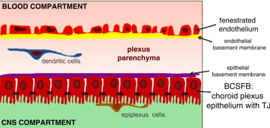
The choroid plexus plays a central role in the formation and regulation of cerebrospinal fluid. The epithelial cells of the choroid plexus hereby form the direct barrier between the blood compartment and the CSF, whereas the capillaries within the choroid plexus parenchyma differ from those of the brain, because they allow free movement of molecules across the endothelial cells through fenestrations and intercellular gaps (reviewed by [109]).
In healthy mice the choroid plexus is composed of a single layer of cuboidal epithelial cells that surrounds the central stroma. The apical plasmalemma of the epithelial cells forms numerous microvilli and some cilia (summa-rized in [7]; Fig.4). Directly underneath the microvilli the adjacent epithelial cells are sealed by tight junctions, which in freeze-fracture replicas appear as parallel strands similar to the tight junctions in oligodendrocytic myelin sheaths [110]. Claudin-11 localizes to both myelin and choroid plexus epithelial tight junctions [111]. In addition, at least claudin-1 and claudin-2, together with occludin, have been described in the choroid plexus epithelial cell tight
junctions of the rat brain [110]. Besides tight junctions restricting permeability across the chrodip plexus epithelium these cells also express a broad range of transporters [112].
Epiplexus cells or Kolmer cells can be observed in direct contact with the epithelial microvilli [7, 113] (Fig. 4). Although the exact origin of the epiplexus cells has been debated, there is evidence that they are of monocytic origin as ultrastructural studies have demonstrated monocyte recruit-ment via the choroidal vessels and subsequent migration across the choroid plexus epithelium [114,115]. Thus, it is tempting to speculate that like antigen-presenting cells in the perivascular spaces of the vascular BBB, these cells may serve as barrier-associated antigen-presenting cells and thus ensure immunosurveillance of the CNS.
It has been difficult to provide direct evidence for the possibility of immune cell entry into the CNS across the BCSFB. Although the choroid plexus shows massive morphological alterations during EAE [7], supporting its involvement in EAE pathogenesis, no regular accumulation of inflammatory cells has been demonstrated within the choroid plexus parenchyma or at the level of the BCSFB during EAE. Circulating immune cells would need to first migrate across the fenestrated choroid plexus capillaries, entering the parenchyma, which still remains outside of the CNS (Fig. 4). From there immune cells need to penetrate the layer of choroid plexus epithelial cells either by passing through the parallel tight junctions strands or by passing through the choroid plexus epithelial cells at a transcellular level. Interestingly, choroid plexus epithelial cells constitu-tively express the adhesion molecules ICAM-1 and VCAM-1 polarized to their apical surface [116]. Both adhesion molecules are upregulated during EAE and can mediate adhesion of immune cells in vitro [117]; however, they would not be directly accessible for leukocytes migrating from the blood into the CSF compartment across the BCSFB. Furthermore, the molecular mechanisms required for immune cells to migrate across the vascular wall of choroid plexus capillaries remain to be defined. While P-selectin has been suggested to be involved in this
CNS COMPARTMENT BLOOD COMPARTMENT plexus parenchyma fenestrated endothelium BCSFB: choroid plexus epithelium with TJ epithelial basement membrane endothelial basement membrane epiplexus cells dendritic cells
Fig. 4 The BCSFB in the cho-roid plexus. Schematic drawing of the choroid plexus pointing out the fenestrated capillaries and the localization of the BCSFB at the level of choroid plexus epithelium
process [118], several studies have failed to detect expression of E- and P-selectin or ICAM-1 and VCAM-1 at the level of choroid plexus endothelial cells [7, 117]. Nevertheless, there is indirect evidence supporting the possibility that the choroid plexus may serve as an entry site for immune cells into the CSF space. A number of studies have demonstrated enhanced leukocyte counts in the CSF during EAE and MS. Furthermore, T cells found in the CSF are mainly central memory T cells and thus distinct from T cell subpopulations present in the circulation or in the inflamed brain parenchyma during EAE or MS, suggesting active recruitment into this compartment [118]. The first molecular evidence for T cell migration into the CNS via the choroid plexus was provided by a recent study demonstrating that Th17 cells may penetrate the BCSFB via CCR6 binding to CCL20 produced by choroid plexus epithelial but not endothelial cells in rodents and man [119] and specifically use the choroid plexus as CNS entry site.
Outlook
Although the BBB and the BCSFB can be localized to highly specialized brain microvascular endothelial cells and choroid plexus epithelial cells, respectively; it has become evident that their unique characteristics depend on the orchestrated interaction of these barrier forming cells with pericytes and astrocytes in their close vicinity. In addition, extracellular matrix components are gaining recognition as novel regulators maintaining brain barrier functions and controlling leukocyte recruitment into the CNS parenchy-ma. Hence, in order to fully understand the cellular and molecular mechanisms involved in the function and dysfunction of the BBB and the BCSFB we need to look beyond the cells constituting the barriers, and to consider also the extracellular matrix networks that the cells lay down at different levels of these barriers. Besides providing a molecular anchor for the barrier forming cells, i.e. endothelial and epithelial cells, astrocytes, and pericytes; the basement membranes of the brain barriers can influence development of these cellular layers and/or maintenance of their normal physiological state, as has been shown for other tissues. These effects can either result from direct cellular interactions with defined basement membrane components or indirectly due to enormous potential of basement membranes to bind and present soluble growth factors and chemokines. While knowledge on the cellular and physio-logical characteristics of the barriers of the brain has advanced enormously, the contribution of acellular barriers and soluble mediators remains largely to be investigated; only by combining these aspects in the future can we fully understand the molecular mechanisms involved in function and dysfunction of the BBB and the BCSFB.
References
1. Ehrlich P (1904) Über die Beziehung chemischer Constitution, Vertheilung, und pharmakologischer Wirkung. Berlin
2. Goldmann EE (1913) Vitalfärbung am Zentralnervensystem. Abh Preuss Wissensch Phys-Math 1:1–60
3. Lewandowsky M (1890) Zur Lehre der Zerebrospinalflüssigkeit. Z Klin Med 40:480–494
4. Biedl A, Kraus R (1898) Über eine bisher unbekannte toxische Wirkung der Gallensäure auf das Zentralnervensystem. Zentralbl Inn Med 19:1185–1200
5. Reese TS, Karnovsky MJ (1967) Fine structural localization of a blood-brain barrier to exogenous peroxidase. J Cell Biol 34:207– 217
6. Leonhardt H (1980) Ependym und circumventriculäre Organe. In: Oksche A, Vollrath L (eds) Handbuch der mikroskopischen Anatomie des Menschen. Springer, Berlin, pp 177–666 7. Engelhardt B, Wolburg-Buchholz K, Wolburg H (2001)
Involve-ment of the choroid plexus in central nervous system inflamma-tion. Microsc Res Tech 52(1):112–129
8. Bouchaud C, Bosler O (1986) The circumventricular organs of the mammalian brain with special reference to monoaminergic innervation. IntRevCytol 105:283–327
9. Alcolado R, Weller RO, Parrish EP, Garrod D (1988) The cranial arachnoid and piamater in man: anatomical and ultrastructural observations. Neuropathol Appl Neurobiol 14:1–17
10. Wolburg H (1995) Orthogonal arrays of intramembranous particles: a review with special reference to astrocytes. J Hirnforsch 36(2):239–258
11. Zhang ET, Inman CBE, Weller RO (1990) Interrelationships of the pia mater and the perivascular (Virchow-Robin) spaces in the human cerebrum. J Anat 170:111–123
12. Sixt M, Engelhardt B, Pausch F et al (2001) Endothelial cell laminin isoforms, laminins 8 and 10, play decisive roles in T cell recruitment across the blood-brain barrier in experimental autoimmune encephalomyelitis. J Cell Biol 153(5):933–946 13. Hawkins BT, Davis TP (2005) The blood-brain barrier/neurovascular
unit in health and disease. Pharmacol Rev 57(2):173–185
14. Wolburg H, Lippoldt A (2002) Tight junctions of the blood-brain barrier. Development, composition and regulation. Vasc Pharma-col 28:323–337
15. Furuse M, Hirase T, Itoh M et al (1993) Occludin: a novel integral membrane protein localizing at tight junctions. J Cell Biol 123(6 Pt 2):1777–1788
16. Saitou M, Furuse M, Sasaki H et al (2000) Complex phenotype of mice lacking occludin, a component of tight junction strands. Mol Biol Cell 22:4131–4142
17. Furuse M, Tsukita S (2006) Claudins in occluding junctions of humans and flies. Trends Cell Biol 16(4):181–188
18. Wolburg H, Wolburg-Buchholz K, Kraus J et al (2003) Localization of claudin-3 in tight junctions of the blood-brain barrier is selectively lost during experimental autoimmune encephalomyelitis and human glioblastoma multiforme. Acta Neuropathol (Berl) 105(6):586–592
19. Martin-Padura I, Lostaglio S, Schneemann M et al (1998) Junctional adhesion molecule, a novel member of the immuno-globulin superfamily that distributes at intercellular junctions and modulates monocyte transmigration. J Cell Biol 142(1):117–127 20. Nasdala I, Wolburg-Buchholz K, Wolburg H et al (2002) A transmembrane tight junction protein selectively expressed on endothelial cells and platelets. J Biol Chem 277(18):16294– 16303
21. Tsukita S, Furuse M, Itoh M (1999) Structural and signalling molecules come together at tight junctions. Curr Opin Cell Biol 11(5):628–633
22. Dejana E, Orsenigo F, Lampugnani MG (2008) The role of adherens junctions and VE-cadherin in the control of vascular permeability. J Cell Sci 121(Pt 13):2115–2122
23. Dejana E, Tournier-Lasserve E, Weinstein BM (2009) The control of vascular integrity by endothelial cell junctions: molecular basis and pathological implications. Dev Cell 16 (2):209–221
24. Breier G, Breviaro F, Caveda L et al (1996) Molecular cloning and expression of murine VE-cadherin in early developing cardiovascular system. Blood 87(2):630–641
25. Taddei A, Giampietro C, Conti A et al (2008) Endothelial adherens junctions control tight junctions by VE-cadherin-mediated upregulation of claudin-5. Nature cell biology 10 (8):923–934
26. Liebner S, Corada M, Bangsow T et al (2008) Wnt/beta-catenin signaling controls development of the blood-brain barrier. J Cell Biol 183(3):409–417
27. Hallmann R, Mayer DN, Berg EL, Broermann R, Butcher EC (1995) Novel mouse endothelial cell surface marker is sup-pressed during differentiation of the blood brain barrier. Dev Dyn 202(4):325–332
28. Graesser D, Solowiej A, Bruckner M et al (2002) Altered vascular permeability and early onset of experimental autoim-mune encephalomyelitis in PECAM-1-deficient mice. J Clin Invest 109(3):383–392
29. Davson H, Oldendorf WH (1967) Symposium on membrane transport. Transport in the central nervous system. Proc R Soc Med 60(4):326–329
30. Janzer RC, Raff MC (1987) Astrocytes induce blood-brain barrier properties in endothelial cells. Nature 325(6101):253–257 31. Bauer HC, Bauer H (2000) Neural induction of the blood-brain
barrier: still an enigma. Cell Mol Neurobiol 20:13–28
32. Abbott NJ, Ronnback L, Hansson E (2006) Astrocyte-endothelial interactions at the blood-brain barrier. Nat Rev Neurosci 7(1):41–53
33. Nicholas DS, Weller RO (1988) The fine anatomy of the human spinal meninges. A light and scanning electron microscopy study. J Neurosurg 69(2):276–282
34. Hutchings M, Weller RO (1986) Anatomical relationships of the pia mater to cerebral blood vessels in man. J Neurosurg 65 (3):316–325
35. Garberg P, Ball M, Borg N et al (2005) In vitro models for the blood-brain barrier. Toxicol In Vitro 19(3):299–334
36. Gerhardt H, Betsholtz C (2003) Endothelial-pericyte interactions in angiogenesis. Cell Tissue Res 314(1):15–23
37. Bondjers C, He L, Takemoto M et al (2006) Microarray analysis of blood microvessels from PDGF-B and PDGF-Rbeta mutant mice identifies novel markers for brain pericytes. Faseb J 20 (10):1703–1705
38. Lindahl P, Johansson BR, Leveen P, Betsholtz C (1997) Pericyte loss and microaneurysm formation in PDGF-B-deficient mice. Science 277:242–245
39. Lindblom P, Gerhardt H, Liebner S et al (2003) Endothelial PDGF-B retention is required for proper investment of pericytes in the microvessel wall. Genes Dev 17(15):1835–1840 40. Timpl R (1989) Structure and biological activity of basement
membrane proteins. Eur J Biochem 180:487–502
41. Hallmann R, Horn N, Selg M et al (2005) Expression and function of laminins in the embryonic and mature vasculature. Physiol Rev 85(3):979–1000
42. del Zoppo GJ, Milner R (2006) Integrin-matrix interactions in the cerebral microvasculature. Arterioscler Thromb Vasc Biol 26 (9):1966–1975
43. Del Zoppo GJ, Milner R, Mabuchi T et al (2006) Vascular matrix adhesion and the blood-brain barrier. Biochem Soc Trans 34(Pt 6):1261–1266
44. Agrawal S, Anderson P, Durbeej M et al (2006) Dystroglycan is selectively cleaved at the parenchymal basement membrane at sites of leukocyte extravasation in experimental autoimmune encephalomyelitis. J Exp Med 203(4):1007–1019
45. Wu C, Ivars F, Anderson P et al (2009) Endothelial basement membrane laminin alpha5 selectively inhibits T lymphocyte extravasation into the brain. Nat Med 15(5):519–527
46. Jucker M, Tian M, Norton DD, Sherman C, Kusiak JW (1996) Laminin alpha 2 is a component of brain capillary basement membrane: reduced expression in dystrophic dy mice. Neurosci-ence 71:1153–1161
47. Zhou FC (1990) Four patterns of laminin-immunoreactive structure in developing rat brain. Brain Res Dev Brain Res 55 (2):191–201
48. Vahedi K, Kubis N, Boukobza M et al (2007) COL4A1 mutation in a patient with sporadic, recurrent intracerebral hemorrhage. Stroke 38(5):1461–1464
49. Gould DB, Phalan FC, Breedveld GJ et al (2005) Mutations in Col4a1 cause perinatal cerebral hemorrhage and porencephaly. Science 308(5725):1167–1171
50. Poschl E, Schlotzer-Schrehardt U, Brachvogel B et al (2004) Collagen IV is essential for basement membrane stability but dispensable for initiation of its assembly during early develop-ment. Development 131(7):1619–1628
51. Wolburg-Buchholz K, Mack AF, Steiner E et al (2009) Loss of astrocyte polarity marks blood-brain barrier impairment during experimental autoimmune encephalomyelitis. Acta Neuropathol 118(2):219–233
52. Brachvogel B, Pausch F, Farlie P et al (2007) Isolated Anxa5+/ Sca-1+ perivascular cells from mouse meningeal vasculature retain their perivascular phenotype in vitro and in vivo. Exp Cell Res 313(12):2730–2743
53. Betsholtz C, Lindblom P, Bjarnegard M et al (2004) Role of platelet-derived growth factor in mesangium development and vasculopathies: lessons from platelet-derived growth factor and platelet-derived growth factor receptor mutations in mice. Curr Opin Nephrol Hypertens 13(1):45–52
54. Durbeej M, Henry MD, Ferletta M, Campbell KP, Ekblom P (1998) Distribution of dystroglycan in normal adult mouse tissues. J Histochem Cytochem 46:449–458
55. Milner R (2009) Microglial expression of alphavbeta3 and alphavbeta5 integrins is regulated by cytokines and the extracellular matrix: beta5 integrin null microglia show no defects in adhesion or MMP-9 expression on vitronectin. Glia 57(7):714–723
56. Proctor JM, Zang K, Wang D, Wang R, Reichardt LF (2005) Vascular development of the brain requires beta8 integrin expression in the neuroepithelium. J Neurosci 25(43):9940–9948 57. Zhu J, Motejlek K, Wang D et al (2002) beta8 integrins are required for vascular morphogenesis in mouse embryos. Devel-opment 129(12):2891–2903
58. Bader BL, Rayburn H, Crowley D, Hynes RO (1998) Extensive vasculogenesis, angiogenesis and organogenesis precede lethal-ity in mice lacking all av integrins. Cell 95:507–519
59. McCarty JH, Monahan-Earley RA, Brown LF et al (2002) Defective associations between blood vessels and brain paren-chyma lead to cerebral hemorrhage in mice lacking alphav integrins. Mol Cell Biol 22:7667–7777
60. Moore SA, Saito F, Chen J et al (2002) Deletion of brain dystroglycan recapitulates aspects of congenital muscular dys-trophy. Nature 418(6896):422–425
61. Ohtsuki S, Terasaki T (2007) Contribution of carrier-mediated transport systems to the blood-brain barrier as a supporting and protecting interface for the brain; importance for CNS drug discovery and development. Pharm Res 24(9):1745–1758 62. Boado RJ, Pardridge WM (1993) Glucose deprivation causes
glucose transporter gene expression via GLUT1 mRNA stabili-zation. J Neurochem 60(6):2290–2296
63. Lyck R, Ruderisch N, Moll AG, et al. (2009) Culture-induced changes in blood-brain barrier transcriptome: implications for amino-acid transporters in vivo. J Cereb Blood Flow Metab 29:1491–1502 64. Lee HJ, Engelhardt B, Lesley J, Bickel U, Pardridge WM (2000)
Targeting rat mouse transferrin receptor monoclonal anti-bodies through blood-brain barrier in mouse. J Pharmacol Exp Ther 292(3):1048–1052
65. Banks WA (2005) Blood-brain barrier transport of cytokines: a mechanism for neuropathology. Curr Pharm Des 11(8):973–984 66. Begley DJ (2004) ABC transporters and the blood-brain barrier.
Curr Pharm Des 10(12):1295–1312
67. Hickey WF, Hsu BL, Kimura H (1991) T-lymphocyte entry into the central nervous system. J Neurosci Res 28(2):254–260 68. Wekerle H, Linington C, Lassmann H, Meyermann R (1986)
Cellular immune reactivity within the CNS. TINS 9:271–277 69. Vajkoczy P, Laschinger M, Engelhardt B (2001)
Alpha4-integrin-VCAM-1 binding mediates G protein-independent cap-ture of encephalitogenic T cell blasts to CNS white matter microvessels. J Clin Invest 108(4):557–565
70. Laschinger M, Vajkoczy P, Engelhardt B (2002) Encephalito-genic T cells use LFA-1 during transendothelial migration but not during capture and adhesion in spinal cord microvessels in vivo. Eur J Immunol 32:3598–3606
71. Kerfoot SM, Norman MU, Lapointe BM et al (2006) Reevalu-ation of P-selectin and alpha 4 integrin as targets for the treatment of experimental autoimmune encephalomyelitis. J Immunol 176:6225–6234
72. Bauer M, Brakebusch C, Coisne C et al (2009) {beta}1 integrins differentially control extravasation of inflammatory cell subsets into the CNS during autoimmunity. Proc Natl Acad Sci U S A 106:1920–1925
73. Engelhardt B, Laschinger M, Schulz M et al (1998) The development of experimental autoimmune encephalomyelitis in the mouse requires alpha4-integrin but not alpha4beta7-integrin. J Clin Invest 102(12):2096–2105
74. Piccio L, Rossi B, Scarpini E et al (2002) Molecular mechanisms involved in lymphocyte recruitment in inflamed brain micro-vessels: critical roles for P-selectin glycoprotein ligand-1 and heterotrimeric G(i)-linked receptors. J Immunol 168:1940–1949 75. Engelhardt B, Ransohoff RM (2005) The ins and outs of T-lymphocyte trafficking to the CNS: anatomical sites and molecular mechanisms. Trends Immunol 26(9):485–495 76. Kerfoot S, Kubes P (2002) Overlapping roles of P-selectin and
alpha 4 integrin to recruit leukocytes to the central nervous system in experimental autoimmune encephalomyelitis. J Immu-nol 169:1000–1006
77. Osmers I, Bullard DC, Barnum SR (2005) PSGL-1 is not required for development of experimental autoimmune enceph-alomyelitis. J Neuroimmunol 166:193–196
78. Engelhardt B, Vestweber D, Hallmann R, Schulz M (1997) E-and P-selectin are not involved in the recruitment of inflamma-tory cells across the blood-brain barrier in experimental autoimmune encephalomyelitis. Blood 90(11):4459–4472 79. Engelhardt B, Kempe B, Merfeld-Clauss S et al (2005) P-selectin
glycoprotein ligand 1 is not required for the development of experimental autoimmune encephalomyelitis in SJL and C57BL/ 6 mice. J Immunol 175(2):1267–1275
80. Doring A, Wild M, Vestweber D, Deutsch U, Engelhardt B (2007) E- and P-selectin are not required for the development of experimental autoimmune encephalomyelitis in C57BL/6 and SJL mice. J Immunol 179(12):8470–8479
81. Uboldi C, Doring A, Alt C et al (2008) L-Selectin-deficient SJL and C57BL/6 mice are not resistant to experimental autoimmune encephalomyelitis. Eur J Immunol 38(8):2156–2167
82. Adamson P, Etienne S, Couraud PO, Calder V, Greenwood J (1999) Lymphocyte migration through brain endothelial cell monolayers involves signaling through endothelial ICAM-1 via a rho-dependent pathway. J Immunol 162(5):2964–2973 83. Lyck R, Reiss Y, Gerwin N et al (2003) T cell interaction with
ICAM-1/ICAM-2-double-deficient brain endothelium in vitro: the cytoplasmic tail of endothelial ICAM-1 is necessary for transendothelial migration of T cells. Blood 102:3675–3683 Epub 2003 Jul 3631
84. Vestweber D (2007) Adhesion and signaling molecules control-ling the transmigration of leukocytes through endothelium. Immunol Rev 218:178–196
85. Carman CV, Springer TA (2008) Trans-cellular migration: cell-cell contacts get intimate. Curr Opin Cell Biol 20(5):533–540 86. Barreiro O, Yanez-Mo M, Serrador JM et al (2002) Dynamic
interaction of VCAM-1 and ICAM-1 with moesin and ezrin in a novel endothelial docking structure for adherent leukocytes. J Cell Biol 157:1233–1245
87. Carman CV, Springer TA (2004) A transmigratory cup in leukocyte diapedesis both through individual vascular endothe-lial cells and between them. J Cell Biol 167:377–388
88. Cross AH, Raine CS (1991) Central nervous system endothelial cell-polymorphonuclear cell interactions during autoimmune demyelination. American 139(6):1401–1409
89. Engelhardt B, Wolburg H (2004) Mini-review: transendothelial migration of leukocytes: through the front door or around the side of the house? Eur J Immunol 34(11):2955–2963
90. Wolburg H, Wolburg-Buchholz K, Engelhardt B (2005) Diape-desis of mononuclear cells across cerebral venules during experimental autoimmune encephalomyelitis leaves tight junc-tions intact. Acta Neuropathol (Berl) 109(2):181–190
91. Geberhiwot T, Assefa D, Kortesmaa J et al (2001) Laminin-8 (alpha4beta1gamma1) is synthesized by lymphoid cells, promotes lymphocyte migration and costimulates T cell prolif-eration. J Cell Sci 114(Pt 2):423–433
92. Thyboll J, Kortesmaa J, Cao R et al (2002) Deletion of the laminin alpha4 chain leads to impaired microvessel maturation. Mol Cell Biol 22(4):1194–1202
93. Colognato H, Yurchenco PD (2000) Form and function: the laminin family of heterotrimers. Dev Dyn 218(2):213–234 94. Yurchenco PD, Smirnov S, Mathus T (2002) Analysis of basement
membrane self-assembly and cellular interactions with native and recombinant glycoproteins. Methods Cell Biol 69:111–144 95. Graesser D, Mahooti S, Madri JA (2000) Distinct roles for
matrix metalloproteinase-2 and alpha4 integrin in autoimmune T cell extravasation and residency in brain parenchyma during experimental autoimmune encephalomyelitis. J Neuroimmunol 109(2):121–131
96. Dubois B, Masure S, Hurtenbach U et al (1999) Resistance of young gelatinase B-deficient mice to experimental autoimmune encephalomyelitis and necrotizing tail lesions. J Clin Invest 104:1507–1515
97. Kieseier BC, Clements JM, Pischel HB et al (1998) Matrix metalloproteinases MMP-9 and MMP-7 are expressed in experimental autoimmune neuritis and the Guillain-Barre syn-drome. Annals 43(4):427–434
98. Toft-Hansen H, Nuttall RK, Edwards DR, Owens T (2004) Key metalloproteinases are expressed by specific cell types in experimental autoimmune encephalomyelitis. J Immunol 173 (8):5209–5218
99. Toft-Hansen H, Babcock AA, Millward JM, Owens T (2007) Downregulation of membrane type-matrix metalloproteinases in the inflamed or injured central nervous system. J Neuroinflam-mation 4:24
100. Nygardas P, Hinkkanen A (2002) Up-regulation of MMP-8 and MMP-9 activity in the BALB/c mouse spinal cord correlates
with the severity of experimental autoimmune encephalomyeli-tis. Clin Exp Immunol 128:245–254
101. Gijbels K, Galardy RE, Steinman L (1994) Reversal of experi-mental autoimmune encephalomyelitis with a hydroxamate inhib-itor of matrix metalloproteases. J Clin Invest 94(6):2177–2182 102. Morini M, Roccatagliata L, Dell’Eva R et al (2004) Alpha-lipoic acid
is effective in prevention and treatment of experimental autoimmune encephalomyelitis. J Neuroimmunol 148(1–2):146–153
103. Parks WC, Wilson CL, Lopez-Boado YS (2004) Matrix metal-loproteinases as modulators of inflammation and innate immu-nity. Nature reviews 4(8):617–629
104. McCawley LJ, Matrisian LM (2001) Matrix metalloproteinases: they’re not just for matrix anymore!. Curr Opin Cell Biol 13:534–540
105. Dean RA, Overall CM (2007) Proteomics discovery of metal-loproteinase substrates in the cellular context by iTRAQ labeling reveals a diverse MMP-2 substrate degradome. Mol Cell Proteomics 6(4):611–623
106. Overall CM, Blobel CP (2007) In search of partners: linking extracellular proteases to substrates. Nature reviews 8(3):245–257 107. Talts JF, Andac Z, Göhring W, Brancaccio A, Timpl R (1999) Binding of G domains of laminin alpha 1 and alpha 2 chains and perlecan to heparin, sulfatides, alpha-dystroglycan and several extracellular matrix proteins. EMBO J 18:863–870
108. Toft-Hansen H, Buist R, Sun XJ et al (2006) Metalloproteinases control brain inflammation induced by pertussis toxin in mice overexpressing the chemokine CCL2 in the central nervous system. J Immunol 177(10):7242–7249
109. Betz LA, Goldstein GW, Katzman R (1989) Blood-brain-cerebrospinal fluid barriers. In: Siegel GJ (ed) Basic neurochem-istry: molecular, cellular, and medical aspects. Raven, New York, pp 591–606
110. Wolburg H, Wolburg-Buchholz K, Liebner S, Engelhardt B (2001) Claudin-1, claudin-2 and claudin-11 are present in tight junctions of choroid plexus epithelium of the mouse. Neurosci Lett 307(2):77–80
111. Bronstein JM, Tiwari-Woodruff S, Buznikov AG, Stevens DB (2000) Involvement of OSP/Claudin-11 in oligodendrocyte membrane interactions: role in biology and disease [review]. J Neurosci Res 59(6):706–711
112. de Lange EC (2004) Potential role of ABC transporters as a detoxification system at the blood-CSF barrier. Adv Drug Deliv Rev 56(12):1793–1809
113. Liao KK, Lu KS (1993) Cast-model and scanning electron microscopy of the rat brain ventricular system. Kao Hsiung I Hsueh Ko Hsueh Tsa Chih 9(6):328–337
114. Ling EA, Kaur C, Lu J (1998) Origin, nature and some functional considerations of intraventrucular macrophages, with special reference to the epiplexus cells. Microsc Res Tech 41:43– 56
115. Lu J, Kaur C, Ling EA (1994) Up-regulation of surface antigens on epiplexus cells in postnatal rats following intraperitoneal injections of lipopolysaccaride. Neurosci 63:1169–1178 116. Wolburg K, Gerhardt H, Schulz M, Wolburg H, Engelhardt B
(1999) Ultrastructural localization of adhesion molecules in the healthy and inflamed choroid plexus of the mouse. Cell Tissue Res 296(2):259–269
117. Steffen BJ, Breier G, Butcher EC, Schulz M, Engelhardt B (1996) ICAM-1, VCAM-1, and MAdCAM-1 are expressed on choroid plexus epithelium but not endothelium and mediate binding of lymphocytes in vitro. American 148(6):1819–1838 118. Kivisakk P, Mahad DJ, Callahan MK et al (2005) Human cerebrospinal fluid central memory CD4+ T cells: evidence for trafficking through choroid plexus and meninges via P-selectin. Proc Natl Acad Sci U S A 100:8389–8394 Epub 2003 Jun 8326 119. Reboldi A, Coisne C, Baumjohann D et al (2009) C-C chemo-kine receptor 6-regulated entry of TH-17 cells into the CNS through the choroid plexus is required for the initiation of EAE. Nat Immunol 10(5):514–523
120. Dickeson SK, Mathis NL, Rahman M, Bergelson JM, Santoro SA (1999) Determinants of ligand binding specificity of the alpha(1) beta(1) and alpha(2)beta(1) integrins. J Biol Chem 274(45):32182– 32191
121. Eble JA, Kassner A, Niland S, et al (2006) Collagen XVI harbors an integrin alpha1 beta1 recognition site in its C-terminal domains. J Biol Chem 28 (35):25745–25756
122. Eble JA (2005) Collagen-binding integrins as pharmaceutical targets. Curr Pharm Des 11(7):867–880
123. Wagner S, Gardner H (2000) Modes of regulation of laminin-5 production by rat astrocytes. Neurosci Lett 284(1–2):105–108