Annals of Oncology 26: 1238–1244, 2015 doi:10.1093/annonc/mdv139 Published online 11 March 2015
Radiosensitization by BRAF inhibitor
therapy—mechanism and frequency
of toxicity in melanoma patients
M. Hecht
1, L. Zimmer
2, C. Loquai
3, C. Weishaupt
4, R. Gutzmer
5, B. Schuster
1, S. Gleisner
1,
B. Schulze
6, S. M. Goldinger
7, C. Berking
8, A. Forschner
9, P. Clemens
10, G. Grabenbauer
11,
T. Müller-Brenne
12, J. Bauch
13, H. T. Eich
13, S. Grabbe
3, D. Schadendorf
2, G. Schuler
14,
P. Keikavoussi
14, S. Semrau
1, R. Fietkau
1, L. V. Distel
1* & L. Heinzerling
141
Department of Radiation Oncology, University Hospital Erlangen, Erlangen;2
Department of Dermatology, University Hospital Essen, Essen;3
Department of Dermatology, University Hospital Mainz, Mainz;4
Department of Dermatology, University Hospital Münster, Münster;5
Department of Dermatology, Hannover Medical School, Hannover;
6
Department of Radiation Oncology, University Hospital Frankfurt, Frankfurt, Germany;7
Department of Dermatology, University Hospital Zurich, Zurich, Switzerland;
8
Department of Dermatology, University Hospital München (LMU), München;9
Department of Dermatology, University Hospital Tübingen, Tübingen, Germany;
10
Department of Radiation Oncology, Hospital Feldkirch, Feldkirch, Austria;11
Department of Radiation Oncology, Hospital Coburg, Coburg;12
Department of Radiation Oncology, University Hospital Mainz, Mainz;13
Department of Radiation Oncology, University Hospital Münster, Münster;14
Department of Dermatology, University Hospital Erlangen, Erlangen, Germany
Received 17 January 2015; revised 26 February 2015; accepted 2 March 2015
Background:
Recent evidence suggests that ionizing radiation may be associated with unexpected side-effects in mel-anoma patients treated with concomitant BRAF inhibitors. A large multicenter analysis was carried out to generate reliable safety data and elucidate the mechanism.Methods:
A total of 161 melanoma patients from 11 European skin cancer centers were evaluated for acute and late tox-icity, of whom 70 consecutive patients received 86 series of radiotherapy with concomitant BRAF inhibitor therapy. To further characterize and quantify a possible radiosensitization by BRAF inhibitors, blood samples of 35 melanoma patients were used for individual radiosensitivity testing byfluorescence in situ hybridization of chromosomal breaks after ex vivo irradiation.Results:
With radiotherapy and concomitant BRAF inhibitor therapy the rate of acute radiodermatitis≥2° was 36% and fol-licular cystic proliferation was seen in 13% of all radiotherapies. Non-skin toxicities included hearing disorders (4%) and dys-phagia (2%). Following whole-brain radiotherapy, rates of radiodermatitis≥2° were 44% and 8% (P < 0.001) for patients with and without BRAF inhibitor therapy, respectively. Concomitant treatment with vemurafenib induced acute radiodermatitis≥2° more frequently than treatment with dabrafenib (40% versus 26%, P = 0.07). In line with thesefindings, analysis of chromo-somal breaks ex vivo indicated significantly increased radiosensitivity for patients under vemurafenib (P = 0.004) and for patients switched from vemurafenib to dabrafenib (P = 0.002), but not for patients on dabrafenib only. No toxicities were reported after stereotactic treatment.Conclusion:
Radiotherapy with concomitant BRAF inhibitor therapy is feasible with an acceptable increase in toxicity. Vemurafenib is a more potent radiosensitizer than dabrafenib.Key words:radiosensitization, radiotherapy, radiation, BRAF, vemurafenib, dabrafenib
introduction
BRAF inhibitors are a standard treatment of patients with
metastatic BRAF V600-mutated melanoma [
1
–
3
]. Frequently,
radiotherapy is also required in these patients [
4
]. Recently,
radiosensitizing effects of both BRAF inhibitors vemurafenib
and dabrafenib have been described [
5
–
10
]. In addition, after
se-quential radiotherapy and BRAF inhibitor treatment, radiation
recall phenomena have been reported [
11
–
13
]. However, some
cancer centers reported good tolerability [
14
,
15
].
Currently, there is no standard approach with regard to
interruption of the systemic therapy with BRAF inhibitors,
while patients undergo radiotherapy. Since the interruption in
treatment could potentially lead to progression, an analysis of
toxicity was called for. The aim of this study was to provide
*Correspondence to: Dr Luitpold V. Distel, Department of Radiation Oncology, UniversityHospital Erlangen, Friedrich-Alexander University Erlangen-Nürnberg, Universitätsstraße 27, D-91054 Erlangen, Germany. Tel: +49-9131-853-2312; Fax: +49-9131-853-9335, E-mail: luitpold.distel@uk-erlangen.de
© The Author 2015. Published by Oxford University Press on behalf of the European Society for Medical Oncology. All rights reserved. For permissions, please email: journals.permissions@oup.com.
reliable data on the frequency and severity of
radiosensiti-zing effects of vemurafenib and dabrafenib in a suf
ficient
num-ber of patients as basis for rational decisions on treatment
algorithms.
methods
patients
In total, 161 metastatic melanoma patients from nine German, one Austrian and one Swiss skin cancer centers were analyzed, retrospectively. Toxicity of 177 radiotherapies in those 161 patients was fully documented. Among these patients, 86 radiotherapies were applied in 70 patients with concomi-tant BRAF inhibitor therapy. Patients’ characteristics are shown in Table1. Regarding the sites of the radiotherapies, the largest subgroup received WBRT with or without stereotactic boost (n = 32). These patients were com-pared with a control group of melanoma patients treated with WBRT without BRAF inhibitors between 1998 and 2014 at the University Hospital Erlangen (n = 91) (Table1).
Individual radiosensitivity was studied in 35 blood samples of melanoma patients with or without BRAF inhibitor therapy. Approval by the Ethics Committee at the University of Erlangen was obtained and all patients gave written informed consent. Blood samples were taken during necessary blood draw at regular follow-up visits.
materials
Acute radiodermatitis of the 177 radiotherapies was scored according to the Common Terminology Criteria for Adverse Events (CTCAE) version 4.0 [16]. Other toxicities were documented descriptively.
Individual radiosensitivity was determined in freshly drawn heparinized peripheral blood from 35 melanoma patients. After dividing the blood sample in two aliquots, one was not irradiated and the other irradiated with a dose of 2 Gy. Ionizing radiation was generated by a 6-MV linear accelerator (Mevatron, Siemens, Germany) with a dose rate of 2.2 Gy per min. After irradiation, lym-phocytes were stimulated with phytohemagglutinin and cultured for 48 h. The preparation for three-colorfluorescence in situ hybridization (FISH) followed a previously described standard technique [17]. Chromosomal aberrations were
Table 1. Patient characteristics
Radiotherapy with concomitant BRAFi WBRT without BRAFi
Number of patients 70 patients 91 patients
Radiotherapies per patient
One 56 patients 91 patients
Two 12 patients
Three 2 patients
Number of radiotherapies 86 100% 91 100%
Mean age (range), years 53 (19–85) 60 (25–87)
Male 50 58.8% 61 67%
Irradiated sites
WBRT 32 37% 91 100%
Bone metastases 19 22%
STX brain 18 21%
Lymph node metastases 8 9%
Soft tissue metastases limbs 4 5%
Mediastinal metastases 3 4% Others 2 2% STX 19 22% WBRT dosage Mean dose 33.6 Gy 33.0 Gy With boost 8 25% of WBRT 26 29% of WBRT
Prior radiotherapy of the same site
All patients 12 14%
Subgroup WBRT 5 16% of WBRT 26 29% of WBRT
Concomitant therapy
Vemurafenib 960 mg b.i.d. 51 59%
Vemurafenib reduced dose 12 14%
Dabrafenib 150 mg b.i.d. 20 23%
Dabrafenib reduced dose 3 4%
Fotemustine 21 23%
Temozolomide 16 18%
Others 13 14%
Characteristics of 86 radiotherapies in 70 patients with any radiotherapy and concomitant BRAF inhibitor therapy and 91 patients with WBRT without BRAF inhibitor therapy.
BRAFi, BRAF inhibitor; STX, stereotactic radiotherapy; WBRT, whole-brain radiotherapy.
scored as breaks per metaphase (B/M). At least 200 metaphase spreads were scored for the unirradiated control and 100 metaphases after 2 Gy. The 0 Gy value was subtracted to correct the influence of spontaneous aberrations. The assessment was carried out in a blinded manner.
statistical analysis
Data analysis was carried out using SPSS 19.0 (IBM Corporation, Armonk, NY) and the Mann–Whitney U-test. Two-sided P values were evaluated and a P value of <0.05 was considered statistically significant.
results
toxicity analysis of all radiotherapies
Any acute or late toxicity appeared in 57% of radiotherapies with
concomitant BRAF inhibitor therapy. Skin toxicity appeared
fre-quently whereas other toxicities were rare (Table
2
). There were
no differences in skin toxicity based on the sites of radiotherapy.
The most frequent toxicities were acute radiodermatitis with
radiodermatitis
≥2° in 36% (Figure
1
A and B) and follicular cystic
proliferation (FCP) in 12.8% (Figure
1
C). One case of hand–foot
syndrome occurred after irradiation of the foot (Figure
1
D) and
one patient developed a maximal form of FCP, which has been
A
C
B
D
Figure 1. Skin toxicities of patients treated with radiotherapy with concomitant BRAF inhibitor therapy. (A) Acute radiodermatitis 3° of a patient treated for axillary metastases. (B) Acute radiodermatitis 3° of a patient treated for a soft tissue metastasis of the ankle. (C) Follicular cystic proliferation (FCP) of a patient after whole-brain radiotherapy. (D) Hand–foot syndrome of a patient treated for a soft tissue metastasis of the foot.
Table 2. Adverse events
Adverse events in 86 radiotherapies (100%) Skin toxicity
Acute radiodermatitis≥ CTCAE 2° 31 36%
Follicular cystic proliferationa 11 13%
Hand–foot syndrome (irradiated area) 1 1%
Impaired wound healing 1 1%
Hyperpigmentation 1 1%
Other toxicities
Hearing disorder 3 4%
Dysphagia 2 2%
Hemorrhagic intracranial metastasis 1 1%
Polyneuropathy 1 1%
Taste disorder 1 1%
Adverse events in 86 radiotherapies of 70 patients treated with radiotherapy and concomitant BRAF inhibitor therapy.
aIncludes one case of cutis verticis gyrate-like toxicity as the maximal
form of FCP.
FCP, follicular cystic proliferation.
described before as cutis verticis gyrate-like toxicity [
10
,
13
]. But
despite this high rate of acute skin toxicities, no severe sequelae
were reported after a mean follow-up time of 6.6 months [95%
confidence interval (CI) 4.8–8.3 months]. Non-skin toxicities were
rare and included hearing disorders (4%) and dysphagia (2%).
BRAF inhibitor therapy was interrupted due to toxicity in 9% and
irradiation was interrupted in 4% of all cases.
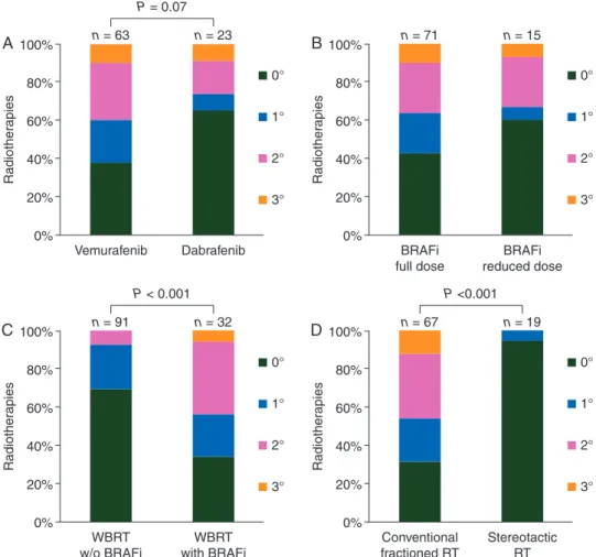
The frequency of radiodermatitis was further analyzed
de-pending on the type of BRAF inhibitor. In patients treated with
vemurafenib (n = 63), acute radiodermatitis
≥°2 occurred in
40%, whereas in the dabrafenib group (n = 23) in only 26%
(P = 0.07) (Figure
2
A). FCPs only appeared in patients taking
vemurafenib. In several patients, the BRAF inhibitor dose was
re-duced precautionary due to the upcoming radiotherapy (n = 5)
or after prior adverse events induced by the BRAF inhibitor
(n = 10). These dose reductions did not reduce
radiation-induced skin toxicity during concomitant treatment compared
with full dosage (P = 0.4) (Figure
2
B).
The largest subgroup of patients treated with radiotherapy
and concomitant BRAF inhibitors received WBRT. These 32
patients were compared with 91 patients treated with WBRT
only. In patients receiving WBRT with concomitant BRAF
in-hibitor therapy acute radiodermatitis
≥°2 according to CTCAE
criteria occurred in 44% of cases compared with 8% of patients
with WBRT only (P < 0.001) (Figure
2
C) [
16
].
Rates of acute radiodermatitis of conventionally fractioned
radiotherapies (n = 67) and stereotactic treatments (n = 19) were
also compared. No increased skin toxicity and no other severe
adverse events were reported after stereotactic radiotherapy with
concomitant BRAF inhibitor therapy (Figure
2
D). In contrast,
acute radiodermatitis
≥°2 was reported in almost every other
patient (46%) who received a conventionally fractioned
radio-therapy with concomitant BRAF inhibitor radio-therapy (P < 0.001).
individual radiosensitivity
ex vivo
Individual radiosensitivity was assessed in peripheral blood
lymphocytes of melanoma patients after ex vivo irradiation.
Three-color FISH was used to analyze the cells
’ ability to
respond to ionizing radiation-induced DNA damage. Misrepair,
0% Vemurafenib n = 63 n = 23 P = 0.07 0° 20% 40% 60% Radiother apies 80% 100%
A
Dabrafenib 1° 2° 3° 0% BRAFi full dose n = 71 n = 15 0° 20% 40% 60% Radiother apies 80% 100%B
BRAFi reduced dose 1° 2° 3° 0% WBRT w/o BRAFi n = 91 n = 32 P < 0.001 0° 20% 40% 60% Radiother apies 80% 100%C
WBRT with BRAFi 1° 2° 3° 0% Conventional fractioned RT n = 67 n = 19 P <0.001 0° 20% 40% 60% Radiother apies 80% 100%D
Stereotactic RT 1° 2° 3°Figure 2. Acute radiodermatitis of patients treated with radiotherapy with concomitant BRAF inhibitor therapy. Acute radiodermatitis of 86 radiotherapies (RT) with concomitant BRAF inhibitor therapy divided in subgroups of BRAF inhibitor (BRAFi) type (A) and BRAF inhibitor dose (B). Acute radiodermatitis after WBRT of 32 patients with and 91 patients without concomitant BRAF inhibitor therapy (C). Acute radiodermatitis of 86 conventionally fractioned or stereotactic radiotherapies with concomitant BRAF inhibitor therapy (D). Grading of skin toxicity according to the Common Terminology Criteria for Adverse Events (CTCAE) version 4.0 (1° Faint erythema or dry desquamation; 2° Moderate to brisk erythema; patchy moist desquamation, mostly confined to skin folds and creases; moderate edema; 3° Moist desquamation in areas other than skin folds and creases; bleeding induced by minor trauma or abrasion; 4° Life-threatening consequences; skin necrosis or ulceration of full thickness dermis; spontaneous bleeding from involved site; skin graft indicated).
impaired signaling and dysfunctional cell cycle control results in
chromosomal aberrations. Color changes along chromosomes
indicate these aberrations (Figure
3
A and B). The chromosomal
aberrations were expressed as mean breaks per metaphase (B/M
value) and were scored in the blood of melanoma patients
without BRAF inhibitor therapy (n = 15), patients taking
vemurafenib (n = 8) or dabrafenib (n = 9) and patients who were
switched from vemurafenib to dabrafenib (n = 3). B/M values
of <0.5 indicate an average radiosensitivity and B/M values
between 0.5 and 0.6 increased radiosensitivity. Patients with B/M
values higher than 0.6 have a clearly increased radiosensitivity
with an increased risk for severe toxicities during radiotherapy
[
17
–
19
]. In the control group, the B/M values of none of the
patients were higher than 0.6 B/M (Figure
3
C). In contrast, 50%
(4/8) of patients under vemurafenib had strongly increased B/M
values. Interestingly, the B/M value was increased only in 11%
(1/9) of patients under dabrafenib. The patient of the dabrafenib
group with the dramatically increased B/M value of 1.0
devel-oped 17 HPV acanthomas and 1 squamous cell carcinoma 3
months after start of therapy with dabrafenib. Patients who were
currently taking dabrafenib and had previously been treated
with vemurafenib, had very high B/M values, even though
vemurafenib treatment was stopped on average 5.2 months
before. Patients under vemurafenib (P = 0.004) and patients
who were switched from vemurafenib to dabrafenib (P = 0.002)
had significantly increased B/M values compared with patients
without BRAF inhibitor therapy. Patients taking vemurafenib
had signi
ficantly higher B/M values than patients under therapy
with dabrafenib (P = 0.04). There was no correlation of B/M
values with BRAF inhibitor dose, dose per body weight or dose
per body mass index. Eight of the patients in which a
radiosensi-tivity testing was carried out were also treated with radiotherapy.
Patients with average B/M values had no skin toxicities, whereas
patients with increased B/M values suffered much more
fre-quently from acute and late skin toxicities
≥2° (Figure
3
D).
discussion
This analysis of a large patient cohort showed an increased rate
of acute radiodermatitis
≥°2 of 36% in patients treated with
radiotherapy and concomitant BRAF inhibitor therapy. Despite
the high rate of acute radiodermatitis, no severe skin-related
late toxicities were reported during an average follow-up time of
6.6 months. FCPs, a characteristic late reaction of concomitant
BRAF inhibitor therapy and WBRT [
9
,
13
], was reported in
13% of our patient cohort. Other reactions like hand
–foot
syn-drome are reported here for the
first time after radiotherapy.
In our patients, these skin reactions were strictly limited to the
irradiated areas. But it has to be considered, that BRAF
inhibi-tors frequently induce follicular dermatitis and hyperkeratosis
without ionizing radiation [
1
–
3
,
20
]. It can be speculated that
some of these adverse events might have also happened without
ionizing radiation. Reports on radiation-induced visceral
reac-tions such as pneumonitis or anorectitis [
7
,
11
] and potentially
liver toxicity exist [
5
]. However, in this patient population,
nonskin toxicity was rare. Another
finding of the study was
that radiation-induced toxicity only appeared in patients, who
received conventionally fractioned radiotherapy with
concomi-tant BRAF inhibitor therapy. No skin or other toxicity appeared
after stereotactic treatment. This is in line with previous case
reports [
15
] and an earlier retrospective analysis of 12 patients
(n = 3 WBRT; n = 3 WBRT + stereotactic boost; n = 6
stereotac-tic radiotherapy) with no reported toxicities except for brain
necrosis in 1 patient [
14
].
0.2 0.4 0.6 Radiosensitivity (B/M) 0.8 P = 0.004 P = 0.04 + P = 0.002 1.0 0.5 0 w/o BRAFi B/M>0.6 0% 50% 11% 100% n = 15 n = 8 n = 9 n = 3Vemurafenib Dabrafenib Dabrafenib after Vemurafenib Distinct increased radiosensitivity Average radiosensitivity
C
A
B
0.2 0.4 0.6 Radiosensitivity (B/M) 0.8 0.5 0 0° 1° 2° 3°Acute or late skin toxicity
D
Figure 3. Individual radiosensitivity testing of melanoma patients with or without BRAF inhibitors ex vivo. Three-color FISH painting of chromo-somes 1 (red), 2 (green) and 4 (yellow). (A) A metaphase without aberra-tions and (B) a metaphase with one dicentric chromosome and two acentric fragments are displayed. The aberrations were scored as 2 breaks per meta-phase (B/M). (C) Lymphocytes were irradiated ex vivo with 2 Gy. B/M found in nonirradiated metaphases were subtracted from those scored in the irra-diated samples. B/M values of patients treated with vemurafenib, dabrafenib and dabrafenib after vemurafenib were compared with melanoma patients without BRAF inhibitor therapy.+The patient with a dramatically increased
B/M value of 1.0 developed 17 HPV acanthomas and one squamous cell car-cinoma 3 months after start of therapy with dabrafenib. (D) Correlation of acute and late skin toxicity of irradiated patients with their B/M values.
So far, it was unclear whether additional toxicity was induced
by BRAF inhibitors and if so, whether this increased toxicity
was mediated by an immunologic boost [
21
] or whether the
effect was direct. To establish the pathogenic mechanism, the
radiosensitivity in patients taking BRAF inhibitors was
investi-gated ex vivo and clearly showed a radiosensitizing effect of
vemurafenib but not of dabrafenib. These ex vivo
findings are in
line with the patient data that also showed a higher rate of acute
radiodermatitis
≥°2 in vemurafenib-treated patients (40%)
com-pared with dabrafenib-treated patients (26%). Interestingly,
photosensitization is almost exclusively reported in
vemurafe-nib-treated patients [
22
]. One might speculate that this is a
con-sequence of the very selective binding af
finity of dabrafenib to
mutant BRAF, whereas vemurafenib also has a low af
finity to
CRAF, wild-type BRAF and possibly other enzymes [
23
].
The radiosensitizing effect of BRAF inhibitors probably also
sensitizes melanoma cells, maybe even to a greater extent than
keratinocytes. In vitro the radiosensitizing effect of BRAF inhibitors
in BRAF-mutated melanoma cells has already been shown [
24
,
25
]. This might enhance the antitumor effect of both
radiother-apy and BRAF inhibitors, which is especially valuable for patients
with multiple brain metastases, when no stereotactic radiotherapy
is possible. Both, whole-brain radiotherapy and BRAF inhibitor
therapy improve cerebral tumor control [
26
–
28
]. Nevertheless,
the prognosis of melanoma patients with multiple brain
metasta-ses is still poor. Synergistic effects of ionizing radiation and BRAF
inhibition within a concomitant treatment regime could improve
the prognosis of these patients.
Whether the BRAF inhibitor therapy should be interrupted
during radiotherapy, has to be discussed in light of these data.
Radiation recall phenomena have been reported up to 1 month
after radiotherapy [
11
–
13
]. Consequently, if maximal safety is
favored, therapy interruption of systemic treatment would last
several weeks and might lead to progression of nonirradiated
metastases. Whereas when radiotherapy is carried out with
con-comitant BRAF inhibitor therapy, systemic tumor control is
maintained. Furthermore, a radiosensitizing effect might
im-prove (local) tumor control. Our data demonstrate that
stereo-tactic radiotherapy with concomitant BRAF inhibitor therapy
does not increase the risk of toxicity. Patients receiving
conven-tionally fractioned radiotherapy with concomitant dabrafenib
have a moderately increased risk of acute radiodermatitis
com-pared with a larger increase in patients taking vemurafenib.
Thus, in patients with planned radiotherapy, the choice of BRAF
inhibitor with respect to toxicity favors dabrafenib. Switching
patients from vemurafenib to dabrafenib before starting
radio-therapy cannot be recommended, as these patients showed the
highest individual radiosensitivity ex vivo. Particularly, patients
under treatment with vemurafenib should be monitored closely
for skin and noncutaneous radiation toxicities and receive early
supportive care, if necessary. Nevertheless, the results of this
ana-lysis show the feasibility of radiotherapy with concomitant BRAF
inhibitor therapy.
disclosure
Seventeen authors report collaborations with different
pharma-ceutical companies, partially with the manufacturers of BRAF
inhibitors, outside the project of this manuscript. All remaining
authors have declared no con
flicts of interest.
references
1. Chapman PB, Hauschild A, Robert C et al. Improved survival with vemurafenib in melanoma with BRAF V600E mutation. N Engl J Med 2011; 364: 2507–2516. 2. McArthur GA, Chapman PB, Robert C et al. Safety and efficacy of vemurafenib in
BRAF(V600E) and BRAF(V600K) mutation-positive melanoma (BRIM-3): extended follow-up of a phase 3, randomised, open-label study. Lancet Oncol 2014; 15: 323–332.
3. Hauschild A, Grob JJ, Demidov LV et al. Dabrafenib in BRAF-mutated metastatic melanoma: a multicentre, open-label, phase 3 randomised controlled trial. Lancet 2012; 380: 358–365.
4. Fonkem E, Uhlmann EJ, Floyd SR et al. Melanoma brain metastasis: overview of current management and emerging targeted therapies. Expert Rev Neurother 2012; 12: 1207–1215.
5. Anker CJ, Ribas A, Grossmann AH et al. Severe liver and skin toxicity after radiation and vemurafenib in metastatic melanoma. J Clin Oncol 2013; 31: e283–e287. 6. Merten R, Hecht M, Haderlein M et al. Increased skin and mucosal toxicity in the
combination of vemurafenib with radiation therapy. Strahlenther Onkol 2014; 190: 1169–1172.
7. Peuvrel L, Ruellan AL, Thillays F et al. Severe radiotherapy-induced extracutaneous toxicity under vemurafenib. Eur J Dermatol 2013; 23: 879–881.
8. Satzger I, Degen A, Asper H et al. Serious skin toxicity with the combination of BRAF inhibitors and radiotherapy. J Clin Oncol 2013; 31: e220–e222. 9. Schulze B, Meissner M, Wolter M, Rödel C, Weiss C. Unusual acute and delayed
skin reactions during and after whole-brain radiotherapy in combination with the BRAF inhibitor vemurafenib. Two case reports. Strahlenther Onkol 2014; 190: 229–232.
10. Harding JJ, Barker CA, Carvajal RD et al. Cutis verticis gyrata in association with vemurafenib and whole-brain radiotherapy. J Clin Oncol 2014; 32: e54–e56. 11. Forschner A, Zips D, Schraml C et al. Radiation recall dermatitis and radiation
pneumonitis during treatment with vemurafenib. Melanoma Res 2014; 24: 512–516. 12. Reigneau M, Granel-Brocard F, Geoffrois L et al. Efflorescence of scalp cysts during vemurafenib treatment following brain radiation therapy: a radiation recall dermatitis? Eur J Dermatol 2013; 23: 544–545.
13. Lang N, Sterzing F, Enk AH, Hassel JC. Cutis verticis gyrata-like skin toxicity during treatment of melanoma patients with the BRAF inhibitor vemurafenib after whole-brain radiotherapy is a consequence of the development of multiple follicular cysts and milia. Strahlenther Onkol 2014; 190: 1080–1081.
14. Narayana A, Mathew M, Tam M et al. Vemurafenib and radiation therapy in melanoma brain metastases. J Neurooncol 2013; 113: 411–416.
15. Rompoti N, Schilling B, Livingstone E et al. Combination of BRAF inhibitors and brain radiotherapy in patients with metastatic melanoma shows minimal acute toxicity. J Clin Oncol 2013; 31: 3844–3845.
16. Institute NC. Common Terminology Criteria for Adverse Events (CTCAE) version 4.0. 2009. http://evs.nci.nih.gov/ftp1/CTCAE/CTCAE_4.03_2010-06-14_Quick Reference_5x7.pdf(19 March 2015, date last accessed).
17. Keller U, Grabenbauer G, Kuechler A, Sauer R, Distel L. Technical report. Radiation sensitivity testing byfluorescence in-situ hybridization: how many metaphases have to be analysed? Int J Radiat Biol 2004; 80: 615–620.
18. Keller U, Grabenbauer G, Kuechler A et al. Cytogenetic instability in young patients with multiple primary cancers. Cancer Genet Cytogenet 2005; 157: 25–32. 19. Distel LV, Neubauer S, Keller U et al. Individual differences in chromosomal
aberrations after in vitro irradiation of cells from healthy individuals, cancer and cancer susceptibility syndrome patients. Radiother Oncol 2006; 81: 257–263. 20. Hauschild A, Grobb J, Demidov LV et al. An update on overall survival (OS) and
follow-on therapies in BREAK-3, a phase III, randomized trial: dabrafenib (D) vs. dacarbazine (DTIC) in patients ( pts) with BRAF V600E mutation-positive metastatic melanoma (MM). Ann Oncol 2014; 25: iv374–iv393.
21. Ridolfi L, de Rosa F, Ridolfi R et al. Radiotherapy as an immunological booster in patients with metastatic melanoma or renal cell carcinoma treated with high-dose interleukin-2: evaluation of biomarkers of immunologic and therapeutic response. J Transl Med 2014; 12: 262.
22. Mattei PL, Alora-Palli MB, Kraft S et al. Cutaneous effects of BRAF inhibitor therapy: a case series. Ann Oncol 2013; 24: 530–537.
23. Menzies AM, Kefford RF, Long GV. Paradoxical oncogenesis: are all BRAF inhibitors equal? Pigment Cell Melanoma Res 2013; 26: 611–615.
24. Dasgupta T, Haas-Kogan DA, Yang X et al. Genotype-dependent cooperation of ionizing radiation with BRAF inhibition in BRAF V600E-mutated carcinomas. Invest New Drugs 2013; 31: 1136–1141.
25. Sambade MJ, Peters EC, Thomas NE et al. Melanoma cells show a heterogeneous range of sensitivity to ionizing radiation and are radiosensitized by inhibition of B-RAF with PLX-4032. Radiother Oncol 2011; 98: 394–399.
26. Dummer R, Goldinger SM, Turtschi CP et al. Vemurafenib in patients with BRAF (V600) mutation-positive melanoma with symptomatic brain metastases: final results of an open-label pilot study. Eur J Cancer 2014; 50: 611–621. 27. Long GV, Trefzer U, Davies MA et al. Dabrafenib in patients with Val600Glu or
Val600Lys BRAF-mutant melanoma metastatic to the brain (BREAK-MB): a multicentre, open-label, phase 2 trial. Lancet Oncol 2012; 13: 1087–1095. 28. Mornex F, Thomas L, Mohr P et al. A prospective randomized multicentre phase III
trial of fotemustine plus whole brain irradiation versus fotemustine alone in cerebral metastases of malignant melanoma. Melanoma Res 2003; 13: 97–103.
Annals of Oncology 26: 1244–1248, 2015 doi:10.1093/annonc/mdv129 Published online 9 March 2015
The impact of docetaxel-related toxicities
on health-related quality of life in patients
with metastatic cancer (QoliTax)
S.-E. Al-Batran
1*, W. Hozaeel
1, F. K. Tauchert
2, R.-D. Hofheinz
3, A. Hinke
4, C.
Windemuth-Kieselbach
5, A. Hübner
6, M. Burmester
7, M. Koenigsmann
8, J. Wiegand
9, G. zur Hausen
1,
B. Linsse
10, R. Kuhl
11& C. Pauligk
1for the Arbeitsgemeinschaft Internistische Onkologie (AIO)
1
Institute of Clinical Cancer Research, Krankenhaus Nordwest, University Cancer Center, Frankfurt;2
Clinic for Oncology and Hematology, Krankenhaus Nordwest, Frankfurt;3
Interdisciplinary Tumour Centre, University Hospital Mannheim, Mannheim;4
WiSP Research Institute, Langenfeld;5
Alcedis GmbH, Gießen;6
Oncology and Urology, Wissenschaftskontor Nord GmbH & Co KG, Rostock;7
Clinic for Urology, Vinzenzkrankenhaus Hannover, Hannover;8
Oncology and Hematology, Onkologisches Ambulanzzentrum, Hannover;9
Oncology and Hematology, Onkologische Praxis, Moers;10
Clinical Study Unit;11
Medical Oncology, Sanofi-Aventis Deutschland GmbH, Berlin, Germany
Received 11 August 2014; revised 19 January 2015; accepted 18 February 2015
Background:
Docetaxel is a widely used cytotoxic agent. This study evaluates the impact of docetaxel toxicities on patient’s health-related quality of life (QoL).Patients and methods:
We conducted a multicenter, prospective, non-interventional trial, in which the QoL was assessed using the EORTC QLQ-C30 questionnaires at baseline and every 4 weeks up to 40 weeks in patients receiving a docetaxel-based chemotherapy for metastatic disease. Treatment-related adverse events were correlated with the cor-responding QoL scores. Uni- and multivariate analyses were applied.Results:
From January 2008 to June 2011, a total of 2659 patients were included. The majority of patients (48.1%) had prostate cancer, followed by breast (17.1%) and non-small-cell-lung cancer (15.8%). Patients received a median of 5 docetaxel cycles with the median dose of 75 mg/m2. The presence of grade 3/4 diarrhea showed the strongest effect on global health status/QoL average scores (50.91 versus 33.06), followed by vomiting (50.91 versus 35.17), dyspnea (50.94 versus 35.81), mucositis/stomatitis (50.88 versus 36.41), nausea (50.91 versus 36.68), infection (50.90 versus 37.14), fatigue (50.90 versus 43.82) and anemia (50.91 versus 41.03), P < 0.05 for all comparisons. Grade 3/4 leuko-penia/neutropenia, alopecia, constipation, neurotoxicity and nail disorders had no significant impact on the global health status/QoL or other items.Conclusion:
In this large non-interventional trial, docetaxel-associated grade 3 or 4 toxicities were shown to have a strong detrimental effect on patient’s QoL. Notably, diarrhea and vomiting had the strongest negative impact on QoL measures. This has to be kept in mind while making therapeutic decisions and providing optimized supportive treatment measures.*Correspondence to: Prof. med. Salah-Eddin Al-Batran, Krankenhaus Nordwest, UC—University Cancer Center Frankfurt, Steinbacher Hohl 2-26, 60488 Frankfurt am Main, Germany. Tel: +49-69-76-01-44-20; Fax: +49-69-76-01-36-55; E-mail: albatran@ aol.com
© The Author 2015. Published by Oxford University Press on behalf of the European Society for Medical Oncology. All rights reserved. For permissions, please email: journals.permissions@oup.com.