JID 1991:164 (September) Correspondence 615
References
I. Gonzalez -Rui z A,Ruiz-Palacios G.Entamoeba hartmanni:missingor misidentified?JInfect Dis1991;164:612-3 .
2. del Muro R, Acosta E,Merino E,GlenderW, Ortiz-OrtizL. Diagnosisof intestinal amebiasis using salivary IgA antibody detection.J Infect Dis 1990; 162:1360-4.
3. Begg CB. Biases in the assessment of diagnostic tests. Stat Med 1987;6:411-23.
4. Gonzalez-Ruiz A. Revision del estado actual del diagnosticodiferencial delas amibas en Mexico. Salud Publica Mex1990:32:589-96.
Primary Human Immunodeficiency Virus Infection
Mimicking Syphilis
Colleagues-Primary exposure to the human immunodefi-ciency virus (HIV) may lead to an acute infectious disease with skin involvement [1-4). In the first review, in 1985 [I), a rash of a mononucleosis type predominantly located on the trunk was present in 50% of patients and seemed characteristic although nonspecific.We report a case with unusual dermatologic mani-festations that were initially mistaken for syphilis.
In September 1990, a 39-year-old homosexual man was re-ferred to us with the diagnosis of secondary syphilis. He pre-sented with a 7-day history of three genital ulcers. A high fever began 5 days later, associated with arthralgias, odynophagia, and a nonpruritic skin rash. Further inquiry disclosed unpro-tected orogenital sex with an unknown partner 17 and 32 days before.
Physical examination revealed a patient in good general con-dition, febrile at 39.5°C,with a widespread symmetric purplish papular eruption in a linear distributionon his face,trunk, and proximal part of the four limbs and palms. Numerous painful oval-shaped ulcers up to I em in diameter were seen on the oropharyngeal mucosa, including the tongue. On the penis, an oblong, nonindurated, painful chancre of 2.5 X I em with a clean base was surrounded by two similar smaller ulcerations. Serous exudate was not obtained when pressure was applied. The scrotal area was also covered by many shallow oval-shaped erosions. Generalized lymphadenopathy was present, including the antecubital and epitrochlear areas. No hepatomegaly was noted, but a slightly enlarged spleen was felt.The rest of the clinical examination was unremarkable.
Findings from laboratory evaluation revealed normal values for sedimentation rate, hemoglobin, white blood count, and bio-chemical profile. Platelet count was 119,000/mm3
•
Repeated darkfield examinations for Treponema pallidum, di-rect examination and culture for Haemophilus ducreyi, and im-munofluorescence staining for herpesviruses (simplex and zos-ter) of the genital ulcer's base were all negative. Serum VDRL
Reprints or correspondence: Dr. A. M.Calza, Clinique de Dermatologie, Hopital Cantonal Universitaire,1211 Geneve 4, Switzerland.
The Journal of Infectious Diseases 1991;164:615-6 ©1991 by The University of Chicago. All rights reserved. 0022-1 899/9 I{6403-0036$0 I.00
5. Aceti A, Pennica A, Celestino D. et al. Salivary IgA antibody detection in invasive amebiasis and in asymptomatic infection.J Infect Dis 1991:164:613-4.
6. MesteckyJ.McGheeJR.Arnold RR, MichalekSM, Prince SJ. Babb JL. Selective induction of an immune response in human external secre-tions by ingestion of bacterial antigen.JClin Invest 1978;61:731-7. 7. Czerkinsky C, Prince SJ,Michalek SM. etal. IgA antibody producing
cells in peripheral blood after antigen ingestion:evidence of a com-mon mucosal immune system in humans.Proc Nat! Acad Sci USA 1987;84:2449-53.
test, fluorescent treponemal antibody test, and Treponema pal/i-dum hemagglutination assay done on several occasions re-mained unreactive. Serologic evaluations for cytomegalovirus. Toxoplasma gondii,Epstein-Barrvirus,rubella, and hepatitisA (IgM) and hepatitis B (antigen) were negative. On the day of referral, search for anti-HlV antibody by screening ELISA and immunoblotting tests was unsuccessful. In contrast, HIV p24 antigen (HIV AG-I; Abbott, North Chicago) was detected in the serum at 39 ng/1. Lymphocyte count was 1113/mmJ
, with a
CD4 count of 323/mm3
and CD8 count of 545/mm3 •
Major features of histologic examination were edema and a perivascular and perifollicular lymphocytic infiltrate in the der-mis. There was minimal basal and suprabasal spongiosis, with lymphocytic exocytosis and rare necrotic keratinocytes in the epidermis.On electron microscopy,cytoplasmic reticulotubular structures were present in most lymphocytes and histiocytes and in rare keratinocytes and Langerhans cells. There was no evi-dence of viral particles either assembling or budding.
The patient's illness resolved spontaneously within 2 weeks. Return to normal body temperature occurred on day 3 and
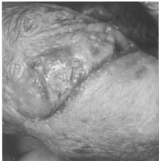
reso-Figure 1. Genital ulcersin a patient with human immunodeficiency virus infection.
616 Correspondence JlD 1991; 164 (September)
lution of the skin rash on day 7 and of the erosive lesions on day 15. HIV antibodies appeared on day 7 and coincided with the disappearance of p24 antigenemia on day 8.
Previous reports have shown that an exanthem, located pre-dominantly on the face and trunk, is probably the most common skin manifestation of primary HIV infection [1-4]. Several re-ports have mentioned oral, esophageal, or genital erosive lesions [3,5). In very rare cases [6-9], concurrent oral and genital ulcers have been documented, but they have often been considered a minor diagnostic feature because of their limited extent.
Owing to the presence of general symptoms associated with mucosal erosive lesions and a skin rash, all suggestiveofsyphilis, the initial clinical picture was misleading [10). The incubation period, morphology, and location of the penile chancre (figure I) were all compatible with primary syphilis, although several features such as the multiplicity of lesions and the absence of induration made this diagnosis unlikely. The papular eruption, which involved the palms as well, could easily have been mis-taken for secondary syphilis, particularly as contamination seemed to have occurred via the orogenital route, unusual for HIV. The diagnosis of primary HIV infection in our case was, however, confirmed by successive serologic investigations.
This case reminds us that primary HIV infection must be in-cluded in the differential diagnosis of genital ulcers, particularly if associated with a skin rash and oral lesions. The use of p24 antigen is useful in this instance. Because of the clinical similari-ties with other sexually transmitted diseases such as herpes, syph-ilis, or H. ducreyi infection, these should be ruled out as well.
Anne-Marie Calza, Sabine Kinloch, Carlo Mainetti, Denis Salomon, and Jean-Hilaire Saurat
Unusually High Level of a Tumor-Associated Antigen in the Serum of Human Immunodeficiency
Virus-Seropositive Individuals
Colleagues-A 90,000-Da molecular mass lipoprotein (90K) recognized by monoclonal antibody Sp-2 [I] has recently been identified in the sera of patients with breast or ovarian cancer or other malignant diseases [2-4]. The 90K, which is now being
biochemically characterized, has an NH2-terminalamino acid
sequence that does not resemble any protein for which sequence information is available (unpublished data). In preparation for a multicenter study on 90K as a prognostic indicator in a broad range of malignancies, we have obtained serum samples from three patients with human immunodeficiency virus (HIV)-re-lated Kaposi's sarcoma. We found that these sera had the high-est 90K-immunoreactive activity ever recorded in our labora-tory. This observation prompted us to investigate the role of this antigen in HIV infection.
Reprints or correspondence: Dr. S. Iacobelli, Cattedra Oncologia Medica, Universita "G. D'Annunzio," Via dei Vestini 5, 66100 Chieti, Italy. The Journal of Infectious Diseases 1991;164:616-7
© 1991 by The University of Chicago. All rights reserved. 0022-1899/91/6403-0037$01.00
Clinic ofDermatology. Hiipital Cantonal Universitaire, Geneva, Switzerland
References
I. Cooper DA. Gold J. Maclean P, et al. Acute AIDS retrovirus infection: definition of clinical illness associated with seroconversion. Lancet 1985;1:537-40.
2. Kessler HA. Blaauw B, Spear J, Deborah AP, Falk LA, LandayA. Diagnosis of human immunodeficiency virus infection in seronega-tive homosexuals presenting with an acute viral syndrome. JAMA 1987;258:1196-9.
3. Tindall B, Barker S. Donovan B. et al. Characterization of the acute clinical illness associated with human immunodeficiency virus infec-tion. Arch Intern Med 1988; 148:945-9.
4. Gaines G. von Sydow M, Pehrson PO, Lundbergh P. Clinical picture of primary HIV infection presenting as a glandular-fever-like illness. BMJ 1988;297: 1363-8.
5. Hulsebosch HJ. Claessen FAP, van Ginkel CJW. Kuiters GRR. Goud-smit J, Lange JMA. Human immunodeficiency exanthem. J Am Acad Dermatol 1990;23:483-6.
6. Bratzke B. Orfanos CEo AkutesPrirnarstadiumeiner HIV infection und Ubergang in AIDS mit Kaposi 24 Monate danach. Hautartzt 1988;39:514-8.
7. Picard C.Oberlin P, Lazareth B, Crickx B. Belaich S. Primo-infection par Ie virus de l'immunodeficience humaine. Ann Dermatol Vener-eoI1990;117:861-4.
8. Biggar RJ, Johnson BK. Musoke SS, et al. Severe illness associated with appearance of antibody to human immunodeficiency virus in an African. BMJ 1986;293:1210-1.
9. Bratzke B, Eichhorn R. Hoffken G, et al. Akute Primarphase als Indika-tor der HIV -l-Infektion. Dtsch Med Wochenschr 1988; 113: 1312-6. 10. Thin RT. Early syphilis in the adult. In: Holmes KK. Sexually
transmit-ted diseases. New York: McGraw-Hili. 1990:221-30.
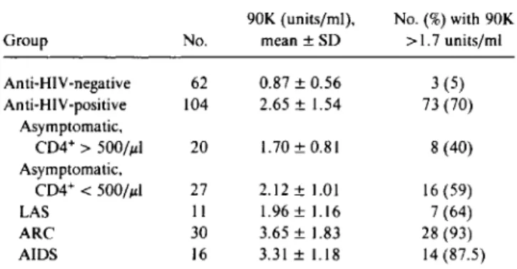
Sera were obtained from 104 anti-HIV-positive subjects: 20 were symptom-free and without decreases in CD4+ lymphocyte number, 27 were also asymptomatic but had decreases in CD4+
lymphocytes«500/ILI), II had lymphadenopathy syndrome,
30 had AIDS-related complex, and 16 had frank AIDS. Control sera were obtained from 62 anti-HI V-negative blood donors
Table 1. Levelsof90-kDa protein (90K) in human
immunodefi-ciency virus (HIV)-seronegative and -seropositive subjects. 90K (units/ml), No.(%)with 90K
Group No. mean ±SO >1.7 units/rnl
Anti-Hlv-negative 62 0.87±0.56 3 (5) Anti-HIV-positive 104 2.65 ±1.54 73 (70) Asymptomatic. CD4+>500//J.I 20 1.70±0.81 8 (40) Asymptomatic, CD4+<500//J.I 27 2.12 ± 1.01 16 (59) LAS II 1.96± 1.16 7 (64) ARC 30 3.65± 1.83 28 (93) AIDS 16 3.31 ±1.18 14 (87.5)
NOTE. LAS=lymphadenopathy syndrome; ARC=AIDS-related complex. All means for anti-HI V-positive subjects and subgroups were significantlygreater than those for anti-HI V-negative subjects(P<.0001, analysis of variance).