HAL Id: hal-02631565
https://hal.inrae.fr/hal-02631565
Submitted on 27 May 2020HAL is a multi-disciplinary open access archive for the deposit and dissemination of sci-entific research documents, whether they are pub-lished or not. The documents may come from teaching and research institutions in France or abroad, or from public or private research centers.
L’archive ouverte pluridisciplinaire HAL, est destinée au dépôt et à la diffusion de documents scientifiques de niveau recherche, publiés ou non, émanant des établissements d’enseignement et de recherche français ou étrangers, des laboratoires publics ou privés.
Coccomorpha, Pseudococcidae) on persimmon fruit
trees (Diospyros kaki) in southern Brazil
Vitor C. Pacheco da Silva, Mehmet Bora Kaydan, Jean-Francois Germain,
Thibaut Malausa, Marcos Botton
To cite this version:
Vitor C. Pacheco da Silva, Mehmet Bora Kaydan, Jean-Francois Germain, Thibaut Malausa, Marcos Botton. Three new species of mealybug (Hemiptera, Coccomorpha, Pseudococcidae) on persimmon fruit trees (Diospyros kaki) in southern Brazil. Zookeys, Pensoft, 2016, pp.61-82. �10.3897/zookeys.584.8065�. �hal-02631565�
Three new species of mealybug (Hemiptera,
Coccomorpha, Pseudococcidae) on persimmon
fruit trees (Diospyros kaki) in southern Brazil
Vitor C. Pacheco da Silva1, Mehmet Bora Kaydan2, Jean-François Germain3,Thibaut Malausa4, Marcos Botton5
1 Plant Protection Graduate Program, Plant Protection Department, UFPel, Campus Universitário Capão
do Leão s/n, Capão do Leão, Rio Grande do Sul, Brazil 2 Imamoglu Vocational School, Çukurova University, 01330, Adana, Turkey 3 Laboratoire de la Santé des Végétaux, Unité d’entomologie et plantes invasives, CBGP 755 avenue du campus Agropolis, CS30016, 34988 Montferrier-sur-Lez, Languedoc-Roussillon, France
4 Institut Sophia Agrobiotech, UMR INRA, Université Nice Sophia Antipolis, CNRS, 400 Route des chappes,
Sophia Antipolis, PACA, France 5 Embrapa Grape and Wine, 515 Rua do Livramento, Bento Gonçalves, Rio Grande do Sul, Brazil
Corresponding author: Vitor C Pacheco da Silva (vitorcezar@gmail.com)
Academic editor: R. Blackman | Received 7 February 2016 | Accepted 19 March 2016 | Published 25 April 2016
http://zoobank.org/9C7E2192-3D64-455A-89EA-B6BF4B4C9CB8
Citation: Pacheco da Silva VC, Kaydan MB, Germain J-F, Malausa T, Botton M (2016) Three new species of mealybug (Hemiptera, Coccomorpha, Pseudococcidae) on persimmon fruit trees (Diospyros kaki) in southern Brazil. ZooKeys 584: 61–82. doi: 10.3897/zookeys.584.8065
Abstract
Brazil has the greatest insect diversity in the world; however, little is known about its scale insect species (Hemiptera: Coccomorpha). Mealybugs (Pseudococcidae) have been found in at least 50% of persimmon orchards Diospyros kaki L. in the southern part of the country. In this study three new mealybug species on persimmon trees located in the Serra Gaúcha Region, RS, Brazil, namely, Anisococcus granarae Pacheco da Silva & Kaydan, sp. n., Ferrisia kaki Kaydan & Pacheco da Silva, sp. n. and Pseudococcus rosangelae Pacheco da Silva & Kaydan, sp. n. are described. In addition, an identification key for the genera occur-ring on fruit orchards and vineyards in Brazil is provided, together with illustrations and molecular data for the new species.
Keywords
Distribution, Neotropical Region, scale insects, taxonomy
http://zookeys.pensoft.net
Copyright Vitor C. Pacheco da Silva et al. This is an open access article distributed under the terms of the Creative Commons Attribution License (CC BY 4.0), which permits unrestricted use, distribution, and reproduction in any medium, provided the original author and source are credited.
Introduction
Southern Brazil is the third largest fruit-producing region in the country. It produces large amounts of temperate fruits, such as grape, apple, stone fruits and persimmon (Fachinello et al. 2011). Persimmon trees (Diospyros kaki L.) (Ebenaceae) were first cultivated in Brazil in the late 19th century, but this crop expanded only after Japanese
immigration, around 1920 (Neuwald et al. 2009). Persimmon is currently grown on about 9,000 ha and about 172,000 tons of fruits are produced annually, for domestic consumption and export (Fachinello et al. 2011). The São Paulo and Rio Grande do Sul states are the main producers of persimmon fruits in Brazil (Camargo-Filho et al. 2003). In Rio Grande do Sul, fruit production occurs mostly in the Serra Gaúcha Region, in which mealybugs (Hemiptera: Coccomorpha: Pseudococcidae) have been detected in at least 50% of production areas (Bavaresco et al. 2005), probably due to increases in insecticide application in recent years, leading to a decrease in the popula-tion of effective natural enemies.
Ten mealybug species have been recorded in association with persimmon trees worldwide: Dysmicoccus brevipes (Cockerell), Hippeococcus wegneri Reyne, Maconellico-ccus hirsutus (Green), PhenacoMaconellico-ccus aceris (Signoret), Ph. pergandei Cockerell, Planococ-cus citri (Risso), Pl. kraunhiae (Kuwana), PseudococPlanococ-cus cryptus Hempel, Ps. longispinus Targioni Tozzetti and Ps. viburni (Signoret) (García et al. 2016).
Live mealybugs are small soft-bodied, sap-sucking insects with an oval, elongated to rounded body, often dorsoventrally compressed, pinkish to grayish in color, covered with a white powdery wax (the source of their common name) (Cox and Pearce 1983). They frequently have waxy filaments, those on the head being shorter than those close to the anus (Gimpel and Miller 1996). These filaments originate from the cerarii – groups of trilocular pores, generally with two conical setae and, in some groups, also auxiliary setae (Williams and Granara de Willink 1992) predominantly found along the margin. The family to which mealybugs belong is the second largest family in infraorder Coccomorpha, in terms of the number of species it contains, almost 2020 species, distributed in 260 genera (García et al. 2016). In the Neotropical Region, only 223 species, from 44 genera, have been recorded (García et al. 2016).
Pseudococcidae can be divided into two subfamilies: Pseudococcinae, character-ized by the presence of: (i) apically knobbed tarsal digitules; (ii) claws without a denti-cle; (iii) antennae generally with eight or fewer segments; (iv) anal ring with setose-like spinules; and (v) absence of quinquelocular pores; and Phenacoccinae, characterized by: (i) setose tarsal digitules; (ii) claws with a denticle; (iii) antennae usually nine-seg-mented; (iv) anal ring with dome-shaped spinules on the outer ring; and (v) presence of quinquelocular pores (Hardy et al. 2008; Kaydan et al. 2015).
In total, 153 species of Pseudococcus (Westwood) have been identified worldwide, 30 of which have been recorded in the Neotropical Region. It can be subdivided into two informal groups according to the presence or absence of simple pores associated with each eye (Gimpel and Miller 1996) — present in Pseudococcus maritimus complex.
It includes 33 species, 21 of which are present in the Neotropical Region (García et al. 2016), and is assumed to have originated from the New World, where some species, such as P. sociabilis Hambleton, P. viburni (Signoret) and P. maritimus (Ehrhorn), are considered to be major pests of fruit crops and vineyards (Casco Mila 2012; Correa et al. 2012; Daane et al. 2012; Pacheco da Silva et al. 2014).
The genus Ferrisia Fullaway, which is of New World origin, includes 18 species, most (12 species) of which occur in the Neotropics (Kaydan and Gullan 2012). The species from this genus are easily separated from the other genera in the Pseudococ-cidae by the presence of robust dorsal enlarged tubular ducts opening to the exterior via an irregularly circular sclerotized area bearing one or more setae and, often, one or more minute pores (Gullan et al. 2010). Furthermore, the living insects have long glassy filaments produced by the enlarged tubular ducts, and, depending on the spe-cies, may have typical dorsal patterns formed by dark areas of cuticle not covered by white wax (Kaydan and Gullan 2012).
The genus Anisococcus Ferris is also believed to have originated from New World and to be closely related to Ferrisia on the basis of both molecular phylogenetic stud-ies (Downie and Gullan 2004; Hardy et al. 2008; Gullan et al. 2010) and morpho-logical studies, as it has minute discoidal pores associated with enlarged tubular ducts and oral collar tubular ducts (Kaydan and Gullan 2012). This genus is found exclu-sively in the Americas, where 11 species have been described, nine in the Nearctic region and two from the Neotropical Region (McKenzie 1967; Williams and Granara de Willink 1992).
Brazil has the greatest biodiversity of any country worldwide and 13% of all spe-cies (including animals, plants, fungi and other organisms) are found only in Brazil (Lewinsohn and Prado 2005). Insect diversity is also greater in Brazil than in any other country, with almost 100 thousand species recorded (almost 10% of all insect species worldwide) (Rafael et al. 2009). It has been estimated that almost 11% of hemipteran insects are present in Brazil. However, only 3.8% of mealybug species have been re-corded in this country, although this should probably be regarded as an underestimate of the percentage actually present.
In Brazil, 530 species, from 20 families of the infraorder Coccomorpha have been recorded (García et al. 2016). In total, 78 species from 21 genera of Pseudococcidae have been detected in Brazil. The most numerous genera are Dysmicoccus Ferris (13) and Pseudococcus (13), followed by Phenacoccus Cockerell (10), Nipaecoccus Sulc (8), Planococcus (Ferris) (5) and Ferrisia (5) (García et al. 2016). Only one Anisococcus species, A. parasitus Williams and Granara de Willink, has been recorded from Brazil (Williams and Granara de Willink 1992).
Three new species of mealybugs sampled from persimmon orchards located in the Serra Gaúcha Region, Rio Grande do Sul, Brazil are described, and an identification key for the genera occurring in fruit orchards and vineyards in Brazil is provided, to-gether with illustrations, molecular data and an identification key for the new species of Anisococcus, Ferrisia and Pseudococcus described here.
Methods
Mealybugs were collected from persimmon orchards during the harvest period in the years 2013–2015. Specimens were collected on fruits and leaves of the trees. Insects at all stages of development were collected (nymphs and adult females) and taken to the laboratory for examination. Nymphs were reared until adulthood on persimmon fruits. Labeled specimens were stored in 95% ethyl alcohol.
Molecular characterization
DNA characterization was performed using the nondestructive method described in Malausa et al. (2011). The DNA region studied was a ~ 760 bp fragment within the mitochondrial region of Cytochrome Oxidase Subunit I previously used in molecular studies on mealybugs (Malausa et al. 2011; Pacheco da Silva et al. 2014). DNA was extracted using the Qiagen DNEasy Tissue kit, following the manufacturer’s recom-mendations. For amplification were used the primers 5’ CCTTCAACTAATCAT-AAAAATATYAG 3’ (Forward) and 5’ TAAACTTCTGGATGTCCAAAAAATCA 3’ (Reverse) PCR was performed using the Qiagen Multiplex PCR kit (QIAGEN, Valen-cia, CA), with a 23 mL reaction mixture and 2 ml of diluted DNA (1–20 ng of DNA matrix). PCR conditions were as follows: initial denaturation at 95°C for 15 mn; 35 cycles of denaturation at 95°C for 30 s, hybridization at 48°C for 90 s, elongation for 60 s; and final extension at 72°C for 10 mn. PCR-amplified fragments were analyzed with the QIAxcel Advanced System with QIAxcel DNA Fast Analysis cartridges (QIAGEN). PCR products were sent to Beckman Genomics (Takeley, United Kingdom) for bidirec-tional sequencing on ABI automatic sequencers (Applied Biosystems, Foster City, CA, USA). Consensus sequences and alignments were generated and checked with Bioedit version 7.01. We carried out BLAST searches (MEGABLAST method) on the NCBI GenBank database (http://www.ncbi.nlm.gov/BLAST).
The DNA results are shown for each species after the morphological descriptions. Additionally, all sequence data are available as Suppl. material 1 (FASTA format). Morphological identification
The DNA voucher specimens plus other preserved adult females were slide-mounted and identified by light microscopy in the Plant Protection Department of Çukurova University, Adana, Turkey and ANSES, Laboratoire de la Santé des Végétaux, Montferri-er-sur-Lez, France, according to a slightly modified version of the method of Kosztarab and Kozár (1988). The mealybugs were examined under a LEICA DM 2500 phase-contrast compound microscope and identified with the keys of Williams and Granara de Willink (1992), Gimpel and Miller (1996) and Kaydan and Gullan (2012).
The slides are stored in the Coccoidea Collection of the Museum Ramiro Gomes Costa (MRGC), Porto Alegre, Brazil (Holotype and some paratypes), Çukurova Uni-versity Coccomorpha collection, Adana, Turkey (KPTC) and Anses, Laboratoire de la Santé des Végétaux, Montferrier-sur-Lez, France (ANSES/LSV).
Morphometric analysis
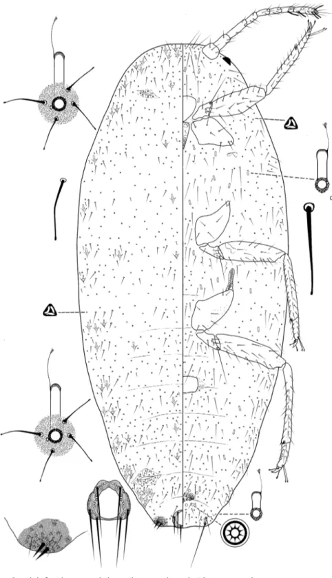
Mealybugs were measured and the main taxonomic characters evaluated and quan-tified under the Leica microscope. Measurements were taken from all the available material. The morphological terms used here are those used by Williams (2004) and Williams and Granara de Willink (1992). All the measurements given are the maxi-mum dimensions (e.g. body width was recorded at the widest part) and are expressed as ranges. Tarsal length excludes the claw. Setal length includes the setal base. Cerarii are numbered as described by Williams and Granara de Willink (1992), with cer-arius 1 on the head, anterior to the antenna, and cercer-arius 17 being on segment VIII. Illustrations are provided for each species. Each figure represents a generalized individual based on several of the specimens used for description. Each illustration is split longitudinally, with the left half representing the dorsum and the right half the venter. Structural details are shown as enlargements around the central draw-ing, and are not drawn to the same scale. The translucent pores on the hind legs are mostly found on the dorsal surface, but they are illustrated ventrally on the main figure for convenience. The illustrations and description were prepared by MBK and VCPS.
Results and discussion
Key to identification of Pseudococcinae genera occurring on fruit trees and in vineyards in Brazil, adapted from Williams (2004) and Williams and Granada de Willink (1992). 1 Dorsal tubular ducts large, each with an orifice surrounded by a round, scle-rotized area containing 1 or more setae within its borders, or with the setae adjacent to the rim ... Ferrisia Fullaway – Dorsal tubular ducts, if present, without this combination of characters ...2 2 Dorsal tubular ducts each with a small adventitious pore or cell adjoining the
main orifice; anal lobe cerarii each with 7-20 conical setae on a sclerotized area; multilocular disc pores always absent ...Anisococcus Ferris – Not with this combination of characters; if there are any pores next to tubular
ducts, then each anal lobe cerarius usually with only 2 conical setae; multi-locular disc pores present or absent ...3 3 Oral rim tubular ducts present somewhere on the body ...4
– Oral rim tubular ducts absent ...6 4 Dorsal surface with setae on posterior segments at least, each broadly lanceo-late or conical in shape, sometimes subequal in size and shape to posterior cerarian setae... Nipaecoccus Sulc – Dorsal surface with all setae flagellate, normally much thinner than cerarian
setae ...5 5 Cerarii anterior to the anal lobe pair mostly with auxiliary setae; with 12–17
distinct pairs of marginal cerarii ... Pseudococcus Westwood – Cerarii anterior to the anal lobe pair without auxiliary setae; with 1–7 distinct
pairs of marginal cerarii ... Maconellicoccus Ezzat 6 With 18 distinct pairs of marginal cerarii; anal lobe bars present ... ... Planococcus Ferris – With 6 to 17 pairs pairs of marginal cerarii; anal lobe bars present or absent .... ... Dysmicoccus Ferris Genus Anisococcus Ferris
Anisococcus Ferris, 1950
Type species. Dactylopius crawii Coquillet by original designation.
Generic diagnosis (adapted from Williams and Granara de Willink 1992; Mc-Kenzie 1967). Body narrowly to broadly oval, 2.0–3.8 mm long, 1.1–2.8 mm wide. Labium with three segments, about as long as the clypeolabral shield. Antennae, 8-seg-mented. Circulus present or absent. Legs well-developed, without translucent pores; apparently with a small denticle on the claw. Both ostioles well developed. Anal lobes well developed. Anal ring rounded, usually large and cellular with six long setae, but sometimes reduced, non-cellular, more or less removed from the posterior apex of the abdomen (Anisococcus ephedrae (Coquillett)).
Dorsum. Dorsal tubular ducts with or without a rim, each orifice associated with one or more minute discoidal pores. Cerarii 13–17 pairs. Anal lobe cerarii, each with 7–20 conical setae on a sclerotized area, often with 3–7 auxiliary setae, remaining cer-arii smaller, each with two or more conical setae plus an associated cluster of trilocular pores. Preocular cerarius always absent. Dorsal setae, slender and flagellate. Trilocular pores evenly distributed. Discoidal pores scattered and associated with tubular ducts, each smaller than trilocular pores. Multilocular disc pores absent.
Venter. Body setae flagellate. Trilocular pores evenly distributed. Discoidal pores scattered or associated with tubular ducts. Multilocular disc pores absent. Oral collar tubular ducts of one or more sizes, of various lengths and widths, with largest ducts, when present, on body margin, often associated with minute discoidal pores.
Key to adult females of Anisococcus found in the Neotropical Region (adapted from Williams and Granada de Willink (1992)).
1 Dorsal oral collar tubular ducts of one size, all large, each about twice the diameter of a trilocular pore, always with a rim ... ...A. milleri Williams & Granara de Willink – Dorsal oral collar tubular ducts of two sizes, the large ducts with a rim, small-er ducts without a rim ...2 2 Ventral oral collar tubular ducts present in rows across medial areas of ab-dominal segments ...3 – Ventral oral collar tubular ducts on abdomen represented by only 1 or 2,
restricted to medially on abdominal segments ... ...A. parasitus Williams & Granara de Willink 3 Oral collar tubular ducts on venter of one size; smaller oral collar tubular
ducts on dorsum without a sclerotized area next to duct opening... ... A. erbi Williams & Granara de Willink – Oral collar tubular ducts on venter of two sizes; smaller oral collar tubular
ducts on dorsum with a sclerotized area next to duct opening ... ...A. granarae Pacheco da Silva & Kaydan, sp. n.
Anisococcus granarae Pacheco da Silva & Kaydan, sp. n.
http://zoobank.org/AB5F7C82-5263-4377-97C7-F98E9FC434F6 Figs 1, 2
Type-locality. Brazil, Farroupilha – Rio Grande do Sul, on fruits in persimmon or-chards, Diospyros kaki, Apr 2015, VC Pacheco da Silva leg.
Type-specimen. Holotype female, Brazil, Farroupilha – Rio Grande do Sul, on Diospyros kaki, on fruits, Apr 2015, coll: VC Pacheco da Silva, MRGC: 2263. Para-types: Brazil, 3 ♀♀ (85, 84, 89) - Farroupilha – Rio Grande do Sul, on D. kaki ‘Fuyu’, Apr 2015, coll: VC Pacheco da Silva and ECW Galzer; 1 ♀ (65) - Bento Gonçalves – Rio Grande do Sul, on D. kaki, May 2015, coll: VC Pacheco da Silva; 2 ♀♀ (112, 114) - Farroupilha – Rio Grande do Sul, on D. kaki ‘Kioto’, Apr 2015, coll: VC Pacheco da Silva and ECW Galzer; 2 ♀♀ (129, 131) - Caxias do Sul – Rio Grande do Sul, on D. kaki ‘Fuyu’, Apr 2015, coll: VC Pacheco da Silva and ECW Galzer; 1 ♀ (142) - Farroupilha – Rio Grande do Sul, on D. kaki ‘Kioto’, Apr 2015, coll: VC Pacheco da Silva and ECW Galzer; 1 ♀ (166) - Farroupilha – Rio Grande do Sul, on D. kaki ‘Kioto’, Apr 2015, coll: VC Pacheco da Silva and ECW Galzer; 3 ♀♀ (190, 191, 192) - Farroupilha – Rio Grande do Sul, on D. kaki ‘Kioto’, Apr 2015, coll: VC Pacheco da Silva and ECW Galzer. ANSES/LSV 3 slides, MBK 2 slide and MRGC 2 slides (2264 and 2265).
Figure 1. Anisococcus granarae Pacheco da Silva & Kaydan, sp. n. Live adult female.
Diagnosis. Anisococcus granarae Pacheco da Silva & Kaydan, sp. n. is character-ized by the following combination of features: (i) dorsal oral collar tubular ducts of 2 sizes, the large type with an indistinct rim, the small type without a rim (but with a sclerotized area next to the ducts opening); (ii) ventral oral collar tubular ducts of two sizes, smaller ducts present in rows across medial areas of abdominal segments, and larger ducts in body margin.
Description. Adult female. Appearance in life.
Body oval, up to 4 mm long at maturity, covered in a layer of white wax; with two longitudinal lines of dorsal patches without wax on the intersegmental areas of the abdo-men, exposing areas of dark gray-to-black subcutaneous pigment (Fig. 1). The margins have 14 small thin lateral filaments plus a long filament produced by anal lobe cerarii.
Body oval, 2.08–3.28 mm long, 1.06–1.82 mm wide. Eye marginal, 60–80 μm wide. Antennae, 8-segmented, 630–730 μm long, with 4 fleshy setae, each 35–70 μm long; apical segment 120–125 μm long, 35 μm wide, with apical setae 60 μm long. Tentorium 190–200 μm long, 175–210 μm wide. Labium 3-segmented, 220–260 μm long, 135–145 μm wide. Anterior spiracles 95–105 μm long, 50–65 μm wide across atrium; posterior spiracles 115–130 μm long, 70–90 μm wide across atrium. Circulus 145–200 μm wide. Legs well-developed; lengths for posterior legs: coxa 280–330 μm,
trochanter + femur 490–560 μm, tibia + tarsus 550–590 μm, claw 35–45 μm. Ratio of length of tibia + tarsus to trochanter + femur, 1.03–1.15:1; ratio of length of tibia to tarsus, 3.00–3.38:1; ratio of length of hind trochanter + femur to greatest width of femur, 3.25–3.77:1. Tarsal digitules capitate, each 60.0–72.5 μm long. Claw digitules capitate, each 45–50 μm. Both pairs of ostioles present; anterior ostioles each with a total for both lips of 55–69 trilocular pores and 25–30 setae; posterior ostioles each with a total for both lips of 49–69 trilocular pores and 17–22 setae. Anal ring 120–125 μm wide, with 6 setae, each setae 260–305 μm long.
Dorsum. Derm membranous, with 16 pairs of cerarii around body margin, each cerarius with 1–6 cerarian setae, each 20.0–22.5 μm long, plus 15–20 trilocular pores be-tween cerarian setae and 3–5 spine-like auxiliary setae. Anal lobe cerarii each with about 12–16 conical setae, each 25.0–32.5 μm long, plus 42–54 trilocular pores and 3–5 spine-like auxiliary setae, all on a sclerotized area about the same size as the anal ring. Dorsal body setae of two kinds, (i) short spine-like slightly flagellate setae, each 20–25 μm long, present in middle of body segments, and (ii) hair-like flagellate setae, each 20–50 μm long, scattered on head and thorax and in single rows on abdominal segments. Trilocular pores each 4–5 μm in diameter, scattered over entire body. Minute discoidal pores, each 2.0–2.5 μm in diameter, also scattered throughout the dorsum and associated with oral collar tubular ducts. Oral collar tubular ducts of two kinds, always with at least 1 minute discoidal pore: (i) larger ducts each 20–25 μm long, 9–10 μm wide at mid-width and with an indistinct rim of duct opening 15 μm wide; totaling 14–21 on the dorsum, with 4 on head, 4 or 5 on thorax and on abdominal segments as follows: II 0–2, III 0–2, IV 0–2, V 2, VI 2, VII 2 and (ii) smaller ducts, each duct 10–15 μm long, 4–5 μm wide at mid-width, with sclerotized area next to duct opening 7.0–7.5 μm wide; scattered throughout on head and thorax, and on abdominal segments as follows: I 12–25, II 12–18, III 14–21, IV 11–21, V 9–13, VI 2–6, VII 25–29, VIII 10–14.
Venter. Setae flagellate, each 12.5–225 μm long, longest setae medially on head. Apical setae of anal lobe each 295–360 μm long. Trilocular pores, each 3–4 μm in di-ameter, frequent throughout the venter. Minute discoidal pores scattered throughout the venter, generally associated with oral collar tubular ducts, each 2–2.5 μm. Oral collar tubular ducts of two sizes: (i) larger ducts concentrated on body margin (same size those on smaller oral collar tubular ducts on dorsum) (2–5 on each side), and (ii) small ducts, each 10.0–12.5 μm long, 2.5–3.0 μm wide, present on head and thorax, and across abdominal segments as follows: I–III 22–31, IV 7–14, V 12–14, VI 6–12, VII 8–10, VIII + IX 0–2.
Comments. Anisococcus granarae Pacheco da Silva & Kaydan sp. n. is most similar to A. erbi Williams & Granara de Willink and A. parasitus Williams & Granara de Willink in having oral collar tubular ducts of two sizes on the dorsum. However, A. granarae can be readily distinguished from A. erbi in having: (i) oral collar tubular ducts of two sizes on the venter, and (ii) 16 cerarii on body margins (13 –15), and from A. parasitus in having: (i) oral collar tubular ducts of two sizes on the venter (A. parasitus has oral collar tubular ducts of only one size), and (ii) ventral oral collar tubular ducts present in rows across medial areas of the abdominal segments (not in rows on A. parasitius).
Etymology. This species is named after Dr. Maria Cristina Granara de Willink who carried out the most valuable studies on the systematics and taxonomy of mealy-bugs in Central and South America.
Host plant. Diospyros kaki.
Distribution. Brazil (Bento Gonçalves, Caxias do Sul and Farroupilha, Rio Grande do Sul).
Molecular characterization. No intraspecific variation was observed at COI (35 replicates). No BLAST hit with high similarity (> 95%) was obtained with GenBank. Genus Ferrisia Fullaway
Ferrisia Fullaway, 1923 Ferrisiana Takahashi, 1929
Type species. Dactylopius virgatus Cockerell, by monotypy and original designation. Generic diagnosis (adapted from Kaydan and Gullan 2012). Adult female. Body elongate to oval, 1.3–5.5 mm long, 0.5–3.0 mm wide. Antennae almost always 8-seg-mented (sometimes 7-seg8-seg-mented in F. milleri Kaydan & Gullan and F. pitcairnia Kaydan & Gullan). Labium 3-segmented, always longer than wide. Posterior pair of spiracles always larger than anterior spiracles. Circulus quadrate, divided by an in-tersegmental line. Legs well-developed, with or without translucent pores on hind coxa, femur and tibia; claw without a denticle; tarsal and claw digitules both capitate, claw digitules thicker than tarsal digitules. Posterior ostioles well-developed; anterior ostioles usually more weakly developed than posterior pair, or absent. Anal lobes well developed. Anal ring typically with 6 anal ring setae.
Description. Dorsum. With long enlarged ducts, each with the orifice surrounded by a circular sclerotized rim, either containing short setae or with setae just outside bor-der. In living insects, these ducts secrete long glassy filaments typical of the genus. Cer-arii confined to anal lobes; each anal lobe usually with 2 enlarged conical setae (more on some specimens of F. dasylirii Cockerell and F. virgata (Cockerell)) plus an associated cluster of trilocular pores and a few auxiliary setae. Body setae slender and flagellate, bluntly tipped to slightly capitate, and of various sizes. Trilocular pores each 3–5 μm in diameter, often slightly larger (4–5 μm diameter) than ventral trilocular pores (typically 3–5 μm), scattered over the dorsum. Minute discoidal pores on the dorsal submargin of the head at base of antennal segment I, usually in a small tight cluster of 3–8 pores (often difficult to see), and also associated with enlarged tubular ducts (generally present within sclerotized area surrounding duct rim). Enlarged tubular ducts present mostly on body margin and submargin in segmental clusters, but often also present medially and submedially; duct opening of each tubular duct with a sclerotized rim surrounded by a circular sclerotized area bearing 0–3 (generally 1 or 2) minute discoidal pores (appearing as clear areas in the cuticle) and with 1–7 (generally 3–5) blunt-tipped to slightly capitated setae. Oral-collar tubular ducts and multilocular pores absent.
Venter. Body setae slender, blunt-tipped to slightly capitate, and of various sizes. Trilocular pores each 2.5–5.0 μm in diameter, scattered over surface. Minute discoidal pores scattered throughout the venter, almost always associated with ventral oral-collar tubular ducts. Enlarged tubular ducts absent. Oral-collar tubular ducts of one or more sizes, of various lengths and widths, shortest ducts often present in marginal clusters, at least on posterior abdominal segments; ducts on anterior abdomen and margins or submargins of posterior abdomen often associated with a minute discoidal pore (rarely 2 pores), usually appearing as a clear circular to oval area in cuticle. Multilocular disc pores generally present (absent in F. meridionalis Williams) on posterior abdominal segments, especially around the vulva.
Key to adult females of Ferrisia from the Neotropical Region (adapted from Kaydan and Gullan (2012)). The key includes only species displaying the following combina-tion of features: (i) ventral oral-collar tubular ducts of at least 2 sizes, smaller ducts present singly or in segmental clusters on the body margin, at least on the last 2 or 3 abdominal segments, and (ii) minute discoidal pores in sclerotized area of enlarged tubular ducts, touching the sclerotized rim of the duct opening.
1 Translucent pores absent from hind coxae; each anal lobe with ≥60 trilocular pores; small oral-collar tubular ducts usually in tight segmental clusters on ventral margins of posterior 2 or 3 abdominal segments, distributed 0–7 on each side of segment VI, 6–25 on each side of VII, and 8–21 on each side of VIII ... F. kondoi Kaydan & Gullan – Translucent pores present on each hind coxa, >20 in number; each anal lobe
with ≤50 trilocular pores; small oral-collar tubular ducts on ventral margins of posterior 2 or 3 abdominal segments either not forming tight clusters or, if perhaps in clusters, these are small, each segment usually with ≤6 ducts on each side ...2 2 Ventral oral-collar tubular ducts on abdominal submargin (not those in pos-terior marginal clusters) sometimes with 2 contiguous elliptical to elongate triangular discoidal pores in sclerotized rim of duct (check with 100x objec-tive) ... F. williamsi Kaydan & Gullan – Ventral oral-collar tubular ducts on abdominal submargin (not those in pos-terior marginal clusters) with a circular discoidal pore in sclerotized rim of duct or on nearby derm in at least some ducts ...3 3 Multilocular disc pores only on abdominal segments VII and VII+IX; 87-99
enlarged tubular ducts present on dorsum; translucent pores on hind legs totaling 16–31 on all segments combined; with 11–15 on each hind coxa; small oral collar tubular ducts on last ventral abdominal segments numbering 1–3 on each side of VII; 0–1 on each side of VIII+IX ... ... Ferrisia kaki Kaydan & Pacheco da Silva, sp. n. – Multilocular disc pores only on abdominal segments VI and VII+IX; 95-113
on all segments combined; with 22–55 on each hind coxa; small oral collar tu-bular ducts on last ventral abdominal segments numbering 3–6 on each side of VII; 3–6 on each side of VIII+IX ...F. cristinae Kaydan & Gullan
Ferrisia kaki Kaydan & Pacheco da Silva, sp. n.
http://zoobank.org/47CFCF98-E37E-40BB-B7DD-634F75242FA1 Fig. 3
Type-locality. Brazil, Caxias do Sul – Rio Grande do Sul, on fruits in persimmon orchards, Diospyros kaki, Apr 2015, VC Pacheco da Silva leg.
Type-specimen. Holotype female, Brazil, Caxias do Sul – Rio Grande do Sul, on Diospyros kaki, on fruits, Apr 2015, coll: VC Pacheco da Silva, MRGC: 2266. Para-types: Brazil, 4 ♀♀ Caxias do Sul – Rio Grande do Sul, on D. kaki ‘Fuyu’, iv.2015, coll: VC Pacheco da Silva and ECW Galzer; 1 ♀ Farroupilha – Rio Grande do Sul, on D. kaki ‘Kioto’, iv.2015, coll: VC Pacheco da Silva and ECW Galzer. ANSES/LSV 1 slide, MBK 3 slides and MRGC 1 slide (2267).
Diagnosis. Ferrisia kaki Kaydan & Pacheco da Silva, sp. n. is characterized by the following combination of features: (i) ventral oral-collar tubular ducts of two sizes, smaller ducts present singly or in segmental clusters on the body margin, on the last two or three abdominal segments; (ii) minute discoidal pores on the sclerotized area of enlarged tubular ducts, almost always touching the sclerotized duct rim, and (iii) both anterior and posterior pairs of ostioles present and well-developed.
Description. Adult female. Appearance in life is unrecorded.
Body oval, 2.76–3.74 mm long, 1.26–1.78 mm wide. Eye marginal, 60–70 μm wide. Antennae 8-segmented, 650–700 μm long, with 4 fleshy setae, each 30– 55 μm long; apical segment 125–130 μm long, 35.0–37.5 μm wide, apical setae 35–45 μm long. Clypeo-labral shield 160–195 μm long, 135–195 μm wide. Labium 3-segmented, 205–215 μm long, 115–130 μm wide. Anterior spiracles 70–75 μm long, 35–45 μm wide across atrium; posterior spiracles 75–85 μm long, 50–60 μm wide across atrium. Circulus 125–130 μm wide. Legs well-developed; length of posterior legs: coxa 260–300 μm, trochanter + femur 470–500 μm, tibia + tarsus 520–570 μm, claw 37–43 μm. Ratio of length of tibia + tarsus to trochanter + femur, 1.06–1.19:1; ratio of length of tibia to tarsus, 2.82–3.14:1; ratio of length of hind trochanter + femur to greatest width of femur, 3.91–4.70:1. Translucent pores present on the coxa (11–15), femur (3–7) and tibia (2–8). Tarsal digitules capitate, each 55–60 μm long. Claw digitules capitate, each 32–45 μm. Both pairs of ostioles present; anterior ostioles each with a total for both lips of 29–32 trilocu-lar pores and 10–12 setae; posterior ostioles each with a total for both lips of 12–16 trilocular pores and 3–5 setae. Anal ring 100–110 μm wide, with 6 setae, each setae 170–193 μm long.
Dorsum. Derm membranous, with only anal lobe pairs of cerarii, each cerarius with 2 cerarian setae, each 30–35 μm long, plus 28–41 trilocular pores between cer-arian setae and 3–5 hair-like auxiliary setae. Dorsal body setae hair-like, flagellate, blunt, each 12.5–60.0 μm long, scattered on head and thorax, and in single rows on abdominal segments. Trilocular pores, each 3–4 μm in diameter, scattered over entire body. Minute discoidal pores each 2.0–2.5 μm in diameter, scattered all over body and also associated with enlarged tubular ducts, almost always touching the sclerotized duct rim. Enlarged tubular ducts, each 35.0–42.5 μm long, 6.5–7.5 μm wide at mid-width; rim of duct opening 8–10 μm wide, sclerotized area 20–30 μm wide, bearing 2–7 hair-like setae, each 15–35 μm long; with 87–99 in total, present on head and thorax, and each side of the abdominal segments and also medially on segments IV-VI, numbering as follows: I 1 or 2, II 1 or 2, III 2, IV 1 or 2, V 2 or 3, VI 2 or 3, VII 6–8.
Venter. Setae flagellate, blunt, each 12.5–210 μm long, longest setae medially on head. Apical setae of anal lobe each 280–300 μm long. Multilocular disc pores each 7–9 μm in diameter, in rows on abdominal segments, as follows: VII 2–4, VIII + IX 4. Trilocular pores, each 3–4 μm in diameter, scattered throughout on the venter. Minute discoidal pores, each 2–2.5 μm wide, scattered throughout and associated with oral collar tubular ducts. Oral collar tubular ducts of two sizes, (i) smaller ducts, each 6.5–7.5 μm long, 3 μm width, present on each side of body margin of abdominal segments, as follows: VI 1, VII 1–3, VIII+IX 0–1, and (ii) larger ducts, each14–16 μm long, 3 μm wide, sparse on head and thorax and across abdominal segments, as follows: III 1 or 2, IV 2 or 3, V 2 or 3, VI 2 or 3, VII 2–4, VIII + IX 0.
Comments. Ferrisia kaki Kaydan & Pacheco da Silva, sp. n. most closely resem-bles F. cristinae Kaydan & Gullan, in having few ventral oral-collar tubular ducts on the abdominal submargin (not those in posterior marginal clusters), and often with a circular discoidal pore in the sclerotized rim of the duct or on nearby derm. However, F. kaki differs from F. cristinae in having: (i) multilocular disc pores only on abdomi-nal segments VII and VIII+IX (VI–VIII+IX in F. cristinae) and (ii) 87–99 enlarged tubular ducts on the dorsum (95–113 in F. cristinae). F. kaki is also similar to F. terani Williams in having a small number of multilocular disc pores and a slender body shape, but F. kaki can be readily distinguished from F. terani in having: (i) two sizes of oral collar tubular ducts on the venter (only one size in F. terani); (ii) enlarged tubular ducts with a minute discoidal pore touching the sclerotized rim of duct opening.
Etymology. This species was named after its host plant, to reflect the high levels of infestation in persimmon orchards.
Host plant. Diospyros kaki.
Distribution. Brazil (Caxias do Sul, Farroupilha, Rio Grande do Sul).
Molecular characterization. No intraspecific variation was observed at COI (7 replicates). A BLAST hit with sequence similarity of 98% was obtained with a se-quence assigned to Ferrisia terani Williams & Granara de Willink from Pacheco da Silva et al. (2014). The alignment between F. kaki and F. terani is shown in Figure 4.
Figure 4. Alignment of the COI DNA sequences obtained for Ferrisia kaki Kaydan & Pacheco da Silva,
sp. n. and F. terani (from Pacheco da Silva et al. 2014). Ferrisia terani is used as reference sequence in the alignment and only the differences to this reference are displayed in the sequence of F. kaki.
Genus Pseudococcus (Westwood) Pseudococcus (Westwood), 1840 Trechocorys Curtis, 1843 Boisduvalia Signoret, 1875 Oudablis Signoret, 1882
Type species. Dactylopius longispinus Targioni Tozzetti.
Generic diagnosis (adapted from Gimpel and Miller 1996; Williams and Granara de Willink 1992). Adult female. Body normally broadly oval, 1.2–4.3 mm long, 0.6–2.6 mm wide. Antennae each normally 8-segmented, occasionally with 7 segments. Labium 3-seg-mented, always longer than wide. Legs well-developed, claw without a denticle; translucent pores generally present on hind legs, on coxae, and/or femora and/or tibiae, rarely on tro-chanter; tarsal and claw digitules both capitate, claw digitules thicker than tarsal digitules. Circulus usually present, well-developed and divided by an intersegmental line; rarely small and not divided, usually wider than long. Quinquelocular pores always absent.
Description. Dorsum. Dorsal setae flagellate. Anterior and posterior ostioles present, well-developed. Cerarii present, 12–17 pairs, preocular pair always absent, each cerarius normally with two conical setae, except for 1 or 2 on head and thorax, each often with
3 or 4 conical setae plus an associated cluster of trilocular pores; anal lobe cerarii well-developed, each often sclerotized, usually with two enlarged conical setae; usually all cer-arii with auxiliary setae, but occasionally auxiliary setae absent anterior to the penultimate pair. Anal ring typically with six setae. Trilocular pores scattered over dorsum. Minute dis-coidal pores usually present, sometimes situated adjacent to rim of oral rim tubular ducts. Oral rim tubular ducts present on body margins and medially and submedially, or in rows across abdominal segments, sometimes associated with minute discoidal pores and setae. Oral-collar tubular ducts often present. Multilocular pores rarely present on dorsum.
Venter. Body setae flagellate. Trilocular pores scattered over entire surface. Min-ute discoidal pores scattered throughout the venter, often of two sizes, larger pores frequently present next to eyes and on venter of anal lobes, sometimes also situated adjacent to rim of oral rim tubular ducts. Oral rim tubular ducts occasionally on ven-ter only. Oral-collar tubular ducts of one or more sizes, of various lengths and widths, shortest ducts often present medially on abdominal segments, and larger ducts often present on margins of abdomen. Multilocular disc pores present on posterior abdomi-nal segments, especially around vulva.
Key to adult females of the Pseudococcus maritimus complex with multilocular disc pores present on dorsum (adapted from Gimpel and Miller (1996)).
1 Oral collar tubular ducts scattered over dorsum ... ...Pseudococcus rosangelae Pacheco da Silva & Kaydan, sp. n. – Oral collar tubular ducts, if present, only on margins or on abdominal seg-ments ...2 2 Dorsal multilocular disc pores scarce, restricted to segments V-VII; fewer
than 10 ventral multilocular disc pores on head and thorax (in total) ...3 – Dorsal multilocular disc pores numerous on segments III-VIII; with more
than 20 ventral multilocular disc pores on head and thorax (in total) ... ...P. peregrinabundus Borchsenius 3 More than 50 translucent pores present on hind tibia; 1–6 large discoidal
pores associated with each eye; 137–258 trilocular pores present on segment VI of venter ...P. nakaharai Gimpel & Miller – Fewer than 20 translucent pores present on hind tibia; 0–2 small discoidal
pore associated with each eye; 42–54 trilocular pores present on segment VI of venter ...P. dasyliriae Gimpel & Miller
Pseudococcus rosangelae Pacheco da Silva & Kaydan, sp. n. http://zoobank.org/D4E09C38-771A-47A2-BACF-8622154C4726 Fig. 5
Type-locality. Brazil, Farroupilha, Rio Grande do Sul, on fruits in persimmon or-chards, Diospyros kaki, 15 Apr 2015, VC Pacheco da Silva leg.
Type-specimen. Holotype female, Brazil, Farroupilha, Rio Grande do Sul, on D. kaki, on fruits, Apr 2015, coll: VC Pacheco da Silva, MRGC: 2262.
Diagnosis. Pseudococcus rosangelae Pacheco da Silva & Kaydan, sp. n. is character-ized by the following combination of features: (i) multilocular disc pores present on the dorsum, and (ii) dorsal oral collar tubular ducts scattered throughout.
Description. Adult female. Appearance if life is unrecorded.
Body oval, elongate, 2.72 mm long, 1.32 mm wide. Eye marginal, 40 μm wide, each with 3 discoidal pores. Antennae 8-segmented, 560–565 μm long, with 4 fleshy setae, each 25.0–42.5 μm long; apical segment 102.5 μm long, 32.5–35.0 μm wide, with apical setae 45.0–47.5 μm long. Clypeolabral shield 175 μm long, 202.5 μm wide. Labium 3-segmented, 175 μm long, 122.5 μm wide. Anterior spiracles 75–80 μm long, 45 μm wide across atrium; posterior spiracles 82.5–90 μm long, 55–60 μm wide across atrium. Circulus 125 μm long, 135 μm wide. Legs well-developed; lengths for posterior legs: coxa 245.0–252.5 μm, trochanter + femur 405–410 μm, tibia + tarsus 460–475 μm, claw 37.5–40.0 μm. Ratio of length of tibia + tarsus to trochanter + femur, 1.13–1.16:1; ratio of length of tibia to tarsus, 2.80–2.84:1; ratio of length of hind trochanter + femur to greatest width of femur, 3.72–3.85:1; translucent pores ab-sent on legs. Tarsal digitules capitate, each 50–57.5 μm long. Claw digitules capitate, 35 μm long. Both pairs of ostioles present; anterior ostioles each with a total for both lips of 29–32 trilocular pores and 6 setae; posterior ostioles each with a total for both lips of 35–39 trilocular pores and 3–6 setae. Anal ring 72.5 μm wide, with 6 setae, each seta 140.0–167.5 μm long.
Dorsum. Derm membranous, with 17 pairs of cerarii around body margin, each cerarius with 2–4 cerarian setae. Setae on each anal lobe cerarius 25.0–27.5 μm long, 10 μm wide, plus 67–76 trilocular pores and 3–4 spine-like auxiliary setae. Dorsal setae short and flagellate, each 5–20 μm long, scattered throughout the dorsum. Tri-locular pores, each 3.7–5.0 μm in diameter, scattered over entire body. A few minute discoidal pores, each 2.5–3.0 μm in diameter, scattered over dorsum. Oral rim tubular ducts, each 12.5 μm long, 3.7 μm wide at mid-width, rim of duct opening 3.7–5.0 μm wide and outer width 7.5–10 μm, seven in total on dorsum, with two ducts on head, two on thorax, and on abdominal segments, as follows: I 2, III 1, IV 1. Oral collar tubular ducts of two sizes: (i) larger ducts, each 5.0 μm long, 3.7–5.0 μm wide, along entire margin of the body; and (ii) smaller ducts, each 5.0–6.2 μm long, 2.5–3.75 μm wide, present throughout the dorsum but in bands on abdominal segments, as follows: I–III 141, IV 88, V 41, VI 32, VII 25, VIII + IX 26. Multilocular disc pores, each 5.0–7.5 μm in diameter, present on abdominal segments, as follows: I–III 6, IV 2, V 6, VI 10, VII 10, VIII + IX 2.
Venter. Setae short and flagellate, each 7.5–145 μm long, longest setae located medially on head. Apical setae of anal lobe each132 μm long. Trilocular pores and minute discoidal pores scattered all over body. Trilocular pores, each 3.7–5.0 μm scat-tered throughout the venter. Oral collar tubular ducts of 2 sizes: (i) larger ducts, each 5.0–6.3 μm long, 3.7–5.0 μm wide in the margin of the body and throughout, and (ii) smaller oral ducts, each 6.2–7.5 μm long, 2.0–2.5 μm wide, present throughout, and
also as bands across abdominal segments, as follows: I–III 110, IV 69, V 81, VI 60, VII 47, VIII + IX 23. Multilocular disc pores, each 5.0–7.5 μm in diameter, present throughout on the venter and on the abdominal segments, as follows: I–III 46, IV 15, V 30, VI 41, VII 29, VIII + IX 21.
Comments. Pseudococcus rosangelae Pacheco da Silva & Kaydan most closely resem-bles P. peregrinabundus, P. nakaharai and P. dasyliriae in having dorsal multilocular disc pores, but P. rosangelae can be distinguished from other species in having: (i) oral collar tubular ducts present over the entire dorsum (on other species not scattered all over the dorsum) and (ii) no translucent pores on the hind legs (present in the other species).
Etymology. This species is named after Rosangela Leme do Prado, mother of the author VCPS.
Host plant. Diospyros kaki.
Distribution. Brazil (Farroupilha, Rio Grande do Sul).
Molecular characterization. No DNA sequence was obtained for P. rosangelae.
Acknowledgment
The first author thanks Ernesto Prado for all his support; Filiz Çalişkan, Alican Kurtuluş and Tair Uulu from Çukurova University and BPI Team from INRA- Institut Sophia Agrobiotech for the help during the internship of VCPS in Turkey and France; to Elisângela Caroline W Galzer for the help during sample collections; and to CNPq for the scholarship of VCPS.
References
Bavaresco A, Botton M, Garcia MS, Nondillo A (2005) Danos e insetos em frutos de caquizeiro em pomares da Serra Gaúcha. Agropecuária Catarinense 18: 56–59.
Camargo-Filho WP, Mazzei AR, Alves HS (2003) Mercado do caqui: Variedades, estacio-nalidade e preços. Informações Econômicas 33: 81–87. http://www.iea.sp.gov.br/OUT/ publicacoes/pdf/seto1-1003.pdf
Casco Mila MN (2012) Pseudococcus sp. próximo a sociabilis (Hemiptera: Pseudococcidae): Desarrolo estacional y determinación de momentos más apropiados para el control en manzanos y perales. PhD thesis, Universidad de la Repúblic, Montevideo. https://www. colibri.udelar.edu.uy/handle/123456789/1832
Correa MCG, Germain J-F, Malausa T, Zaviezo T (2012) Molecular and morphological char-acterization of mealybugs (Hemiptera: Pseudococcidae) from Chilean vineyards. Bulletin of Entomological Research 102: 524–530. doi: 10.1017/S0007485312000053
Cox JM, Pearce MJ (1983) Wax produced by dermal pores in three species of mealybug (Hom-optera: Pseudococcidae). International Journal of Insect Morphology and Embryology 12: 235–248. doi:10.1016/0020-7322(83)90020-X
Daane KM, Almeida RPP, Bell VA, Walker JTS, Botton M, Fallahzadeh M, Mani M, Miano JL, Sforza R, Walton VM, Zaviezo T (2012) Biology and management of mealybugs in vineyards. In: Bostanian NJ, Vincent C, Isaacs R (Eds) Arthropod Management in Vine-yards. Springer Netherlands, Dordrecht, 271–306. doi: 10.1007/978-94-007-4032-7_12 Downie DA, Gullan PJ (2004) Phylogenetic analysis of mealybugs (Hemiptera: Coccoidea:
Pseudococcidae) based on DNA sequences from three nuclear genes, and a review of the higher classification. Systematic Entomology 29: 238–259. doi: 10.1111/j.0307-6970.2004.0 0241.x
Fachinello JC, Pasa MS, Schmitiz JD, Betemps DL (2011) Situação e perspectivas da fruti-cultura de clima temperado no Brasil. Revista Brasileira de Frutifruti-cultura E: 109–120. doi: 10.1590/S0100-29452011000500014
García M, Denno B, Miller DR, Miller GL, Ben-Dov Y, Hardy NB (2016) ScaleNet: A Literature-based model of scale insect biology and systematics. http://www.scalenet.info
Gimpel WF, Miller DR (1996) Systematic analysis of the mealybugs in the Pseudococcus
mariti-mus complex (Homoptera: Pseudococcidae). International Contributions on Entomology
2: 1–163.
Gullan PJ, Kaydan MB, Hardy NB (2010) Molecular phylogeny and species recognition in the mealybug genus Ferrisia Fullaway (Hemiptera: Pseudococcidae). Systematic Entomology 35: 329–339. doi: 10.1111/j.1365-3113.2009.00513.x
Hardy NB, Gullan PJ, Hodgson CJ (2008) A subfamily-level classification of mealybugs (He-miptera: Pseudococcidae) based on integrated molecular and morphological data. System-atic Entomology 33: 51–71. doi: 10.1111/j.1365-3113.2007.00408.x
Kaydan MB, Gullan PJ (2012) A taxonomic revision of the mealybug genus Ferrisia Fullaway (Hemiptera: Pseudococcidae), with descriptions of eight new species and a new genus. Zootaxa 3543: 1–65. http://www.mapress.com/zootaxa/list/2012/3543.html
Kaydan MB, Kozár F, Hodgson C (2015) A review of the phylogeny of Palaearctic mealy-bugs (Hemiptera: Coccomorpha: Pseudococcidae). Arthropod Systematics & Phylogeny 73: 175–195. http://www.senckenberg.de/files/content/forschung/publikationen/arthro-podsystematics/asp_73_1/09_asp_73_1_kaydan_175-195.pdf
Kosztarab M, Kozár F (1988) Scale Insects of Central Europe. W. Junk Publishers, Dordrecht, 456 pp.
Lewinsohn TM, Prado PI (2005) How many species are there in Brazil? Conservation Biology 19: 619–624. doi: 10.1111/j.1523-1739.2005.00680.x
Malausa T, Fenis A, Warot S, Germain J-F, Ris N, Prado E, Botton M, Vanlerberghe-Masutti F, Sforza R, Couloux A, Kreiter P (2011) DNA markers to disentangle complexes of cryp-tic taxa in mealybugs (Hemiptera: Pseudococcidae). Journal of Applied Entomology 135: 142–155. doi: 10.1111/j.1439-0418.2009.01495.x
McKenzie HL (1967) Mealybugs of California with Taxonomy, Biology and Control of North American Species (Homoptera: Coccoidea: Pseudococcidae). University of California Press, Berkeley, 526 pp.
Neuwald D, Sestari I, Saquet A (2009) Persimmon production and commercialization in Brazil: An Overview. Acta Horticulturae 833: 51–56. doi: 10.17660/ActaHortic.2009.833.7
Pacheco da Silva VC, Bertin A, Blin A, Germain J-F, Bernardi D, Rignol G, Botton M, Malausa T (2014) Molecular and morphological identification of mealybug species (Hemiptera: Pseu-dococcidae) in Brazilian vineyards. PLoS ONE 9: 1–7. doi: 10.1371/journal.pone.0103267 Rafael JA, Aguiar AP, Amorim DDS (2009) Knowledge of insect diversity in Brazil: chal-lenges and advances. Neotropical Entomology 38: 565–570. doi: 10.1590/S1519-566X2-0090 00500001
Williams DJ (2004) Mealybugs of Southern Asia. The Natural History Museum, Kuala Lumpur, 896 pp.
Williams DJ, Granara de Willink MC (1992) Mealybugs of Central and South America. CAB International, Wallingford, 635 pp.
Supplementary material 1
COI DNA sequences obtained for Anisococcus granarae Pacheco da Silva & Kaydan, sp. n. and Ferrisia kaki Kaydan & Pacheco da Silva, sp. n.
Authors: Vitor C. Pacheco da Silva, Mehmet Bora Kaydan, Jean-François Germain, Thibaut Malausa, Marcos Botton
Data type: FASTA file
Explanation note: This supplementary file contais the senquences of a fragment from the mitochondrial region of Cytochrome Oxidase Subunit I of two new species of mealy-bugs found on persimmon trees in Southern Brazil, Anisococcus granarae Pacheco da Silva & Kaydan, sp. n. and Ferrisia kaki Kaydan & Pacheco da Silva, sp. n.
Copyright notice: This dataset is made available under the Open Database License (http://opendatacommons.org/licenses/odbl/1.0/). The Open Database License (ODbL) is a license agreement intended to allow users to freely share, modify, and use this Dataset while maintaining this same freedom for others, provided that the original source and author(s) are credited.