HAL Id: inserm-00354730
https://www.hal.inserm.fr/inserm-00354730
Submitted on 21 Jan 2009HAL is a multi-disciplinary open access
archive for the deposit and dissemination of sci-entific research documents, whether they are pub-lished or not. The documents may come from teaching and research institutions in France or abroad, or from public or private research centers.
L’archive ouverte pluridisciplinaire HAL, est destinée au dépôt et à la diffusion de documents scientifiques de niveau recherche, publiés ou non, émanant des établissements d’enseignement et de recherche français ou étrangers, des laboratoires publics ou privés.
ZFPIP/Zfp462 is maternally required for proper early
Xenopus laevis development.
Audrey Laurent, Julie Massé, Francis Omilli, Stéphane Deschamps, Laurent
Richard-Parpaillon, Isabelle Chartrain, Isabelle Pellerin
To cite this version:
Audrey Laurent, Julie Massé, Francis Omilli, Stéphane Deschamps, Laurent Richard-Parpaillon, et al.. ZFPIP/Zfp462 is maternally required for proper early Xenopus laevis development.. Developmental Biology, Elsevier, 2009, 327 (1), pp.169-76. �10.1016/j.ydbio.2008.12.005�. �inserm-00354730�
ZFPIP/Zfp462 is maternally required for proper early Xenopus laevis
development
Audrey Laurenta, Julie Massea, Francis Omillia, Stéphane Deschampsa, Laurent Richard-Parpaillona, Isabelle Chartraina and Isabelle Pellerina*
a
Institut de Génétique et Développement, UMR CNRS 6061, IFR 140, Université de Rennes 1, 35043 Rennes, France
*Corresponding author: Isabelle Pellerin, UMR CNRS 6061, Génétique et Développement, IFR 140, Université de Rennes 1, Campus Villejean, 2 avenue du Professeur Léon Bernard, CS34317, F-35043 Rennes Cedex, France; Tel. + (33) 2 23 23 44 63; Fax. + (33) 2 23 23 44 78
Abstract
ZFPIP (Zinc Finger Pbx1 Interacting Protein) has been recently identified in our laboratory in a yeast two hybrid screen using an embryonic mouse cDNA library and PBX1 as a bait. This gene encodes a large protein (250 kDa) that contains a bipartite NLS, numerous C2H2 zinc fingers and is highly conserved amongst vertebrates. In order to address the role of ZFPIP during embryonic development, we analysed the expression pattern of the gene and performed morpholinos injections into Xenopus laevis embryos. We first showed that the ZFPIP protein was maternally present in oocytes. Then, ZFPIP was detected from morula to neurula stages in the nucleus of the cells, with a gradient from animal to vegetal pole. By injection of ZFPIP morpholinos, we showed that morphant embryos were unable to undergo proper gastrulation and subsequently exhibited a persistent opened blastopore. Analysis of molecular and cellular events that were altered in morphant embryos highlighted an impairment of cell division processes as illustrated by atypical mitosis with aberrant metaphase, anaphase or telophase, incomplete chromosome segregation or conjointed nuclei. The overall data presented here demonstrated that ZFPIP was a major developing gene that acts in the very first steps of embryonic development of Xenopus laevis.
Introduction
Homeodomain proteins play crucial roles in the developmental processes of many multicellular organisms. As transcription factors, they contribute with specific partners to modulate several genetic programs throughout organism life. Amongst homeodomain proteins, PBX1 is essential for the development of most area of the embryo. PBX1 was initially identified as a proto-oncogene in human leukaemia induced by the expression of the oncogenic fusion E2a-PBX1 protein (Kamps et al., 1990; Nourse et al., 1990). PBX1 belongs to the TALE family of proteins characterized by a Three Amino Acids Loop Extension within their homeodomain. Demonstration was made that PBX proteins were able to interact with a subset of HOX proteins and as such were considered as essential HOX cofactors involved in developmental gene regulation (Moens and Selleri, 2006). However, increasing amount of data revealed that PBX1 can act as cofactor of other non homeodomain containing developmental regulators (Laurent et al., 2008). We have recently identified a new PBX1 interacting factor that we named ZFPIP for Zinc Finger PBX1 Interacting Protein (Laurent et al., 2007). This factor corresponds to a large protein of 250 kDa containing several zinc fingers that is expressed mainly in the forebrain, midbrain and the limb buds of mouse embryo. The ZFPIP cDNA was also isolated by a microarray-based approach aiming to identify genes involved in the process of cortical arealization and the gene has been referred to as Zfp462 (Chang et al., 2007). In order to gain insights into the role of this gene during vertebrate development, we used the Xenopus laevis model. Indeed, the xenopus embryo is well suited for investigations of developmental and cellular processes because of its well characterized invariant cell fate map and the ability to assess the functional effects of proteins on developmental pathways. Surprisingly, we observed that the ZFPIP/Zfp462 protein was present in xenopus egg, a stage at which no PBX1 expression is detected (Maeda et al., 2002). These data suggested that ZFPIP/Zfp462 is involved in non-PBX1 dependant mechanisms which might be crucial for early xenopus development. Using a morpholinos based strategy, we showed that knock-down expression of ZFPIP/Zfp462 impaired very early development of xenopus. Indeed, during the cleavage phase, cells under-expressing ZFPIP/Zfp462 exhibited altered cell division, resulting in aberrant DNA repartition in daughter cells. These altered processes led to defects in the subsequent gastrulation of the embryos. We thus have identified a new developmental gene which is instrumental as maternal factor during cleavage phase of xenopus development.
Materials and methods
Materials
Specific ZFPIP and control Morpholinos were purchased from Gene-tools (Gene-tools, Inc). Two different antisense Morpholino oligonucleotides against xZFPIP mRNA were designed as follows:
MO1 xZFPIP: 5’GACUUCCAUAUUCCUAAACCAGG 3’ MO2 xZFPIP: 5’TTCCTAAACCAGGATTTTCATTAGG 3’
Two different controls were used in the experiments: the sequence of the standard Control Morpholino oligonucleotide (ctrl MO) provided by Gene-tools and a Morpholino oligonucleotide corresponding to the MO1 sequence except for 5 bases (ctrl MO1). The sequences of these 2 Morpholinos were designed as follows:
Ctrl MO: 5’CCTCTTACCTCAGTTACAATTTATA 3’ Ctrl MO1: 5’GAgUUCgAUAUUgCUAAAgCAcG 3’
The cDNA encoding GFP, hZFPIP, hZFPIP-FLAG were inserted into the pT7TS vector (generous gift from Jean-Pierre Tassan, IGDR France). The resulting vectors were linearized and in vitro transcription assays were performed using mMessage mMachine kit according to manufacturer’s protocol (Ambion).
Sequence analysis
The data base search was performed using the BLAST (Basic Local Alignment Search Tool) program (Altschul et al., 1997). The alignments were obtained using the MultiAlin program (Multiple sequence Alignment, INRA, Institut National de Recherche Agronomique, Toulouse, France) (Corpet, 1988).
Embryos manipulations
Xenopus laevis embryos were obtained by in vitro fertilization after stimulation of female with 500 i.u. of human chorionic gonadotropin hormon.
Morpholinos (ctrl MO, ctrl MO1, xZFPIP MO1 and xZFPIP MO2) and RNAs (hZFPIP mRNA, hZFPIP-FLAG mRNA, GFP mRNA) were injected in one or two blastomeres of two-cell stage embryos. GFP mRNAs were co-injected as a lineage tracer. Amounts injected ranged from 25 to 50 ng of morpholinos or 1 to 5 ng of RNAs in a maximal volume of 9.2 nL. Injected or not injected embryos were allowed to develop at 16°C and were regularly observed. For whole mount immunohistochemistry and TUNEL assays, embryos were fixed in MEMFA buffer (0.1M MOPS pH 7.4; 2mM EGTA; 1mM MgSO4; 3.7%
formaldehyde) or stored at -80°C for RNA extraction. Studies were performed at several stages according to the table of Nieuwkoop and Faber (1994).
RNA extractions and quantitative RT-PCR
Total RNAs were extracted from whole embryos using Nucleospin kit (Macherey-Nagel). One microgram of total RNA from each time point was reverse transcribed to cDNA using random primer hexamers (New England Biolabs). For RT-PCR experiments (Xbra, Sox17α and Sox2), PCR conditions were 94°C for 45 s, annealingfor 45 s at 50°C during 25 cycles. PCR reactions were completed by a final extension at 72°C for 10 minutes. For quantitative Real-Time PCR, primers were designed with Primer ExpressTM software (Applied Biosystems). Samples along with primers and Syber Green Master Mix (Applied Biosystems) were run in an ABI Prism 7000 SDS (Applied Biosystems) according to the manufacturer’s protocol. Relative quantification of ZFPIP mRNA in each sample was calculated by comparison of their Ct values previously normalized with Ct values obtained for ODC (Ornithine Decarboxylase) internal control amplification. Experiments were run three times from different RNA extractions. The standard deviations reflect variability within triplicates. Primers used in Real-Time PCR experiments were designed as follows:
ODC-F: 5’AAAATGGATGACTGCGAGATGGG 3’ ODC-R: 5’AATGAAGATGCTGACTGGCAAAAC 3’; xZFPIP-F: 5’GCAGCAGATTGGTAATGATTGAGT 3’ xZFPIP-R: 5’AAACTACAAGGATGGGCAAGGA 3’
Xbra-F: 5’TGGCACCAGAGAATGATCAC 3’ Xbra-R: 5’TGCGGTCACTGCTATGAACTG 3’ Sox17α-F: 5’TACTGCAACTACCCCAGTGC 3’ Sox17α-R: 5’AGAGCCCGTCCTTCTCAATA 3’ Sox2-F: 5’CTCTGCACATGAAGGAGCAT 3’ Sox2-R: 5’CCCGGGCAGAGTGTACTTAT 3’
Protein extracts and Western blot analysis
Oocytes were treated or not with 10 µg/ml cycloheximide and collected at different times. Embryos or oocytes were disrupted and homogenized in lysis buffer (10 mM Hepes pH 7.5; 0.25 mM KCl; 0.25 M Sucrose; 10 % Glycerol; 0.2 mM EDTA; 0.15 mM EGTA) containing a mix of proteases inhibitors (Sigma). The proteins were then Freon extracted, separated on SDS–polyacrylamide gel and electro-transferred onto PVDF membrane (Immobilon). Western blots were performed using an anti-PCNA (α-PCNA) antibody (Sigma) and with an anti-ZFPIP serum (α-ZFPIP; (Laurent et al., 2007)).
Whole mount expression analysis
For whole-mount immunohistochemistry, wild type embryos were depigmented in hydrogen peroxide and permeabilized in PBS + 0.5 % Triton X-100. After blocking in PBS + 10 % Goat serum + 10 % BSA, they were incubated with α-ZFPIP immune serum or with preimmune serum control overnight at 4°C. The secondary antibody used was HRP- or AP-conjugated and staining was performed with Diaminobenzidine or BM-purple (Roche).
Microscopy
For fluorescence microscopy, explants were dissected from fixed stage 12 embryos injected with hZFPIP-FLAG mRNA, xZFPIP MO1, xZFPIP MO2, ctrl MO or ctrl MO1. Immunostaining was carried out using either an anti-FLAG antibody (α-FLAG, Sigma) or an anti-phosphohistone H3 antibody (α-PH3, Upstate Biotechnology, 1:1000). Fluorochrome-conjugated goat anti-rabbit IgG (Alexa Fluor® 555 goat anti-rabbit IgG, Invitrogen, 1:1000) was used as a secondary antibody. F-actin staining was obtained with Phalloidin-Fluoroprobe 547H procedure (Interchim). Nuclei staining was obtained with 0.5 µg/mL DAPI. Samples were viewed with a fluorescence microscope (DMRXA; Leica) with a 40x NA 1.32 lens equipped with standard fluorescence filters or a confocal Leica TCS SP2 microscope. Confocal images were processed using LCS LEICA confocal softwares.
Whole mount t unnel assay
Injected Xenopus albino embryos were collected when controls reached stage 20-22 and processed for TUNEL (TdT-mediated dUTP-X nick end labelling) assays using a protocol adapted from Hensey and Gautier (1999). Following removal of the vitelline membrane in PBS, embryos were sequentially washed in PBS + 0.2% Tween 20 and TdT buffer (Invitrogen). End labeling of breaks in DNA was carried out overnight at room temperature in TdT buffer containing 150 U/µl terminal deoxynucleotidyl transferase (TdT,
Invitrogen) and 0.5 µM digoxygenin-dUTP (Roche diagnostics). Embryos were then washed in PBS + 1mM EDTA (2 x 1h, at 65°C) and PBS (4 x 1h). After blocking in PBS containing 0.1 % Triton X-100 + 2 mg/ml BSA + 20 % goat serum for 1h, embryos were incubated overnight at 4°C with alkaline phosphatase-conjugated anti-digoxygenin Fab fragments (1/2000; Roche diagnostics). Following multiple washes in PBS + 0.1 % Triton X-100, the chromogenic reaction was performed in NBT/BCIP solution (Interchim). When reactions reached the desired intensity, embryos were transferred to MEMFA for 3h and stored in methanol.
Results and discussion
ZFPIP/Zfp462 is a large protein containing multiple Zinc Finger motifs
Data bases searches revealed that the mouse ZFPIP/Zfp462 cDNA encodes a protein that is highly conserved between vertebrates (figure 1A, B). As previously mentioned (Laurent et al., 2007), the protein contains 34 zinc finger motifs amongst which 26 are conserved from zebrafish to Human (figure 1A). Furthermore, three of these zinc finger modules constitute a triplet at the C-terminus end of the protein that is close but not identical to the DNA binding domain found in Krüppel like factors. The presence of numerous zinc finger motifs within a large protein (250 kDa) suggests that this protein could play multiple roles such as DNA/RNA binding factor or partner of several different proteins. In addition, the protein contains at least one bipartite NLS which is sufficient to target the protein to the nucleus (Laurent et al., 2007).
In order to reconstitute the xenopus ZFPIP cDNA, we first used Xenopus laevis and Xenopus tropicalis ESTs available in data bases. Since parts of the cDNAs were missing in the two species, we performed partial cDNAs sequencing of the Xenopus laevis cDNA and we were able to get an hybrid xenopus cDNA. By comparing the overlapping sequences of Xenopus laevis and tropicalis cDNAs, the corresponding proteins were predicted to be at least 94% homologous (figure 1C).
Expression pattern of ZFPIP in Xenopus laevis embryos
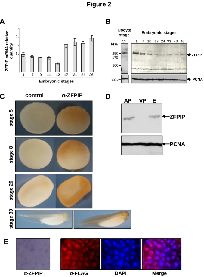
Real-Time Reverse-Transcription PCR assays indicated that the amount of ZFPIP transcripts was quite stable in whole embryo from fertilization (stage 1) to early gastrulation stage (stage 11). This amount then decreased slightly at stage 12. Compared to this stage, the level of ZFPIP mRNAs exhibited a 4 fold increase at stage 36. These data indicated that ZFPIP mRNAs were maternally stored in xenopus oocyte and then degraded. After MBT, these RNAs are zygotically transcribed and we observed a significant increase of the mRNAs level at stage 17 when neurulation occurs. This level of ZFPIP mRNAs was maintained until stage 36 (figure 2A).
Western blots analysis revealed that the ZFPIP protein was also maternally stored in oocyte, since ZFPIP was detected as early as stage VI postvitellogenic oocytes and was abundant at stage 1. After stage 7, the relative amount of the protein was then lower in the embryo from stage 10 to stage 33. A slight increase was then observed after stage 33 (figure 2B). By dissecting the animal and vegetative poles (stage 9), we observed that the protein was essentially localised at the animal pole of the embryos (figure 2D).
In order to complete this expression pattern, we performed whole-mount immunochemistry using a specific ZFPIP antibody (figure 2C). We confirmed that at the
morula and blastula stages, the protein preferentially localised at the animal pole of the embryo. Later in the neurula stage, ZFPIP seemed to be present in the neural folds, neural plate, neural crest and presumptive eyes region. This preferential expression in nervous and sensory region was more evident in tail bud stage, where ZFPIP was detected in the head including otic vesicules and eyes, cardiac region, spinal cord and somites. This regionalization of ZFPIP during neurulation explains the decreasing amount of the protein observed in Western blot.
In addition, the immunostaining of the endogenous protein observed in stages 8 (not shown) and 20 embryos was clearly nuclear (figure 2E). This nuclear localization was confirmed by an additional experiment that has consisted in over-expressing an hZFPIP-FLAG protein into xenopus embryos and that has shown an immunostaining in the nucleus of embryo cells (figure 2E).
The particular pattern of expression of ZFPIP suggested the involvement of the protein at two crucial moments during xenopus development. Firstly, the high amount of maternally stored protein indicated that it could play a key role during the cleavage phase. Indeed, considering the structure of ZFPIP with several zinc finger motifs, it likely interacts with DNA, RNA and different proteins and could therefore participate in multiple fundamental processes during pre-MBT xenopus development such as RNA stability, DNA conformation, replication, transcription inhibition or chromosome segregation. Secondly, the regionalization of the protein observed during neurulation might indicate its involvement in neural cell differentiation process. Accordingly, previous studies have demonstrated that ZFPIP is mostly present in the central and peripheral nervous system of the mouse embryo (Laurent et al., 2007; Chang et al., 2007). In addition, our previous work had indicated that ZFPIP interacts with PBX1 (Laurent et al., 2007) but also with some HOX proteins (data not shown). This ability to strongly interact with PBX1 and HOX proteins indicates that ZFPIP might be a more general partner of homeodomain proteins and might be involved in several developmental processes. In order to explore the role of this gene, we undertook an experiment aiming to knock-down its expression in vertebrate embryo.
ZFPIP morpholinos disrupt Xenopus laevis early development
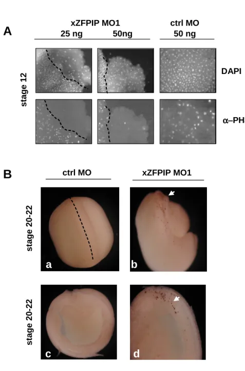
Since the ZFPIP protein was already present at stage 1 of xenopus development, it was necessary to evaluate the turn over of the protein before starting the knock-down of the gene by a morpholinos strategy. Indeed, a high stability of the protein could have rendered inefficient this approach. We thus estimated the turn over of the protein by incubating stage VI oocytes in cycloheximide and observed that the ZFPIP protein began to be degraded after 24 hours of treatment and was fully degraded at 84 hours (figure 3A). This relative instability of the protein in oocytes suggested that the morpholinos strategy was relevant to knock-down the expression of the gene in the embryo. We thus started a series of experiments aiming to knock-down the gene by a morpholinos strategy. For this purpose, we used two different specific morpholinos (MO1 and MO2, see methods for the design) and two different controls (ctrl MO and ctrl MO1, see methods for the design). Most of the data presented in figures 4 to 6 are those obtained with xZFPIP MO1 but identical results were obtained with xZFPIP MO2.
The first experiment has consisted in injecting specific xenopus ZFPIP morpholinos (xZFPIP MO1) in stage VI oocytes. Following morpholinos injection, we observed the complete disappearance of the ZFPIP protein within 48 hours (figure 3B) whereas no variation of the protein was observed in oocytes injected with control morpholinos (ctrl MO). Considering that the stability of ZFPIP was compatible with a morpholinos strategy and since the designed xZFPIP morpholinos were efficiently targeting the ZFPIP mRNA, we performed
the knock-down of the gene in xenopus embryo. We performed the injection of ZFPIP morpholinos in one blastomere at two-cell stage and compared the development of one blastomere versus the other. Injection of these specific morpholinos of xZFPIP mRNAs (xZFPIP MO1) caused abnormal gastrulation and failure of blastopore closure in at least 54% of the embryos (figure 3C-D, figure 4). This abnormal development of the injected part of the embryo was correlated to a significant decrease of ZFPIP protein as shown by Western blot in figure 3E. Injection of xZFPIP MO1 in the two blastomeres of the embryo was also performed and led to a high percentage of mortality (data not shown). The experiment was reproduced using another specific xZFPIP morpholinos, designated as xZFPIP MO2. The same phenotype was observed in xZFPIP MO2 morphants than in xZFPIP MO1 injected embryos with 90% of the embryos showing abnormal phenotype. This abnormal development was correlated to the nearly depletion of ZFPIP protein as shown by Western blot (figure 3E).
In order to ensure that the phenotype observed was linked to the absence of ZFPIP protein, we attempted to rescue the effects of the xZFPIP MOs by coinjection of the human ZFPIP mRNA (hZFPIP mRNA) which is not targeted by xZFPIP MOs. As illustrated in figure 4, coinjection of xZFPIP MO1 with hZFPIP mRNA restored a normal phenotype for 70 % of the embryos. Interestingly, over-expression of hZFPIP RNA did not induce any particular phenotype.
The data presented above clearly demonstrated that ZFPIP was essential for proper early stages of xenopus embryogenesis. The crucial role of this gene in development has been noticed by others. Indeed, in a gene trap approach performed in mouse embryonic stem cells, the ZFPIP/Zfp462 gene has been disrupted and the resulting mice either died or presented growth deficiency. However, in this work, the gene trap procedure had introduced an insertion that caused the expression of a dominant negative form of the protein but not the loss of function of the gene (Skarnes et al., 1992). Unfortunately, no further experiment has been performed by these authors or by others that could highlight the developmental role of the gene in mammalians. Therefore, generation of knock-out mice for ZFPIP/Zfp462 would be a useful tool to study the role of the gene during mammal’s development.
Knock-down expression of ZFPIP impairs cell division and triggers cell death
Using an amphibian model, we showed that the knock-down of ZFPIP/Zfp462 triggered severe abnormal phenotype demonstrating that the gene was essential for embryogenesis. Since the effects of the knock-down of the protein appear to be very drastic, we first tested the expression of some markers of germ layers specification. By analysing the expression of early markers of mesoderm (Xbra), neuroderm (Sox2) and endoderm (Sox17
α
), we did not observe any significant differences in morphant and control embryos, suggesting that germ layer specification was correctly induced (figure 5A). In order to further understand the involvement of the gene in development, we tried to determine the earliest stage at which abnormal phenotype was visualized. In fact, cells presenting a bigger size than normal blastomeres were observed in xZFPIP MOs injected embryos, as soon as stage 5 (figure 5B). This phenotype was directly observable but was also visible after confocal microscopy analysis performed on stage 12 embryos (figure 5B). These first data suggested an impairment of cell division. Indeed, using DAPI staining, we observed several aberrant mitosis or interphases in xZFPIP MOs injected embryos. As shown in figure 5C, several cells exhibited none, bigger or micro nuclei. In addition, mitotic cells underwent atypical mitosis, showing aberrant metaphase, anaphase, incomplete chromosome segregation, conjoined nuclei (figure 5C). Amongst these abnormal cell phenotypes observed in morphant embryos, cells exhibiting none or abnormal nuclei (with conjoined chromatin or nuclei) were particularly abundant(30%, n=50). Such altered mechanisms inevitably induce cell death and are thus deleterious for embryos.
To study the impact of ZFPIP MOs on cell division and/or cell death in the xenopus embryo, we performed two complementary analyses. Firstly, using the mitotic marker Phospho histone H3 (PH3), we evaluated the division rate of embryo cells and observed a striking decrease of cell mitosis in ZFPIP/Zfp462 depleted cells (90% of the animal cap explants exhibited no more PH3 immunostaining, n=11) (figure 6A). Secondly, we showed that xZFPIP MO1 injected cells did not significantly exhibit cell death earlier than in control until stage 20, as demonstrated by TUNEL assay (figure 6B). The results thus demonstrated that the knock-down of ZFPIP/Zfp462 in embryos impaired cell cycle and induced later on cell death. Indeed, these data could be interpreted by a mechanism of cell death, known as mitotic catastrophe, which is an event occurring after microtubules inhibition or DNA damage. In this mechanism, DNA damage can arrest cells in G1/G2, transcription is inhibited and subsequently, cells die by secondary apoptosis (Blagosklonny, 2007).
Further experiments will be necessary to precise the molecular mechanisms triggered by ZFPIP/Zfp462 during early embryogenesis. However, the available data obtained so far in our laboratory indicated that the protein could play a role in chromatin conformation. Indeed, the protein was found in the germinal vesicle of oocyte (data not shown) and remained nuclear in the blastomeres of xenopus embryo during the early stages of its development. In addition, embryonic cells in which the knock-down of the gene has been performed, exhibited a relaxed chromatin compared to control cells (Masse et al., in preparation). In order to better understand the function of ZFPIP/Zfp462, additional experiments will be performed using the model of embryonic cell in culture, which is a more convenient model to investigate molecular and cellular mechanisms. Indeed, xenopus cells contain high amount of vitellus which makes difficult the analysis of markers by microscopy approaches.
The overall data on ZFPIP/Zfp462 reveal that this newly identified gene has an instrumental role during vertebrate development. Playing a role in cell division, this large protein containing numerous zinc fingers might act on chromatin structure and therefore be determinant in the control of genetic programs during vertebrate development.
Acknowledgements
We are grateful to Stéphanie Dutertre from the microscopy platform of IFR 140. We are also indebted to Dr. JP Tassan and Dr. F. Chesnel for helpful comments on the experiments and the manuscript. AL and JM were supported by a grant from the “Conseil Régional de Bretagne”. This work was supported by the CNRS and by grants from the “Conseil Régional de Bretagne”.
References
Altschul, S. F., Madden, T. L., Schaffer, A. A., Zhang, J., Zhang, Z., Miller, W., and Lipman, D. J. 1997. Gapped BLAST and PSI-BLAST: a new generation of protein database search programs. Nucleic Acids Res 25, 3389-402.
Blagosklonny, M. V. 2007. Mitotic arrest and cell fate: why and how mitotic inhibition of transcription drives mutually exclusive events. Cell Cycle 6, 70-4.
Chang, Y. S., Stoykova, A., Chowdhury, K., and Gruss, P. 2007. Graded expression of Zfp462 in the embryonic mouse cerebral cortex. In "Gene Expr Patterns", Vol. 7, pp. 405-12.
Corpet, F. 1988. Multiple sequence alignment with hierarchical clustering. Nucleic Acids Res 16, 10881-90.
Hensey C, Gautier J. 1999. Developmental regulation of induced and programmed cell death in Xenopus embryos. Ann N Y Acad Sci. 887, 105-19.
Kamps, M. P., Murre, C., Sun, X. H., and Baltimore, D. 1990. A new homeobox gene contributes the DNA binding domain of the t(1;19) translocation protein in pre-B ALL. Cell 60, 547-55.
Laurent, A., Bihan, R., Deschamps, S., Guerrier, D., Dupe, V., Omilli, F., Burel, A., and Pellerin, I. 2007. Identification of a new type of PBX1 partner that contains zinc finger motifs and inhibits the binding of HOXA9-PBX1 to DNA. Mech Dev.
Laurent, A., Bihan, R., Omilli, F., Deschamps, S., and Pellerin, I. 2008. PBX proteins: much more than Hox cofactors. Int J Dev Biol 52, 9-20.
Maeda R, Ishimura A, Mood K, Park EK, Buchberg AM, Daar IO. 2002. Xpbx1b and Xmeis1b play a collaborative role in hindbrain and neural crest gene expression in Xenopus embryos. Proc Natl Acad Sci U S A 99, 5448-53.
Moens, C. B., and Selleri, L. 2006. Hox cofactors in vertebrate development. Dev Biol 291, 193-206.
Nieuwkoop, P. D, and Faber, J. 1994. Normal Table 1 of Xenopus laevis (Daudin): A Systematical and Chronological Survey of the Development from the Fertilized Egg till the End of Metamorphosis. 3rd ed. Garland Publishing, New York, NY.
Nourse, J., Mellentin, J. D., Galili, N., Wilkinson, J., Stanbridge, E., Smith, S. D., and Cleary, M. L. 1990. Chromosomal translocation t(1;19) results in synthesis of a homeobox fusion mRNA that codes for a potential chimeric transcription factor. Cell 60, 535-45. Skarnes WC, Auerbach BA, Joyner AL. 1992. A gene trap approach in mouse embryonic
stem cells: the lacZ reported is activated by splicing, reflects endogenous gene expression, and is mutagenic in mice. Genes Dev. 6, 903-18
Figure legends
Fig. 1. ZFPIP is a highly conserved zinc finger protein. ZFPIP is a 250 kDa protein that contains 26 conserved C2H2 motifs and one NLS (A). Amino acid sequence is conserved from Danio rerio to Mus musculus; Mn: Mus musculus; Rn: Ratus norvegicus; Hs: Homo sapiens; Cf: Canis familiaris; Bt: Bovis Taurus; Gg: Gallus gallus; Tn: Tetraodon nigroviridis (B). Sequences available in amphibian models and conservation between Xenopus laevis (Xl) and Xenopus tropicalis (Xt). Overlapping sequences of cDNAs between the two species were compared and were determined to be at least 94% homologous (C).
Fig. 2. ZFPIP is maternally and zygotically expressed during Xenopus laevis development. RT-PCR experiments using Xenopus laevis embryo RNA extracts show that ZFPIP mRNAs are present in stage 1 and then decrease until stage 12. Transcription of mRNAs is enhanced at stage 17 (A). The ZFPIP protein is maternally present in oocytes. The amount of protein is then diluted during the cleavage phase. The α–PCNA antibody is used as an internal quantitative control (B). Whole mount immunohistochemistry assays are performed on Xenopus laevis embryos using an α–ZFPIP or preimmune serum at stages 5, 8, 20 and 39. The pictures show that ZFPIP is mostly present in the animal pole of blastula and progressively localises in developing neural tissues (C). Western blots confirm that the protein is preferentially localised in the animal pole. AP, VP and E correspond respectively to animal pole, vegetal pole and embryo protein extracts (D). Nuclear localization of endogenous ZFPIP or hZFPIP-FLAG protein is observed in embryos or in hZFPIP mRNA injected embryos. The immunohistochemistry is performed with α–ZFPIP or α–FLAG antibodies on Xenopus laevis embryos explants (stage 20) (E).
Fig. 3. ZFPIP depletion impairs xenopus development. Stage VI oocytes are incubated either in 10 µg/mL cycloheximide or in a cycloheximide-free culture medium. Total protein extracts of oocytes collected after different times of incubation are analyzed by Western blotting using an α–ZFPIP serum and α–PCNA antibody (A). Stage VI oocytes are injected with 50 ng of ctrl MO or xZFPIP MO1 and allowed to develop for different times. Western blots of total protein extracts of oocytes are analyzed using an α–ZFPIP serum and α–PCNA antibodies; they demonstrate the specificity of the xZFPIP MO1 (B). Embryos are injected in one blastomere at the two-cell stage with 50 ng of ctrl MO (D, H, L, P) or 25-50 ng of xZFPIP MO1 (A-C, E-G, I-K, M-O) and are allowed to develop (C). Embryos are injected with 50 ng of xZFPIP MO1 and 1ng of GFP mRNA. They grow until stage 12 and are observed using a fluorescence binocular (D). Embryos are either injected with 25-50 ng of ctrl MO, ctrl MO1, xZFPIP MO1, xZFPIP MO2 or not injected and allowed to develop until stage 8. Depletion of ZFPIP protein is demonstrated by Western blot performed with total protein extracts and analyzed using an α-ZFPIP serum and α–PCNA antibody. Depletion of ZFPIP by MO1 has been performed by injecting one blastomere at the 2 cell-stage and depletion of ZFPIP by MO2 has been performed by injecting the 2 blastomeres at the same developmental stage (E).
Fig. 4. hZFPIP mRNA rescues the gastrulation defect observed in embryos injected with xZFPIP MO. Xenopus laevis embryos are injected with either 50 ng of ctrl MO, 1-5 ng of hZFPIP RNA, 25 ng of xZFPIP MO1 or 25 ng of xZFPIP M1 and 1-5 ng of hZFPIP RNA. The percentage of normal embryos obtained after the different injections are given in the table and are represented by histograms.
Fig. 5. xZFPIP knock-down does not alter germ layer specification but impairs cell divisions in Xenopus laevis embryos. RT-PCR experiments demonstrate that Xbra, Sox2 and Sox17
α
are correctly induced in morphant embryos at stage 7, 10 and 12.5. RNAs are extracted from xZFPIP MO1 injected embryos (Mo) or ctrl MO injected (Ct). Enzymatic reactions are performed with (RT+) or without (RT-) reverse transcriptase. Standard PCR protocol is used with the primers indicated in methods (A). Stage 5 Xenopus laevis morphant shows aberrant cell size and shape (B, b1). Stage 12 Xenopus laevis explants stained with DAPI (nuclei staining) and phalloïdine-Fluoroprobe 547H (F-actin staining) exhibit bigger cells containing altered DNA content (B, b2). Scale bar represents 100µm (B). DAPI stained animal caps of morphant embryos contain cells with variable nucleus shape (C, c1-3), conjoined chromatin or nuclei (C, c1-2), micronuclei (C, c3), tripolar metaphases and anaphases (C, c4-6) or anuclear cells (C,c2).
Fig. 6. xZFPIP knock-down prevents cell mitosis and triggers cell death in Xenopus laevis embryos. Xenopus laevis embryos are injected in one blastomere at the two-cell stage with 25-50 ng of xZFPIP MO1 or ctrl MO. Embryos are then allowed to develop until stage 12. Animal cap explants are prepared, immunostained using an α-PH3 antibody and nuclei stained with DAPI (A). Xenopus laevis embryos are injected in one blastomere at the two-cell stage with 25-50 ng of xZFPIP MO or ctrl MO and are allowed to develop until stage 20-22. The TUNEL assay performed on whole (a, b) or sectioned embryos (c, d) shows a staining in the presumptive neural region (arrows).
Figure 1
Sequences not currently available 1 2 3 4
A
B
1 Mus musculus> 70% identity between the 4 species 2 Homo sapiens
3 Gallus gallus > 65% identity between the 4 species 4 Danio rerio Conserved C2H2 motif
bipartite NLS
AA that are absent in the protein 100 AA Rn Hs Cf Bt Gg Tn Dr Mm 97.0% 92.6% 91.9% 90.9% 80.5% 50.4% 43.7% Rn 93.2% 92.5% 91.5% 80.5% 50.0% 43.6% Hs 95.4% 94.1% 82.1% 50.5% 43.2% Cf 95.0% 82.5% 50.2% 43.6% Bt 81.1% 50.5% 43.6% Gg 51.1% 45.9% Tn 48.8%
C
Xl Xt 93% 200 AA 96% 94%Figure 2
ZFPIP PCNAB
A
C
s ta g e 5 s ta g e 2 0 s ta g e 8 control αααα-ZFPIP s ta g e 3 9 Oocyte stage 1 7 10 17 24 33 43 46 250 175 100 kDa VI 32.5 Embryonic stagesD
Z F P IP m R N A re la ti v e q u a n ti ty 1 7 9 11 12 17 21 24 36 1 7 9 11 12 17 21 24 36 1 2 Embryonic stages Merge α αα α-FLAG DAPI α αα α-ZFPIP ZFPIP PCNA AP VP E ZFPIP PCNA AP VP EE
Figure 3
ZFPIP PCNA s ta g e 1 2 m o rp h a n tsxZFPIP MO1 + GFP mRNA
D
A
s ta g e 7 s ta g e 1 2xZFPIP MO1 injected embryos
s ta g e 2 0 ctrl MO s ta g e 5 A B C D H G F E L K J I P O N M 0 15 24 48 84 - cyclo (h) 0 15 24 48 84 + cyclo (h) PCNA ZFPIP
C
E
250 32.5 c tr l M O M O 1 ( 5 0 n g ) kDa NIB
24 48 72 24 48 72 ctrl MO (h) xZFPIP MO1 (h) PCNA ZFPIP c tr l M O M O 2 ( 2 5 n g ) M O 2 ( 5 0 n g ) c tr l M O 1 MO1 MO2Figure 4
Injected
molecule Quantity % mortality
None ctrl MO
hZFPIP RNA hZFPIP RNA
xZFPIP MO1
xZFPIP MO1+hZFPIP RNA xZFPIP MO1+hZFPIP RNA
50 ng 1 ng 5 ng 25 ng 25 ng + 1 ng 25 ng + 5 ng % affected
embryos % wild type embryos 3 0 13 6 17 1 2 23 23 2 8 54 44 26 74 77 85 86 29 55 72 Number of embryos 111 74 54 56 58 71 50 0 50 100 NI ctrl MO hZFPIP RNA 1 ng hZFPIP RNA 5ng
xZFPIP MO1 xZFPIP MO1 + hZFPIP RNA 1ng xZFPIP MO1 + hZFPIP RNA 5ng Mortality Gastrulation defect Wild type