HAL Id: hal-03230295
https://hal.umontpellier.fr/hal-03230295
Submitted on 25 May 2021
HAL is a multi-disciplinary open access
archive for the deposit and dissemination of
sci-entific research documents, whether they are
pub-lished or not. The documents may come from
teaching and research institutions in France or
abroad, or from public or private research centers.
L’archive ouverte pluridisciplinaire HAL, est
destinée au dépôt et à la diffusion de documents
scientifiques de niveau recherche, publiés ou non,
émanant des établissements d’enseignement et de
recherche français ou étrangers, des laboratoires
publics ou privés.
Distributed under a Creative Commons Attribution| 4.0 International License
Inhibition of G-protein signalling in cardiac dysfunction
of intellectual developmental disorder with cardiac
arrhythmia (IDDCA) syndrome
Pasquelena de Nittis, Stephanie Efthymiou, Alexandre Sarre, Nicolas Guex,
Jacqueline Chrast, Audrey Putoux, Tipu Sultan, Javeria Raza Alvi, Zia Ur
Rahman, Faisal Zafar, et al.
To cite this version:
Pasquelena de Nittis, Stephanie Efthymiou, Alexandre Sarre, Nicolas Guex, Jacqueline Chrast, et al..
Inhibition of G-protein signalling in cardiac dysfunction of intellectual developmental disorder with
cardiac arrhythmia (IDDCA) syndrome. Journal of Medical Genetics, BMJ Publishing Group, In
press, �10.1136/jmedgenet-2020-107015�. �hal-03230295�
ORIGINAL RESEARCH
Inhibition of G- protein signalling in cardiac
dysfunction of intellectual developmental disorder
with cardiac arrhythmia (IDDCA) syndrome
Pasquelena De Nittis ,
1Stephanie Efthymiou,
2Alexandre Sarre,
3Nicolas Guex,
4Jacqueline Chrast,
1Audrey Putoux,
5Tipu Sultan,
6Javeria Raza Alvi,
6Zia ur Rahman,
6Faisal Zafar,
7Nuzhat Rana,
7Fatima Rahman,
8Najwa Anwar,
8Shazia Maqbool,
8Maha S Zaki ,
9Joseph G Gleeson ,
10David Murphy,
2Hamid Galehdari,
11Gholamreza Shariati,
12Neda Mazaheri,
11Alireza Sedaghat,
13SYNAPS Study Group,
Gaetan Lesca,
14Nicolas Chatron,
1,14Vincenzo Salpietro,
2Marilena Christoforou,
2Henry Houlden,
2William F Simonds,
15Thierry Pedrazzini,
16Reza Maroofian,
2Alexandre Reymond
1To cite: De Nittis P,
Efthymiou S, Sarre A, et al. J Med Genet Epub ahead of print: [please include Day Month Year]. doi:10.1136/ jmedgenet-2020-107015
►Additional material is published online only. To view, please visit the journal online (http:// dx. doi. org/ 10. 1136/ jmedgenet- 2020- 107015). For numbered affiliations see end of article.
Correspondence to
Professor Alexandre Reymond, Center for Integrative Genomics, University of Lausanne, Lausanne 1015, Switzerland; alexandre. reymond@ unil. ch Received 19 March 2020 Revised 30 August 2020 Accepted 4 September 2020
© Author(s) (or their employer(s)) 2020. Re- use permitted under CC BY. Published by BMJ.
ABSTRACT
Background Pathogenic variants of GNB5 encoding
the β5 subunit of the guanine nucleotide- binding
protein cause IDDCA syndrome, an autosomal recessive neurodevelopmental disorder associated with cognitive disability and cardiac arrhythmia, particularly severe bradycardia.
Methods We used echocardiography and telemetric
ECG recordings to investigate consequences of Gnb5 loss in mouse.
Results We delineated a key role of Gnb5 in heart
sinus conduction and showed that Gnb5- inhibitory signalling is essential for parasympathetic control of heart rate (HR) and maintenance of the sympathovagal balance. Gnb5−/− mice were smaller and had a smaller
heart than Gnb5+/+ and Gnb5+/−, but exhibited better
cardiac function. Lower autonomic nervous system modulation through diminished parasympathetic control and greater sympathetic regulation resulted in a higher baseline HR in Gnb5−/− mice. In contrast, Gnb5−/− mice
exhibited profound bradycardia on treatment with carbachol, while sympathetic modulation of the cardiac stimulation was not altered. Concordantly, transcriptome study pinpointed altered expression of genes involved in cardiac muscle contractility in atria and ventricles of knocked- out mice. Homozygous Gnb5 loss resulted in significantly higher frequencies of sinus arrhythmias. Moreover, we described 13 affected individuals, increasing the IDDCA cohort to 44 patients.
Conclusions Our data demonstrate that loss of
negative regulation of the inhibitory G- protein signalling causes HR perturbations in Gnb5−/− mice, an effect
mainly driven by impaired parasympathetic activity. We anticipate that unravelling the mechanism of Gnb5 signalling in the autonomic control of the heart will pave the way for future drug screening.
INTRODUCTION
Intellectual developmental disorder with cardiac arrhythmia (IDDCA, OMIM (Online Mendelian
Inheritance in Man): #617173) is an autosomal recessive neurodevelopmental disorder with onset in early childhood. Inactivating and hypomorphic mutations in the β5 subunit of guanine nucleotide-
binding protein (GNB5), respectively, cause severe and mild forms of the disorder.1 The former is
associated with cognitive disability, poor or absent speech and/or severe cardiac arrhythmias. The moderate manifestation of the syndrome, also named language delay and ADHD/cognitive impair-ment with or without cardiac arrhythmia (LADCI) syndrome (OMIM: #617182), consists of mild intellectual impairment, language delay, attention deficit hyperactivity disorder (ADHD) and, in about half the cases, severe cardiac arrhythmia.1 2 Some
patients with IDDCA also showed retinal dysfunc-tion and nystagmus, epilepsy, hypotonia and gastro-intestinal problems.1–10 The GNB5 retinopathy
is a unique combination of dual retinal signalling defects reminiscent of features of both bradyopsia and rod ON- bipolar dysfunction,5 while the IDDCA
epilepsy is characterised by early seizure onset (~3 months of age) with focal seizures rapidly evolving into epileptic spasms and consequent generalised multifocal discharges.4
The heart rate (HR) is established by the sinoatrial node, the pacemaker of the cardiac muscle, and controlled by the autonomic nervous system. This autonomic nervous system consists of two anatomi-cally and functionally distinct divisions: the sympa-thetic and the parasympasympa-thetic branches, whose functions are often antagonistic but work together to maintain balance. In the heart, the postganglionic fibres of the sympathetic trunk stimulate the β-ad-renoreceptors, thereby increasing HR and force of contraction. The parasympathetic modulation of the heart is primarily mediated by acetylcholine release, which activates the M2- muscarinic receptors (M2R) present on cells innervated by parasympathetic postganglionic neurons, including sinoatrial node cells. The activation of M2R triggers Gi/o subfamily G- proteins, which turn on G- protein- gated
copyright.
on May 25, 2021 at Univ of Montpellier. Protected by
http://jmg.bmj.com/
inwardly rectifying K+ channels (GIRK) resulting in membrane hyperpolarisation and decrease in HR. Regulator of G- pro-tein signalling (RGS) propro-teins negatively regulate the timing of this M2R- GIRK signalling. GNB5, a divergent member of the Gβ family, has the unique property of forming complexes with R7- RGS proteins.11–16 In particular, the GNB5- RGS6 complex is
involved in cardiac GIRK deactivation kinetics. Rgs6- null mice manifested heart conduction anomalies and hypersensitivity to parasympathomimetics.17 Zebrafish model defective for gnb5
gene correspondingly showed reduced heartbeat on reinforced parasympathetic stimulation, eye movement defects and altered swimming behaviour1 and cardiomyocytes differentiated from
human induced pluripotent stem cells (iPSCs) edited to engineer the GNB5- Ser81Leu missense variant associated with LADCI showed a decrease in spontaneous activity on stimulation with carbachol compared with normal cells.7
Whereas homozygous Gnb5- null mice recapitulated many of the corresponding human disease phenotypes such as learning deficiencies, hyperactivity, impaired motor coordination and perturbed vision,18–22 a systematic cardiac evaluation has never
been performed in a mammalian model. Here, we assessed heart electrophysiology of Gnb5 mice models. We detected an increased frequency of sinus arrhythmias in Gnb5−/− animals,
which have a smaller heart than wild- type and Gnb5+/−, but
exhibited better cardiac function. Gnb5−/− mice also displayed
enhanced parasympathetic sensitivity on stimulation with a cholinergic agonist. Consistent with this, transcriptome profiling of atria and ventricles revealed overexpression of genes involved in cardiac muscle contractility, along with reduced ventricular expression of genes required for development of pacemaker cells in Gnb5−/− mice. Finally, we expanded the number of
ascer-tained IDDCA individuals and the GNB5 mutational spectrum. MATERIALS AND METHODS
Enrolment
All affected individuals and their family members were recruited in Pakistan (families R–V), France (family W), Egypt (families X and Y) and Iran (family Z) after signing a written informed consent according to ethical review boards policies. Clinical ascertainment included physical examinations, medical history interviews and specialised consultations by a certified neurolo-gist and cardioloneurolo-gist as appropriate. Venous blood was collected in EDTA for DNA extraction according to standard procedures. Exome sequencing
Whole- exome sequencing of families R–V and Z was performed by Macrogen, Korea, as described in reference.23 Briefly, target
enrichment was performed with 2 µg genomic DNA using the SureSelectXT Human All Exon Kit version 6 (Agilent Tech-nologies, Santa Clara, California, USA) to generate barcoded whole- exome sequencing libraries. Libraries were sequenced on the HiSeqX platform (Illumina, San Diego, California, USA) with 50× coverage. Quality assessment of the sequence reads was performed by generating QC statistics with FastQC.24 The
filtering strategy included screening for only exonic and donor/ acceptor splicing variants. In accordance with the pedigree and phenotype, priority was given to variants rare or absent in public databases (1000 Genomes project, National Heart, Lung, and Blood Institute Exome Variant Server, Complete Genomics 69 and Exome Aggregation Consortium V.0.2).
Trio exome sequencing was performed in proband 37 (family W) and her parents using SeqCap EZ Medexome library prepa-ration kit following manufacturer’s recommendations (Roche,
Indiana, Indianapolis, USA). Libraries were sequenced on a NextSeq500 (Illumina) at a mean depth coverage of 73× with 93.3% of the target bases above 30×. Genomic alignment against the hg19/GRCh37 assembly and variant calling were, respectively, done with BWAMEM V.0.7.12 and GATK Haplo-typeCaller V.3.4 (Broad Institute, Boston, Massachusetts, USA). Only highly confident variants were kept for analysis (total depth >9, alternative allele depth >4, no strand bias, mosaicism >10%). Rare variants were considered as having a frequency of <1% in GnomAD v2 dataset. Whole- exome sequencing of families X and Y was performed as described in Makrythanasis et
al.25 Sanger sequencing in each family confirmed the segregation
of GNB5 variants with the phenotype. Mouse husbandry
The Gnb5 mouse line was recovered from cryopreserved sperm using in vitro fertilisation. The knockout allele was engineered in a C57BL/6J inbred genetic background by heterozygous deletion of exon 3 in the germline, as previously described.18 22
Genet-ically modified animals were born and housed in the Animal Facility of the Centre for Integrative Genomics, under controlled temperature conditions and a 12- hour light–dark cycle with free access to water, normal chow and nest building material. Mouse genomic DNA was extracted from ear biopsies using the hot shot protocol26 and used for genotyping as described.22 To
prevent the previously documented high mortality of Gnb5−/−
pups at weaning,18 heterozygous breeding couples used to
obtain knockout pups were given breeding food pellet enriched for proteins and vitamins (Kliba 3336, extrudate). Additionally, litters including Gnb5−/− pups were fed from 14 to 28 days of age, that is, starting 1 week before weaning, with powdered wet maintenance food (Kliba 3436) in a Petri dish placed directly onto the floor of the cages, an expedient that should provide easier access to the food for the pups.
In vivo transthoracic ultrasound imaging protocol
Transthoracic echocardiography was performed using a 30 MHz probe and the Vevo 2100 Ultrasound machine (VisualSonics, Toronto, Ontario, Canada). A light anaesthesia was achieved with 1%–1.5% isoflurane, maintaining HR at 400–500 beats/ min. The mice were placed in decubitus dorsal on a heated 37°C platform to maintain body temperature. The heart was imaged in the 2D mode in the parasternal long- axis view. From this view, an M- mode cursor was positioned perpendicular to the inter-ventricular septum and the posterior wall of the left ventricle, at the level of the papillary muscles. Diastolic and systolic inter-ventricular septa, left inter-ventricular posterior wall thickness and left ventricular internal end- diastolic and end- systolic chamber dimensions were measured. Three separate M- mode images were measured and averaged. Left ventricular fractional ening and ejection fraction were also calculated. Fractional short-ening and ejection fraction were assessed from M- mode based on the percentage changes of left ventricular end- diastolic and end- systolic diameters and volumes, respectively. We used male mice at 9 weeks of age of three different genotypes (Gnb5+/+,
Gnb5+/− and Gnb5−/−).
In vivo electrocardiography measurements
For in vivo electrocardiography monitoring we have subcutane-ously implanted biopotential telemetric transponders (ETA- F10, Data Sciences International) allowing continuous monitoring in conscious freely moving animals at 12 weeks of age. The nega-tive electrode was implanted at the top of the right pectoral
copyright.
on May 25, 2021 at Univ of Montpellier. Protected by
http://jmg.bmj.com/
muscle, and the positive one was anchored at the level of the last left rib (at about 1 cm of the xiphoid appendix), thus leading to a normal lead II trace. Baseline ECG was recorded 10 days after device implantation, over a period of 86 hours. After 30 min of basal measurements, we injected mice with 0.9% saline solution (intraperitoneal NaCl, 10 mL/kg) as a vehicle control; next, the following compounds were administered one at a time: atropine (PubChem CID: 174174, intraperito-neal, 1 mg/kg), carbachol (PubChem CID: 5831, intraperitoneal 0.1 mg/kg) and in a subset of mice isoprenaline (PubChem CID: 3779, intraperitoneal 4 mg/kg) and atenolol (PubChem CID: 2249, intraperitoneal 2 mg/kg) as well, with a one night interval between each injection. The amounts of isoprenaline and aten-olol were chosen after testing 10 doses ranging from 4 µg/kg to 4 mg/kg and 2 µg/kg to 2 mg/kg, respectively. While atropine and carbachol respectively inhibit and activate the parasympa-thetic system atenolol and isoprenaline respectively block and promote the sympathetic response.
At the end of the experiment, mice were sacrificed by CO2 inhalation; the heart was digitally imaged both within its thoracic position and after excision. Heart weight was recorded and tibia length was measured to normalise the heart weight to body size. Images were taken by a Leica DCF295 digital colour camera with 3M pixels mounted on a MZ6 stereomiscroscope (Leica, Switzerland). The length of the tibia was measured using a preci-sion calliper after removal from the left leg (without patella nor articular cartilage).
Baseline ECG traces were analysed as follows: 10 min of recording were analysed using ECG- Auto software in shape recognition mode (EMKA Technology, France) every 30 min, during night and day phases. Mean values were reported for each analysed parameter. During pharmacological challenges, ECG recording was analysed continuously, with 10 min steps; thus, one mean of each parameter was calculated every 10 min. On ECG traces we analysed: (1) RR interval (measured at R peak, expressed in millisecond; (2) HR: heart beating rate, calcu-lated as 60/(RR/1000), expressed in beats/min; (3) PR interval (interval between beginning of P wave and R peak, expressed in millisecond); (4) QT duration: duration of the QT complex; (5) QTc (corrected QT, calculated from QT/√(RR/100)27 (all
expressed in millisecond) that allows correction of QT from HR variations).
Temperature and activity were also recorded. Activity was estimated by displacement of the telemetric device from the antennas of the recording plate. This measurement was only used as a qualitative index of mouse activity/movement. Temperature and activity values are mean values of 1- hour interval.
Time-domain heart rate variation (HRV) analysis
Twelve- hour segments of day/night phases were selected from the baseline recording period and analysed separately for the HRV analysis. Specifically, R wave detection was performed and R- R interval time series were obtained. To ensure inclu-sion of sinus beats only, values not included between R–R intervals±2 SD were excluded as reported in Thireau et al.28
The analysed time- domain HRV parameters, computed using Kubios HRV Standard software V. 3.2.0, were: (1) mean R–R intervals (NN, in millisecond); (2) SD of all normal R–R inter-vals (SDNN, in millisecond); (3) square root of the mean square differences (RMSSD) between successive normal inter-vals (in millisecond); and (4) percentage of normal consecutive R–R intervals differing by >xms (pNNx, in %, in this study x=6 ms).
Arrhythmia assessment
Arrhythmias were identified based on ECG trace (RR interval) and counted. Observed arrhythmias were defined as follows: (1) escape atrial beat, with a P wave morphology different from that of the sinus P wave, classified as ‘long’ and ‘short’ according to the location of the P wave on the ECG trace (specifically, long was an escape beat whose duration was longer than two normal PP intervals, and short were those escape beats that lasted less than two normal PP intervals); (2) atrioventricular block defined on ECG by more than one P wave for one QRS complex; (3) premature beats; and (4) episodes of tachy-cardia followed by bradytachy-cardia, with HR oscillation between high and low values within a few seconds, independently of other type of arrhythmias.
Statistical tests
Each parameter measured was reported as mean±SD. Standard t- test was used to assess differences between two groups. Anal-ysis of variance tests, followed by Tukey post hoc tests, were also calculated and are displayed. A p value of <0.05 was considered significant, and stars on the plots represent the level of signif-icance (*p≤0.05, **p≤0.01, ***p≤0.001, ****p≤0.0001; p>0.05 was considered not significant).
Transcriptome profiling, data processing and differential expression analysis
A total of 72 RNA sequencing libraries were generated from two heart tissues, atria and ventricles, and three brain regions, cerebellum, hippocampus and cerebral cortex. Both the entire ventricles (left and right) or atria (left and right) were used for the sample processing; therefore, the atrial tissue represents a mixture of heart muscle cells, as well as sinoatrial and atrioventricular cells. The whole cerebellum, the hippocampi from each hemisphere of the brain and the whole cerebral cortex were dissected for RNA extraction. For this experi-ment, we created a cohort of adult (18 weeks) male mice including 6 Gnb5−/−, 6 Gnb5+/− and 6 Gnb5+/+ animals. Brain transcriptome
was only assessed in Gnb5−/− and Gnb5+/+ animals. Tissue
collec-tion and processing procedures were designed to minimise biolog-ical and technbiolog-ical variation. Specifbiolog-ically, tissues were dissociated in QIAzol Lysis Reagent (Qiagen) using the gentleMACS Dissociator (Miltenyi Biotec). Cell suspension was used to obtain total RNA. Genomic DNA contamination was removed by digestion with RNase- free Deoxyribonuclease I (Qiagen). RNA concentration and purity were measured by ND-1000 spectrophotometer (Thermo Scientific, Wilmington, North Carolina, USA), and RNA integrity was verified by fragment analyser automated CE system (Advanced Analytical Technologies) according to manufacturer’s instructions. Libraries were then prepared with TruSeq Stranded RNA Library Prep Kit (Illumina) and sequenced on multiple lanes of an Illumina HiSeq4000 platform, generating an average of 50M single- end 125- cycle reads for each sample. Quality of sequence was assessed by FastQC V.0.11.4.24 Purity- filtered reads were adapters- trimmed
and quality- trimmed with Cutadapt V.1.8.29 Reads matching to
ribosomal RNA sequences were removed with fastq_screen V.0.11.1. Remaining reads were further filtered for low complexity with reaper V.15–065.30 Reads were then aligned against Mus Musculus. GRCm38.92 genome using STAR V.2.5.3a.31 The number of read
counts per gene locus was summarised with htseq- count V.0.9.132
using Mus Musculus.GRCm38.92 gene annotations. Quality of the RNA- seq data alignment was assessed using RSeQC V. 2.3.7.33 Reads
were also aligned to the Mus Musculus.GRCm38.92 transcriptome using STAR V. 2.5.3a,31 and the estimation of the isoforms
abun-dance was computed using RSEM V.1.2.31.34 To assess differential
expression between genotypes within each tissue, we compared
copyright.
on May 25, 2021 at Univ of Montpellier. Protected by
http://jmg.bmj.com/
Gnb5−/− and Gnb5+/−± to wild- type specimens. Data analysis was
performed with the R Bioconductor package DESeq2 V.1.14.1.35
Differentially expressed genes (DEGs) were identified at the Benjamini- Hochberg adjusted p<0.05 level, using Wald test under design ~genotype. For gene set enrichment analysis, no direction criterion on fold change was applied. Enriched Gene Ontology (GO) categories were identified using the enrichment analysis package in R/Bioconductor, clusterProfiler,36 considering only categories with at
least 10 and maximum 500 annotated genes. Nominally significant enriched terms were retained for results interpretation.
Western blotting analysis
Tissue lysates, including atria, ventricles, cerebral cortex, cerebellum and hippocampi, were prepared in RIPA buffer (Millipore) supple-mented with protease inhibitors (Thermo Fisher Scientific). Tissues were homogenised using the gentleMACS Dissociator (Miltenyi Biotec). After SDS- polyacrylamide gel electrophoresis (SDS- PAGE) and transfer to nitrocellulose membrane, blots were incubated with anti- Gnb5, anti- Rgs7 (both a generous gift from Dr William F Simonds) and anti- Gnb3 (Cell Signalling Technology) antibodies, separately, and with an antiactin antibody (Sigma), used for loading control. Horseradish peroxidase- conjugated anti- rabbit antibody (Santa Cruz) and the ECL chemiluminescence system (Millipore) were used for detection.
RESULTS
Clinical and molecular features of thirteen novel patients with IDDCA
We identified 13 additional IDDCA cases (online supplemental figure S1, table S1 and table 1) through exome sequencing of nine consanguineous families and data aggregation of multiple laboratories and clinical centres via GeneMatcher37 38 or direct
contacts. Consistent with previous reports,1–10 the carrier of a
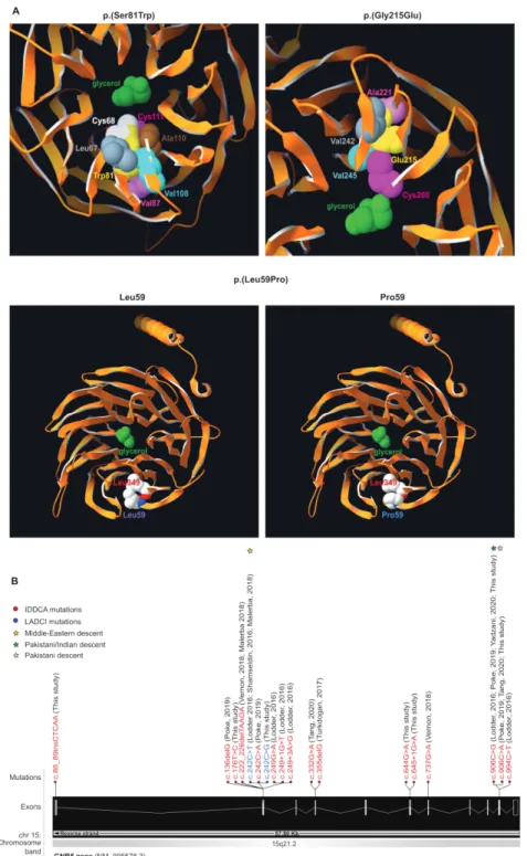
homozygous GNB5 missense variant on Ser81 presented with LADCI, the mild form of IDDCA (family W), while the nine individuals with biallelic loss- of- function (LoF) alleles due to truncating or splicing mutations displayed phenotypes corre-sponding to the severe end of the disease spectrum (families R and S, U and V, and X and Z). The remaining three individuals (families T and Y) carrying novel homozygous missense variants on Gly215 (family T) and on Leu59 (family Y) similarly presented with the severe IDDCA phenotypical spectrum. Clinical features of affected individuals are summarised in table 1 and detailed in the online supplemental note and table S1. The three missense variants (c.644G>A, p.(Gly215Glu), family T, c. 242C>G, p.(Ser81Trp), family W and c.176T>C, p.(Leu59Pro), family Y; transcript NM_006578.3) were not reported before (figure 1). They are predicted by a majority of prediction tools to be likely damaging to protein function (online supplemental table S2) and are absent from GnomAD V.3.39 To assess possible impact, we
modelled these three substitutions using the crystal structure of the GNB5- RGS9 complex40 and found that each variant could
impact the potential binding properties of the GNB5 central pore (p.(Ser81Trp) and p.(Gly215Glu)) or the protein folding (p.(Le-u59Pro)) (figure 1A). Specifically, Serine 81 is buried inside a β strand of the first WD40 repeat close to the central pore of the β propeller structure1 2 where a glycerol molecule is observed
in pbi structure 2pbi (figure 1A, top left panel). Reminiscent of the Ser81Leu variant previously documented,1 a tryptophan at
position 81 cannot be accommodated without disrupting the structure and potential binding properties of the pore. Our model suggests that the rearrangements necessary to settle such a bulky sidechain will change the channel characteristics. We have investigated the related rotamers, emphasising the steric
Table 1 Overlapping clinical features of individuals with IDDCA and LADCI syndromes
Individuals with phenotype/ individuals (total n) Phenotype severity (IDDCA) Phenotype severity (LADCI) Phenotype severity (intermediate) Gender 23F, 21M 15F, 19M 7F, 2M 1F Clinical examination Dysmorphic features 11/44 (25%) 10/34 – 1/1
Congenital malformations 4/44 (9%) 4/34 (heart) – – Neurological manifestations
Intellectual disability 37/44 (84%) 32/34 4/9 1/1
Speech delay 34/44 (77%) 27/34 6/9 1/1
Hypotonia 33/44 (75%) 29/34 3/9 1/1
Seizures 25/44 (57%) 25/34 – –
Behavioural disorders 6/44 (14%) 3/34 (ASD) 3/9 (ADHD) –
MRI anomalies 8/44 (18%) 8/34 – –
Sleep disturbance 2/44 (5%) 2/34 – –
Cardiac manifestations
Sinus sick syndrome 27/44 (61%) 22/34 4/9 1/1
Pacemaker implantation 6/44 (14%) 4/34 1/9 1/1 Ophthalmological findings Nystagmus 26/44 (59%) 26/34 – – Strabismus 6/44 (14%) 5/34 – 1/1 Retinal disease 14/44 (32%) 14/34 – – Gastrointestinal problems
Pathological gastric reflux 18/44 (41%) 18/34 – –
Pedigree charts and variants of single individuals are detailed in the corresponding published reports1–4 6 8–10 81 and in online supplemental figure S1. Detailed phenotypical
information of each affected individuals is reported in online supplemental table S1 and supplemental note.
ADHD, attention deficit hyperactivity disorder; ASD, autism spectrum disorder; F, female; IDDCA, Intellectual developmental disorder with cardiac arrhythmia; LADCI, language delay and ADHD/cognitive impairment; M, male.
copyright.
on May 25, 2021 at Univ of Montpellier. Protected by
http://jmg.bmj.com/
Figure 1 Variants modelling and IDDCA mutational spectrum. (A, top left) Top view of the Gnb5 (orange, PDB entry 2pbi) 3D protein model, showing the
mutated Trp81 (yellow) and the glycerol molecule (green) in the centre of the pore. The rearrangements necessary to accommodate a tryptophan residue at position 81 will change the channel characteristics. The rotamer displayed here highlights clashes of Trp81 with Cys68 (white) and Cys111 (magenta). In additional rotamers, the bulky tryptophan sidechain will severely bump into Leu67 (grey), Val87 (pink), Val108 (cyan) and Ala110 (brown). (A, top right) As shown in this view of the beta propeller from above, the ‘wild- type’ Gly215 (not shown) lays in a beta- sheet, with on top Ala221 (pink), at the bottom Cys200 (magenta), and in front a beta- strand harbouring Val242 (grey) and Val245 (cyan). The presence of Glu215 cannot be tolerated, as it will encroach into one of the residues previously enumerated. Another rotamer shows clashes into Val245 (cyan). Overall, all rotamers may also force the sidechain of Cys200 (magenta) to reorient itself toward the internal part of the channel to provide space to accommodate glutamine at position 215 (yellow). In this position, the Cys200 sidechain will occupy the space dedicated to the glycerol (green), thus changing the properties of the channel. (A, bottom) The Leu59 (purple label, left panel) is positioned closely to the Leu349 residue just above (red label) in an antiparallel beta- sheet. This secondary structure will likely be broken in presence of a proline at that position (blue label, right panel), which in turn will disrupt the protein folding as the proline sidechain will collide into Leu349 (red label). (B) Distribution of the IDDCA published and novel variants along the schematically represented 11 exons of the human GNB5 gene (transcript NM_006578.3; Ensembl (release 98, September 2019). The variants of IDDCA affected individuals are represented in red (LoF) and the missense LADCI variants in blue. The yellow star marks the variant of Middle Eastern descent, while green stars indicate the amber and ochre variants from the Indian subcontinent (dark green) and of Pakistani descent (light green), respectively. IDDCA, intellectual developmental disorder with cardiac arrhythmia; LADCI, language delay and ADHD/cognitive impairment; LoF, loss of function.
copyright.
on May 25, 2021 at Univ of Montpellier. Protected by
http://jmg.bmj.com/
hindrance- induced local rearrangements associated with the tryptophan replacement, whose perturbations were evaluated using the backbone- dependent rotamer library implemented in the Swiss- PdbViewer.41 Depending on the rotamer, tryptophan
81 will severely encroach with leucine 67, cysteine 68, valine 87, cysteine 111, valine 108 and/or alanine 110 (figure 1A, top left panel).
Pedigree charts and variants of single individuals are detailed in the corresponding published reports1–4 6 8–10 43 and in (online
supplemental figure S1). Detailed phenotypic information of each affected individuals is reported in (online supplemental table S1 and Supplemental Note). Abbreviations are as follows: M: male; F: female, ASD: Autism Spectrum Disorder, ADHD: Attention Deficit Hyperactivity Disorder.
Glycine 215 (figure 1A, top right panel) lies in a beta- sheet, between alanine 221 and cysteine 200. It faces a beta- strand harbouring valine 242 and valine 245. There is not enough space to accommodate the glutamic acid sidechain as it would encroach into one or more sidechains of the above- enumerated residues. All rotamers would also probably force the sidechain of cysteine 200 to reorient itself toward the interior of the channel to accom-modate the glutamic acid 215 sidechain. This will infringe on the glycerol molecule space40 changing the channel characteristics.
Additionally, the c.644G>A, p.(Gly215Glu) variant that affects the second to last nucleotide of exon 6 might alter the activity of the donor splice site as well as create cryptic exonic splicing enhancers or silencers according to the NNSplice, NetGene2 and Splicing Finder prediction tools (online supplemental table S2).
Leucine 59 (figure 1A, bottom left panel) is in close contact with leucine 349, and both residues, linked by a hydrogen bond, belong to an antiparallel beta- sheet. A proline at position 59 (figure 1A, bottom right panel) will likely destabilise (break) this hydrogen bond, thus representing a structural conundrum. Locally, the presence of a proline might disrupt the overall protein fold as there will not be enough space to accommodate this residue, its cyclic sidechain clashing into Leu349.
To date a total of 18 pathogenic GNB5 variants and one homo-zygous deletion at 15q21.2 encompassing GNB5 gene have been identified in 44 individuals with IDDCA (online supplemental ffigure 1S 1B and table S1). Suggestive of a founder effect, the eight affected individuals from three families (families E–G) carrying the Ser81Leu variant all originate from Arab coun-tries (Morocco, Algeria and Saudi Arabia). Of note, the Greater Middle East Variome Project42 (http:// igm. ucsd. edu/ gme/) did
not identify this variant within 2497 individuals. Similarly, the patients from six families (families D, L, P, R, S and V) harbouring the amber nonsense c.906C>G (Tyr302*) variant are from the Indian subcontinent (one from India and five from Pakistan). Another variant modifying the Tyr302 codon in an ochre codon (c.906C>A) was found in two additional Pakistani families (families N and U), suggesting again a possible founder effect. Of note, the same ochre variant was shown to be de novo on the paternal allele of the proband of the Chinese descent family O.9
The 34 IDDCA individuals present with the severe end of the disease spectrum, which is characterised by severe ID (32 out of 34) with poor or absent speech (27/34), early onset sinus node dysfunction (22/34) with 4/22 who had a pacemaker implanted, variable visual abnormalities (26/34), seizures (25/34), hypo-tonia (29/34) and gastrointestinal problems (18/34). Addition-ally, 9/34 individuals showed different types of dysmorphic features (table 1 and online supplemental table 1), and MRI evaluation revealed altered brain structure in 8/34 children, with four having thinner corpus callosum, two having long posterior and hypogenesis of corpus callosum, respectively, one cerebral
atrophy and one cerebral and cerebellar cortical atrophy. Three individuals (individual 16, family H; individual 39, family X; and individual 44, Family Z) showed autistic features, and the other two displayed sleep disturbances (individual 27, family N, and individual 29, family P). All severely affected individuals carry either biallelic truncation mutations or biallelic missense variants that probably result in LoF. Nine patients displayed the milder LADCI syndrome and biallelic missense variants at posi-tion 81: 4/9 presented with mild ID with 6/9 showing language deficits; 4/9 were noted to have sinus node dysfunction (one of which with pacemaker implantation); and 3/9 were reported with impaired fine motor skills. Behaviorally, 3/9 patients exhib-ited ADHD. The remaining patient (family J) is compound heterozygous for the LoF p.Asp74Glufs52* and the Ser81Leu variants.3 She presented with an intermediate manifestation of
the symptoms with mild ID accompanied by speech delay, hypo-tonia and sinus bradycardia (table 1). Like patient 21 from family I who carries the same LoF p.Asp74Glufs52* combined with a different missense, p.(Arg246Gln),8 she is affected by hearing
loss (online supplemental table S1). Gnb5 knockout mouse cohort
To model the cardiac manifestations occurring in IDDCA syndrome, we used the mouse model knockout for Gnb5 (Gnb5– /–), thus mimicking a complete LoF. Whereas ~66% of the pups
carrying the homozygous null allele were previously reported to die prior to or at weaning,18 preweaning mortality was very
low in our husbandry setting (see Materials and methods). We experienced only 5%, 11% and 6% of losses in the three cohorts we generated by mating heterozygous parents (figure 2A). We recorded two to eight breeding events during three generations of husbandry with an average litter size of 4–10 pups. The vast majority of preweaning lethality appears to be associated with
Gnb5–/– as shown by the quasi- Mendelian distribution of
geno-types (figure 2A). We longitudinally monitored the body weight of Gnb5+/+ (wild type), Gnb5+/– and Gnb5–/– male and female
mice from 3 to 46 weeks of age (figure 2C,D). We confirmed previous reports18 22 that showed that female and male knockout
animals are smaller (figure 2B–D) and that heterozygote male mice are heavier (figure 2). Importantly, knockout mice had a smaller heart (figure 2E), even smaller than expected when the heart weight was normalised to the tibia length, a proxy for animal size (figure 2F). During animal handling, no obvious gender differences were observed regarding development, behaviour or other gross phenotypes.
Ultrasound scans pinpointed increased cardiac function in Gnb5 knockout mice
To characterise the Gnb5 knockout mouse line at cardiac level, we first performed ultrasound scans in baseline conditions. We used 16 Gnb5+/+, 8 Gnb5+/– and 16 Gnb5–/– male mice (9 wo (weeks
of age)) and analysed heart morphology and function (see Mate-rials and methods section). Echocardiography confirmed that
Gnb5–/– animals had smaller hearts, as demonstrated by reduced
ventricular chambers both in diastole and systole (figure 3A). Consequently, ventricular volume was also smaller (figure 3B). Estimated left ventricular weight was significantly lower in
Gnb5–/– compared with Gnb5+/– and wild- type (figure 3C). Left
posterior ventricular wall and interventricular septum thickness were not substantially modified (online supplemental figure S2A, B). Interestingly, Gnb5–/– mice demonstrated improved
cardiac function, as judged by increased fractional shortening (figure 3D) and ejection fraction (figure 3E). However, stroke
copyright.
on May 25, 2021 at Univ of Montpellier. Protected by
http://jmg.bmj.com/
volume (figure 3F) and cardiac output (figure 3G) were similar to those measured in wild- type mice. Notably, and unexpect-edly, Gnb5+/–have a bigger heart (figure 3A–C) compared with
wild type, while their cardiac function remains unchanged (figure 3D–E). Therefore, the increased stroke volume (figure 3F) and cardiac output (figure 3G) reflected an increased volume of blood pumped by the ventricle.
Taken together, these results indicated that Gnb5–/– mouse
hearts were smaller than that of the other two genotypes but compensated their smaller size by increased cardiac efficiency. Loss of functional Gnb5 determines the onset of sinus arrhythmias
As cardiac arrhythmia in the form of bradycardia and ectopic beats is one of the core symptoms in IDDCA, we examined HRVs in Gnb5 mouse models with in vivo ECG monitoring. ECG was performed at 12 weeks on the same male mice used for echocardiography. Baseline ECG parameters were not different among Gnb5+/+, Gnb5+/– and Gnb5–/– animals, except for HR in
knocked- out animals that showed a trend toward higher values (online supplemental figure S2C, minimum and maximum HR values registered in wild- type were 313 and 741 beats/min, while lowest and greatest HR values in knockout were 349 and 744 beats/min, significance varying between p=1.28E-04 and p=0.9971, over 36 daylight time points).
Close inspection of baseline ECG over a 24- hour window allowed the quantification and characterisation of small changes in the intervals between successive heartbeats (RR interval) corre-sponding to cardiac arrhythmias (Materials and methods). The 24- hour ECGs unearthed a significant increase in arrhythmic events in Gnb5–/– mice compared with heterozygous and wild- type
littermates. We counted on average 53 short atrial escape beats in
Gnb5+/+ and 204 in Gnb5–/– animals over 24 hours (figure 4B–E,
p=4.936e-06). Long atrial escape beats (figure 4C–F; 1 vs 117 events, p=7.792e-06) and atrioventricular blocks (figure 4D–G; 0.6 vs 30 events, p=0.04078) were similarly observed significantly more frequently in knockout animals. Few episodes of tachycardia followed by bradycardia and premature beats were also observed in homozygous knockouts.
These results demonstrated that Gnb5–/– mice have a defect at
the level of sinus node as well as cardiac conduction anomalies linked to the atrioventricular node. Of note, we did not assess for other anomalies.
Gnb5-deficient mice exhibit higher cholinergic sensitivity and normal sympathetic activity
Human homozygote carriers of GNB5 pathogenic variants show severe bradycardia at rest with a maximal HR unchanged during exercise. Zebrafish and human cell modelling supported predominantly parasympathetic modulation in the aetiology of
Figure 2 Gnb5 mouse line features. (A) Mouse mating strategy and gender and genotype distribution over three successive generations. Preweaning and
postweaning mortality is reported for each colony. (B) Size of Gnb5+/+ and Gnb5−/− mice. (C,D) Body weights profile monitored from 3–46 weeks of age. All mice were weaned on week 3. Data are shown as mean±SD. (C,D) Panels separate body weights according to sex. Gnb5+/+ is depicted in grey, Gnb5+/− in blue and Gnb5−/− in red. (E) At sacrifice, neither significant morphology difference nor thoracic position of the heart were observed among groups. (F) Bar plot showing that Gnb5−/− hearts (red, n=16) are smaller compared with the other genotypes (Gnb5+/−, blue (n=8) and Gnb5+/+, grey (n=16)). Data are shown as mean of the ratio between heart weight and tibia length (used to normalise for animal size)±SEM. Asterisks on the plots represent the level of significance: *p≤0.05, **p≤0.01, ***p≤0.001, ****p≤0.0001; p>0.05 was considered not significant.
copyright.
on May 25, 2021 at Univ of Montpellier. Protected by
http://jmg.bmj.com/
HR disturbances in IDDCA individuals.1 7 To better assess the
possible involvement of the autonomic innervation, we used an in vivo mammalian model system whose physiology is closer to humans. We used ECG telemetry to monitor HR and observed higher HR in baseline conditions (online supplemental figure S2C) possibly reflecting a higher rate of activity of the Gnb5–/–
mice, as measured qualitatively here and reported previously22
(online supplemental figure S2D,E), or alternatively differences in the HR regulation.
Parasympathetic blockade with atropine (1 mg/kg) had a positive chronotropic effect; that is, HR increased (HR
Gnb5+/+=690 beats/min±68, HR Gnb5+/–=700 beats/min±34,
HR Gnb5–/–=758 beats/min±28 (p
+/+ vs. +/–=0.63, p+/+ vs –/–=2.62E-03, p+/– vs –/–=2.38E-03)) (online supplemental figure
S3A). In contrast, carbachol administration (0.1 mg/kg) triggered a rapid decrease of the HR in the three groups, with a significant effect in Gnb5–/– mice, whose HR dropped until 335 beats/min
(HR Gnb5+/+=448 beats/min±147, HR Gnb5+/–=415 beats/
min±157, HR Gnb5–/–=335 beats/min±141 (p
+/+ vs –/–=3E-02;
online supplemental figure S3B). HR quickly recovered in all genotypes. The duration of the bradycardia was similar in the
three groups (~1 hour). Moreover, since the baseline HR of knockout animals was higher, when expressed in relation to the basal values, the effect of atropine was not different in the three groups of mice (p+/+ vs +/–=0.22, p+/+ vs –/–=0.07, p+/– vs –/–=0.77; figure 5A), while the carbachol- induced bradycardia was more severe in Gnb5–/– (p=1.79E-03, figure 5B).
To mimic the sympathetic response and investigate a possible role of the β-adrenergic response in the heart rhythm pertur-bations of IDDCA syndrome, we challenged Gnb5+/+ and
Gnb5–/– animals with either isoprenaline or atenolol. Injection
of the sympathetic agonist isoprenaline (4 mg/kg) resulted in a prolonged (~1 hour) increase of HR, with values comparable in both genotypes (p=0.41, online supplemental figure S3C). HR slowly decreased to baseline; this reduction reached lower than baseline values in Gnb5–/– mice (online supplemental figure S3C).
However, when expressed in percentage of the baseline, the tachycardia seemed stronger in wild- type (p=0.09, figure 5C).
The sympathetic antagonist atenolol (2 mg/kg) induced a similar decrease of HR in both groups (p=0.73, p=0.22 when compared with baseline (online supplemental figure S3D and figure 5D).
Figure 3 Morphological and functional parameters measured by ultrasound scan in Gnb5+/+, Gnb5+/− and Gnb5−/− male mice. (A) Left ventricular internal diameter at diastole (left) and systole (right). (B) Left ventricular volume in diastole (left) and systole (right). (C) Mass of the left ventricle. (D–G) Cardiac function expressed as fractional shortening (D), ejection fraction (E), cardiac output (F) and stroke volume (G). Parameters unchanged between the three genotypes are shown in online supplemental figure S2A,B.
copyright.
on May 25, 2021 at Univ of Montpellier. Protected by
http://jmg.bmj.com/
Figure 4 Cardiac arrhythmias recorded in Gnb5 mouse line. (A) Normal ECG trace recorded in wild- type male mice with the main spikes specified as used
in the text. (B,C) Gnb5−/− male mice ECG traces showing escape atrial beats classified as short (B) and long (C) and characterised by the occurrence of a late P- wave (red arrow). (D) Gnb5−/− male mice ECG trace demonstrating atrioventricular blocks, with more than one P- wave per QRS complex (consecutive red arrows). (E–G) Respective box plots indicating the number of arrhythmias, that is, the number of short (E) and long (F) escape atrial beats and atrioventricular blocks (G) per 24 hours. ns, not significant.
copyright.
on May 25, 2021 at Univ of Montpellier. Protected by
http://jmg.bmj.com/
These results indicate that the Gnb5–/– mice bradycardia results
from enhanced parasympathetic (cholinergic) stimulation/reflex. Our data also suggest an increased sympathetic activation when animals are under stress, that is, when they are challenged by the injection of drugs.
In contrast to individuals affected with IDDCA, Gnb5–/– mice
showed a higher HR in baseline conditions (online supplemental figure S2C and S3A- D, first three data points). We hypothesised that such discrepancy could be linked to different autonomic nervous system tone between human and mouse.43 We
there-fore analysed the response to the administration of drugs to determine which of the parasympathetic and sympathetic system mostly influences baseline HR. Atropine- mediated parasympa-thetic inhibition induced an increase in HR with a variation from the baseline, which was smaller in knockout animals than in wild types (figure 5E), pinpointing that the parasympathetic tone is lower in Gnb5–/– mice. Conversely, the sympathetic blockade by
atenolol induced a greater reduction of the HR in Gnb5–/– than
control littermates (figure 5E) evocative of the greater sympa-thetic tone observed in basal conditions. Of note, the control responses mediated by NaCl injection also showed a difference in HR attributable to increased parasympathetic/sympathetic balance; the HR increase, due to the stress caused by the injection
and animal handling, was less pronounced in Gnb5–/– mice. Our
results suggest that higher basal HR in Gnb5–/– mice could be
due to lower parasympathetic tone and higher sympathetic tone. To further assess the functioning of cardiac autonomic regulation, we performed time domain analysis of HRV in
Gnb5+/+ and Gnb5–/– mice. We measured NN, SDNN, RMSSD
and pNN6 during light (day) and dark (night) phases in each genotype. Whereas all parameters differ in the wild- types, the knockouts showed no significant HRV differences between light and dark phases (table 2) confirming that differences in RR intervals (NN), and hence in HR, were greater in Gnb5+/+
(pNN=1.25E-08, table 2) than in Gnb5–/– mice (p
NN=0.72,
table 2). This suggests abnormal autonomic regulation, in
particular via the parasympathetic system in the Gnb5–/– mouse.
Concordantly, RMSSD, which reflects short- term variations in HR, and pNN6, both measures of the parasympathetic nervous system regulation, were significantly different between night and day in Gnb5+/+ mice (p
RMSSD=4.38E-05, ppNN6=9.71E-06;
table 2), while they were almost unchanged in Gnb5–/– mice
(pRMSSD=0.65, ppNN6=0.9, table 2). Similarly, SDNN that indi-cates the total autonomic variability, fluctuated in Gnb5+/+ mice
(pSDNN=1.16E-02, table 2) but not in Gnb5–/– mice (p
SDNN=0.61,
table 2) at night.
Figure 5 Pharmacological administration of compounds mimicking parasympathetic and sympathetic stimulation. (A) HR monitoring after injection of
atropin (intraperitoneal 1 mg/kg). (B) Bradycardia measured in response to carbachol (intraperitoneal 0.1 m/kg). (C) HR variation in response to atenolol (intraperitoneal 2 mg/kg). (D) Increased HR after isoprenaline administration (intraperitoneal 4 mg/kg). Data points are expressed as percentage of the baseline values. (E) Smaller parasympathetic and bigger sympathetic blockade in Gnb5−/− mice (red), compared with wild- type littermates (black), indicative of lower parasympathetic and higher sympathetic tones in basal conditions. HR, heart rate.
copyright.
on May 25, 2021 at Univ of Montpellier. Protected by
http://jmg.bmj.com/
All parameters were significantly reduced during the light phase when comparing Gnb5+/+ and Gnb5–/– mice (p
NN=3.45E-03,
pSDNN=4.76E-02, pRMSSD=9.07E-03, ppNN6=7.34E-04; table 2). Taken together, these results demonstrated an impaired auto-nomic regulation in Gnb5–/– mice, in particular a lower
modula-tion of the parasympathetic activity. Transcriptome analysis of Gnb5 mice
To explore the transcriptional consequences of Gnb5 loss in the heart, we profiled the transcriptomes of atria and ventricles of
Gnb5–/–, Gnb5+/– and wild- type male mice at 18 weeks of age
using RNA- sequencing (RNA- seq) (online supplemental table S3). RNA- seq libraries were sequenced to a median depth of ~50 000 000 single- end reads per samples. Samples clustering (using Poisson model44) to define the global relationship among
all samples, showed a very clear separation of atria and ventricles (online supplemental figure S4A) as well as of hippocampi, cere-bellum and cerebral cortex (online supplemental figure S4B).
Transcripts quantification confirmed that Gnb5, the orthologue of GNB5, is expressed in the brain (online supplemental figure S5) and cardiac tissues (atria and ventricles; figure 6A,B, top) of adult mice and that its expression levels correlated with gene dosage. We identified 98 significantly DEGs in atria (online supplemental table S4) and 63 in ventricles (online supplemental table S5) applying a false discovery rate method for multiple testing with a 5% threshold. Consistent with the phenotype described in Gnb5–/– mice, we found altered expression of
genes involved in cardiac muscle contractility, HR regulation and cardiac conduction. Among the upregulated genes falling into these categories in atria were Npr3 (p=9.71E-04), Comp (p=3.7E-05) and Scn10a (p=2.88E-02), while downregulated transcripts were Myh7 (p=1.31E-02), Lmod2 (p=1.04E-04) and
Agt (p=7.97E-03) (figure 6A, top). Profiling of the ventricles demonstrated significant upregulation of Lrrc10 (p=3.91E-02),
Tnnt2 (cardiac troponin, p=2.88E-02), Scn10a (p=1.84E-02)
and Drd2 (p=8.28E-04) (figure 6B, top), and reduced expression
Table 2 Results of HRV analysis
Genotype Gnb5+/+ Gnb5−/− Gnb5+/+ vs Gnb5−/−
Night/day Night Day P value Night Day P value P value P value
Mean SD SEM Mean SD SEM Night versus
day Mean SD SEM Mean SD SEM Night versus
day Night Day
Mean RR (NN) 112.27 10.32 2.66 136.81 13.25 3.31 1.25E-08 110.00 7.45 1.92 111.63 24.18 6.04 0.72 0.21 3.45E-03 SDNN 23.47 3.44 0.89 26.56 5.02 1.25 0.012 24.05 4.16 1.11 23.17 3.31 0.83 0.61 0.94 4.76E-02 RMSSD 4.77 1.74 0.45 6.54 2.10 0.53 4.38E-05 5.05 2.49 0.64 4.69 1.05 0.26 0.65 0.86 9.07E-03 pNN6 13.81 8.96 2.31 24.13 10.49 2.62 9.71E-06 13.33 7.98 2.06 13.21 5.00 1.25 0.90 0.88 7.34E-04 HRV parameters measured in time- domain HRV analysis over 12- hour nocturnal and 12- hour diurnal intervals. Values are reported as mean; SD and SEM were calculated and reported. Standard t- test was used to assess differences between Gnb5+/+ and Gnb5−/− groups, as well as for differences between day and night. Description of the parameters
used can be found in the Materials and Methods section.
HRV, heart rate variation; NN, mean R–R interval; RMSSD, square root of the mean square difference; SDNN, SD of all normal R–R intervals.
Figure 6 Comparison of transcriptome profiles of atria and ventricles in Gnb5+/+, Gnb5+/ and Gnb5−/− male mice. (A) Atrial expression profiles of Gnb5 (top, left) and other DEGs (top Lane); Gnb and Rgs transcripts quantification in atria (bottom lane). (B) Ventricular expression profiles of Gnb5 gene (top, left) and other DEGs (top lane); Gnb and Rgs transcripts quantification in ventricles (bottom). DEG, differentially expressed gene.
copyright.
on May 25, 2021 at Univ of Montpellier. Protected by
http://jmg.bmj.com/
of Tbx18 (p=1.32E-04). Notably, the expression level of TNNT2 gene was not modified in GNB5- Ser81Leu hiPSC.7
Correspond-ingly, nominally enriched gene sets were associated with devel-opment of the cardiac conduction system (GO:0003161, online supplemental table S6, S7), regulation of HR (GO:0002027, online supplemental table S6, S7) and cardiac muscle contraction (GO:0060047, GO:0060048; online supplemental table S6, S7).
MYH7 and TNNT2 encode sarcomeric proteins, essential for
contraction and relaxation of the heart muscle, and mutations in these genes have been linked to cardiomyopathy.45
Scn10a encodes the voltage- gated sodium channel NaV1.8. Its human orthologue was associated with HR regulation and genome- wide association studies suggested it as a modulator of PR interval duration, that is, atrial conduction time.46 Lrrc10
and Lmod2 are associated with dilated cardiomyopathy in both human and mice47–50; while Lrrc10 functions in the
sarco-meric Z- disc and the T- tubule components, involved in muscle contraction,47 loss of functional Lmod2 has been linked to short
thin filaments and reduction in maximum calcium- activated force production.48 Rodent ventricular cardiomyocytes are
converted into spontaneous firing cells (eg, sinoatrial- node pace-maker cells) by expression of the transcription factor Tbx18,51
whereas a polymorphism in DRD2 has been associated with motor learning and HR.52 RYR1 has a dominant role in muscles
contractility, even though it is more prominently associated with skeletal muscle.53 Common variants in RYR1 are associated with
left ventricular hypertrophy.54
Given that (1) Gnb5 has four paralogous genes, with ~50% sequence identity55 56; (2) the respective encoded proteins may
have redundant function; (3) the propagation of the Gnb5- mediated signal is controlled by RGS15 16 57; and (4) Gnb5 and
Rgs (ie, Rgs6 and Rgs7) are coexpressed both at RNA (online supplemental figure S6A,B) and protein levels,17 we
investi-gated the expression patterns of these two families of genes in our transcriptome profiles to investigate possible compensatory mechanisms. We found significant upregulation of Gnb3 in ventricles (figure 6B, bottom, p=5.21E-03) and a trend toward increased expression level in atria (figure 6A, bottom, p=0.24). The genes encoding the other Gβ subunits, named Gnb1, Gnb2 and Gnb4, did not change expression (figure 6A,B, bottom). In ventricles, the three R7- Rgs expressed in heart, Rgs6, Rgs7 and Rgs11, showed a trend towards increased expression when Gnb5 was knocked out (figure 6B, bottom). Only Rgs6 showed a similar trend in atria (figure 6A, bottom). Gnb5+/−
mice showed no or very subtle changes of their transcriptome compared with controls; we detected only six DEGs in atria (online supplemental table S8) and one in ventricles (online supplemental table S9) beside the engineered Gnb5. Profiling Gnb5 protein expression revealed detectable levels in the heart of wild- type mice, along with abundant presence in three brain regions (cortex, cerebellum and hippocampi), while immuno-blotting of Gnb5−/− tissues verified the complete absence of the
protein (online supplemental figure S6C). Notably, we detected no compensatory changes in either Gnb3 or Rgs7 levels; the two proteins were undetectable in Gnb5−/− atria and
ventri-cles (online supplemental figure S6C). This suggests that Gnb3 protein stability may be decreased and further demonstrates that the physical association with Gnb5 is critical for the stability of the Gnb5/R7- Rgs complex, as previously described for Rgs6.17
Gnb5 and Rgs7 proteins were enriched in atria compared with ventricles in wild- type mice (online supplemental figure S6C).
We further investigated transcriptome signatures in three brain regions relevant to the IDDCA pathology: the cortex because of its role in higher cognitive function, the hippocampus as it
participates in the formation of long- term and spatial memories and the cerebellum for its motor control and language processing. Cerebellar and hippocampal anomalies were documented in
Gnb5 knockout pups.22 Overall, we observed 206 DEGs in
cere-bellum, 105 in hippocampus and 48 in cerebral cortex (at false discovery rate (FDR) 5%) (online supplemental table S10- S12). These genes resulted in over- representation of gene ontology (GO) terms involved in regulation of excitatory postsynaptic membrane potential (GO:0060079), regulation of neurotrans-mitters secretion (GO:0046928), learning (GO:0007612) and synapse organisation (GO:0050808), regulation of inositol 3- phosphate (GO:0014065), regulation of cAMP- mediated signalling (GO:0043951) (in cerebellum, online supplemental table S13); visual perception (GO:0007601), phototransduc-tion (GO:007602), guanylate cyclase activity (GO:0031282) and regulator of signalling receptors (GO:2000272) (in hippo-campus, online supplemental table S14); and sensory percep-tion of light stimuli (GO:0050953) and lens development (GO:0002088) (in cortex, online supplemental table S15).
Quantitative RT- PCR and microarray data previously indi-cated altered expression levels of genes impliindi-cated in neuronal development and function, such as down- regulation of Grid2 (glutamate ionotropic receptor, delta 2) and upregulation of
Synpo (synaptopodin),22 as well as increased expression of Guca1a and Guca1b, calcium- binding protein activating
photo-receptor guanylate cyclases. Guca1a (phippocampus=1.81E-03) and
Guca1b (pcerebellum=1.56E-302, phippocampus=1.9589E-196, p
cor-tex=2.209E-178) were DEGs in the brain of Gnb5 knockout
mice; they showed significant upregulation (online supplemental figure S5). Synpo showed a trend of increased expression in the hippocampus (online supplemental figure S5B, p=0.55).
A subset of additional DEGs included Grin2a (p=1.044E-06),
Snca (p=7.96E-03), Gnai2 (p=7.79E-03) and Kcnj2 (p=0.04), in
the cerebellum (online supplemental figure S5A), Hes5 (p=0.04) and Sox6 (p=0.02) in the hippocampus (online supplemental figure S5B) and Ntrk3 (p=9.69E-04) and Cacna2d4 (p=0.03) in the cortex (online supplemental figure S5C). The glutamate- gated ion channel Grin2a protein plays important roles in long- term potentiation and in efficient synaptic transmission. Disruption of this gene is associated with focal epilepsy and speech disorder with or without cognitive disability.58 Hes5 and Sox6 genes, respectively, are a transcriptional repressor and
acti-vator, required for the regulation of transition timing of neuro-genesis and glioneuro-genesis in mammalian neocortical development59
and in the normal development of the central nervous system.60 Cacna2d4 encodes for a calcium channel whose mutations are
associated with retinal dysfunction in human.61 The expression
of transcripts encoding for different Gβ subunits and R7- RGS genes remained unchanged (online supplemental figure S5 A- C, bottom), suggesting that the brain could be less proficient than the heart in compensating dysregulated pathways. Rgs7 protein level was greatly reduced in Gnb5−/− brain, further emphasising
the Gnb5/R7- Rgs codependence (online supplemental figure S6C).
DISCUSSION
The advent of high- throughput sequencing allowed identifica-tion of the cause of hundreds of Mendelian diseases,62 in
partic-ular those involving intellectual disability.63 For example, data
aggregation of exome sequencing uncovered a link between variants in genes encoding the Gβ subunits of the heterotrimeric G- proteins with a group of neuropsychiatric conditions with cardiac manifestations and ophthalmic pathologies.1–4 6 8–10 64–79
copyright.
on May 25, 2021 at Univ of Montpellier. Protected by
http://jmg.bmj.com/
Mutations in GNB5, encoding the divergent Gβ5 subunit of the
guanine–nucleotide binding protein family, were recently shown to be causative of the autosomal recessive IDDCA syndrome. Differently from Gβ1–4, Gβ5 forms irreversible dimer with the
G- protein γ-like domain80 present in the R7 regulator group of
G- protein signalling proteins (R7 RGS). Dimerisation with Gβ5
is absolutely required for the stability of the R7 RGS proteins16. Genotype–phenotype correlation showed that carriers of truncating GNB5 variants present with the severe form of this syndrome, while missense alleles are associated with milder phenotypical manifestations. Recently, an individual carrying a homozygous deletion spanning three genes, BCL2L, GNB5 and MYO5C, was reported to display a phenotype which over-laps significantly with the IDDCA manifestations,81 further
confirming that this phenotype is associated with loss of function of GNB5. In this work, we increased the number of reported individuals with IDDCA and associated variants, through identification of nine additional families and five novel caus-ative variants. The core symptoms of IDDCA include cogni-tive disability, epilepsy, retinopathy and cardiac arrhythmia.1–10
This is a life- long condition with a risk of sudden death. Six of the seven deceased IDDCA individuals are suspected to have died from a sudden cardiac arrest secondary to an arrhythmia. Although the mutational spectrum of this syndrome has already been partly defined, the overall molecular mechanism by which perturbations of GNB5 translate into IDDCA phenotypical manifestations remains unclear. Modelling in zebrafish and cardiomyocytes differentiated from human iPSCs provided some initial answers1 7; however, we thought that the mouse would
be a more appropriate tool to investigate consequences of this human syndromic neurodevelopmental condition. As little was known about possible cardiac conduction anomalies in mice, we characterised both the cardiac phenotype and molecular outcome of knocking- out Gnb5. Gnb5−/− animals were smaller
and had smaller ventricles, and therefore the chambers would contain less blood as a consequence. However, they exhibited an increase in fractional shortening and ejection fraction, a sign of compensatory cardiac function. The quantity of blood ejected by the ventricles, as defined by the cardiac output and stroke volume, remained unchanged between Gnb5−/− and Gnb5+/+
mice, indicating that Gnb5−/− heart has adapted to ensure that it
efficiently meets the body’s demands for perfusion.
ECG measurements showed that, conversely to IDDCA affected individuals who present with severe bradycardia at rest, Gnb5−/− mice have higher HRs in basal condition,
espe-cially during the day, the mouse sleeping phase. Zebrafish knocked- out for all gnb5a and gnb5b copies present in the fish genome, showed a similar trend of increased basal HR.1 It is
plausible that these differences are linked to a lower parasym-pathetic tone in fish and rodents compared with humans. Of note, we showed that the parasympathetic and sympathetic tones of Gnb5−/− mice were, respectively, lower and higher than those of controls (figure 5E), thus possibly influencing the basal HR. Whereas we cannot conclude that this higher basal HR is exclusively due to changes in HR regulation as we and others observed an increase in activity of Gnb5−/− mice21 22 (online
supplemental figure S2D), the zebrafish model showed no sign of hyperactivity.1 In normal conditions, HR is balanced by the
synergic interactions between the sympathetic and parasympa-thetic nervous systems. Our HRV analysis, aimed at assessing such sympathovagal balance, confirmed a reduced modulation of the parasympathetic stimulation in the heart of Gnb5−/− mice (table 2).
ECG recordings additionally revealed a high number of arrhythmias in knockout animals, including escape beats, tachy-cardia/bradycardia episodes and atrioventricular block. These sinus arrhythmias and conduction problems, reminiscent of arrhythmias observed in IDDCA subjects, further corroborate the involvement of Gnb5 gene in altered cardiac function and irregular heartbeat. Finally, our mice study showed that Gnb5- inhibitory signalling is essential for the parasympathetic control of the HR as suggested by previous studies in other models.1 7
Knockout mice treated with a parasympathomimetic presented with bradycardia, while injection of an antiparasympathetic drug atropine had the same effect as in wild type. The β-adrenergic activity of Gnb5−/− mice was unaltered, suggesting a normal sympathetic modulation of the cardiac stimulation, and further confirming that the more efficient cardiac function is an adapta-tion to counteract the reduced size of the Gnb5−/− heart.
Consistent with this observation, transcriptome profiling provided insights into modifications of cardiac contraction properties, along with reduced ventricular expression of genes required for development of pacemaker cells. Instead of specific signals (eg, pacemaker function genes in atria), genes related to heart muscle contraction (eg, Myh7 and Lmod2 in atria, Tnnt2 and Lrrc10 in ventricles) and conduction system functions (eg,
Agt and Npr3 in atria, Tbx18 in ventricles and Snc10a in both)
were identified as DEGs in both atria and ventricles in Gnb5−/− animals. These transcriptome alterations may shed light on the mechanisms that result in the pathology of IDDCA. Approaches like single- cell RNA- sequencing combined with the isolation of sinoatrial node cells are warranted to complement our findings in the future. Globally, these data further challenge our study, since we cannot unravel whether the transcriptome modifications are cause or consequence of the augmented contractility. We argue, in line with the aforementioned results, that we are possibly investigating a compensatory mechanism. Interestingly, mice lacking Gnb5 overexpress Gnb3, a gene encoding a different Gβ subunit and involved in the activation of GIRK channels.82 In
contrast to Gnb1 and Gnb4 that have a well- documented role in the nervous system,76 83 Gnb3- null mice presented with cardiac
manifestations, including slower HRs. However, their isolated hearts responded equivalently to muscarinic receptor- adrenergic and β-adrenergic receptor- stimulations, thus suggesting that
Gnb3 is unlikely to be involved directly in the G- protein
signal-ling controlsignal-ling heart pacemaker activity.84 Nonetheless, its
higher expression may play a role in coping with the loss of
Gnb5; further studies are warranted to elucidate such
compen-satory mechanisms. The expression of Gnb2 is unchanged in the cardiac tissue of Gnb5 knockouts; however, it was also linked to heart functions in mouse and human66 69 with Gnb2 knockout
mice having increased HR, and ECG trace revealing shortened RR and PQ intervals and ST segment (https://www. mousepheno-type. org/ data/ genes/ MGI: 95784).85
The transcriptome data provided molecular data supporting an increased cardiac function of Gnb5−/− mice. It probably results from both a compensatory mechanism, as shown by misexpres-sion of genes involved in cardiac muscle contractility, and an alteration of the expression of genes involved in the regulation of the HR. We also documented transcriptional changes in three brain regions of knockout mice which correlate with the IDDCA neuropsychiatric disease spectrum.
HR regulation by the parasympathetic and sympathetic branches of the autonomic nervous system takes place in the pacemaker cells of the sinoatrial node. While sympathetic modu-lation increases pacemaker cell firing rate, the vagal parasym-pathetic activity decreases the HR. Both activities are mediated
copyright.
on May 25, 2021 at Univ of Montpellier. Protected by
http://jmg.bmj.com/


![[PDF] Materiel bureautique PDF document de formation | Cours Bureautique](data:image/gif;base64,R0lGODlhAQABAIAAAP///wAAACH5BAEAAAAALAAAAAABAAEAAAICRAEAOw==)