HAL Id: inserm-00843353
https://www.hal.inserm.fr/inserm-00843353
Submitted on 11 Jul 2013HAL is a multi-disciplinary open access archive for the deposit and dissemination of sci-entific research documents, whether they are pub-lished or not. The documents may come from teaching and research institutions in France or abroad, or from public or private research centers.
L’archive ouverte pluridisciplinaire HAL, est destinée au dépôt et à la diffusion de documents scientifiques de niveau recherche, publiés ou non, émanant des établissements d’enseignement et de recherche français ou étrangers, des laboratoires publics ou privés.
Redox-sensitive stimulation of type-1 ryanodine
receptors by the scorpion toxin maurocalcine.
Michel Ronjat, José Pablo Finkelstein, Paola Llanos, Luis Montecinos,
Hicham Bichraoui, Michel de Waard, Cecilia Hidalgo, Ricardo Bull
To cite this version:
Michel Ronjat, José Pablo Finkelstein, Paola Llanos, Luis Montecinos, Hicham Bichraoui, et al.. sensitive stimulation of type-1 ryanodine receptors by the scorpion toxin maurocalcine.: Redox-sensitive RyR stimulation by Maurocalcine. Cell Calcium, Elsevier, 2013, 53 (5-6), pp.357-65. �10.1016/j.ceca.2013.03.004�. �inserm-00843353�
REDOX-SENSITIVE STIMULATION OF TYPE-1 RYANODINE RECEPTORS BY THE SCORPION TOXIN MAUROCALCINE
Michel Ronjata,b,c, José Pablo Finkelsteind,e, Paola Llanosd, Luis Montecinosd,e, Hicham
Bichraouia,b, Michel De Waarda,b,c, Cecilia Hidalgod,e, Ricardo Bulld,e.
a
Unité Inserm 836, Grenoble Institute of Neuroscience, Site Santé, BP 170, 38042 Grenoble, bUniversité Joseph Fourier, Grenoble, France; cLab. Ex ICST, dCentro de
Estudios Moleculares de la Célula; ePrograma de Fisiología y Biofísica, Instituto de
Ciencias Biomédicas, Facultad de Medicina, Universidad de Chile, Independencia 1027, Santiago 7, Chile.
Corresponding author: Dr. Ricardo Bull, Programa de Fisiología y Biofísica, ICBM, Facultad de Medicina, Universidad de Chile, Independencia 1027, Santiago 7, Chile. Postal Code: 838-0453.
Phone: (56-2) 2978-6313; FAX: (56-2) 2777-6916. E-mail: rbull@med.uchile.cl
Co-corresponding author: Dr. Michel Ronjat, Unité Inserm 836, Grenoble Institute of Neuroscience, Site Santé, BP 170, 38042, Grenoble, France. Phone: 33-(0)4 56 52 05 65; FAX: 33-(0)4 56 52 05 72. E-mail: michel.ronjat@ujf-grenoble.fr
Running title: Redox-sensitive RyR stimulation by Maurocalcine
Keywords: Ca2+ release; sulfhydryl oxidation; ryanodine binding; planar lipid bilayers;
Mg2+ inhibition; channel sub-conductance.
*Manuscript
ABSTRACT
The scorpion toxin maurocalcine acts as a high affinity agonist of the ryanodine receptor isoform expressed in skeletal muscle. Here, we investigated the effects of the reducing agent dithiothreitol or the oxidizing reagent thimerosal on type-1 ryanodine receptor stimulation by maurocalcine. Pre-incubation of skeletal sarcoplasmic reticulum vesicles with dithiothreitol did not affect maurocalcine-induced Ca2+ release compared to native
vesicles, but thimerosal prevented this response. Maurocalcine enhanced more effectively equilibrium [3H]-ryanodine binding to dithiothreitol-treated than to native reticulum
vesicles, and increased 5-fold the apparent Ki for Mg2+ inhibition of [3H]-ryanodine binding
to native vesicles. Single calcium release channels incorporated in planar lipid bilayers displayed a long-lived open sub-conductance state after maurocalcine addition. The fractional time spent in this sub-conductance state decreased when lowering the cytoplasmic Ca2+ concentration from 10 µM to 0.1 µM or at cytoplasmic Mg2+ concentrations ≥ 30 µM. At 0.1 µM Ca2+ concentration, only channels that displayed poor
activation by Ca2+ were readily activated by 5 nM maurocalcine; subsequent incubation
with thimerosal abolished the sub-conductance state induced by maurocalcine. We interpret these results as an indication that maurocalcine acts as a more effective type-1 ryanodine receptor channel agonist under reducing conditions.
1. INTRODUCTION
Ryanodine receptors (RyR) are intracellular Ca2+ release channels with a key role in
skeletal muscle excitation-contraction coupling [1]. Physiological activation of type-1 RyR (RyR1) channels, the prevalent isoform expressed in adult mammalian skeletal muscle, occurs as a consequence of conformational changes of the interacting dihydropyridine receptors (DHPR) induced by membrane depolarization [2]. Several cellular components and covalent modifications of the protein modify RyR1 activity, including ATP, cyclic ADP ribose, H+ and Mg2+, phosphorylation or changes in RyR1 redox state [1]. In
particular, RyR redox state markedly influences the effects of physiological RyR agonists or inhibitors, such as Ca2+, Mg2+ and ATP, on RyR activity [3, 4]. Skeletal RyR1 channels
possess a few cysteine residues with high reactivity to redox agents at physiological pH [5, 6]. Modification of essential sulfhydryl residues involved in the gating of RyR1 channels modulates the apparent Ca2+ affinity of both, the activating high affinity and the inhibitory
low affinity Ca2+/Mg2+ binding sites [7-9]. Oxidizing agents decrease significantly the
inhibitory effects of Mg2+ on RyR1 activity [10, 11].
Under conditions of sustained or strenuous exercise, skeletal muscle cells increase the production of reactive oxygen species that may affect RyR1 activity in vivo [12]. Consequently, to explore if the redox changes that occur in skeletal muscle during exercise-associated contractile activity modify RyR1-mediated Ca2+ release, it becomes important to
study RyR1 channels under different redox conditions, which affect in particular their Ca2+
useful tools to study RyR1 function in a cellular context. The scorpion toxin maurocalcine (MCa), a 33-mer basic peptide cross-linked by three disulfide bridges that crosses cell membranes within 1-2 min [13, 14] is a high-affinity and reversible RyR1 agonist. Addition of MCa at nanomolar concentrations to RyR1 channels incorporated in planar lipid bilayers induces a well-defined channel sub-conductance state, with long-lasting openings to a level equivalent to 50-60% of maximal current [15]. Maurocalcine also activates channels modified with ryanodine or lacking the FK506-binding protein [16, 17]. At 10 µM free Ca2+ concentration ([Ca2+]), MCa concentrations in the nM range (EC50=12 nM) stimulate
[3H]-ryanodine binding to RyR1 [16]. Maurocalcine shifts the stimulation of [3
H]-ryanodine binding by Ca2+ to lower concentrations and decreases the inhibitory effects of
high [Ca2+] [17]. These findings correlate well with the marked stimulation of Ca2+ release
induced by MCa addition to skeletal SR vesicles (EC50=17.5 nM) or cultured myotubes
[17].
In this work, we used isolated skeletal SR vesicles enriched in triads to test the effects of MCa on RyR1-mediated Ca2+ release and [3H]-ryanodine binding, under the different
redox conditions produced by the reducing agent dithiothreitol (DTT) or the oxidizing agent thimerosal. We also tested the effects of DTT, thimerosal and of varying free Mg2+
concentrations ([Mg2+]) on the sub-conductance state induced by MCa in single RyR1
channels incorporated in planar lipid bilayers. Our results strongly suggest that, under reducing conditions, MCa is an effective high-affinity RyR1 channel agonist. In addition, we show that incubation with thimerosal hinders RyR1 channel activation by MCa at resting Ca2+ concentration.
2. MATERIALS AND METHODS
2.1 Materials
All reagents used were of analytical grade. Lipids were from Avanti Polar Lipids, Inc. (Birmingham, AL). Bovine serum albumin, DTT, ryanodine, thimerosal, and protease inhibitors (leupeptin, pepstatin A, benzamidine, trypsin inhibitor, phenylmethylsulfonyl fluoride) were from Sigma Chemical Co. (St. Louis, MO). Calcium Green-2 was from Molecular Probes, Inc (Eugene, OR) and [3H]-ryanodine from NEN Life Sciences (Boston,
MA). Maurocalcine was synthesized as described [15].
2.2 Membrane preparations
SR vesicles were isolated from fast skeletal muscle of New Zealand rabbits as described [18]. Aliquots of vesicle-containing suspensions frozen in liquid nitrogen were stored at -80ºC for up to 30 days. The Bioethics Committee for Investigation in Animals of the Facultad de Medicina, Universidad de Chile approved all experimental protocols used in this work.
2.3 Calcium release experiments
Ca2+ release was determined in a fluorescence spectrometer, using the fluorescent
triad-enriched SR vesicles were diluted to 0.2 mg per ml in a solution containing (in mM): 100 KCl, 10 phosphocreatine plus 15 IU/ml creatine phosphokinase, 20 MOPS/Tris pH 7.2 and 0.09 µM Calcium Green-2. After addition of SR vesicles, the [Ca2+] of the above medium (Mg2+-free) was determined with a Ca2+ electrode (Orion, Beverly, MA) calibrated
with a commercial kit (WPI, Sarasota, FL). Ca2+ uptake was initiated at 25°C by
simultaneous addition of 2.0 mM MgCl2 plus 2.0 mM ATP ([Mg2+] = 0.35 mM). Ca2+
release was induced by addition of MCa, or caffeine. Assuming a Kd of 0.4 µM, the [Ca2+]
was calculated from Calcium Green-2 fluorescence (F) according to the equation: [Ca2+] = Kd (Fmax-F)/(F-Fmin)
The maximal (Fmax) and the minimal (Fmin) values of fluorescence were determined
after addition of 1.0 mM CaCl2 or 10 mM ethyleneglycol-bis(ß-aminoethyl ether) N, N, N',
N'-tetraacetic acid (EGTA), respectively. In Mg2+-free solutions the values of [Ca2+]
calculated according to the above equation from determinations of Calcium Green-2 fluorescence, coincided with the [Ca2+] values determined with the Ca2+ electrode. Caffeine
addition produced some quenching of Calcium Green-2 fluorescence (10% at 5 mM caffeine), which was corrected to calculate extravesicular [Ca2+] levels.
2.4 Equilibrium [3H]-ryanodine binding
SR vesicles (1 mg/ml) were incubated at 37°C for 2 h in a solution containing (in mM): 150 KCl, 20 MOPS-Tris, pH 7.2, 1 EGTA, 1 CaCl2, 10 nM [3H]-ryanodine,
plus/minus MCa at the concentrations indicated in the text. In some experiments, vesicles were pre-incubated for 15 to 30 min with DTT (0.5 or 1 mM). Non-specific binding was
determined by addition of 10 µM ryanodine to the above incubation solution. After incubation, [3H]-ryanodine binding was determined by filtration as described [19].
2.5 Single channel experiments
Channel recordings were obtained as reported [20-22]. The cis (cytoplasmic) solution contained 0.5 mM Ca2+-HEPES and 225 mM HEPES-Tris, pH 7.4. To set the desired [Ca2+]
and/or [Mg2+], N-(2-hydroxyethyl)-ethylenediamine-triacetic acid (HEDTA) and/or EGTA
were added to the cis compartment. Total concentrations of HEDTA, EGTA, Ca2+ and Mg2+
required for each [Ca2+] and/or [Mg2+] were calculated with the WinMAXC program
(www.stanford.edu/~cpatton).The trans (luminal) solution contained 40 mM Ca2+-HEPES,
15 mM Tris-HEPES, pH 7.4; therefore, in all experiments the charge carrier was Ca2+. The
lipid bilayer was held at 0 mV; channel recordings were obtained at 22 ± 2°C. Current data were filtered at 400 Hz (-3 dB) using an eight-pole low-pass Bessel type filter (902 LPF, Frequency Devices Inc., Haverhill, MA). Data were digitized at 2 kHz with a 12-bit A/D converter (Labmaster DMA interface, Scientific Solutions, Inc., Solon, OH) using the commercial software Axotape (Molecular Devices Corporation, Sunnyvale, CA).
We defined Po as the fraction of time a single RyR1 channel spent in the open state in
the absence of MCa. Addition of MCa induced a well-defined sub-conductance state; we defined PMCa as the fractional time the channel spent in this sub-conductance state, and PoB
as the fractional time a channel not dwelling in the MCa-induced sub-conductance state spent in the open state (see Fig. S1). Fractional times were computed with the pClamp
commercial software (Molecular Devices Corporation, Sunnyvale, CA). Nonlinear fitting of data was performed using the SigmaPlot software (Systat Software Inc., Richmond, CA).
2.6 Data expression and statistical analysis
Data are expressed as Mean ± Standard Error (SE). Student’s t-test was used for statistical analysis, unless specified otherwise.
3. RESULTS
3.1 Effects of maurocalcine and caffeine on vesicular Ca2+ release
Active Ca2+ uptake, mediated by the sarcoplasmic/endoplasmic reticulum Ca2+
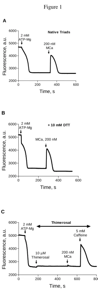
-ATPase (SERCA), was started by adding Mg-ATP to triad-enriched SR vesicles isolated from rabbit fast skeletal muscle. Addition of Mg-ATP to native SR vesicles produced a fast decrease in extravesicular [Ca2+] (Fig. 1A), which decreased on average from 23.8 ± 5.9 µM (N = 7, measured with a calcium electrode) to < 0.15 µM, calculated from the decrease in probe fluorescence. Addition of MCa to native SR vesicles actively loaded with calcium induced fast Ca2+ release, as illustrated in the fluorescence record from the representative
experiment shown in Figure 1A.To favor the reduced state of RyR1, SR vesicles were pre-incubated with the reducing agent DTT before initiating Ca2+ uptake. Addition of MCa
effectively promoted Ca2+ release from calcium-loaded SR vesicles pre-incubated with
Ca2+ from native or DTT-treated vesicles was transient, indicating both that RyR1 channels
closed/inactivated rapidly after activation with MCa and that pre-incubation with DTT did not modify SERCA activity. Similar behaviors were observed in four independent experiments carried out with native or DTT-treated vesicles. To test the effects of RyR1 oxidation, we added thimerosal (10 µM) after Ca2+ uptake completion. In contrast to the
behavior displayed by native or DTT-treated vesicles, subsequent addition of MCa (200 nM) did not stimulate release, whereas further addition of caffeine effectively induced transient Ca2+ release (Fig. 1C), showing that thimerosal did not inhibit RyR1 or SERCA
function. This same behavior was observed in five independent experiments. Addition of a higher concentration of MCa (400 nM) did not induce Ca2+ release from thimerosal-treated
vesicles (not shown), suggesting that - at the low [Ca2+] present in the extravesicular
solution after Ca2+ uptake - oxidized RyR1 channels have significantly decreased MCa
affinity compared to native or reduced channels.
The results illustrated in Figure 1 show that the reducing agent DTT did not hinder MCa-induced Ca2+ release through RyR1 channels while incubation with the SH-oxidizing
agent thimerosal prevented MCa-induced but not caffeine-induced Ca2+ release. Thus,
redox agents have opposite effects on Ca2+ release induced by MCa or caffeine, since
caffeine is a more effective agonist of RyR1-mediated Ca2+ release under oxidizing than
under reducing conditions (Beltrán et al, manuscript in preparation). To test the hypothesis that redox agents modify the effectiveness of MCa as RyR1 agonist, we measured under different redox conditions the effects of MCa on vesicular [3H]-ryanodine binding and on
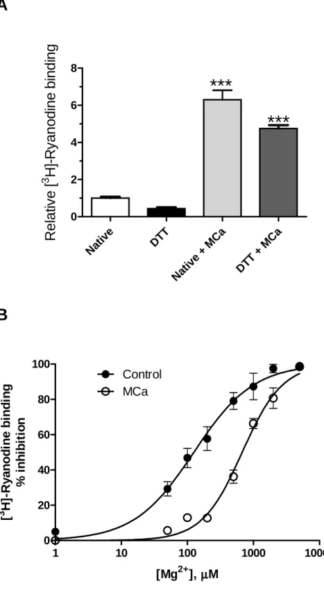
3.2 Maurocalcine increases equilibrium [3H]-ryanodine binding. Effects of DTT and Mg2+
The plant alkaloid ryanodine is a highly selective reagent, considered the gold standard to monitor RyR channel function. A good correlation between single RyR1 channel activity and [3H]-ryanodine binding density to skeletal SR vesicles has been
reported [23, 24]. Maurocalcine stimulated [3H]-ryanodine binding density when added to
native SR vesicles (Fig. 2A), which correlates with the stimulation of Ca2+ release
described above. Native (untreated) SR vesicles incubated with MCa (50 nM) exhibited an increase of 6.3 ± 0.5 fold (Mean ± SE, N=3) in equilibrium [3H]-ryanodine binding relative
to the values determined in the absence of MCa. Vesicles incubated with DTT displayed a 60% reduction, on average, of [3H]-ryanodine binding relative to native vesicles (Fig. 2A),
confirming the inhibitory effects of reducing agents on [3H]-ryanodine binding to RyR
channels [3]. Addition of 50 nM MCa to DTT-treated SR vesicles, however, resulted in higher stimulation of [3H]-ryanodine binding density compared with native vesicles (Fig.
2A). In DTT-treated vesicles MCa increased [3H]-ryanodine binding 11.1 ± 1.4 fold (N=3)
relative to vesicles incubated only with DTT, about twice the stimulation induced by MCa in native vesicles (p = 0.048). Therefore, DTT enhanced the effect of MCa on [3
H]-ryanodine binding, which could reflect an increase in apparent affinity of RyR1 for channel activation by MCa. Irreversible RyR1 inhibition induced by long exposure to thimerosal, such as that required to measure equilibrium [3H]-ryanodine binding density, precluded a
Several reports indicate that Mg2+ is a potent inhibitor of RyR1 activity [1].
Interestingly, oxidizing reagents decrease the inhibitory effects of Mg2+ on RyR1 activity
[11] while reducing agents increase the Mg2+ inhibition of [3H]-ryanodine binding to
endoplasmic reticulum vesicles isolated from rat brain cortex [3]. Here, we investigated if MCa modified the inhibition of [3H]-ryanodine binding to RyR1 produced by Mg2+. As
illustrated in Figure 2B, incubation of native skeletal SR vesicles with 200 nM MCa significantly decreased the inhibitory effects of Mg2+ on equilibrium [3H]-ryanodine
binding, which became somewhat cooperative since MCa increased nHill from 1.0 ± 0.1 to
1.4 ± 0.1 (p = 0.047). Native SR vesicles incubated with MCa displayed a 5-fold higher Ki
value for Mg2+-mediated inhibition of [3H] ryanodine binding relative to the Ki value for
Mg2+ exhibited by control vesicles (p = 0.008; see Fig. 2).
3.3 Effects of maurocalcine on RyR1 single channel activity
An earlier study showed that MCa induces a distinct long-lasting sub-conductance state in native RyR1 channels incorporated in planar lipid bilayers [15]. We have previously reported that single RyR1 channels display three different responses to cytoplasmic [Ca2+] changes (Fig. S2), which depend on RyR channel redox state [3, 8, 22].
Incubation with reducing agents favors the appearance of channels with “low activity”, characterized by Po values < 0.1 in the [Ca2+] range 0.1-500 µM. Partial RyR oxidation of
“low activity” channels usually generates the “moderate activity” response, the typical bell-shaped response to cytoplasmic calcium previously reported for native skeletal RyR1 channels [25], with maximal activity in the 10-30 µM [Ca2+] range and Po values < 0.1 at
0.1 or 500 µM [Ca2+]. Further oxidation increases channel activity at low cis [Ca2+], increases markedly Po to values as high as 0.99 at [Ca2+] > 100 µM and suppresses the
inhibitory effect of 500 µM cytoplasmic [Ca2+]; we call these channel behavior the “high activity” response. Here, we tested the effects of MCa on RyR1 single channels incorporated into lipid bilayers that displayed either the low or the moderate activity behavior (Fig. S2). The effects of MCa were evaluated using two parameters, i) PMCa that
represents the fractional time the channel spends in the MCa induced sub-conductance state and ii) PoB that represents the fractional time spent in the open state in the presence of MCa,
during the intervals bridging two openings at the sub-conductance level (See Fig. S1). The low activity single RyR1 channel depicted in Figure 3A displayed at 10 µM [Ca2+] a Po value < 0.01. Addition of 5 nM MCa induced channel openings to a
well-defined sub-conductance state, with PMCa = 0.50 and PoB = 0.01 (Fig. 3B, top trace). In the
continuous presence of MCa, lowering cytoplasmic [Ca2+] to 1 µM by addition of HEDTA as chelating agent decreased PMCa to a value of 0.31 while PoB stayed at 0.01 (Fig. 3B,
center record). Further lowering cytoplasmic [Ca2+] to 0.1 µM decreased PMCa even more,
to a value of 0.24, while PoB decreased to 0.00 (Fig. 3B, lower record).
The single moderate activity RyR1 channel, illustrated in Figure 4A, displayed Po
values of 0.01 at 500 µM [Ca2+] (top trace) and of 0.65 at 10 µM [Ca2+] (lower trace), respectively. Addition of 5 nM MCa at 10 µM cytoplasmic [Ca2+] prompted the emergence of the sub-conductance state, with PMCa = 0.84 and PoB = 0.71 (Fig. 4B, top trace). In the
continuous presence of MCa, lowering cytoplasmic [Ca2+] to 1 µM decreased PMCa to a
to 0.1 µM decreased both PMCa and PoB to 0.00 (Fig. 4B, lower record). Therefore, the
reduction in cytoplasmic [Ca2+] in the range of 10 to 0.1 µM decreased both PMCa and PoB.
Average PMCa and PoB values obtained at different cytoplasmic [Ca2+] from several
single channels that spontaneously displayed either low or moderate activity are illustrated in Figure 5. At 10 µM [Ca2+], addition of 5 nM MCa induced the sub-conductance state in
both low and moderate activity channels, which displayed PMCa values of 0.51 ± 0.10 (N=6)
and 0.78 ± 0.05 (N=5), respectively. In contrast, addition of 5 nM MCa at near resting [Ca2+] (0.1 µM) modified only low activity channels, which reached PMCa values of 0.24 ±
0.04 (N=11) that differed significantly from the PMCa values of 0.03 ± 0.03 (N=4) exhibited
by moderate activity channels (p = 0.001). The Po values were similar to the PoB values
obtained in low or moderate activity channels, respectively, suggesting that 5 nM MCa had no effect on channel activity between the sub-conductance events (Fig. 5). As expected, however, low activity channels displayed much lower Po and PoB values than moderate
activity channels at 10 µM [Ca2+], but not at 0.1 µM [Ca2+] (see Fig. 5).
Thimerosal increases markedly the response of low activity RyR1 channels to cytoplasmic [Ca2+] and sequentially promotes the moderate and the high activity responses
[8]. To test if thimerosal affects the single channel response to MCa, we added thimerosal to low activity channels treated with MCa. The single channel illustrated in Figure 6 displayed at 10 µM cytoplasmic [Ca2+] a Po value < 0.01 (Fig. 6A, top trace), typical of a
low activity channel. After lowering cytoplasmic [Ca2+] to 0.1 µM and adding 5 nM MCa, the channel exhibited the MCa-induced sub-conductance state, with PMCa = 0.08 and PoB =
0.01 (Fig. 6A, lower trace). Thimerosal was added to the cytoplasmic side to a final concentration of 200 µM; after 80 s, the cytoplasmic chamber was perfused with 20 ml of
225 mM HEPES-Tris, pH 7.4. Subsequent addition of 5 nM MCa at 0.1 µM cytoplasmic [Ca2+] failed to elicit the sub-conductance state observed before incubation with thimerosal,
although PoB reached 0.02 (Fig. 6B, lower trace). In three independent experiments carried
out at cytoplasmic [Ca2+] = 0.1 µM, single low activity RyR1 channels displayed average values of PMCa = 0.07 ± 0.01 and PoB = 0.00 ± 0.00 after addition of 5 nM MCa. Subsequent
addition of thimerosal as above decreased PMCa to 0.01 ± 0.01 and increased PoB to 0.02 ±
0.01 (N=3). Therefore, incubation of low activity channels with thimerosal precludes RyR1 activation by 5 nM MCa at 0.1 µM cytoplasmic [Ca2+], a response probably due to modification of RyR1 cysteine residues by thimerosal.
3.4 Mg2+ inhibits the effects of Maurocalcine on RyR1 single channel activity
In view of the fact that SR vesicles incubated with MCa displayed 5-fold higher Ki
for Mg2+ inhibition of [3H] ryanodine binding when compared to control vesicles (Fig. 2),
we investigated next the joint effects of Mg2+ and MCa on low activity single RyR1
channels. As illustrated in Figure 7A, after addition of 5 nM MCa at 10 µM cytoplasmic [Ca2+] a low activity channel displayed PMCa = 0.68 and PoB = 0.06 (top record). Subsequent
increase of cytoplasmic [Mg2+] to 30 µM, decreased PMCa to 0.35 and PoB to 0.02 (center
record). Increasing cytoplasmic [Mg2+] to 300 µM further decreased PMCa to 0.09 and PoB to
0.00 (lower record). In this experiment, increasing cytoplasmic [Mg2+] reduced PMCa with
K0.5 = 30 ± 8 µM. Similar results were obtained with another low activity channel.
The moderate activity channel illustrated in Figure 7B displayed PMCa = 0.95 and PoB
record). Subsequent increase of cytoplasmic [Mg2+] to 30 µM had negligible effect on both PMCa and PoB (center record), whereas further increase to 100 µM free [Mg2+] decreased
PMCa to 0.24 and PoB to 0.09 (lower record). In three independent experiments with
moderate activity channels, cytoplasmic [Mg2+] reduced PMCa and PoB with K0.5 of 72 ± 19
µM and 61 ± 13 µM, respectively. Therefore, the apparent affinity values of cytoplasmic [Mg2+] for inhibition of PMCa and PoB were comparable (p=0.662), favoring the possibility
that both effects are related. Moreover, the apparent affinity of Mg2+ inhibitory effects is
seemingly higher in low than in moderate activity RyR1 channels.
4. DISCUSSION
As pointed out earlier [26], Ca2+ release from the SR is significantly more sensitive
to redox agents than Ca2+ uptake. The remarkable effects of redox agents on SR Ca2+
release reside in the great sensitivity of RyR1 channels to oxidizing/reducing agents, which by changing RyR1 redox state define the response of the channel to agonists and inhibitors. Several authors have proposed that RyR channels act as cellular redox sensors [26-29]. The redox state of a few highly reactive cysteine residues, defined as such because they react with redox agents at physiological pH, determines RyR channel activation/inhibition by physiological agonists/inhibitors, and in particular, it conditions how RyR channels respond to changes in cytoplasmic [Ca2+] or [Mg2+] [4].
The results presented in this work show that redox reagents modify the agonist effects of MCa on skeletal RyR1 in a complex manner. The sulfhydryl-reducing reagent DTT, which strongly inhibits RyR1 channel activation by Ca2+ [8], did not prevent the
stimulation of Ca2+ release, or the opening of RyR1 single channels to a sub-conductance
state, produced by MCa addition at near resting [Ca2+] levels (0.1 µM [Ca2+]). Moreover,
MCa increased [3H]-ryanodine binding to DTT-treated vesicles two-fold when compared
with native vesicles. In contrast, at 0.1 µM [Ca2+] the oxidizing reagent thimerosal
prevented both MCa-induced Ca2+ release from SR vesicles and the emergence of the
typical sub-conductance state induced by MCa in single RyR1 channels. Thus, the present results concerning the effects of redox agents on the agonist action of MCa on Ca2+ efflux,
ryanodine binding and single channel sub-conductance opening, provide a consistent framework.
The fact, however, that PMCa decreased as single channel activity diminished when
lowering cytoplasmic [Ca2+] (Figs. 2-4), discards the possibility that the redox-sensitivity of
the agonist effects of MCa is simply due to an increase in the apparent affinity of Ca2+
binding site(s) involved in RyR1 activation. If this were the case, an oxidizing agent such as thimerosal that favors channel activation by low [Ca2+], should have increased PMCa;
likewise, moderate activity channels should have displayed more activation by MCa than low activity channels, exactly the opposite effects as those observed. Therefore, we propose that in spite of the inhibition of RyR1 channel activation by Ca2+ produced by reducing
agents, the reduction of RyR1 cysteine residues enhances the binding affinity of MCa to RyR1. Accordingly, the stimulation by MCa of RyR1-mediated Ca2+ release at 0.1 µM
[Ca2+] under reducing conditions would arise from increased MCa binding to reduced
RyR1 (or low activity) channels, which are poorly activated by low [Ca2+] but would have
moderate activity) channels would exhibit scant MCa binding in spite of their higher activity, due to their lower affinity for MCa.
The simplest way to explain how both low and moderate activity channels decreased PMCa when channel activity diminished on lowering [Ca2+] or increasing [Mg2+] (see below)
is to propose that the binding site(s) for MCa in RyR1 are freely accessible in the open channel state. This feature may explain why MCa does not elicit Ca2+ release when injected
into resting skeletal muscle fibers [30], in which RyR1 channels are closed due to their mechanical interaction with the II-III loop of the neighboring DHPR [31-33]. A previous study suggested that MCa binds to RyR1 channels that open on membrane depolarization and that this interaction specifically alters the process of repolarization-induced closure of the channels [30]. Furthermore, voltage-activated low-amplitude local Ca2+ signals persist
after fiber repolarization in the presence of MCa [34]. Based on these combined results, we propose that MCa binding to open RyR1 channels gives rise to the MCa-induced sub-conductance state that transiently keeps the channels refractory to DHPR-induced closing after repolarization.
Single channel experiments show that cytoplasmic [Mg2+] reduced the agonist effect
of MCa on RyR1. Since the apparent affinity of [Mg2+] for the inhibitory effects on PMCa
and on PoB were comparable, the most straightforward mechanism of inhibition of the
agonist effect of MCa on RyR1 channel is the inhibition of Po by Mg2+, with no
modification in RyR1 affinity for MCa binding. We report here that MCa induces a significant decrease in Mg2+ inhibition of [3H]-ryanodine binding to native RyR1 channels,
suggesting that MCa binding to the RyR1 protein produces a decrease in the Mg2+ affinity
decreases RyR1 sensitivity to Mg2+ inhibition [9, 11]. Moreover, reducing agents seem to
increase markedly the affinity of RyR1 channels for MCa, suggesting the presence of cysteine residues in the MCa binding site(s). Maurocalcine binds to two discrete RyR1 regions, fragment 3 (residues 1021– 1631) and fragment 7 (residues 3201– 3661) [35]. Noteworthy, one cysteine residue susceptible to S-glutathionylation (Cys1591) is present in fragment 3 whereas fragment 7 contains a cysteine residue (Cys3635) that is susceptible to S-glutathionylation, S-nitrosylation and disulfide oxidation [6]. In addition, another cysteine residue (Cys3193) susceptible to S-glutathionylation is situated only eight amino acids away from the beginning of fragment 7. Based on the present results, we suggest that MCa binding to RyR1 modifies the environment of one or more of these cysteine residues, resulting in a decreased affinity for Mg2+. Conversely, modification of these residues by
oxidation with thimerosal would decrease the affinity of MCa binding to RyR1, albeit we cannot rule out other possibilities. Accordingly, it would be of interest to study if mutations of these three particular cysteine residues affect the stimulation of RyR1 activity by MCa.
ACKNOWLEDGMENTS
This work was supported by FONDECYT-FONDAP 15010006, ECOS-CONICYT C05B03 and ECOS-SUD. Hicham Bichraoui was a recipient from the Association Francaise contre les Myopathies.
ABREVIATIONS
[Ca2+] = free calcium concentration
[Mg2+] = free magnesium concentration
DHPR = dihydropyridine receptor
DTT = dithiothreitol
EGTA = ethyleneglycol-bis(ß-aminoethyl ether) N, N, N', N'-tetraacetic acid HEDTA = N-(2-hydroxyethyl)-ethylenediamine-triacetic acid
MCa = Maurocalcine
PMCa = fractional time spent by the channel in the sub-conductance state
induced by Maurocalcine
Po = fractional time spent by the channel in the open state
PoB = fractional time spent by the channel in the open state, excluding the
time spent in the sub-conductance state induced by Maurocalcine
RyR = Ryanodine receptor
RyR1 = type-1 Ryanodine receptor
SE = standard error
SERCA = sarcoplamic/endoplasmic Ca2+-ATPase
REFERENCES
1. M. Fill, J.A. Copello, Ryanodine receptor calcium release channels, Physiol Rev. 82 (2002) 893-922.
2. E. Rios, G. Brum, Involvement of dihydropyridine receptors in excitation-contraction coupling in skeletal muscle, Nature. 325 (1987) 717-720.
3. R. Bull, J.P. Finkelstein, A. Humeres, M.I. Behrens, C. Hidalgo, Effects of ATP, Mg2+, and redox agents on the Ca2+ dependence of RyR channels from rat brain cortex, Am J Physiol Cell Physiol. 293 (2007) C162-171.
4. C. Hidalgo, P. Donoso, Crosstalk between calcium and redox signaling: from molecular mechanisms to health implications, Antioxid Redox Signal. 10 (2008) 1275-1312.
5. A.A. Voss, J. Lango, M. Ernst-Russell, D. Morin, I.N. Pessah, Identification of hyperreactive cysteines within ryanodine receptor type 1 by mass spectrometry, J Biol Chem. 279 (2004) 34514-34520.
6. P. Aracena-Parks, S.A. Goonasekera, C.P. Gilman, R.T. Dirksen, C. Hidalgo, S.L. Hamilton, Identification of cysteines involved in S-nitrosylation, S-glutathionylation, and oxidation to disulfides in ryanodine receptor type 1, J Biol Chem. 281 (2006) 40354-40368. 7. J. Suko, G. Hellmann, Modification of sulfhydryls of the skeletal muscle calcium release channel by organic mercurial compounds alters Ca2+ affinity of regulatory Ca2+ sites in single channel recordings and [3H]ryanodine binding, Biochim Biophys Acta. 1404 (1998) 435-450.
8. J.J. Marengo, C. Hidalgo, R. Bull, Sulfhydryl oxidation modifies the calcium dependence of ryanodine-sensitive calcium channels of excitable cells, Biophys J. 74 (1998) 1263-1277.
9. P. Donoso, P. Aracena, C. Hidalgo, Sulfhydryl oxidation overrides Mg2+ inhibition of calcium-induced calcium release in skeletal muscle triads, Biophys J. 79 (2000) 279-286. 10. J. Suko, G. Hellmann, H. Drobny, Modulation of the calmodulin-induced inhibition of sarcoplasmic reticulum calcium release channel (ryanodine receptor) by sulfhydryl oxidation in single channel current recordings and [(3)H]ryanodine binding, J Membr Biol. 174 (2000) 105-120.
11. P. Aracena, G. Sanchez, P. Donoso, S.L. Hamilton, C. Hidalgo, S-glutathionylation decreases Mg2+ inhibition and S-nitrosylation enhances Ca2+ activation of RyR1 channels, J Biol Chem. 278 (2003) 42927-42935.
12. H. Westerblad, D.G. Allen, Emerging roles of ROS/RNS in muscle function and fatigue, Antioxid Redox Signal. 15 (2011) 2487-2499.
13. A. Mosbah, R. Kharrat, Z. Fajloun, J.G. Renisio, E. Blanc, J.M. Sabatier, M. El Ayeb, H. Darbon, A new fold in the scorpion toxin family, associated with an activity on a ryanodine-sensitive calcium channel, Proteins. 40 (2000) 436-442.
14. E. Esteve, K. Mabrouk, A. Dupuis, S. Smida-Rezgui, X. Altafaj, D. Grunwald, J.C. Platel, N. Andreotti, I. Marty, J.M. Sabatier, M. Ronjat, M. De Waard, Transduction of the scorpion toxin maurocalcine into cells. Evidence that the toxin crosses the plasma membrane, J Biol Chem. 280 (2005) 12833-12839.
15. Z. Fajloun, R. Kharrat, L. Chen, C. Lecomte, E. Di Luccio, D. Bichet, M. El Ayeb, H. Rochat, P.D. Allen, I.N. Pessah, M. De Waard, J.M. Sabatier, Chemical synthesis and characterization of maurocalcine, a scorpion toxin that activates Ca2+ release channel/ryanodine receptors, FEBS Lett. 469 (2000) 179-185.
16. L. Chen, E. Esteve, J.M. Sabatier, M. Ronjat, M. De Waard, P.D. Allen, I.N. Pessah, Maurocalcine and peptide A stabilize distinct subconductance states of ryanodine receptor type 1, revealing a proportional gating mechanism, J Biol Chem. 278 (2003) 16095-16106. 17. E. Esteve, S. Smida-Rezgui, S. Sarkozi, C. Szegedi, I. Regaya, L. Chen, X. Altafaj, H. Rochat, P. Allen, I.N. Pessah, I. Marty, J.M. Sabatier, I. Jona, M. De Waard, M. Ronjat, Critical amino acid residues determine the binding affinity and the Ca2+ release efficacy of maurocalcine in skeletal muscle cells, J Biol Chem. 278 (2003) 37822-37831.
18. C. Hidalgo, J. Jorquera, V. Tapia, P. Donoso, Triads and transverse tubules isolated from skeletal muscle contain high levels of inositol 1,4,5-trisphosphate, J Biol Chem. 268 (1993) 15111-15117.
19. R. Bull, J.J. Marengo, B.A. Suarez-Isla, P. Donoso, J.L. Sutko, C. Hidalgo, Activation of calcium channels in sarcoplasmic reticulum from frog muscle by nanomolar concentrations of ryanodine, Biophys J. 56 (1989) 749-756.
20. R. Bull, J.J. Marengo, Sarcoplasmic reticulum release channels from frog skeletal muscle display two types of calcium dependence, FEBS Lett. 331 (1993) 223-227.
21. J.J. Marengo, R. Bull, C. Hidalgo, Calcium dependence of ryanodine-sensitive calcium channels from brain cortex endoplasmic reticulum, FEBS Lett. 383 (1996) 59-62.
22. R. Bull, J.J. Marengo, J.P. Finkelstein, M.I. Behrens, O. Alvarez, SH oxidation coordinates subunits of rat brain ryanodine receptor channels activated by calcium and ATP, Am J Physiol Cell Physiol. 285 (2003) C119-128.
23. R. Coronado, J. Morrissette, M. Sukhareva, D.M. Vaughan, Structure and function of ryanodine receptors, Am J Physiol. 266 (1994) C1485-1504.
24. G. Meissner, Ryanodine receptor/Ca2+ release channels and their regulation by endogenous effectors, Annu Rev Physiol. 56 (1994) 485-508.
25. M. Fill, R. Coronado, J.R. Mickelson, J. Vilven, J.J. Ma, B.A. Jacobson, C.F. Louis, Abnormal ryanodine receptor channels in malignant hyperthermia, Biophys J. 57 (1990) 471-475.
26. I.N. Pessah, K.H. Kim, W. Feng, Redox sensing properties of the ryanodine receptor complex, Front Biosci. 7 (2002) a72-79.
27. J.P. Eu, J. Sun, L. Xu, J.S. Stamler, G. Meissner, The skeletal muscle calcium release channel: coupled O2 sensor and NO signaling functions, Cell. 102 (2000) 499-509.
28. R. Xia, T. Stangler, J.J. Abramson, Skeletal muscle ryanodine receptor is a redox sensor with a well defined redox potential that is sensitive to channel modulators, J Biol Chem. 275 (2000) 36556-36561.
29. C. Hidalgo, Cross talk between Ca2+ and redox signalling cascades in muscle and neurons through the combined activation of ryanodine receptors/Ca2+ release channels, Philos Trans R Soc Lond B Biol Sci. 360 (2005) 2237-2246.
30. S. Pouvreau, L. Csernoch, B. Allard, J.M. Sabatier, M. De Waard, M. Ronjat, V. Jacquemond, Transient loss of voltage control of Ca2+ release in the presence of maurocalcine in skeletal muscle, Biophys J. 91 (2006) 2206-2215.
31. T. Tanabe, K.G. Beam, B.A. Adams, T. Niidome, S. Numa, Regions of the skeletal muscle dihydropyridine receptor critical for excitation-contraction coupling, Nature. 346 (1990) 567-569.
32. X. Lu, L. Xu, G. Meissner, Activation of the skeletal muscle calcium release channel by a cytoplasmic loop of the dihydropyridine receptor, J Biol Chem. 269 (1994) 6511-6516. 33. J. Nakai, T. Tanabe, T. Konno, B. Adams, K.G. Beam, Localization in the II-III loop of the dihydropyridine receptor of a sequence critical for excitation-contraction coupling, J Biol Chem. 273 (1998) 24983-24986.
34. L. Csernoch, S. Pouvreau, M. Ronjat, V. Jacquemond, Voltage-activated elementary calcium release events in isolated mouse skeletal muscle fibers. J Membr Biol. 226 (2008) 43-55.
35. X. Altafaj, W. Cheng, E. Esteve, J. Urbani, D. Grunwald, J.M. Sabatier, R. Coronado, M. De Waard, M. Ronjat, Maurocalcine and domain A of the II-III loop of the dihydropyridine receptor Cav 1.1 subunit share common binding sites on the skeletal ryanodine receptor, J Biol Chem. 280 (2005) 4013-4016.
FIGURE LEGENDS
Figure 1. Effects of MCa on Ca2+ release from actively loaded triad-enriched SR vesicles.
Active loading with Ca2+ was initiated by addition of ATP-Mg, as indicated in the figure.
Changes in extravesicular [Ca2+] were determined with Calcium Green-2, as detailed in
Materials and Methods. In the three representative experiments illustrated (Panels A-C), addition of ATP-Mg promoted fast Ca2+ uptake, and decreased probe fluorescence to a
steady value corresponding to an extravesicular [Ca2+] < 150 nM (see Results). Subsequent
addition of MCa caused effective and transient Ca2+ release from native vesicles (Panel A)
or from vesicles pre-incubated for 30 min with 10 mM DTT (Panel B). Addition of the sulfhydryl oxidizing agent thimerosal after completion of Ca2+ uptake prevented Ca2+
release induced by MCa but not by caffeine (Panel C).
Figure 2. Maurocalcine enhances [3H] ryanodine binding to triad-enriched SR vesicles
and decreases the inhibitory effects of Mg2+. As illustrated in Panel A, incubation of
native vesicles with 50 nM MCa stimulated equilibrium [3H]-ryanodine binding density
(light grey bar) relative to the values determined in the absence of MCa (empty bar). Vesicles incubated with DTT displayed lower [3H]-ryanodine binding relative to native
vesicles (Panel A, black bar). Addition of 50 nM MCa to DTT-treated SR vesicles increased [3H]-ryanodine binding relative to vesicles incubated only with DTT (Panel A,
dark grey bar). ***: p < 0.0001 calculated by One-way ANOVA followed by Dunnett's multiple comparison test. As illustrated in Panel B, incubation of native skeletal SR
equilibrium [3H]-ryanodine binding. Fitting the experimental points to a non-linear Hill
function yielded nHill =1.0 ± 0.1 and Ki = 125.3 ± 8.8 µM for native vesicles, and nHill = 1.4
± 0.1 and Ki = 676.4 ± 49.0 µM for native SR vesicles incubated with 200 nM MCa.Values
represent Mean ± SE (N=3).
Figure 3. Effects of MCa on a low activity RyR1 channel. Representative current
recordings obtained with the same low activity single RyR1 channel in the absence (Panel A) or at 5 nM MCa (Panel B). PMCa and PoB values, calculated for the whole recorded
periods (at least 180 s) and the free cytoplasmic [Ca2+] are depicted above each current
trace. Addition of MCa induced channel openings to a well-defined sub-conductance current level, indicated by an arrow at the right of the current traces. Sequential reduction of cytoplasmic [Ca2+] using HEDTA and/or EGTA (see Materials and Methods),
progressively reduced both PMCa and PoB (second and third trace).
Figure 4. Effects of MCa on a moderate activity RyR1 channel. Representative current
recordings of the same moderate activity single RyR1 channel obtained in the absence (Panel A) or at 5 nM MCa (Panel B). PMCa and PoB values, calculated for the whole
recorded periods (at least 150 s) and the free cytoplasmic [Ca2+] are depicted above each
current trace. Addition of MCa prompted the emergence of the sub-conductance current level, as indicated by an arrow at the right of the top and middle traces of Panel B. Sequential reduction of cytoplasmic [Ca2+] using HEDTA and/or EGTA (see Materials and
Methods), progressively reduced both PMCa and PoB (second and third trace). Notice that
Figure 5. Comparative effects of MCa on low and moderate activity RyR1 channels. Bars
represent mean Po, PoB and PMCa values (± SE) obtained from several single channels that
spontaneously displayed either low (empty bars) or moderate activity (black bars). As illustrated in Panel A, addition of 5 nM MCa at 10 µM [Ca2+] induced the sub-conductance
state in both low and moderate activity channels. In contrast, as illustrated in Panel B, addition of 5 nM MCa at 0.1 µM [Ca2+] modified only low activity channels. Numbers
above or on each bar represent the number of single channel data included in each condition. *: p < 0.05; **: p = 0.001.
Figure 6. Thimerosal treatment inhibited MCa activation of a single RyR1 channel at 0.1
µM [Ca2+]. Panel A shows representative current recordings obtained from a single low
activity channel at the indicated cytoplasmic [Ca2+] before (upper trace) and after addition
of MCa (lower trace). Sub-conductance current level is indicated, as usual, with an arrow at the right of lower trace. PMCa and PoB values, calculated for the whole recorded periods (at
least 70 s) and cytoplasmic [Ca2+] are displayed above each trace. Panel B was obtained
with the same channel recorded in Panel A, after incubation with 200 µM thimerosal for 80 s followed by extensive perfusion of the cis chamber to eliminate non reacted thimerosal (for further details, see Results). At 0.1 µM [Ca2+], addition of 5 nM MCa did not elicit the
sub-conductance state displayed by the channel before incubation with thimerosal (compare trace in Panel B with lower trace in Panel A).
Figure 7. Mg2+ inhibits the effects of Maurocalcine on low and moderate activity RyR1
channels. Representative current recordings were obtained, at 10 µM cytoplasmic [Ca2+]
and in the presence of 5 nM MCa, from single channels that spontaneously displayed either low (Panel A) or moderate activity (Panel B). In each case, the same channel was sequentially exposed to increasing cytoplasmic [Mg2+], as indicated near each current trace.
PMCa and PoB values, calculated from the whole recorded periods (at least 180 s), are
depicted above the recordings. The arrows indicate the sub-conductance current level induced by MCa.
Figure 1 0 200 400 600 2000 3000 4000 5000 6000 200 nM MCa 2 mM ATP-Mg Native Triads A Time, s F lu o re s c e n c e , a .u . 0 200 400 600 2000 3000 4000 5000 6000 MCa, 200 nM 2 mM ATP-Mg + 10 mM DTT B Time, s F lu o re s c e n c e , a .u . 0 200 400 600 800 2000 3000 4000 5000 6000 10µM Thimerosal 200 nM MCa 5 mM Caffeine 2 mM ATP-Mg Thimerosal C Time, s F lu o re s c e n c e , a .u .
Figure 2
A
Nat ive DTT Nat ive + M Ca DTT + M Ca 0 2 4 6 8***
***
R
e
la
tiv
e
[
3H
]-R
y
a
n
o
d
in
e
b
in
d
in
g
B
1 10 100 1000 10000 0 20 40 60 80 100 Control MCa [Mg2+], µµµµM [ 3 H ]-R y a n o d in e b in d in g % i n h ib it io nSUPPLEMENTARY MATERIAL
Figure legends
Figure S1. Graphical representation of PMCa and PoB. To calculate PMCa values we added
all individual dwell times in which the channel spent in the MCa-induced sub-conductance state, defined as si, and divided this sum by the total recorded time. To calculate PoB values
we added all oj values, which represent the channel dwell time in the full open state, and
divided this sum by the sum of the bi values, which represent the dwell times in which the
channel did not reside in the MCa-induced sub-conductance state.
Figure S2. Single RyR1 channels spontaneously display three different responses to
cytoplasmic [Ca2+]. These responses include the low activity response (filled triangles),
characterized by Po values < 0.1 in the [Ca2+] range 0.1-500 µM; the moderate activity
response (open circles) with maximal Po in the 10-30 µM [Ca2+] range and Po < 0.1 at 0.1
or 500 µM [Ca2+], and the high activity response (filled circles) characterized by significant activity at 0.1 µM [Ca2+] and Po close to 1.0 in the 1.0-500 µM [Ca2+] range. The low