HAL Id: inserm-00759004
https://www.hal.inserm.fr/inserm-00759004
Submitted on 29 Nov 2012
HAL is a multi-disciplinary open access
archive for the deposit and dissemination of
sci-entific research documents, whether they are
pub-lished or not. The documents may come from
teaching and research institutions in France or
abroad, or from public or private research centers.
L’archive ouverte pluridisciplinaire HAL, est
destinée au dépôt et à la diffusion de documents
scientifiques de niveau recherche, publiés ou non,
émanant des établissements d’enseignement et de
recherche français ou étrangers, des laboratoires
publics ou privés.
How do T-type calcium channels control low-threshold
exocytosis?
Norbert Weiss, Gerald Zamponi, Michel de Waard
To cite this version:
Norbert Weiss, Gerald Zamponi, Michel de Waard. How do T-type calcium channels control
low-threshold exocytosis?. Communicative and Integrative Biology, Taylor & Francis Open, 2012, 5 (4),
pp.377-80. �10.4161/cib.19997�. �inserm-00759004�
How do T-type calcium channels control low-threshold exocytosis?
Norbert Weiss,1,* Gerald W. Zamponi1and Michel De Waard2,3
1Hotchkiss Brain Institute; Department of Physiology and Pharmacology; University of Calgary; Calgary, AB Canada;2Grenoble Institute of Neuroscience;
La Tronche, France;3University Joseph Fourier; Grenoble, France
Keywords: calcium channel, T-type, Cav3.2 channel, SNARE, syntaxin-1A,
SNAP-25, exocytosis, neuron, chromaffin cell, MPC cell Submitted: 03/11/12 Accepted: 03/12/12
http://dx.doi.org/10.4161/cib.19997
*Correspondence to: Norbert Weiss; Email: [email protected] or [email protected]
Addendum to: Weiss N, Hameed S, Fernández-Fernández JM, Fablet K, Karmazinova M, Poillot C, et al. A Ca(v)3.2/syntaxin-1A signaling complex controls T-type channel activity and low-thresh-old exocytosis. J Biol Chem 2012; 287:2810–8; PMID:22130660; http://dx.doi.org/10.1074/jbc. M111.290882
L
ow-voltage-activated T-type calcium channels act as a major pathway for calcium entry near the resting mem-brane potential in a wide range of neuronal cell types. Several reports have uncovered an unrecognized feature of T-type channels in the control of vesi-cular neurotransmitter and hormone release, a process so far thought to be mediated exclusively by high-voltage-acti-vated calcium channels. However, the underlying molecular mechanisms link-ing T-type calcium channels to vesicular exocytosis have remained enigmatic. In a recent study, we have reported that Cav3.2 T-type channel forms a signalingcomplex with the neuronal Q-SNARE syntaxin-1A and SNAP-25. This inter-action that relies on specific Cav3.2
molecular determinants, not only modu-lates T-type channel activity, but was also found essential to support low-threshold exocytosis upon Cav3.2 channel
expres-sion in MPC 9/3L-AH chromaffin cells. Overall, we have indentified an unrecog-nized regulation pathway of T-type calcium channels by SNARE proteins, and proposed the first molecular mecha-nism by which T-type channels could mediate low-threshold exocytosis.
Depolarization-evoked synaptic transmis-sion relies on the calcium (Ca2+)-regulated
release of quantal packets of neuro-transmitters following fusion of synaptic vesicles with the presynaptic plasma mem-brane.1It is well established that neuronal
voltage-gated Ca2+channels, by converting
electrical signals into intracellular Ca2+
concentration elevations, play a key role in triggering evoked neurotransmitter
release.2-4 Hence, Ca2+ entry through
high-voltage-activated (HVA) channels (N-, P/Q-, and in some extent and particular cell populations, L- and R-type) into presynaptic nerve terminals in response to action potentials supports a transient Ca2+microdomain5essential for
synaptic exocytosis. However, the obser-vation that some neurons can release functionally significant amounts of neuro-transmitter below the threshold of action potentials6 questioned the possible
involvement of another source of Ca2+
ions, independent of HVA channels activation.
In contrast to HVA channels, low-voltage-activated (LVA) T-type Ca2+
channels activate in response to subthres-hold membrane depolarizations between -65 mV and -50 mV and thus represent an important source of Ca2+ entry near the
resting membrane potential. Hence, besides controlling important physiological processes by regulating neuronal excitabi-lity, pacemaker activity and post-inhibi-tory rebound burst firing, mounting evidences from various neuronal cell types suggest an efficient role of T-type channels in fast and low-threshold exocytosis.7-10
Until now, however, the mechanism whereby these channels support exocytosis events at the molecular level remained a mystery. In a recent study, we provided compelling evidence of the existence of a Cav3.2/syntaxin-1A
mole-cular complex essential for T-type-depend-ent exocytosis.11
In mammalian synapses, interaction of several members of the vesicle-docking / release machinery (including syntaxin-1A/ 1B and SNAP-25) onto a synprint (syn-aptic protein interaction site) domain
ARTICLE ADDENDUM
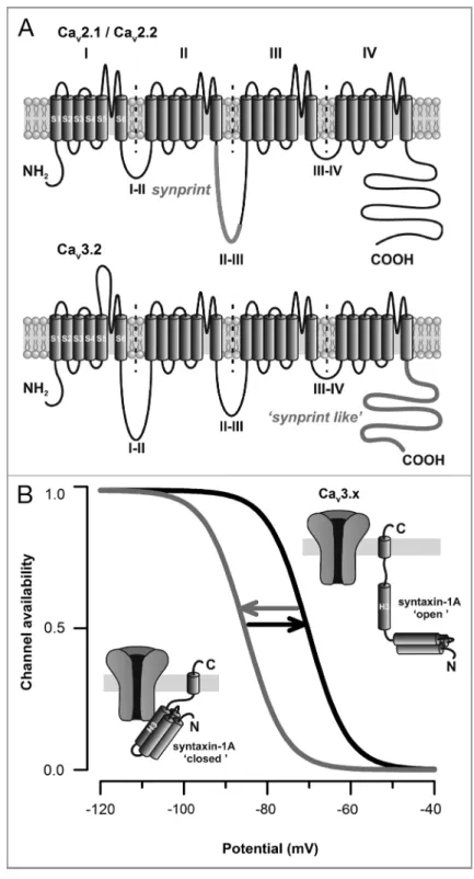
located within the intracellular loop between domains II and III of the Cav2.112 and Cav2.213channels (Fig. 1A,
top panel) ensures a close localization of the secretory vesicles near the Ca2+source.
In turn, both synatxin-1A/1B and SNAP-25 modulate calcium channel activity14,15
to fine tune Ca2+ entry and synaptic
strength. However, in contrast to HVA channels, all of the three T-type channel members (i.e Cav3.1, Cav3.2 and Cav3.3)
lack the consensus synprint site, making the molecular understanding of the involvement of these channels in the exocytosis process quite difficult. Our observation that Cav3.2 channels associate
with syntaxin-1A in central neurons prompted us to investigate the possible existence of specific Cav3.2 channel
molecular determinants other than the consensus HVA synprint domain. Using biochemical and cellular trafficking approaches, we demonstrated that syn-taxin-1A, as well as SNAP-25, interact with the C-terminal domain of Cav3.2
channel (Fig. 1A, lower panel). Moreover, using patch-clamp recordings performed on tsA-201 cells expressing Cav3.2
chan-nels, we demonstrated that co-expression of a syntaxin-1A in its “closed” conforma-tional state (i.e the conformation adopted by the syntaxin-1A in isolation or in interaction with Munc18)16,17 potently
decreases Cav3.2 channel availability by
shifting the voltage-dependence of inac-tivation toward more hyperpolarized membrane potentials, similarly to what was previously reported for N- and P/Q-type channels.14,15,18-20 Interestingly, this
regulation was abolished upon co-expres-sion of SNAP-25, and not observed with a constitutively “open” syntaxin-1A (i.e the conformation adopted upon its association with SNAP-25)18 (Fig. 1B). Given that
syntaxin-1A undergoes a conformational switch from a “closed” to an “open” conformation during the vesicle release cycle,16,21,22 this suggests that syntaxin-1A
may be able to dynamically regulate T-type channel availability during various stages of exocytosis. Interestingly, although T-type channels utilize distinct molecular determinants to interact with SNARE proteins (the C-terminal domain vs. the classical synprint of the II-III linker), they are subjected to a similar SNARE
regulation. Does this observation question the molecular mechanism by which bind-ing of syntaxin-1A produces changes in channel gating? Earlier reports have shown
that reorganization of intramolecular inter-actions among the main intracellular loops of Cav2 channels critically influence
channel inactivation.23-30 Mapping the
Figure 1. SNARE proteins modulate high- and low-voltage-gated calcium channels via distinct molecular determinants. (A) Membrane topology of voltage-gated calcium channels highlighting the localization of thesynprint site located within the intracellular linker between domains II and III of Cav2.1/Cav2.2 channels (top panel), and the“synprint like” domain of Cav3.x channels
(bottom panel) located within the C-terminal domain of the channel. (B) Voltage-dependence of Cav3.x channel availability during the conformational switch of syntaxin-1A.
intramolecular interactions of T-type channels along with the characterization of the minimal sequence engaged in the interaction with SNARE proteins will provide important structural information on how syntaxin-1A modulates channel gating.
It is well known that direct interaction of SNARE proteins with Cav2.1 and
Cav2.2 channels is critical for
depolariza-tion-evoked neurotransmitter release. Hence, disruption of the Ca2+
channel-SNARE proteins coupling by deletion of thesynprint domain or by peptides derived from thesynprint sequence, alters synaptic transmission.31-34 We revealed that
similarly to HVA channels, T-type channels-mediated exocytosis relies on a channel-SNARE protein interaction. Indeed, membrane capacitance recordings performed on MPC 9/3L-HA chromaffin cells expressing Cav3.2 channels revealed
robust voltage-dependent exocytosis which was totally prevented by co-expression of the Cav3.2 C-terminal domain (i.e the
synaptic protein interaction site of Cav3.2). Ablation of Cav3.2-dependent
exocytosis most likely results from the specific uncoupling of the channel with SNARE proteins and not from a side
alteration of the exocytosis machinery by itself because no alteration was observed when exocytosis was induced by direct intracellular Ca2+ elevation. Hence, we
showed that similarly to HVA channels, a physical coupling between SNARE pro-teins and T-type channels is critical for T-type-dependent exocytosis. Considering the relative small conductance of T-type channels35 and the restricted diffusion of
Ca2+ due to the high Ca2+ buffering
capacity of neuronal cells,36it is
conceiv-able that this interaction allows the close localization of the vesicle-docking / release machinery in close proximity to the Ca2+
source in order to efficiently sense Ca2+
elevation. However, we cannot exclude the possibility that interaction of T-type channels with SNARE proteins could form a macromolecular complex through which channel conformational changes following membrane depolarization would work as an on/off molecular switch of secretion by controlling the ultimate conformational change of the releasing complex as pre-viously proposed for HVA channels.37,38
Although this concept still requires further investigation, the use on a non-conducting channel to investigate T-type-dependent secretion would definitively provide
interesting information about the func-tional importance of T-type channel interaction with SNARE proteins.
Overall, we revealed an unrecognized regulation of low-voltage-activated T-type Ca2+ channels by SNARE proteins, and
provide the first evidence for a molecular mechanism by which these channels could mediate low-threshold exocytosis. We revealed that, although T-type Ca2+
chan-nels differ from HVA chanchan-nels by their molecular constituents, they possess the same ability to functionally interact with SNARE proteins, highlighting a key evolutionary mechanism for specialized fast and spatially delimited exocytosis.
Disclosure of Potential Conflicts of Interest
No potential conflicts of interest were disclosed.
Acknowledgments
N.W is supported by a postdoctoral fellowship from Alberta Innovates Health Solutions (AIHS) and Hotchkiss Brain Institute. G.W.Z is funded by the Canadian Institutes of Health Research, is a Canada Research Chair and AIHS Scientist.
References
1. Edwards RH. The neurotransmitter cycle and quantal size. Neuron 2007; 55:835-58; PMID:17880890; http://dx.doi.org/10.1016/j.neuron.2007.09.001 2. Llinás R, Sugimori M, Silver RB. Microdomains of
high calcium concentration in a presynaptic terminal. Science 1992; 256:677-9; PMID:1350109; http://dx. doi.org/10.1126/science.1350109
3. Neher E, Sakaba T. Multiple roles of calcium ions in the regulation of neurotransmitter release. Neuron 2008; 59:861-72; PMID:18817727; http://dx.doi.org/ 10.1016/j.neuron.2008.08.019
4. Weber AM, Wong FK, Tufford AR, Schlichter LC, Matveev V, Stanley EF. N-type Ca2+ channels carry the largest current: implications for nanodomains and transmitter release. Nat Neurosci 2010; 13:1348-50; PMID:20953196; http://dx.doi.org/10.1038/nn.2657 5. Schneggenburger R, Neher E. Presynaptic calcium and
control of vesicle fusion. Curr Opin Neurobiol 2005; 15:266-74; PMID:15919191; http://dx.doi.org/10. 1016/j.conb.2005.05.006
6. Ivanov AI, Calabrese RL. Intracellular Ca2+ dynamics during spontaneous and evoked activity of leech heart interneurons: low-threshold Ca currents and graded synaptic transmission. J Neurosci 2000; 20:4930-43; PMID:10864951
7. Carabelli V, Marcantoni A, Comunanza V, de Luca A, Díaz J, Borges R, et al. Chronic hypoxia up-regulates alpha1H T-type channels and low-threshold catecho-lamine secretion in rat chromaffin cells. J Physiol 2007; 584:149-65; PMID:17690152; http://dx.doi.org/10. 1113/jphysiol.2007.132274
8. Pan ZH, Hu HJ, Perring P, Andrade R. T-type Ca(2+)
channels mediate neurotransmitter release in retinal bipolar cells. Neuron 2001; 32:89-98; PMID: 11604141; http://dx.doi.org/10.1016/S0896-6273 (01)00454-8
9. Egger V, Svoboda K, Mainen ZF. Mechanisms of lateral inhibition in the olfactory bulb: efficiency and modulation of spike-evoked calcium influx into granule cells. J Neurosci 2003; 23:7551-8; PMID:12930793 10. Tang AH, Karson MA, Nagode DA, McIntosh JM,
Uebele VN, Renger JJ, et al. Nerve terminal nicotinic acetylcholine receptors initiate quantal GABA release from perisomatic interneurons by activating axonal T-type (Cav3) Ca²⁺ channels and Ca²⁺ release from stores. J Neurosci 2011; 31:13546-61; PMID: 21940446; http://dx.doi.org/10.1523/JNEUROSCI. 2781-11.2011
11. Weiss N, Hameed S, Fernández-Fernández JM, Fablet K, Karmazinova M, Poillot C, et al. A Ca(v)3.2/ syntaxin-1A signaling complex controls T-type channel activity and low-threshold exocytosis. J Biol Chem 2012; 287:2810-8; PMID:22130660; http://dx.doi. org/10.1074/jbc.M111.290882
12. Rettig J, Sheng ZH, Kim DK, Hodson CD, Snutch TP, Catterall WA. Isoform-specific interaction of the alpha1A subunits of brain Ca2+ channels with the
presynaptic proteins syntaxin and SNAP-25. Proc Natl Acad Sci U S A 1996; 93:7363-8; PMID:8692999; http://dx.doi.org/10.1073/pnas.93.14.7363 13. Sheng ZH, Rettig J, Takahashi M, Catterall WA.
Identification of a syntaxin-binding site on N-type calcium channels. Neuron 1994; 13:1303-13; PMID: 7993624; http://dx.doi.org/10.1016/0896-6273(94) 90417-0
14. Bezprozvanny I, Scheller RH, Tsien RW. Functional impact of syntaxin on gating of N-type and Q-type calcium channels. Nature 1995; 378:623-6; PMID: 8524397; http://dx.doi.org/10.1038/378623a0
15. Zhong H, Yokoyama CT, Scheuer T, Catterall WA.
;
Reciprocal regulation of P/Q-type Ca2+ channels by SNAP-25, syntaxin and synaptotagmin. Nat Neurosci 1999; 2:939-41; PMID:10526329; http://dx.doi.org/ 10.1038/1472116. Dulubova I, Sugita S, Hill S, Hosaka M, Fernandez I, Südhof TC, et al. A conformational switch in syntaxin during exocytosis: role of munc18. EMBO J 1999; 18:4372-82; PMID:10449403; http://dx.doi.org/10. 1093/emboj/18.16.4372
17. Brunger AT. Structure of proteins involved in synaptic vesicle fusion in neurons. Annu Rev Biophys Biomol Struct 2001; 30:157-71; PMID:11340056; http://dx. doi.org/10.1146/annurev.biophys.30.1.157 18. Jarvis SE, Barr W, Feng ZP, Hamid J, Zamponi GW.
Molecular determinants of syntaxin 1 modulation of N-type calcium channels. J Biol Chem 2002; 277:44399-407; PMID:12221094; http://dx.doi.org/ 10.1074/jbc.M206902200
19. Wiser O, Bennett MK, Atlas D. Functional interaction of syntaxin and SNAP-25 with voltage-sensitive L- and N-type Ca2+channels. EMBO J 1996; 15:4100-10;
PMID:8861939
20. Sutton KG, McRory JE, Guthrie H, Murphy TH,
<
Snutch TP. P/Q-type calcium channels mediate the activity-dependent feedback of syntaxin-1A. Nature 1999; 401:800-4; PMID:10548106; http://dx.doi.org/ 10.1038/4458621.
=
Fiebig KM, Rice LM, Pollock E, Brunger AT. Folding intermediates of SNARE complex assembly. Nat Struct Biol 1999; 6:117-23; PMID:10048921; http://dx.doi. org/10.1038/580322. Richmond JE, Weimer RM, Jorgensen EM. An open form of syntaxin bypasses the requirement for UNC-13 in vesicle priming. Nature 2001; 412:338-41; PMID: 11460165; http://dx.doi.org/10.1038/35085583 23. Restituito S, Cens T, Barrere C, Geib S, Galas S, De
Waard M, et al. The [beta]2a subunit is a molecular groom for the Ca2+ channel inactivation gate. J
Neurosci 2000; 20:9046-52; PMID:11124981 24. Geib S, Sandoz G, Cornet V, Mabrouk K, Fund-Saunier
O, Bichet D, et al. The interaction between the I-II loop and the III-IV loop of Cav2.1 contributes to voltage-dependent inactivation in a beta -voltage-dependent manner. J Biol Chem 2002; 277:10003-13; PMID:11790766; http://dx.doi.org/10.1074/jbc.M106231200 25. Sandoz G, Lopez-Gonzalez I, Stamboulian S, Weiss N,
Arnoult C, De Waard M. Repositioning of charged I-II loop amino acid residues within the electric field by beta subunit as a novel working hypothesis for the control of fast P/Q calcium channel inactivation. Eur J Neurosci 2004; 19:1759-72; PMID:15078550; http:// dx.doi.org/10.1111/j.1460-9568.2004.03216.x 26. Agler HL, Evans J, Tay LH, Anderson MJ, Colecraft
HM, Yue DT. G protein-gated inhibitory module of N-type (ca(v)2.2) ca2+channels. Neuron 2005;
46:891-904; PMID:15953418; http://dx.doi.org/10.1016/j. neuron.2005.05.011
27. Raghib A, Bertaso F, Davies A, Page KM, Meir A, Bogdanov Y, et al. Dominant-negative synthesis suppression of voltage-gated calcium channel Cav2.2 induced by truncated constructs. J Neurosci 2001; 21:8495-504; PMID:11606638
28. Page KM, Heblich F, Davies A, Butcher AJ, Leroy J, Bertaso F, et al. Dominant-negative calcium channel suppression by truncated constructs involves a kinase implicated in the unfolded protein response. J Neurosci 2004; 24:5400-9; PMID:15190113; http://dx.doi.org/ 10.1523/JNEUROSCI.0553-04.2004
29. Page KM, Heblich F, Margas W, Pratt WS, Nieto-Rostro M, Chaggar K, et al. N terminus is key to the dominant negative suppression of Ca(V)2 calcium channels: implications for episodic ataxia type 2. J Biol Chem 2010; 285:835-44; PMID:19903821; http://dx. doi.org/10.1074/jbc.M109.065045
30. Bucci G, Mochida S, Stephens GJ. Inhibition of synaptic transmission and G protein modulation by synthetic CaV2.2 Ca²⁺ channel peptides. J Physiol 2011; 589:3085-101; PMID:21521766; http://dx.doi. org/10.1113/jphysiol.2010.204735
31. Mochida S, Sheng ZH, Baker C, Kobayashi H, Catterall WA. Inhibition of neurotransmission by peptides containing the synaptic protein interaction site of N-type Ca2+channels. Neuron 1996; 17:781-8;
PMID:8893034; http://dx.doi.org/10.1016/S0896-6273(00)80209-3
32. Rettig J, Heinemann C, Ashery U, Sheng ZH, Yokoyama CT, Catterall WA, et al. Alteration of Ca2+
dependence of neurotransmitter release by disruption of Ca2+ channel/syntaxin interaction. J Neurosci 1997;
17:6647-56; PMID:9254677
33. Harkins AB, Cahill AL, Powers JF, Tischler AS, Fox AP. Deletion of the synaptic protein interaction site of the N-type (CaV2.2) calcium channel inhibits secretion in mouse pheochromocytoma cells. Proc Natl Acad Sci U S A 2004; 101:15219-24; PMID:15471993; http:// dx.doi.org/10.1073/pnas.0401001101
34. Keith RK, Poage RE, Yokoyama CT, Catterall WA, Meriney SD. Bidirectional modulation of transmitter release by calcium channel/syntaxin interactions in vivo. J Neurosci 2007; 27:265-9; PMID:17215385; http://dx. doi.org/10.1523/JNEUROSCI.4213-06.2007 35. Perez-Reyes E. Molecular physiology of
low-voltage-activated t-type calcium channels. Physiol Rev 2003; 83:117-61; PMID:12506128
36. Foehring RC, Zhang XF, Lee JC, Callaway JC. Endogenous calcium buffering capacity of substantia nigral dopamine neurons. J Neurophysiol 2009; 102:2326-33; PMID:19675297; http://dx.doi.org/10. 1152/jn.00038.2009
37. Atlas D. Signaling role of the voltage-gated calcium channel as the molecular on/off-switch of secretion. Cell Signal 2010; 22:1597-603; PMID:20388539; http://dx.doi.org/10.1016/j.cellsig.2010.04.003
38. Weiss N. Control of depolarization-evoked presynaptic