HAL Id: hal-03023550
https://hal.archives-ouvertes.fr/hal-03023550
Submitted on 25 Nov 2020
HAL is a multi-disciplinary open access
archive for the deposit and dissemination of sci-entific research documents, whether they are pub-lished or not. The documents may come from teaching and research institutions in France or abroad, or from public or private research centers.
L’archive ouverte pluridisciplinaire HAL, est destinée au dépôt et à la diffusion de documents scientifiques de niveau recherche, publiés ou non, émanant des établissements d’enseignement et de recherche français ou étrangers, des laboratoires publics ou privés.
How much energy do we need to ablate 1 mm3 of stone
during Ho:YAG laser lithotripsy? An in vitro study
Frédéric Panthier, Eugenio Ventimiglia, Laurent Berthe, Catherine Chaussain,
Michel Daudon, Steeve Doizi, Olivier Traxer
To cite this version:
Frédéric Panthier, Eugenio Ventimiglia, Laurent Berthe, Catherine Chaussain, Michel Daudon, et al.. How much energy do we need to ablate 1 mm3 of stone during Ho:YAG laser lithotripsy? An in vitro study. World Journal of Urology, Springer Verlag, 2020, 38 (11), pp.2945-2953. �10.1007/s00345-020-03091-5�. �hal-03023550�
How much energy do we need to ablate 1 mm
3of stone during Ho:YAG
laser lithotripsy? An in vitro study
Frédéric Panthier1,2,3 · Eugenio Ventimiglia1,2,4 · Laurent Berthe3 · Catherine Chaussain5 · Michel Daudon1,6 ·
Steeve Doizi1,2 · Olivier Traxer1,2
Abstract
Introduction Holmium:yttrium–aluminium–garnet (Ho:YAG) is currently the gold standard for lithotripsy for the treatment of all known urinary stone types. Stone composition and volume are major determinants of the lithotripsy. This in vitro study evaluated the required energy to ablate 1 mm3 of various stone types with different laser settings using Ho:YAG.
Methods 272 µm core-diameter laser fibers (Boston Scientific©) were connected to a 30 Watt MH1 Ho:YAG generator (Rocamed®). An experimental setup consisting of immerged human stones of calcium oxalate monohydrate (COM), uric acid (UA) or cystine (Cys) was used with a single pulse lasing emission (0.6/0.8/1 J), in contact mode. Stones were dried out before three-dimensional scanning to measure ablation volume per pulse (AVP) and required energy to treat 1 mm3 (RE).
Results All settings considered, ablation volumes per pulse (AVP) for COM were significantly lower than those for UA and Cys (p = 0.002 and p = 0.03, respectively), whereas AVP for Cys was significantly lower than those for UA (p = 0.03). The mean REs at 0.6 J pulse energy (PE) for COM, Cys and UA were 34, 8.5 and 3.2 J, respectively The mean REs at 1 J PE for COM, Cys and UA were 14.7, 6.4 and 2 J, respectively. At 0.6 J PE, RE for COM was more than tenfold and fivefold higher than those for UA and Cys, respectively.
Conclusion This in vitro study shows for the first time a volumetric evaluation of Ho:YAG efficiency by the ablation volume per pulse on human stone samples, according to various pulse energies. The REs for COM, UA and Cys should be considered in clinical practice.
Keywords Holmium YAG · Laser · Lithotripsy · Volume
Introduction
Over the past three decades, holmium:yttrium–alumin-ium–garnet (Ho:YAG) laser has become the main player among lasers currently used for lithotripsy, due to its effectiveness, versatility, and safety profile [1]. Although Ho:YAG represents an effective alternative for the treat-ment of all known urinary stone types [2], stone composi-tion itself is still one of the main determinants of lithotripsy
efficacy itself [3]. Despite this, stone composition and stone volume are not taken in consideration in pre-operative plan-ning tools [4-8]. Moreover, international guidelines as well do not include stone composition and volume in the chart of surgical options [4, 5]. Consequently, surgical planning in stone surgery is limited. The main reason for this lack of appraisal is that the actual energy needed to ablate a spe-cific unit of volume of stone, together with its determinants, remains unknown. Moreover, no previous reliable and repro-ducible methodology has ever been developed to estimate the lithotripsy duration among laser settings and stone vol-ume, despite that available technology would allow to do it [9]. Against this background, we aimed to estimate in a laboratory setting the minimum amount of energy required to ablate 1 mm3 of stone, according to different stone com-positions and energy parameters, with the ultimate purpose that these estimates will be useful to improve pre-operative planning strategies and predictive tools.
Frédéric Panthier and Eugenio Ventimiglia Equal contribution
* Olivier Traxer olivier.traxer@aphp.fr
Methods
Ho:YAG laser generator
A low-power (30 Watts), short-pulse MH1 Ho:YAG gen-erator (Rocamed®) with a 272 µm core-diameter laser fiber (Sureflex, Boston Scientific©) was used in this study. Three different laser pulse energies (PE) were chosen: 0.6, 0.8 and 1 J. The laser generator was modified to deliver a single pulse emission. Pulse duration was verified before our study using an amplified photodetector (InGaAs PDA10D2, Thorlabs®, Newton, USA) with a peak spectral response from 900 to 2600 nm connected to an oscillscope (InfiniiVision DSO5014A, Agilent Technologies®, Santa Clara, USA).
Kidney stones
We used human stone samples from our institutional stone bank and classified according to the Daudon classification [10]. We selected pure stones composed of calcium oxalate monohydrate (COM), uric acid (UA), and cystine (Cys). For each type, three samples were chosen. We focused on COM, UA, and Cys, since they represent different stone popula-tions and a large panel of patients. Before laser emission, the samples were immerged into a saline solution for 30 min. After experiments, the stones were dried at ambient tem-perature for 3 days before three-dimensional acquisition.
Experimental setup
Immerged stones were immobilized into a bench model and filled with saline solution (NaCl 0.9%) at ambient tem-perature. No irrigation was required for our study. The laser fiber was placed vertically in contact with the sample, controlled by a micrometric screw. A specific fiber sup-port was manufactured to assure perfect immobility during laser emission (Fig. 1). The laser machine was modified to deliver a single pulse emission. After each emitted pulse, we cleaved the laser fiber with ceramic scissors. Figure 1
shows the experimental conditions for in vitro tests. All laser pulses were repeated three times per stone sample. After lithotripsy, stones were dried as previously described and scanned (micro-CT Quantum FX, Perkin Elmer©) at the Life Imaging Platform (Paris Descartes University, Montrouge, France) with a visual range of 20 mm and a 10 µm resolution. Ablation volumes per pulse (mm3) were assessed by 3D segmentation using 3D Slicer (NIH©) (Fig. 2) [11]. The required energy (RE) to ablate 1 mm3 was then calculated from PE and ablation volumes.
Measurement method
Figure 3 presents the stone samples and the three-dimen-sional reconstruction by segmentation for each type of stone (COM, UA, and Cys). We separately quantified each ablation volume per laser pulse, using 3D Slicer [11], a free open-source software designed for segmentation (MRI, CT scan). Figure 2 presents the sequence of logi-cal operations used for volume measurement. Using the segment editor of 3D Slicer, we used the program feature "threshold effect" to create two segments labelled "air" and "stone": it uses an adjustable density scale, based on the Hounsfield units, to define a segment (volume) that will fit with the sample (“stone” segment). The “air” segment is then created, with an opposite threshold density scale, to obtain a segment that fills the three craters. Finally, the “logical operator” tool subtracted air and stone to get the three ablation craters made by the single pulse laser emis-sions. Then, we used the “split into segment” module to separate the different volumes into three independent seg-ments. At the end of the measurement, we got each volume by “segment quantification” module. Given the ablation volume per pulse (AVP), we calculated the required energy to treat 1 mm3 of stone (division of PE by AVP).
Time of laser lithotripsy estimation
Given the REs, we evaluated the theoretical lasing time for various maximum diameters and volumes, using the sphere formula (4/3πR3). The estimated volume repre-sents a sphere included in a cube (R3/2). We provide two
Fig. 1 Experimental setup: filled cuvette with a vertically immobi-lized 272 µm core-diameter laser fiber in contact with the stone for single-pulse laser emission
durations: T and T + 30%. The second one intends to esti-mate the duration of lithotripsy, including the inaccuracy rate due to the dissipation of energy, the “start and stop” effect (pedal activation of the laser generator) and the movements of the patient due to ventilation. We provide 0.6, 0.8 and 1 J PE with 15 Hz of pulse rate to estimate the lasing time to fit with “dusting” and “popcorning” laser settings.
Statistical analysis
For the ablation volumes and the RE, a two-tailed Student’s
t test was used to show statistical significance between laser
settings and stone types. P values of less than 0.05 were regarded as statistically significant.
Results
Ablation volumes
All settings considered, ablation volumes per pulse (AVP) for COM were significantly lower than those for UA and Cys (p = 0.002 and p = 0.03, respectively). AVP for Cys was sig-nificantly lower than those for UA (p = 0.03). Those results remained consistent after stratifying according to the energy delivered (Table 1).
Considering the specific pulse energies, 1 J and 0.8 J resulted, respectively, in fourfold and twofold higher AVPs for COM than 0.6 J. AVPs with 1 J and 0.8 J were 2-fold and 1.5-fold higher for Cys, and 2.5-fold and 2-fold higher for UA than 0.6 J, respectively. At 0.6 J pulse energy, AVP for COM was more than tenfold and fivefold lower than those for UA and Cys, respectively. This difference decreased non-proportionally with the elevation of pulse energy (Table 1).
Energy needed to ablate 1 mm3 of stone (RE) (Fig. 4, Table 2)
All settings considered, mean (± SD) REs for COM (24 ± 9 J) were significantly higher than those for Cys (7.6 ± 3 J) and UA (2.5 ± 0.7 all p < 0.001, Table 1).
Considering the specific pulse energies, 1 J pulse energy resulted in a twofold lower RE compared to 0.6 J and a 50% lower RE compared to 0.8 J for COM. For Cys, 1 J pulse energy resulted in a 25% lower RE compared to 0.6 J and a 20% lower RE compared to 0.8 J. For UA, 1 J pulse energy resulted in a 50% lower for UA compared to 0.6 J and a 15% lower RE compared to 0.8 J (Fig. 2). At 0.6 J pulse energy, RE for COM was more than tenfold and fourfold higher than those for UA and Cys, respectively.
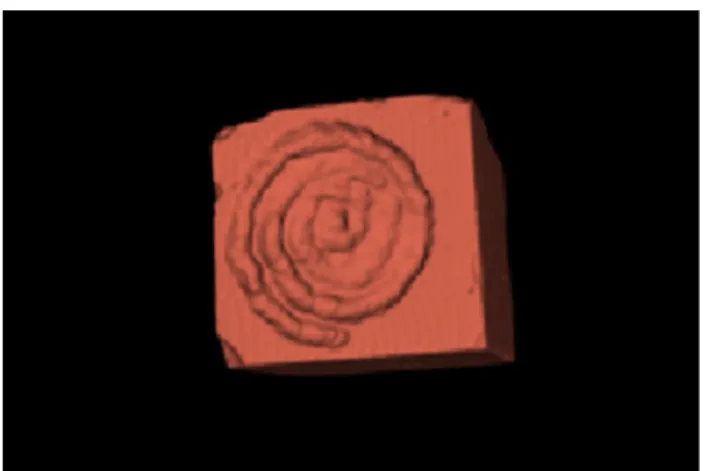
Fig. 2 Method of segmentation using 3D Slicer to assess ablation volumes based on DICOMs’ non-enhanced CT scan. First, segmenta-tion of the stone (a), then segmentasegmenta-tion of the air of the spiral crater (b) and compilation of both to get ablation volume (c)
Relying on RE values, we were able to obtain hypotheti-cal treatment duration times for stones of different compo-sitions and sizes, as reported in Supplementary Fig. 5 and Table 3.
Discussion
We estimated in this laboratory study the ablation volumes for each singe laser pulse according to both pulse energy and stone composition. Ablation volumes increased, although not proportionally, with the increase of pulse energy, and were higher in COM stones and lower in UA ones, with Cys stones in-between. As a consequence, we were able to
calculate the volume needed to ablate 1 mm3 of stone in ideal experimental conditions according to the aforemen-tioned parameters.
Three‑dimensional evaluation of stone burden and ablation volume
The improvements in the 3D evaluation of stone volume and the ongoing evolution from maximum diameter to 3D stone burden have not been translated yet into current clin-ical practice [12-14]. To the best of our knowledge, laser ablating efficacy was never precisely estimated in terms of stone volume, and no clear and reproducible methodol-ogy is available at this regard [15-17]. To overcome this
Fig. 3 Stone samples (upper part) after laser emission and 3D Slicer reconstruction (lower part) by segmentation and a separate quantification of
each pulse ablation volume
Table 1 Ablation volume per pulse (µm3) beyond stone type
and pulse energy in contact mode
Stone type Volumes (µm3) (mean ± SD)
Energy (joules) All energies combined
0.6 0.8 1 Calcium oxalate monohydrate 17.7 ± 2 35.6 ± 8 (p = 0.01) 71.1 ± 17 (p = 0.01) 35.9 ± 20 Cystine 85,3 ± 8 136.9 ± 71 (p = 0.6) 168.3 ± 48 (p = 0.04) 101.1 ± 47 Uric acid 198.1 ± 44 360.1 ± 123 (p = 0.012) 507.4 ± 120 (p = 0.07) 126.2 ± 30 p value COM vs cystine 0.01 0.004 0.1 0.03 COM vs UA 0.08 0.016 0.025 0.002 Cystine vs UA 0.05 0.07 0.07 0.03
knowledge gap, we developed an experimental model based on software-based 3D volume segmentation and single pulse delivery of the laser energy. The main chal-lenge of this study was represented by the accurate assess-ment of small volumes in irregular human stones. In this regard, 3D volume segmentation served as a reliable solu-tion for both these purposes (Figs. 2, 3). This method of 3D volume segmentation uses density (threshold) segmen-tation because we can easily differentiate kidney stones (+ 500 UH) from urine (clinical situation) or air (labora-tory experiments) on computerized tomography (CT scan). CT scan is well known in clinical practice for its accuracy, especially for urinary stone pre-operative stone evalua-tion, including low-dose CT scan. Consequently, we tried retroactively from clinical practice to use a CT scan-based method to evaluate the ablation volumes. Moreover, our method is also supported by the fact that stone HU den-sity on non-contrast CT scan is recognized as an effective
method to predict stone type, except for differentiating COM and calcium oxalate dihydrate (COD) [18].
Required energy to treat 1 mm3 of stone
In stone surgery, RE has been gaining attention, based on the same design than in benign prostatic obstruction laser procedures: Holmium and Greenlight Laser Enucleation of the Prostate (HoLEP and GreenLEP) has adopted the deliv-ered energy as an effectiveness criteria, even more during the learning curve [19, 20]. Consequently, the evaluation of an endoscopic stone procedure and surgeons may rely on the delivered energy for a stone volume. In this regard, our results are in line with the previous report by Mekayten et al. [21], which reported that in average 13 J is needed to ablate 1 mm3 of a stone of ~ 1000 HU.
We also showed that COM stones required more RE than Cys and UA stones. Previous studies compared the mechani-cal properties of human and artificial kidney stones (begos-tone). Because of the density (kg/m3), it was harder to create a fissure in COM stones than is UA stones, as well as hard versus soft BegoStones [22]. Drawing a parallel, hard stones may be tougher to break than soft ones. That could explain our findings on the higher RE for COM than for Cys and UA. More recently, Molina et al. found that only calcium phos-phate stones needed less energy than COM and UA [23]. In this study (100 patients), only seven cases presented with UA stones and one with cystine stone. Most of the cases were calcium oxalate stones (64%), revealing monohydrate ones needed more laser energy than dihydrate stones. This confirms that the less dense a stone, the less is the laser energy required. Teichman et al. supported the same results and proposed that COM stones decompose at a higher tem-perature than Cys and UA stones (204 versus 100 °C), to explain the higher pulse energy required to fragment COM stones [24]. They also confirmed a significant correlation between the cumulative laser energy and stone location (kidney), volume and calcium component. Finally, power was also associated with RE in their study, and traducing high pulse energy will need less total delivered energy for treatment (dust or fragment). This represents a compromise between the size of produced fragments and the lasing time, following the idea that “dusting” (high-frequency–low pulse rate) produces smaller fragments than “fragmentation” set-tings (low-frequency–high pulse rate) [25]. This may be rel-evant also on the surgeon’s technique, but it is well admit-ted that “dusting” mode tries to create small fragments that could be spontaneously evacuated [26].
Surgical planning of endoscopic stone procedures
The estimation of the energy needed to ablate that we provide in this current study may turn out useful for
0,6 0,8 1 0 10 20 30 40
single pulse energy (J)
Energy /m m 3 COM Cystine UA
Delivered Energy to treat 1mm3of stone
Fig. 4 Required energy to treat 1 mm3 beyond stone type and pulse
energy in contact mode
Table 2 Required total energy for 1 mm3 lithotripsy beyond stone
type and pulse energy
Stone type Required energy for 1 mm3 lithotripsy (joules)
Energy (J) 0.6 0.8 1 All combined Calcium oxa-late mono-hydrate 34 ± 3.3 23,2 ± 5.2 14.7 ± 3.9 24 ± 9 cystine 8.5 ± 1.8 7.8 ± 5.8 6.4 ± 2.2 7.6 ± 3 Uric acid 3.2 ± 0.6 2.4 ± 0.7 2 ± 0.5 2.5 ± 0.7 p-value COM vs cystine 24 ± 9 vs 7,6 ± 3 < 0,0001 COM vs UA 24 ± 9 vs 2,5 ± 0,7 < 0,0001 UA vs cystine 2,5 ± 0,7 vs 7,6 ± 3 < 0,0001
pre-operative planning of laser lithotripsy procedures: by knowing the stone volume and of the stone composition or density, and hypothesizing a 20–30% dissipation of energy due to inefficiently delivered pulses (inaccuracy or “stop and start” effect) and patient movements, it is theoretically possi-ble to estimate the duration of surgical procedures (Tapossi-ble 3). This percentage could be greater or lower regarding the sur-geon’s experience or technique and the surgical conditions (localization of the stone, visualization, adverse events). Moreover, the lower the gap between the estimated and observed total energy during the procedure itself might serve as a proxy of lithotripsy efficiency, as well as the proficiency of the surgeon. Supplementary Fig. 5 shows an exponential
relation between the ablation time and the diameter of the stone. These results are supported by our volumetric evalu-ation: doubling the diameter of the cube means multiplying by eight the volume of the sphere included in the cube. That may justify again the importance of a volumetric assessment.
Limitations
Our study is not devoid of limitations. First, our study evalu-ated ablation volume using ideal experimental conditions, which are substantially different from the current clinical practice; each pulse was delivered cutting the laser fiber beforehand, whereas during intracorporeal lithotripsy the
Table 3 Energy and lithotripsy duration besides stone type, maximum diameter or volume and laser settings For COM Diameter
(mm)
Volume (mm3)
0,6J-15Hz 0,8J-15Hz 1J-15Hz
Total J Time Time+30% Total J Time Time+30% Total J Time Time+30%
1 0,5 17 1,1sec 1,5sec 11 0,7sec 1sec 7 0,5sec 0,6sec
2 4 135 9sec 12sec 90 6sec 8sec 56 3,8sec 4,9sec
3 13,5 460 30sec 40sec 300 20sec 26sec 190 13sec 17sec
4 32 1080 1,2min 1,56min 720 48sec 1min 450 30sec 39sec
5 62,5 2120 2,35min 3min 1400 1,6min 2min 879 59sec 1,3min
6 108 3660 4min 5,3min 2400 2,7min 3,5min 1519 1,7min 2,2min
7 171,5 5800 6,5min 8,4min 3850 4,3min 5,6min 2412 2,7min 3,5min
8 256 8680 9,6min 12,5min 5750 6,4min 8,3min 3600 4min 5,2min
9 364,5 12350 13,7min 17,8min 8200 9min 11,8min 5127 5,7min 7,4min
10 500 16950 18,8min 24,5min 11230 12,5min 16,2min 7032 7,8min 10,2min
For Cys Diameter (mm)
Volume (mm3)
0,6J-15Hz 0,8J-15Hz 1J-15Hz
Total J Time Time+30% Total J Time Time+30% Total J Time Time+30%
1 0,5 4 0,2sec 0,3sec 2,9 0,2sec 0,3sec 3 0,2sec 0,3sec
2 4 28 1,9sec 2,4sec 23 1,6sec 2sec 24 1,6sec 2sec
3 13,5 95 6,3sec 8,2sec 79 5,2sec 6,8sec 80 5,3sec 7sec
4 32 225 15 sec 19sec 187 12sec 16sec 190 13sec 16sec
5 62,5 440 29sec 38sec 365 24sec 32sec 371 25sec 32sec
6 108 760 51 sec 66sec 631 42sec 55sec 642 43sec 56sec
7 171,5 1206 1,3min 1,7min 1002 1,1min 1,4min 1019 1,1min 1,5min
8 256 1801 2min 2,6min 1496 1,7min 2,2min 1521 1,7min 2,2min
9 364,5 2564 2,8min 3,7min 2130 2,4min 3,1min 2166 2,4min 3,3min
10 500 3517 3,9min 5,1min 2922 3,2min 4,2min 2971 3,3min 4,3min
For UA Diameter (mm)
Volume (mm3)
0,6J-15Hz 0,8J-15Hz 1J-15Hz
Total J Time Time+30% Total J Time Time+30% Total J Time Time+30%
1 0,5 1,5 0,1sec 0,1sec 1,1 0,1sec 0,1sec 1 0,1sec 0,1sec
2 4 12 0,8sec 1sec 9 0,6sec 0,8sec 8 0,5sec 0,7sec
3 13,5 41 2,7sec 3,5sec 30 2sec 2,6sec 27 1,8sec 2,3sec
4 32 97 6,5sec 8,4sec 71 4,7sec 6,2sec 63 4,2sec 5,5sec
5 62,5 189 12sec 16sec 139 9,3sec 12sec 123 8,2sec 11sec
6 108 327 22sec 28sec 240 16sec 21sec 213 14sec 18sec
7 171,5 519 35sec 45sec 381 25sec 33sec 338 23sec 29sec
8 256 775 52sec 1,1min 569 38sec 49sec 505 34sec 44sec
9 364,5 1104 1,2min 1,6min 810 54sec 1,2min 718 48sec 1min
laser fiber gradually deteriorates without the possibility of cutting it after each pulse. Moreover, we used contact mode for the delivery of laser energy: if on the one hand this pro-vided the highest delivery of energy to the stone, on the other we might have overestimated the ablation volume as compared to clinical practice. In such conditions, the fiber tip is not always in contact with a clear locus at the sur-face of the stone for various reasons: surgical conditions, visualization, irrigation and retropulsion, surgeon’s ability or mode of treatment. Hence, limiting contact mode may reduce fiber degradation or fracture and burnback effects [27-29]. It should be noted that our choice was driven by the necessity to guarantee the highest reproducibility in the experimental setting: it would have not been possible to observe the same distance between the laser fiber tip and the stone due the irregularity in both surface and dimensions of the real human stones that we used in our experiments. Conversely, the possibility of using human stones allowed us to have more realistic estimates.
For the specific purpose of this study, we used a low-power, short-pulse laser generator that may represent a limi-tation. First, numerous practitioners use low-power genera-tors for endocorporeal lithotripsy. Then, even high-power frequency dusting techniques have been more effective than low-power frequencies in a laboratory study. However, we cannot state about the superiority of high-power over low-power devices. In clinical practice, microbleeding, visualiza-tion of the fiber tip degradavisualiza-tion, and producvisualiza-tion of fragments > 1 mm are the limitations of high-power Ho:YAG genera-tors, including Moses technology, which will be probably managed in the near future (suction devices, optimization of the vapor bubble shapes) [30-34]. The Moses technol-ogy has recently showed a greater ablation weight in soft artificial BegoStones (contact mode or 1 mm distance) than short- or long-pulse mode. Those results were not main-tained against hard stones, at any distance, which is contra-dictory to Aldoukhi et al.’s study [35, 36]. Considering these controversial data, we recognize the Moses technology could be associated with greater ablation volume, especially in a distance mode. Concerning the long pulse duration (LP), Sroka et al. found similar ablation volumes compared to short pulse but a deeper crater with LP, regardless of the laser settings [37]. LP presents controversial data about the popcorning efficiency; so we cannot conclude on this spe-cific aspect [31, 38]. On the contrary, LP has shown a lower retropulsion and fiber degradation than during short pulse [25, 39]. Regarding those findings, we recognize the interest of an LP Ho:YAG generator, but we cannot assure that an LP Ho:YAG device would have presented higher ablation volumes and, as a consequence, lower RE. Furthermore, we did not evaluate other laser fibers than 272 µm core-diameter laser fiber (200 µm or 365 µm), which could provide addi-tional data for surgical planning.
Conclusion
This in vitro study shows for the first time a volumetric eval-uation of Ho:YAG efficiency by the ablation volume per pulse on human stone samples. Calcium oxalate monohy-drate needs tenfold and fivefold higher laser energy to treat the same volume than uric acid and cystine, respectively. High pulse energy will need less total delivered energy to dust but will produce bigger fragments, traducing the compromise which has to be done in clinical practice. The required energy to treat may represent a valuable information in surgical planning and post-operative accuracy evaluation.
Acknowledgements In vivo imaging was performed at Life
Imag-ing Facility of Paris Descartes University (Plateforme Imageries du Vivant), supported by France Life Imaging (grant ANR-11-INBS-0006) and Infrastructures Biologies-Santé.
Author contributions FP and EV: equal contribution: protocol devel-opment, data collection and management, data analysis, manuscript writing and editing. LB: protocol development, data collection and management, manuscript editing. CC: protocol development, data collection. MD: protocol development, data collection. SD: protocol development, manuscript writing and editing. OT: protocol develop-ment, data analysis, manuscript writing and editing.
Funding Frédéric Panthier received a French Association of Urology Research Grant (2018).
Compliance with ethical standards
Conflicts of interest The authors declare that they have no conflict of interest. However, Olivier Traxer has declared as being a consultant for Boston Scientific Corporation, Coloplast, Wolf, B-Braun, IPG photon-ics, Lumenis, Olympus and Rocamed. Steeve Doizi has declared as being a consultant for Boston Scientific Corporation and Coloplast.
Research involving human participants or animals This article does not contain any studies with human participants or animals performed by any of the authors.
Informed consent This article does not contain any studies with human participants or animals performed by any of the authors. Human stone samples were used in this study, with the informed consent of each patient.
References
1. Gupta PK (2007) Is the holmium:YAG laser the best intracor-poreal lithotripter for the ureter? A 3-year retrospective study. J Endourol 21(3):305–309
2. Kronenberg P, Somani B (2018) Advances in lasers for the treat-ment of stones—a systematic review. Curr Urol Rep. 19(6):45 3. Kadihasanoglu M, Yucetas U, Culha MG, Erkan E, Toktas MG
(2017) Effect of stone composition on the outcomes of semi-rigid ureteroscopy using holmium: yttrium–aluminum–garnet laser or pneumatic lithotripsy. J Coll Physicians Surg-Pak JCPSP 27(4):227–231
4. Türk C, Petřík A, Sarica K, Seitz C, Skolarikos A, Straub M et al (2016) EAU guidelines on interventional treatment for urolithiasis. Eur Urol 69(3):475–482
5. EAU guidelines on interventional treatment for urolithiasis.—Pub-Med—NCBI [Internet]. [cité 26 juin 2019]. Disponible sur: https :// www.ncbi.nlm.nih.gov/pubme d/26344 917
6. Pradère B, Doizi S, Proietti S, Brachlow J, Traxer O (2018) Evalu-ation of guidelines for surgical management of urolithiasis. J Urol 199(5):1267–1271
7. Assimos D, Krambeck A, Miller NL, Monga M, Murad MH, Nelson CP et al (2016) Surgical management of stones: American Uro-logical Association/EndouroUro-logical Society guideline. Part I. J Urol. 196(4):1153–1160
8. Assimos D, Krambeck A, Miller NL, Monga M, Murad MH, Nelson CP et al (2016) Surgical Management of stones: American Urologi-cal Association/EndourologiUrologi-cal Society guideline, part II. J Urol. 196(4):1161–1169
9. Wilhelm K, Miernik A, Hein S, Schlager D, Adams F, Benndorf M et al (2018) Validating automated kidney stone volumetry in CT and mathematical correlation with estimated stone volume based on diameter. J Endourol 32(7):659–664
10. Daudon M (2004) Analysis and classification of calculi: contribu-tion to the etiology of calculous disease. Rev Med Suisse Romande 124(8):445–453
11. D Slicer [Internet]. https ://www.slice r.org/. Cited 10 May 2019 12. De Coninck V, Traxer O (2018) The time has come to report stone
burden in terms of volume instead of largest diameter. J Endourol. 32(3):265–266
13. Merigot de Treigny O, Bou Nasr E, Almont T, Tack I, Rischmann P, Soulié M et al (2015) The cumulated stone diameter: a limited tool for stone burden estimation. Urology. 86(3):477–481
14. Patel SR, Nakada SY (2011) Quantification of preoperative stone burden for ureteroscopy and shock wave lithotripsy: current state and future recommendations. Urology 78(2):282–285
15. Lidén M, Andersson T, Broxvall M, Thunberg P, Geijer H (2012) Urinary stone size estimation: a new segmentation algorithm-based CT method. Eur Radiol 22(4):731–737
16. Sridhar S, Kumaravel N (2001) Automatic segmentation of medi-cal images for renal medi-calculi and analysis. Biomed Sci Instrum. 37:405–409
17. Marsousi M, Plataniotis KN, Stergiopoulos S (2014) Shape-based kidney detection and segmentation in three-dimensional abdominal ultrasound images. Conf Proc Annu Int Conf IEEE Eng Med Biol Soc IEEE Eng Med Biol Soc Annu Conf. 2014:2890–2894 18. Hassani H, Raynal G, Spie R, Daudon M, Vallée J-N (2012)
Imag-ing-based assessment of the mineral composition of urinary stones: an in vitro study of the combination of Hounsfield unit measure-ment in noncontrast helical computerized tomography and the twinkling artifact in color Doppler ultrasound. Ultrasound Med Biol 38(5):803–810
19. Peyronnet B, Robert G, Comat V, Rouprêt M, Gomez-Sancha F, Cornu J-N et al (2017) Learning curves and perioperative out-comes after endoscopic enucleation of the prostate: a comparison between GreenLight 532-nm and holmium lasers. World J Urol 35(6):973–983
20. Panthier F, Pasquier J, Bruel S, Azancot V, De La Taille A, Gas-man D (2019) En bloc greenlight laser enucleation of prostate (GreenLEP): about the first hundred cases. World J Urol. https :// doi.org/10.1007/s0034 5-019-02941 -1
21. Mekayten M, Lorber A, Katafigiotis I, Sfoungaristos S, Leotsakos I, Heifetz EM et al (2019) Will stone density stop being a key factor in endourology? The impact of stone density on laser time using Lumenis Laser p120w and standard 20 W laser: a comparative study. J Endourol 33(7):585–589
22. Esch E, Simmons WN, Sankin G, Cocks HF, Preminger GM, Zhong P (2010) A simple method for fabricating artificial kidney stones of different physical properties. Urol Res 38(4):315–319
23. Molina WR, Marchini GS, Pompeo A, Sehrt D, Kim FJ, Monga M (2014) Determinants of holmium:yttrium–aluminum–garnet laser time and energy during ureteroscopic laser lithotripsy. Urology 83(4):738–744
24. Teichman JM, Vassar GJ, Bishoff JT, Bellman GC (1998) Holmium:YAG lithotripsy yields smaller fragments than lithoclast, pulsed dye laser or electrohydraulic lithotripsy. J Urol 159(1):17–23 25. Kronenberg P, Traxer O (2015) Update on lasers in urology 2014: current assessment on holmium:yttrium–aluminum–garnet (Ho:YAG) laser lithotripter settings and laser fibers. World J Urol 33(4):463–469
26. Doizi S, Keller EX, De Coninck V, Traxer O (2018) Dust-ing technique for lithotripsy: what does it mean? Nat Rev Urol. 15(11):653–654
27. Talso M, Emiliani E, Haddad M, Berthe L, Baghdadi M, Montanari E et al (2016) Laser fiber and flexible ureterorenoscopy: the safety distance concept. J Endourol. 30(12):1269–1274
28. Peplinski B, Faaborg D, Miao E, Alsyouf M, Myklak K, Kelln W et al (2016) The effect of laser fiber cleave technique and lithotripsy time on power output. J Endourol. 30(6):678–684
29. Haddad M, Emiliani E, Rouchausse Y, Coste F, Berthe L, Doizi S et al (2017) Impact of laser fiber tip cleavage on power output for ureteroscopy and stone treatment. World J Urol. 35(11):1765–1770 30. Ventimiglia E, Traxer O (2019) Is very high power/frequency really necessary during laser lithotripsy? RE: understanding the popcorn effect during holmium laser lithotripsy for dusting (Aldoukhi et al, Urology. 2018 Dec;122:52–57). Urology 127:135. https ://doi. org/10.1016/j.urolo gy.2019.01.032
31. Aldoukhi AH, Roberts WW, Hall TL, Teichman JMH, Ghani KR (2018) Understanding the popcorn effect during holmium laser litho-tripsy for dusting. Urology Déc 122:52–57
32. Aldoukhi AH, Ghani KR (2019) Reply to: letter-to-the-editor: under-standing the popcorn effect during holmium laser lithotripsy for dusting. Urology. 127:135–136
33. Ventimiglia E, Traxer O (2019) What Is Moses effect: a historical perspective. J Endourol. mai 33(5):353–357
34. Keller EX, de Coninck V, Audouin M, Doizi S, Bazin D, Daudon M et al (2019) Fragments and dust after holmium laser lithotripsy with or without « Moses technology »: How are they different? J Biophotonics 12(4):e201800227
35. Winship B, Wollin D, Carlos E, Li J, Peters C, Simmons WN et al (2018) Dusting efficiency of the Moses holmium laser: an automated in vitro assessment. J Endourol 32(12):1131–1135
36. Aldoukhi AH, Roberts WW, Hall TL, Ghani KR (2019) Watch your distance: the role of laser fiber working distance on fragmentation when altering pulse width or modulation. J Endourol 33(2):120–126 37. Sroka R, Pongratz T, Scheib G, Khoder W, Stief CG, Herrmann T et al (2015) Impact of pulse duration on Ho:YAG laser litho-tripsy: treatment aspects on the single-pulse level. World J Urol 33(4):479–485
38. Emiliani E, Talso M, Cho S-Y, Baghdadi M, Mahmoud S, Pinheiro H et al (2017) Optimal Settings for the Noncontact Holmium:YAG Stone Fragmentation Popcorn Technique. J Urol. 198(3):702–706 39. Wollin DA, Ackerman A, Yang C, Chen T, Simmons WN,
Preminger GM et al (2017) Variable pulse duration from a new holmium:YAG laser: the effect on stone comminution, fiber tip deg-radation, and retropulsion in a dusting model. Urology 103:47–51
Affiliations
Frédéric Panthier1,2,3 · Eugenio Ventimiglia1,2,4 · Laurent Berthe3 · Catherine Chaussain5 · Michel Daudon1,6 ·
Steeve Doizi1,2 · Olivier Traxer1,2 Frédéric Panthier fredericpanthier@gmail.com Eugenio Ventimiglia eugenio.ventimiglia@gmail.com Laurent Berthe laurent.berthe@ensam.eu Catherine Chaussain Catherine.chaussain@parisdescartes.fr Steeve Doizi steeve.doizi@aphp.fr
1 GRC No 20, Groupe de Recherche Clinique Sur La Lithiase
Urinaire, Hôpital Tenon, Sorbonne Université, 75020 Paris, France
2 Service D’Urologie, Assistance-Publique Hôpitaux de Paris,
Hôpital Tenon, Sorbonne Université, 4 rue de la Chine, 75020 Paris, France
3 PIMM, UMR 8006 CNRS-Arts Et Métiers ParisTech, 151 bd
de l’Hôpital, 75013 Paris, France
4 Division of Experimental Oncology/Unit of Urology,
URI-Urological Research Institute, IRCCS Ospedale San Raffaele, 20132 Milan, Italy
5 Plateforme D’imagerie du Vivant, EA 2496 Orofacial
Pathologies, Imagery and Biotherapies, Dental School Faculty, University Paris Descartes and Life Imaging Plateform (PIV), Montrouge, France
6 Service Des Explorations Fonctionnelles, Hopital TENON, 4