HAL Id: hal-01556782
https://hal.sorbonne-universite.fr/hal-01556782
Submitted on 5 Jul 2017
HAL is a multi-disciplinary open access
archive for the deposit and dissemination of
sci-entific research documents, whether they are
pub-lished or not. The documents may come from
teaching and research institutions in France or
abroad, or from public or private research centers.
L’archive ouverte pluridisciplinaire HAL, est
destinée au dépôt et à la diffusion de documents
scientifiques de niveau recherche, publiés ou non,
émanant des établissements d’enseignement et de
recherche français ou étrangers, des laboratoires
publics ou privés.
Distributed under a Creative Commons Attribution| 4.0 International License
Comparison of preoperative imaging and pathological
findings for pancreatic head adenocarcinoma
Marine Gilabert, Jean-Marie Boher, Jean-Luc Raoul, François Paye, Philippe
Bachellier, Olivier Turrini, Jean Robert Delpero
To cite this version:
Marine Gilabert, Jean-Marie Boher, Jean-Luc Raoul, François Paye, Philippe Bachellier, et al..
Comparison of preoperative imaging and pathological findings for pancreatic head adenocarcinoma.
Medicine, Lippincott, Williams & Wilkins, 2017, 96 (24), pp.e7214. �10.1097/MD.0000000000007214�.
�hal-01556782�
Comparison of preoperative imaging and
pathological
findings for pancreatic head
adenocarcinoma
A retrospective analysis by the Association Française de Chirurgie
Marine Gilabert, MD, PhD
a, Jean-Marie Boher, PhD
b, Jean-Luc Raoul, MD, PhD
a,∗, François Paye, MD, PhD
c,
Philippe Bachellier, MD, PhD
d, Olivier Turrini, MD, PhD
e, Jean Robert Delpero, MD, PhD
e, the Association
Française de Chirurgie
Abstract
Initial imaging of pancreatic ductal adenocarcinoma is of crucial importance in the decision-making process. The aim of this study was to compare preoperative imaging, pathological data, and outcomes in a series of patients who underwent resection for pancreatic head cancer.
From January 2004 to December 2009, data were collected by the Association Française de Chirurgie on 1044 patients who receivedfirst-line R0 resection of pancreatic head cancer.
On imaging (computed tomography scan 97%, echoendoscopic ultrasound 61.3%, magnetic resonance imaging 46.5%), arterial, venous, or lymph node invasion was suspected in 20, 161, and 197 patients, respectively; arterial, venous, or lymph node invasion was observed histologically in 11, 116, and 736 cases, respectively. In the patients for whom both imaging and pathological data were available, the concordance, sensitivity, specificity, positive predictive value, and negative predictive value were as follows: 97.5%, 27.3%, 98%, 20%, and 99%, for arterial invasion; 86.5%, 54%, 91%, 47.8%, and 93.2%, for venous invasion; and 38%, 21%, 86%, 78%, and 41%, respectively, for lymph node invasion. Imaging of arterial invasion had no prognostic value, while histological evidence of invasion was associated with a poor prognosis. Venous and lymph node invasion, as demonstrated by imaging and by pathological analysis, had an adverse prognostic value.
Imaging gives a fair positive predictive value for venous or arterial invasion; venous invasion on imaging and histology was associated with a poor prognosis; arterial invasion on imaging does not have any significant prognostic value.
Abbreviations: CI= confidence interval, CT = computed tomography, DFS = disease-free survival, EUS = echoendoscopic ultrasound, FDG-PET= fluorodeoxyglucose positron emission tomography, IPMN = intraductal papillary mucinous neoplasm, MRI = magnetic resonance imaging, NPV= negative predictive value, OS = overall survival, PDAC = pancreatic ductal adenocarcinoma, PPV= positive predictive value, Se = sensitivity, Sp = specificity, US = ultrasound.
Keywords:imaging, pancreatic cancer, pathology, survival, vascular invasion
1. Introduction
Pancreatic adenocarcinoma represents a public health issue.[1] Surgery, the only curative treatment, is possible in fewer than 20% of cases. However, despite carcinologic resection, survival is bleak, with a median overall survival (mOS) of approximately 30 months.[2]After the exclusion of cancer extension, the primary causes of tumor’s unresectability[3] are due to the close and
complex vascular connections between the head of the pancreas, the mesenterico-portal venous axis, and the celiac and superior mesenteric arterial axes. Therefore, preoperative imaging helps in the decision of whether a complete resection is possible and whether upfront surgery is recommended. When tumoral vascular connections are very close in proximity, preoperative treatment could allow for a complete resection (R0) via a decrease in the tumor size; these“borderline” cases are frequent. This shows the importance of an examination by imaging, and the necessity of a standardization[4]of these explorations.
The purpose of this study was to compare preoperative imaging data with data obtained from the final pathological analysis from a large group of patients who underwent resection for pancreatic head ductal adenocarcinoma (PHDAC) without preoperative treatment.
Editor: Maria Kapritsou.
MG and J-LR designed the study and wrote the paper. MG, J-LR, FP, JMB, PB, OT, and JRD collaborated on the paper’s conception, reviewed the paper, and approved thefinal version of the article to be published.
SIRIC (grant INCa-DGOS-INSERM 6038) and Paoli-Calmettes Institute Marseille. The authors have no conflicts of interest to disclose.
a
Department of Medical Oncology, Paoli-Calmettes Institute,bClinical Trial Office
and Biostatistics Unit, Paoli-Calmettes Institute, Marseille,c
Department of Digestive Surgery, Hôpital Saint-Antoine, Paris,dHepatobiliary Surgery and Liver Transplantation, Hôpital Universitaire de Strasbourg, Strasbourg,e
Department of Digestive Surgery, Paoli-Calmettes Institute, Marseille, France.
∗
Correspondence: Jean-Luc Raoul, Department of Medical Oncology, Paoli-Calmettes Institute, Marseille, France (e-mail: raouljl@ipc.unicancer.fr). Copyright© 2017 the Author(s). Published by Wolters Kluwer Health, Inc. This is an open access article distributed under the terms of the Creative Commons Attribution-Non Commercial License 4.0 (CCBY-NC), where it is permissible to download, share, remix, transform, and buildup the work provided it is properly cited. The work cannot be used commercially without permission from the journal.
Medicine (2017) 96:24(e7214)
Received: 28 February 2017 / Received infinal form: 27 April 2017 / Accepted: 19 May 2017
http://dx.doi.org/10.1097/MD.0000000000007214
Observational Study
Medicine
2. Methods
From January 2004 to December 2009, the Association Française de Chirurgie collected thefiles of 1886 patients who underwent curative surgery for PDAC at 37 centers in France, Switzerland, Belgium, and Monaco.[5] Every institutional review board’s center approved the study. A standardized document was completed (retrospectively) for each patient in terms of preoperative (medical history, clinical presentation, biology, imaging, and drainage), perioperative (observations, techniques), postoperative (complications and mortality), and pathological findings (TNM/UICC 2002, assessment of venous, arterial, pancreatic, and posterior resection margins) as well as follow-up data (adjuvant therapy, relapse, and survival). The final conclusion of the preoperative imaging was based on the different techniques used (ultrasound [US], computed tomogra-phy [CT] scan, magnetic resonance imaging [MRI], echoendo-scopic ultrasound [EUS], and fluorodeoxyglucose positron emission tomography [FDG-PET] scan) after evaluation by the surgeon in charge or during a multidisciplinary board meeting.
Were included in this retrospective analysis all patients who underwent resection for PDAC without preoperative treatment. The following patients were excluded from this large database (some of them were excluded for several reasons):
- Patients who did not undergo surgery of a curative intent (R2 surgery and patients with metastatic cancer) (n=12). - Patients whose data lacked preoperative imaging assessment
conclusions (n=270).
- Patients who underwent resection after neoadjuvant treatment (n=201).
- Patients who underwent pancreatic surgery for a tumor located on the body or the tail (n=466).
Finally, data were available for 1044 patients with PDAC who receivedfirst-line resections.
Surgery was indicated according to the attending surgeon’s choice and was determined after a careful assessment of the preoperative imaging that was performed at his center and according to his usual practice.
The histopathological analysis focused on the tumor size and on the arterial, venous, or lymphatic invasions. Invasions into the arteries or veins were defined either by vascular wall invasion or by a positive margin (i.e., tumor cells observed at the edge of the tumor by microscopy), meaning that perivascular and vascular invasion were both considered as invasion.
2.1. Statistical analysis
All statistical analyses were performed with SAS v9.3 software (SAS Institute Inc., Cary, NC). The surgical procedures, perioperative treatments, and tumors characteristics of the patients were described as means, medians, and as percentages for categorical variables. The survival rates (median, 95% confidence interval [CI]) were estimated by the Kaplan–Meier method. The concordance, sensitivity (Se), specificity (Sp), positive predictive value (PPV), and negative predictive value (NPV) of the preoperative imaging were estimated[6]with 95% exact CIs. The concordance was defined as a good correlation between 2 examinations, and the Se was determined based on the capacity of the examination to give a positive result when the disease was present. The Sp was determined based on the capacity of an examination to give a negative result when the disease was not present. The predictive values were defined by the probability
that the disease was present when the test was positive (PPV) and not present when the test was negative (NPV).
3. Results
3.1. Characteristics of the patients and the preoperative imagingfindings
A total of 43% of the population (1044 patients) was female and the median age 66 years (range, 27–87). The clinical signs that led to the diagnosis were jaundice (77.3%), pain (38%), angiocho-litis (6.8%), pancreatitis (8.8%), or digestive stenosis (3.3%). Pre-existing diabetes was noted in 8.8% of patients. An endoprosthesis was applied preoperatively in 243 patients. Preoperative imaging assessment included contrast-enhanced CT scan (97%), echography (66.6%), EUS (61.3%), and pancreatic MRI (46.5%). An FDG-PET scan was performed in 80 patients. Through various types of imaging modalities, venous invasion (portal or superior mesenteric vein) was demonstrated in 161 patients and arterial invasion (celiac trunk, hepatic artery, or superior mesenteric artery) in 20 patients; lymph node invasion (small diameter >15mm) was demonstrated in 197 (18.8%) patients. The median tumor size was 2.5 cm (range, 0–12). The surgical procedure performed in all cases was pancreaticoduo-denectomy. It was associated with venous resection in 260 cases, arterial resection in 10 cases, and extension to other viscera in 49 cases. The postoperative mortality rate was 4%. Adjuvant therapy was given to 74% of the patients.
Pathologicalfindings were as follows: median tumor diameter of 3.0 cm (0–12); intraductal papillary mucinous neoplasm (IPMN) found in 238 surgical samples (22.7%); tumors were well (34.8%), moderately (52.7%), or poorly (12.5%) differentiated; lymphatic or venous emboli were frequent (54.5%) as was perinervous extension (74.6%); lymph node invasion was observed in 736 cases; arterial invasion in 11 cases and venous invasion in 116 cases. After pathological examination (142 patients had incomplete data), 6 patients were classified as stage 0, 30 (3.3%) as stage IA, 77 (8.5%) as stage IB, 145 (16%) as stage IIA, 589 (65.3%) as stage IIB, and 55 (6.1%) as stage III. An excellent correlation (P<.0001) was observed between tumor size as measured by radiology and as measured by the surgical samples.
3.2. Evaluation of the concordance between the preoperative imaging data and arterial, venous, and lymphatic invasion as well as thefinal histopathological data
Imaging and histological data about arterial invasion were available for 813 patients. Among those, 15 demonstrated arterial invasion by imaging, histologically confirmed in 3 patients (20% of the cases) and disproved in 12 (80%) patients; nevertheless, for the remaining 798 patients who showed no arterial invasion by imaging, damage was found in 8 surgical samples (1%). The concordance between the 2 techniques was 97.5% (96–99), the imaging Se was 27.3% (6–61), the Sp was 98% (97–99), the PPV was 20% (4–48), and the NPV was 99% (98–99) (Tables 1 and 2).
Imaging and histological data with regard to venous invasion were available for 928 patients. Venous invasion was described in 136 cases, and confirmed by histology in 65 (48%); imaging for venous invasion was negative in 792 patients, but demonstrated histologically in 54 cases (7%). The concordance was 86.5% Gilabert et al. Medicine (2017) 96:24 Medicine
(38–44), the Se was 54% (45–63), the Sp was 91% (89–93), the imaging PPV was 47.8% (39–56), and the NPV was 93.2% (91–95).
Lymph node invasion was suspected on imaging in 195 patients and histologically confirmed in 152 patients (77%). However, among the 820 patients without imaging data, 555 (67%) have lymph node invasion on histology. The concordance was 38% (66–78), the Se was 21% (18–25), the Sp was 86% (82–90), the imaging PPV was 78% (71–84), and the imaging PPV was 41% (38–44).
These data were similar for patients with associated IPMN (small group size; radiological and pathological data available in 102 patients); the correlation between tumor size measured by radiology or pathology was excellent (P<.0001); in 2 patients, imaging demonstrated an arterial invasion, not found on resected specimen and in 1 case, arterial invasion, not suspected preoperatively was found on pathological examination; in 17 cases, there existed a venous invasion on pathology, suspected preoperatively in only 9 cases and in 22 cases, imaging was considered as positive while that was demonstrated only in 9 cases.
3.3. Survival
The mOS was 29.1 months. The 1- and 3-year OS rates were 78.8% and 42.8%, respectively. The median disease-free survival (mDFS) was 15.0 months, with a 1- and 3-year DFS of 57.4% and 27.1%, respectively. Identification of arterial invasion by imaging (n=20) was of no prognostic value. The mOS and mDFS were, respectively, 23.6 months (range, 8.7 to NR) and 14.5
months (range, 8.7–25.4), compared with 27.4 months (range, 24.7–35.0) and 15.2 months (range, 13.9–17.0), respectively, depending on whether or not arterial invasion was suspected on imaging.
However, the identification of arterial invasion (Fig. 1) by histology (n=11) was significantly associated with a less favorable mOS of 13.0 months (range, 6.0–25.4) (P=.05) and a less favorable mDFS of 10.0 months (range, 6.0–23.6) (P=.04) compared with the absence of histological arterial invasion: mOS of 28.5 months (range, 24.5–36.6) and mDFS of 15.2 months (range, 13.7–17.0), respectively.
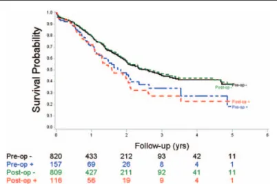
The identification of venous invasion (Fig. 2) by imaging (n= 161) had an adverse prognostic value, and the mOS and mDFS were, respectively, 22.1 months (range, 17.5–28.2) and 11.8 months (range, 9.9–14.1) if venous invasion was suspected, compared with 31.3 months (range, 25.2–36.6) (P<.01) and 16.3 months (range, 14.3–19.1) (P=.0004) if venous invasion was not suspected. Pathological identification of venous invasion (n=116) was highly significantly associated with less favorable mOS. The mOS and mDFS were 19.0 months (range, 15.5–25.1)
Table 2
Evaluation of concordance, sensitivity, specificity, positive pre-dictive value, and negative prepre-dictive value between preoperative imaging and histological data (pancreatic head adenocarcinoma).
Arterial involvement, % Venous involvement, % Lymph nodes, % Concordance 97.5 86.5 41.1 Sensitivity 27.3 54 21 Specificity 98 91 86
Positive predictive value 20 47.8 78 Negative predictive value 99 93.2 41
Figure 1. Overall survival of patients who underwent resection for pancreatic head cancer following suspected arterial involvement on imaging (pre-op) or on pathologic analysis (post-op) ; pre-op : no arterial lesion on preoperative imaging, pre-op +: suspected involvement on preoperative imaging; post-op : no arterial lesion on pathology; and post-op +: arterial lesion on pathology.
Figure 2. Overall survival of patients who underwent resection for pancreatic head cancer following suspected venous involvement on imaging (pre-op) or on pathologic analysis (post-op); pre-op : no venous lesion on preoperative imaging, pre-op + : suspected involvement on preoperative imaging; post-op : no venous lesion on pathology; and post-op + : venous lesion on pathology.
Table 1
Comparison between preoperative imaging andfinal pathological conclusions in patients resected from a pancreatic head adeno-carcinoma.
Preoperative imaging: suspected involvement
Pathological conclusion Yes No Total Arterial invasion Yes 3 8 11 No 12 790 802 Total 15 798 813 Venous invasion Yes 65 54 119 No 71 738 809 Total 136 792 928
Lymph node invasion
Yes 152 555 707
No 43 265 308
and 11.8 months (range, 8.3–13.7), respectively, compared with 31.7 months (range, 26.9–39.8) (P=.001) and 16.5 months (range, 14.6–19.0) (P=.0004), respectively, depending on whether or not venous invasion was detected histologically.
The prognostic value of lymph node invasion as detected by imaging was found for both OS (P=.02) and DFS (P=.003): the mOS and mDFS were 56.1 months (range, 36.0 to NR) and 35.6 months (range, 22.3–53.8), respectively, compared with 23.7 months (range, 21.2–25.2) (P<.0001) and 13.1 months (range, 11.8–14.1) (P<.0001), respectively, in the absence or presence of histological lymph node invasion.
4. Discussion
Surgical resection is the only approach with the potential to cure patients with PDAC. Preoperative imaging is therefore of crucial importance.[7]It is quite easy to diagnose liver or lung metastases, but more difficult to diagnose peritoneal carcinomatosis. The main problem is the differentiation of those with resectable tumors, those with locally advanced unresectable tumors, and those with“borderline resectable” tumors. For patients who have resectable tumor, the current guidelines propose resection followed by adjuvant chemotherapy. For patients with unre-sectable tumors, systemic chemotherapy and radiotherapy are advised, whereas patients with borderline resectable tumors require multimodality treatment.[8,9] Imaging procedures are
important in the distinction of these 3 groups. As recommended by the National Comprehensive Cancer Network, all centers of our study used multidetector CT to assess vascular invasion of tumors.[10] MRI can be useful in the detection of cystic
neoplasms.[11]Since 18F-FDG-PET is not recommended, endo-scopic US is useful to complement other imaging techniques[12]
and when a tissue diagnosis is needed.[3] The definitions of resectability and borderline resectability are still not clear and certainly differ among centers and surgeons.[3]A venous resection
is feasible.[13,14]A resection is questionable in the case of arterial invasion; a histological confirmation of arterial invasion is associated with a very poor prognosis, even though it is not considered by some as a strict contraindication to resection if otherwise feasible.[15,16]
Our large retrospective analysis of more than 1000 cases showed that, in real-life situations and in large-sized surgical centers, major discordances exist between the imaging results and the pathological data of resected specimens. The study of preoperative imaging performance is complex because it requires histological evidence, and can only be performed in patients who underwent resection. Obviously, if those patients underwent resection despite a negative imaging assessment, there is a major bias, surgery being performed due to young age, doubt about radiologic conclusions, etc. Then our study has some major limitations because retrospective, declarative, and quite old, but the number of included patients gives some credibility to the conclusions. PPV and NPV of the preoperative imaging techniques, even in unicentric series, are far from perfect leading to a standardization of preoperative imaging examination procedures for patients with PDAC.[4,17,18]Japanese authors[19] have proposed a classification by portography or contrast-enhanced CT scan of radiological types of portal vein invasion in cases of pancreatic head cancer. In a large series of 671 patients, they found an excellent correlation between pathological portal vein invasion and type. Despite its importance, arterial invasion is less well described. Recently, a novel scoring system[20] was
produced; the 3 major imaging parameters associated with
arterial wall invasion were length of tumor contact (per 16 mm), circumferential involvement (per 180°), and deformity of the vascular diameter.
To the best of our knowledge, our series is the largest series that compares a real-life situation, imaging, and pathology. We can draw 3 sets of conclusions from our series: PPV of imaging with regard to arterial invasion was poor (20%) but the PPV was fair (47.8%) for venous invasion; patients with positive imaging for arterial invasion who underwent surgery had a similar outcome than those without such imaging findings. The outcome of patients with histologic demonstration of arterial invasion was dismal; and venous invasion on imaging and on pathology was associated with a poor outcome.
In most series, the predictive value of imaging of arterial involvement was poor or fair.[21,22] Many series that contain cases of“false-positive” patients have also been described.[23,24] These studies showed an overestimation of arterial invasion by CT, especially in cases of degenerated IPMN. In our series, the presence of IPMN was not associated with an overestimation of tumor extension. A recent trial demonstrated an excellent agreement among radiologists with respect to the local staging of PDAC.[25]Our series is a major element in favor of exploratory surgery[26]with biopsies of artery sheaths in some patients who had suspected arterial involvement on preoperative imaging without any other negative finding. If the biopsy results are negative, resection is reasonable, if they are positive, neoadjuvant medical therapeutics might be preferable.
Venous invasion detected by imaging or by pathology is associated with a poor prognosis. However, given the dismal prognosis for those patients with suspected venous invasion, one can question the purpose of preoperative therapy for such patients; nevertheless, phase III trials are mandatory.
In summary, we propose a consideration of patients with PDAC who have venous invasion on preoperative imaging as high-risk patients, these patients may not be excellent candidates forfirst-line resection and must be reevaluated after a first line of medical treatment (systemic chemotherapy? radiochemother-apy?). In case of suspected arterial invasion as only negative imaging data, an exploratory laparotomy with biopsy can be proposed. The presence of IPMN was not a confounding factor in our series. The conclusions drawn from this large multicenter series are obviously subject to various critiques: this series is retrospective, declarative, multicentric, and fairly “old,” and lacks a large amount of important data. Yet based on the high number of medicalfiles that were analyzed and its multicentric character, this study remains relevant and in agreement with the results of smaller unicentric series. The generalized use of a reporting template[4]by large volume centers is mandatory.
References
[1] De Angelis R, Sant M, Coleman MP, et al. Cancer survival in Europe 1999–2007 by country and age: results of EUROCARE-5-a-population-based study. Lancet Oncol 2014;15:23–34.
[2] Jemal A, Siegel R, Xu J, et al. Cancer statistics 2010. CA Cancer J Clin 2010;60:277–300.
[3] Hartwig W, Jager D, Debus J, et al. Improvement of surgical results for pancreatic cancer. Lancet Oncol 2013;14:e476–85.
[4] Al-Hawary MM, Francis IR, Chari ST, et al. Pancreatic ductal adenocarcinoma radiology reporting template: consensus of the Society of Abdominal Radiology and the American Pancreatic Association. Radiology 2014;270:248–60.
[5] Delpero JR, Paye F, Bachelier P, et al. Cancer du Pancréas. Monographies de l’Association Française de Chirurgie, Arnette, Wolters Kluwer, France; 2010.
[6] Jaeschke R, Guyatt GH, Sackett DL. User’s guide to medical literature. How to use an article about a diagnostic test. Are the results of the study valid? Evidence-based medicine working group. JAMA 1994;271:389–91. [7] Soriano A, Castells A, Ayuso C, et al. Preoperative staging and tumor resectability assessment of pancreatic cancer: prospective study compar-ing endoscopic ultrasonography, helical computed tomography, mag-netic resonance imaging, and angiography. Am J Gastroenterol 2004;99: 492–501.
[8] Callery MP, Chang KJ, Fishman EK, et al. Pretreatment assessment of resectable and borderline resectable pancreatic cancer: expert consensus statement. Ann Surg Oncol 2009;16:1727–33.
[9] Bockhorn M, Uzunoglu FG, Adham M, et al. Borderline resectable pancreatic cancer: a consensus statement by the International Study Group of Pancreatic Surgery (ISGPS). Surgery 2014;155:977–88. [10] Tempero MA, Arnoletti JP, Behrman SW, et al. National Comprehensive
Cancer Networks. Pancreatic Adenocarcinoma, version 2.2012: featured updates to the NCCN Guidelines. J Natl Compr Canc Netw 2012;10: 703–13.
[11] Tamburrino D, Riviere D, Yaghoobi M, et al. Diagnostic accuracy of different imaging modalities following computed tomography (CT) scanning for assessing the resectability with curative intent in pancreatic and periampullary cancer. Cochrane Database Syst Rev2016;9: CD011515.
[12] Tellez-Avila FI, Chavez-Tapia NC, Lopez-Arce G, et al. Vascular invasion in pancreatic cancer: predictive values for endoscopic ultra-sound and computed tomography imaging. Pancreas 2012;41:636–8. [13] Bachellier P, Nakano H, Oussoultzoglou PD, et al. Is
pancreaticoduo-denectomy with mesentericoportal venous resection safe and worth-while? Am J Surg 2001;182:120–9.
[14] Murakami Y, Uemura K, Sudo T, et al. Benefit of portal or superior mesenteric vein resection with adjuvant chemotherapy for patients with pancreatic head carcinoma. J Surg Oncol 2013;107:414–21. [15] Bachellier P, Rosso E, Lucescu I, et al. Is the need for an arterial resection
a contraindication to pancreatic resection for locally advanced pancreatic adenocarcinoma? A case-matched controlled study. J Surg Oncol 2011; 103:75–84.
[16] Martin RCII, Scoggins CR, Egnatashvili V, et al. Arterial and venous resection for pancreatic adenocarcinoma: operative and long-term outcomes. Arch Surg 2009;144:154–9.
[17] Tamm EP, Balachandran A, Bhosale PR, et al. Imaging of pancreatic adenocarcinoma: update on staging/resectability. Radiol Clin North Am 2012;50:407–28.
[18] Tamm EP, Bhosale PR, Vikram R, et al. Imaging of pancreatic ductal adenocarcinoma: state of the art. World J Radiol 2013;28:98–105. [19] Nakao A, Kanzaki A, Fujii T, et al. Correlation between radiographic
classification and pathological grade of portal vein wall invasion in pancreatic head cancer. Ann Surg 2012;255:103–8.
[20] Watanabe S, Kobayashi N, Kubota K, et al. A novel scoring system for arterial invasion of pancreatic body and tail cancer based on multi-detector row computed tomography and biomarkers. Pancreatology 2013;13:161–9.
[21] Lu DS, Reber HA, Krasny RM, et al. Local staging of pancreatic cancer: criteria for unresectability of major vessels as revealed by pancreatic-phase, thin-section helical CT. Am J Roentgenol 1997;168: 1439–43.
[22] Lee JK, Kim AY, Kim PN, et al. Prediction of vascular involvement and resectability by multidetector-row CT versus MR imaging with MR angiography in patients who underwent surgery for resection of pancreatic ductal adenocarcinoma. Eur J Radiol 2010;73:310–6. [23] Yekebas EF, Bogoevski D, Cataldegirmen G, et al. En bloc vascular
resection for locally advanced pancreatic malignancies infiltrating major blood vessels: perioperative outcome and long-term survival in 136 patients. Ann Surg 2008;247:300–9.
[24] Catalano C, Laghi A, Fraioli F, et al. Pancreatic carcinoma: the role of high-resolution multislice spiral CT in the diagnosis and assessment of resectability. Eur Radiol 2003;13:149–56.
[25] Loizou L, Albiin N, Ansorge C, et al. Computed tomography staging of pancreatic cancer: a validation study addressing interobserver agree-ment. Pancreatology 2013;13:570–5.
[26] Buchs NC, Chilcott M, Poletti PA, et al. Vascular invasion in pancreatic cancer: imaging modalities, preoperative diagnosis and surgical management. World J Gastroenterol 2010;16:818–31.