Introduction
The cervical spine has been an interest area in orthodontics for several reasons. Firstly, natural head position has commonly been assessed on lateral cephalograms using the cervical spine as the reference structure (Kylamarkula and Huggare, 1985; Huggare, 1986). Secondly, skeletal age estimation can be made by the cervical spine method, where the morphology of the upper cervical vertebrae can be associated with the somatic growth curve of the patient and used in predicting growth potential for planning orthodontic treatment (Hassel and Farman, 1995M; Baccetti et al., 2002). Thirdly, different anomalies of cervical vertebrae (fusions and posterior arch deficiency) have been reported to occur in patients with cleft lip and palate (Ross and Lindsay, 1965; Sandham, 1986; Horswell, 1991; Ugar and Semb, 2001; Rajion et al., 2006), craniofacial syndromes (Gray et al., 1964; Hensinger, 1991; Guille and Sherk, 2002; Tracy et al., 2004; Kaplan et al., 2005), sleep apnea (Sonnesen et al., 2008), and different dentoskeletal malocclusions (Sonnesen and Kjaer, 2007a,b, 2008a,b;
Koletsis and Halazonetis, 2009).
Cervical vertebrae anomalies in subjects with Class II
malocclusion assessed by lateral cephalogram and cone beam
computed tomography
Dominika Bebnowski*, Michael P. Hänggi*, Goran Markic*, Malgorzata Roos** and
Timo Peltomäki***
*Clinic for Orthodontics and Pediatric Dentistry, **Biostatistics Unit, Institute of Social and Preventive Medicine, University of Zurich, Switzerland and ***Dental and Oral Diseases Outpatient Clinic, Department of Eye, Ear and Oral Diseases, Tampere University Hospital and Department of Otolaryngology, University of Tampere, Tampere, Finland
Correspondence to: Dr Timo Peltomäki, Dental and Oral Diseases Outpatient Clinic, Tampere University Hospital, Biokatu 6B, FI-33520 Tampere, Finland. E-mail: [email protected]
SUMMARY A high prevalence of cervical vertebrae anomalies (CVA) has been recently associated with various malocclusions. Our aim was to study the prevalence of CVA on lateral cephalograms in Class II subjects and to compare the findings with those obtained from cone beam computed tomography (CBCT). Standardized cephalograms of 238 Class II patients were analysed for CVA. Cephalogram and CBCT were available for an additional 21 subjects. Cephalometric values were correlated with vertebrae morphology; logistic regressions and intraobserver agreement were evaluated. Inspection of lateral cephalograms could exclude CVA in 90.3 per cent of the subjects, while 9.7 per cent showed potential fusions. No correlations were found between the cephalometric values and potential vertebrae anomalies. In the 21 patients with a CBCT and a lateral cephalogram, the visual assessment of the cephalogram yielded a potential fusion in nine cases. None could be confirmed by CBCT. A low number of potentially fused cervical vertebrae could be detected on lateral cephalograms. The possible fusions did not correlate to any cephalometric values nor could they be confirmed by CBCT, the gold standard for assessing CVA. Visual examination of a cephalogram may result in a false-positive finding and does not allow reliable diagnosis of CVA.
Fusion of the upper cervical vertebrae occurs most commonly between the second and the third (C2–C3) vertebrae. They are probably the result of a failure in normal embryological segmentation due to locally decreased blood supply during foetal development. Patients are generally asymptomatic, but increasing age or injury may precipitate symptoms (Yochum and Rowe, 2005). According to one line of recent studies, the prevalence of cervical spine anomalies, particularly vertebral fusion, is very high (41.5– 61.4 per cent) in patients with skeletal deep bite (Sonnesen and Kjaer, 2007b), Class II (Sonnesen and Kjaer, 2008a), Class III (Sonnesen and Kjaer, 2007a), or open bite (Sonnesen and Kjaer, 2008b), while the prevalence is lower, 14.3 per cent, in patients with normal dentoskeletal relationship (Sonnesen and Kjaer, 2007a,b, 2008a,b). Considerably, lower prevalence numbers of under 0.9 per cent in normal population have been reported by other studies (Tetradis and Kantor, 1999; Yochum and Rowe, 2005; Koletsis and Halazonetis, 2009).
Concerning skeletal Class II with increased overjet, a 52.9 per cent prevalence of cervical vertebrae fusions has
associations between cephalometric measurements and cervical column morphology (Sonnesen and Kjaer, 2008a). These findings have been challenged due to difficulties in reliably determining cervical vertebrae anomalies (CVA) on a single lateral cephalogram (Koletsis and Halazonetis, 2009). This difficulty is also known in radiological literature and has been critically discussed already some years ago (Massengill et al., 1997; Yochum and Rowe, 2005). The aim of our study was to evaluate the prevalence of CVA on lateral cephalograms in Class II subjects, to study possible correlations between cervical column and craniofacial morphology, and to compare the findings with those obtained from cone beam computed tomography (CBCT).
Subjects and methods
The material consisted of pre-treatment lateral cephalograms of 238 subjects, 129 males and 109 females, with an age range of 7–15 years (mean 10.4 years, SD 1.6 years). The subjects were randomly selected from the archive of the Clinic for Orthodontics and Pediatric Dentistry at the University of Zurich. Inclusion criteria for the subjects were 1. healthy subjects, 2. Caucasian ethnicity, 3. at least three of four class II molar relationship on both sides, 4. more than or equal to 4 mm overjet, and 5. existence of at least two consecutive lateral cephalograms of good quality including the first pre-treatment cephalogram, showing the first four cervical vertebrae. Excluding criteria were patients with syndromes or other developmental deformities. The resulting group had a mean overjet of 8.6 ± 2.3 mm and a mean ANB of 5.7 ± 1.8 degrees.
Additionally, subjects with a lateral cephalogram and a CBCT made for other diagnostic purposes were included. These diagnostic purposes were (ordered in frequency of occurrence): localization of impacted teeth, presurgical assessments, asymmetries, temporomandibular joint diagnostics, and clarification of cementoma. Because of the retrospective nature of this study, no additional x-rays were taken to study the cervical vertebrae and therefore, no ethical approval was needed; however, informed consent was obtained from all patients. Only patients with skeletal and dental Class I or Class II were included. Patients were excluded when the first four cervical vertebrae were not visible on the CBCT or the lateral cephalogram or if they had syndromes or other developmental deformities. Lateral cephalograms were studied first and only those subjects where the cervical vertebrae were not separated by a large distance were studied extensively with the KaVo 3D eXam Vision software. This resulted in 21 studied CBCT’s and their corresponding lateral cephalograms. The subjects consisted of 10 males and 11 females with an age range of 11.2–46.5 years (mean 20.8 years, SD 10.6 years).
All lateral cephalograms were taken with the Frankfort horizontal plane parallel to the floor. The cephalograms were hand-traced and conventional linear and angular cephalometric measurements representing various vertical and sagittal craniofacial relations were determined. To assess repeatability and measurement error, 20 radiographs were randomly selected and re-traced by the same investigator after a period of at least 2 weeks.
Cervical vertebrae morphology
The first four cervical vertebrae in the lateral cephalogram were structurally traced with the method described by
Vastardis and Evans (1996). The abnormalities of the cervical vertebrae were defined by visual assessment directly on the lateral cephalograms and structural tracings. They were classified according to the method of Sandham (1986) and divided into two categories: posterior arch deficiency and fusion anomalies. The posterior arch deficiency of the atlas was recorded when a uniform radio-opacity without an internal cortical outline was observed at the distal margin of the posterior arch (Farman et al., 1979;
Farman and Escobar, 1982; Sandham, 1986; Jones, 1998;
Koletsis and Halazonetis, 2009). Potential fusions between the cervical vertebrae were identified as osseous continuities without complete separation at the intervertebral disc or at the articular surfaces (Farman et al., 1979; Farman and Escobar, 1982; Sandham, 1986; Jones, 1998; Koletsis and Halazonetis, 2009; example given in Figure 1). For the diagnosis, consensus was achieved between the first and the second author.
CBCT analysis
The KaVo 3D eXam Cone Beam three-dimensional Dental Imaging System was used in this study. The settings were as followed: standard scan mode with an imaging volume of 16 × 13 cm, scan speed of 8.5 seconds, and voxel size of 0.4 or 0.3 mm.
The three-dimensional images of the cervical vertebrae were analysed for vertebral anomalies using the KaVo 3D eXam Vision software. For the diagnosis, consensus was achieved between the first and the second author.
Statistics
Logistic regression analysis was used to calculate the odds ratios (ORs) and associated 95 per cent confidence intervals (95% CI) to determine the association between presence or absence of cervical spine anomalies and lateral cephalometric measurements describing craniofacial dimensions. Sample size of 238 observations provides a power of 95 per cent of detecting an OR larger than 1.8 when the alpha level equal to 5 per cent is assumed.
Kappa (k) statistic (Landis and Koch, 1977) was used to analyse the intraobserver agreement of repeated measurements. k values higher than 80 per cent were considered to have good agreement. The statistical analysis was performed with SPSS software (version 16.0.2; SPSS, Chicago, Illinois, USA). Results were considered significant at P < 0.05.
Results
Repeatability examination of the lateral cephalograms showed good agreement (0.80) as assessed by the kappa coefficient (Landis and Koch, 1977). No cervical vertebrae anomaly was found in 90.3 per cent (n = 215) of the subjects, while 9.7 per cent (n = 23) showed potentially fused cervical vertebrae judged on lateral cephalograms. No posterior arch deficiency or blockfusion was found. All potential fusions were detected between C2 and C3 on the lateral cephalograms. The prevalence of possible cervical vertebrae fusions in this Class II population was thus 9.7 per cent (95% CI: 6.5–14.1).
Logistic regression analysis determining the association between presence or absence of CVA and lateral cephalometric measurements describing craniofacial dimensions of the cephalometric measurements is shown in Table 1. No statistically significant associations were found between the cephalometric variables and the possible CVA. The ORs ranged between 0.88 (95% CI: 0.75–1.04) and 1.11 (95% CI: 0.93–1.34).
In the 21 patients with the CBCT and the lateral cephalogram, the visual assessment of the lateral cephalogram yielded a possible fusion in nine cases. However, none could be confirmed with the CBCT data. On all CBCT scans, continuous joint spaces were clearly visible in all sagittal, transversal, and vertical sections (example of the CBCT scans given in Figure 1B–F).
Discussion
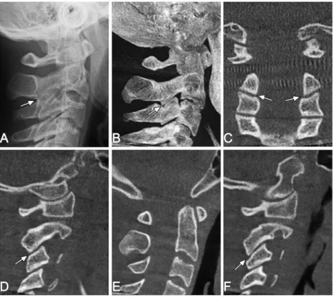
Association between upper cervical vertebrae morphology, craniofacial malformations, and dentoskeletal malocclusions has been extensively studied. The findings have, however, Figure 1 Conventional lateral cephalogram (A) and cone beam computed tomography (CBCT)
images (B-F) of the same patient: (A) lateral cephalogram with an absence of a continuous radiolucent area between the cervical vertebrae C2 and C3 (possible fusion). (B) lateral view of the most intensive projection calculated from CBCT. (C) coronal section of the CBCT through the left and right articular process. Note the intervertebral joint space between C2 and C3 and how this joint is oriented more oblique to the transversal plane than the caudal joints. In the projection of a conventional lateral cephalogram, this results in a more pronounced overlapping of C2 and C3. (E) median section of cervical vertebrae C1, C2, C3, and C4. (D) sagittal section of the right and (F) of the left articular process. Note the clearly visible separation of all vertebral segments and the joint spaces, marked by arrows.
revealed varying and contradicting results regarding fusions. Explanations for the differing prevalence in literature may include true interpopulation diversity, differences in methodological reliability, subjectivity, and lack of interobserver calibration (Koletsis and Halazonetis, 2009). Low fusion prevalence, ranging from 0 to 0.9 per cent (Brown et al., 1964; Ross and Lindsay, 1965; Sandham, 1986; Massengill et al., 1997; Tetradis and Kantor, 1999;
Rajion et al., 2006; Koletsis and Halazonetis, 2009) has been reported, while prevalence above 14 per cent and exceeding 60 per cent at highest for skeletal Class III individuals has also been quoted (Sonnesen and Kjaer, 2007a,b, 2008a,b, 2008). Of particular interest for the present study is the reported 52.9 per cent prevalence of cervical vertebrae fusions in subjects (mean age 25) with skeletal Class II with increased overjet (Sonnesen and Kjaer, 2008a). In our sample, only 9.7 per cent of the 238 Class II subjects (mean age 10.4) showed potentially fused cervical vertebrae C2–C3 on the conventional cephalogram. It is clear that the two samples differ in age and that older subjects have larger vertebrae, thus also more overlapping in conventional cephalograms. However, since real fusions already occur at a very early stage (embryological development), they should also be visible in children (Sandham, 1986; Yochum and Rowe, 2005).
On a two-dimensional radiograph, the exclusion of a fusion (clearly visible joint space without overlapping) is straightforward, while in patients with overlapping joint facets, the difference between a real anatomic fusion and a radiographic overlapping of the facets often could not be reliably determined. Therefore, an accurate discrimination between a fusion and a superimposition was not possible
and those patients were included in the ‘potentially fused cervical vertebrae’ group. In the group where a CBCT and a lateral cephalogram were available (n = 21), none of the possible fusions (n = 9) based on the lateral cephalogram judgment could be confirmed with CBCT data because continuous joint spaces were clearly visible (example given in Figure 1A–F).
A recent study (Koletsis and Halazonetis, 2009) reported no prevalence of CVA in general orthodontic population. The authors speculated that the sample may not have included sufficiently extreme facial types to produce a detectable number of fusions. In the present study, the sample was large with great variation. Concerning overjet and mandibular retrognathism, which have been claimed to be particularly associated with fusion of the cervical vertebrae (Sonnesen and Kjaer, 2008a), the variation was great. Range of overjet was 4–15 mm, and extreme overjets of more than 11 mm consisted nearly 20 per cent of the sample. The same holds true for the ANB angle with range 1.4–11.0 degrees and 13 per cent with more than or equal to 8.0 degrees. Therefore, our sample can be considered as representative and yet a low prevalence of CVA was found. The power analysis revealed that due to the high sample size (n = 238), an OR of at least 1.8 should have been detected with a power of 95 per cent.
The topic of C2–C3 facet joint ‘pseudo-fusions’ on two-dimensional radiographs has been discussed in radiological literature already some years ago (Massengill et al., 1997;
Yochum and Rowe, 2005). Absence of a continuous
radiolucent area between the articular or spinosus processus as the only criteria may not be a valid method to identify fusions. Oblique orientation of the cervical facet joints cervical vertebrae anomalies.
Variable Potential fusion (n = 23) No fusion (n = 215) Logistic regression analysis (95% CI)
Mean SD Mean SD Odds ratio Lower Upper P value
Sagittal dimensions SNA (°) 80.5 4.3 79.8 3.7 1.06 0.94 1.18 0.37 SNB (°) 75.1 4.5 74.1 3.4 1.08 0.96 1.22 0.21 ANB (°) 5.4 1.4 5.7 1.8 0.93 0.73 1.19 0.58 ANPg (°) 4.2 1.9 4.5 2.3 0.95 0.78 1.15 0.58 SNPg (°) 76.1 4.4 75.2 3.6 1.08 0.96 1.21 0.23 Wits (mm) 3.2 3.5 3.2 2.5 0.99 0.84 1.17 0.93 Vertical dimensions SpaSpp–Mgo (°) 28.3 3.8 29 5 0.97 0.89 1.06 0.52 SN–SpaSpp (°) 5.7 2.8 6.6 2.6 0.88 0.75 1.04 0.14 SN–Mgo (°) 34 4.6 35.6 5.4 0.95 0.87 1.03 0.18
Cranial base angle
SN–Ba (°) 130 5.7 131.2 4.6 0.95 0.87 1.04 0.25
Incisor relationships
Overjet (mm) 9.1 2.1 8.5 2.3 1.11 0.93 1.34 0.26
Overbite (mm) 3.6 2.2 3.9 2.4 0.96 0.80 1.14 0.61
relative to the X-ray beam, flexion, or extension of the spine and other morphological variations could result in superposition of structures giving an analogous appearance of fusions (Massengill et al., 1997; Yochum and Rowe, 2005; Koletsis and Halazonetis, 2009). More valid methods for assessing fusions and other anomalies are direct observation (on autopsy material or during surgical exploration; Brown et al., 1964; Templeton and Brown, 1964) and three-dimensional radiographs (CT or CBCT;
McAfee et al., 1986; Hensinger, 1991; Massengill et al., 1997; Guille and Sherk, 2002; Yochum and Rowe, 2005;
Rajion et al., 2006; Carreon et al., 2007; Shi et al., 2007). The present study with both, CBCT and lateral cephalogram, is in line with these previous studies.
In a study by Massengill et al. (1997), 81 consecutive trauma patients (who had both plain radiographs and a CT scan) were evaluated to identify the anatomic basis for the apparent C2–C3 facet joint fusions. While they could not detect any real fusions on the CT scans, they found that the degree of obliquity of the facet joints (measured on the CT scans relative to transverse plane) correlated directly with the apparent degree of fusion seen on lateral cephalograms. They concluded, that this pseudo-fusion is due to oblique orientation of these facet joints relative to the X-ray beam and is usually unaffected by patient position. It was suggested that the anatomic orientation of the facet joints is the most important factor for the appearance of the C2–C3 facet joints on lateral radiographs and it illustrates a potential interpretation pitfall of a normal anatomic variant (Massengill et al., 1997; Yochum and Rowe, 2005).
In an anatomical study on articular facets of the human spine (n = 276), the three-dimensional geometry from C2 to L5 vertebrae was assessed (Panjabi et al., 1993). From the results of the study, it becomes clear that the angle of the articular facets increases from approximately 35 to 75 degrees from C2 to T4 in the transversal plane. This means that the angle of the joints facet relative to the transversal plane starts relatively oblique with C2/C3 and then becomes more horizontal in the lower parts of the spine (Figure 1C). Evidently, this results in the most pronounced overlapping at C2–C3 in comparison to the lower facet joints on a conventional cephalogram.
In order to improve reliability, more sophisticated methods have been investigated, including flexion– extension radiograph pairs (Guille and Sherk, 2002; Taylor
et al., 2007), growth-based superimpositional (Koletsis and Halazonetis, 2009), computed tomography (Rajion et al., 2006; Carreon et al., 2007), and computer-aided quantitative motion analysis (Taylor et al., 2007; Fassett et al., 2008). The present study and previous studies (Hensinger, 1991;
Massengill et al., 1997; Guille and Sherk, 2002; Yochum and Rowe, 2005; Rajion et al., 2006; Carreon et al., 2007;
Shi et al., 2007) suggest the use of three-dimensional imaging for assessing fusions of upper cervical vertebrae to avoid false-positive findings by visual examination of
lateral cephalograms. Interpretation of lateral cephalograms is challenging and a frequent pitfall exists in patients where a normal C2–C3 facet joint appears as a pseudo-fusion but is actually a normal anatomic variant of overlapping joint facets (Massengill et al., 1997; Yochum and Rowe, 2005). Conclusions
A low number of potentially fused cervical vertebrae were detected using lateral cephalograms in Class II patients. The possible fusions did not correlate to any cephalometric values nor could they be confirmed in those patients with a CBCT, the gold standard for assessing CVA. Visual examination of a single cephalogram may result in a false-positive finding and does not allow reliable diagnosis of cervical vertebrae fusion. The degree of obliquity seems to correlate directly with the apparent degree of pseudo-fusion. References
Baccetti T, Franchi L, McNamara J A Jr 2002 An improved version of the cervical vertebral maturation (CVM) method for the assessment of mandibular growth. Angle Orthodontist 72: 316–323
Brown M W, Templeton A W, Hodges F J III 1964 The incidence of acquired and congenital fusions in the cervical spine. American Journal of Roentgenology, Radium Therapy, and Nuclear Medicine 92: 1255–1259 Carreon L Y, Djurasovic M, Glassman S D, Sailer P 2007 Diagnostic
accuracy and reliability of fine-cut CT scans with reconstructions to determine the status of an instrumented posterolateral fusion with surgical exploration as reference standard. Spine 32: 892–895
Farman A G, Escobar V 1982 Radiographic appearance of the cervical vertebrae in normal and abnormal development. British Journal of Oral Surgery 20: 264–274
Farman A G, Nortje C J, Joubert J J 1979 Radiographic profile of the first cervical vertebra. Journal of Anatomy 128: 595–600
Fassett D R, Apfelbaum R I, Hipp J A 2008 Comparison of fusion assessment techniques: computer-assisted versus manual measurements. Journal of Neurosurgery Spine 8: 544–547
Gray S W, Romaine C B, Skandalakis J E 1964 Congenital fusion of the cervical vertebrae. Surgery, Gynecology and Obstetrics 118: 373–385 Guille J T, Sherk H H 2002 Congenital osseous anomalies of the upper and
lower cervical spine in children. Journal of Bone and Joint Surgery. American volume 84-A: 277–288
Hassel B, Farman A G 1995 Skeletal maturation evaluation using cervical vertebrae. American Journal of Orthodontics and Dentofacial Orthopedics 107: 58–66
Hensinger R N 1991 Congenital anomalies of the cervical spine. Clinical Orthopaedics and Related Research 264: 16–38
Horswell B B 1991 The incidence and relationship of cervical spine anomalies in patients with cleft lip and/or palate. Journal of Oral and Maxillofacial Surgery 49: 693–697
Huggare J 1986 Head posture and craniofacial morphology in adults from northern Finland. Proceedings of the Finnish Dental Society 82: 199–208 Jones E T 1998 The cervical spine. Lippincott-Raven Publishers, Philadelphia Kaplan K M, Spivak J M, Bendo J A 2005 Embryology of the spine and
associated congenital abnormalities. Spine Journal 5: 564–576 Koletsis D D, Halazonetis D J 2009 Cervical vertebrae anomalies in
orthodontic patients: a growth-based superimpositional approach. European Journal of Orthodontics 32: 36–42
Kylamarkula S, Huggare J 1985 Head posture and the morphology of the first cervical vertebra. European Journal of Orthodontics 7: 151–156
categorical data. Biometrics 33: 159–174
Massengill A D, Huynh S L, Harris J H Jr 1997 C2-3 facet joint “pseudo-fusion”: anatomic basis of a normal variant. Skeletal Radiology 26: 27–30
McAfee P C, Bohlman H H, Han J S, Salvagno R T 1986 Comparison of nuclear magnetic resonance imaging and computed tomography in the diagnosis of upper cervical spinal cord compression. Spine 11: 295–304 Panjabi M M, Oxland T, Takata K, Goel V, Duranceau J, Krag M 1993
Articular facets of the human spine. Quantitative three-dimensional anatomy. Spine 18: 1298–1310
Rajion Z A et al. 2006 A three-dimensional computed tomographic analysis of the cervical spine in unoperated infants with cleft lip and palate. Cleft Palate-Craniofacial Journal 43: 513–518
Ross R B, Lindsay W K 1965 The cervical vertebrae as a factor in etiology of cleft palate. Cleft Palate Journal 36: 273–281
Sandham A 1986 Cervical vertebral anomalies in cleft lip and palate. Cleft Palate Journal 23: 206–214
Shi H, Scarfe W C, Farman A G 2007 Three-dimensional reconstruction of individual cervical vertebrae from cone-beam computed-tomography images. American Journal of Orthodontics and Dentofacial Orthopedics 131: 426–432
Sonnesen L, Kjaer I 2007a Cervical column morphology in patients with skeletal Class III malocclusion and mandibular overjet. American Journal of Orthodontics and Dentofacial Orthopedics 132: 427–e12
Sonnesen L, Kjaer I 2007b Cervical vertebral body fusions in patients with skeletal deep bite. European Journal of Orthodontics 29: 464–470 Sonnesen L, Kjaer I 2008a Anomalies of the cervical vertebrae in patients
with skeletal Class II malocclusion and horizontal maxillary overjet.
188 e115–188 e120
Sonnesen L, Kjaer I 2008b Cervical column morphology in patients with skeletal open bite. Orthodontics and Craniofacial Research 11: 17–23 Sonnesen L, Petri N, Kjaer I, Svanholt P 2008 Cervical column morphology
in adult patients with obstructive sleep apnoea. European Journal of Orthodontics 30: 521–526
Taylor M, Hipp J A, Gertzbein S D, Gopinath S, Reitman C A 2007 Observer agreement in assessing flexion-extension X-rays of the cervical spine, with and without the use of quantitative measurements of intervertebral motion. Spine Journal 7: 654–658
Templeton A W, Brown M W 1964 The transverse processes of the cervical vertebral segment. A correlation of oblique roentgenograms with skeletonized material. Radiology 82: 912–915
Tetradis S, Kantor M L 1999 Prevalence of skeletal and dental anomalies and normal variants seen in cephalometric and other radiographs of orthodontic patients. American Journal of Orthodontics and Dentofacial Orthopedics 116: 572–577
Tracy M R, Dormans J P, Kusumi K 2004 Klippel-Feil syndrome: clinical features and current understanding of etiology. Clinical Orthopaedics and Related Research 424: 183–190
Ugar D A, Semb G 2001 The prevalence of anomalies of the upper cervical vertebrae in subjects with cleft lip, cleft palate, or both. Cleft Palate-Craniofacial Journal 38: 498–503
Vastardis H, Evans C A 1996 Evaluation of cervical spine abnormalities on cephalometric radiographs. American Journal of Orthodontics and Dentofacial Orthopedics 109: 581–588
Yochum T R, Rowe L J 2005 Essentials of skeletal radiology. Lippincott Williams & Wilkins, Baltimore