Stimulation of DNA synthesis in rat and mouse liver by various
tumor promoters
Marie-Therese Biisser and Werner K.Lutz
Institute of Toxicology, Swiss Federal Institute of Technology and University of Zurich, CH-8603 Schwerzenbach, Switzerland
'To whom reprint requests should be sent
In order to investigate whether the stimulation of liver DNA
synthesis might be used to detect one class of hepatic tumor
promoters, the incorporation of orally administered
radio-labelled thymidine into liver DNA was determined in rats and
mice 24 h after a single oral gavage of test compounds at
various dose levels. Three DNA-binding hepatocarcinogens,
aflatoxin B,~ benzidine and carbon tetrachloride, did not
stimulate but rather inhibited DNA synthesis (not for CC1
4).
Four hepatic tumor promoters, clofibrate, DDT,
phenobar-bital and thioacetamide, gave rise to a stimulation in a
dose-dependent manner. Single oral doses between 0.02 and
0.3 mmol/kg were required to double the level of thymidine
incorporation into liver DNA (= doubling dose, DD).
Dif-ferences between species or sex as observed in long-term
car-cinogenicity studies were reflected by a different stimulation
of liver DNA synthesis. In agreement with the bioassay data,
aldrin was positive only in male mice (DD = 0.007 mmol/kg)
but not in male rats or female mice. 2,3,7,8-TCDD was
positive in male mice (DD = 10 ~
6mmol/kg) and in female
rats (DD = 2 x 10~
6mmol/kg) but not in male rats. The
assay was also able to distinguish between structural isomers
with different carcinogenicities. [alpha]
Hexachlorocyclo-hexane stimulated liver DNA synthesis with a doubling dose
of about 0.2 mmol/kg in male rats whereas the
[gamma]-isomer was ineffective even at 1 mmol/kg. So far, only one
result was inconsistent with carcinogenicity bioassay data. The
different carcinogenicity of di(2-ethylhexyl)adipate (negative
in rats) and di(2-ethylhexyl)phthalate (positive) was not
detec-table. Both plasticizers were positive in this short-term system
with DD's of 0.7 mmol/kg for DEHA and 0.5 mmol/kg for
DEHP. The proposed assay is discussed as an attempt to
devise short-term assays for carcinogens not detected by the
routine genotoxicity test systems.
Introduction
A number of short-term tests exist for the assessment of the
cinogenic potential of chemicals. Most of these detect only
car-cinogens which interact with nucleic acids (1), or potentially
induce DNA repair synthesis (2) or mutations in bacteria or
mam-malian cells (3,4). An increasing number of compounds are
be-ing found to induce tumors in a long-term bioassay although they
generally produce negative results in the above genotoxicity tests.
•Abbreviations: CLF, clofibrate; PB, phenobarbital; DD, doubling dose; AFB,, aflatoxin B,; BZD, benzidine; [alpha]HCH, [alphajhexachlorocyclohexane; [gamma]HCH, [gamma]hexachlorocyclohexane; ALD, aldrin; DDT, 1,1,1-trichloro-2,2-bis(p-chlorophenyl)ethane; DEHP, di(2-ethylhexyl)phthalate; TAA, thioacetamide; DEHA, di(2-ethylhexyl)adipate; TCDD, 2,3,7,8-tetrachlorodi-benzo-p-dioxin; [l4C]TdR, [methyl-14C]thymidine; [3H]TdR, [methyl-3H]-thymidine; TdRII, thymidine incorporation index; SF, stimulation factor.
They are often active as tumor promoters in two-stage
ex-periments and exhibit biological activities as hormones (e.g.
ethinylestradiol), peroxisome proliferators [e.g. clofibrate
(CLF*)] or enzyme inducers [e.g. phenobarbital (PB)]. A
cor-relation of these activities with the carcinogenicity of these
com-pounds might be empirical but it could form the basis for
short-term assays. At the present time only the initiation —
promotion assay is employed routinely in the liver (5—7). The
test compounds are examined for their ability to promote tumor
or foci formation after initiation with a known genotoxic agent.
In the search for a potentially common biological activity of some
tumor promoters, our decision fell on the stimulation of DNA
synthesis. Although not a sufficient condition (8,9), cell division
seems to be a prerequisite in a number of stages in the process
of carcinogenesis. Firstly, the replication of damaged DNA is
a necessary event for a successful initiation (10-13). Secondly,
the mitotic activity plays an important role during the period of
tumor promotion and progression (14).
In this report we investigated the ability of different
hepato-carcinogens to stimulate DNA synthesis in the liver of rats and
mice. The experimental system elaborated by Schulte-Hermann
and co-workers (15) was applied. Radiolabelled thymidine
in-corporation was measured in the peak phase of the diurnal cycle
of liver DNA synthesis, 24 h after administration of the test
com-pounds at various dose levels. The dose which produced a
doubl-ing of the control level DNA synthesis was named the doubldoubl-ing
dose (DD) and was used for quantitative comparisons. In a first
set of experiments, DNA-binding carcinogens were compared
with classical hepatic tumor promoters to check whether it is
possible to distinguish between the two classes. The system was
tested further by using carcinogens with different potencies in
different species (rats versus mice) or sex. Non-carcinogenic
structural isomers of hepatocarcinogens were also investigated.
Materials and methods
Animals
Rats were from Ivanovas, Kisslegg, FRG (Iva:SIV-50 SD), from the University Hospital Zurich, Switzerland (Osbome-Mendel) or from Charles River, Sulzfeld, FRG [Crl:CD(SD)BR or CDF(F-344)/CrlBR]. Mice were from Charles River, Sulzfeld, FRG (B6C3F,/CrlBR).
The animals were kept in Marcolone cages, the rats in pairs, the mice in groups of four. The illumination time was from 9 a.m. to 9 p.m. Food pellets (Haltungsdiat Nr. 343, Klingental Miihle AG, Kaiseraugst, Switzerland) in unlimited quantity were available only between 9 p.m. and 2 a.m. For certain control experiments, food availability was extended to 15 h (9 p.m. to 12 a.m.). Drinking water was available at all times. The animals were kept untreated for an acclimatization period of at least 2 weeks, whereafter weight gain was as high as without restricted diet. Treatment groups usually consisted of four animals, in some cases there were only two animals per group (aflatoxin B, [AFB,], ben-zidine [BZD], CCL4, CLF, PB, [alphajhexachlorocyclohexane |[alpha]HCH|, [gamma]hexacnlorocyclohexane ([gamma]HCH)). The animals' weight was recorded twice a week.
Chemicals
The following test compounds were used: AFB( (Senn AG, Dielsdorf, Switzerland), aldrin (ALD) (Riedel De Haen AG, Seelze, FRG), BZD (Fluka AG, Buchs, Switzerland), CCI4 (Merck, Darmstadt, FRG), CLF, 1,1,1-tri-chloro-2,2-bis(p-chlorophenyl)ethane (DDT), di(2-ethylhexyl)phthalate (DEHP)
and thioacetamide (TAA) (all from Fluka AG, Buchs, Switzerland), di(2-ethyl-hexyl)adipate (DEHA) (Merck, Darmstadt, FRG), [alpha]HCH and [gamma]-HCH (Celamerck GmbH Co., Ingelheim/Rhein, FRG), PB (Siegfried AG, Zofingen, Switzerland), 2,3,7,8-tetrachlorodibenzo-p-dioxin (TCDD) (Givaudan AG, Diibendorf, Switzerland).
Application solutions were made up in corn oil (ALD, CLF, CC14, DEHA, DEHP, [alpha]HCH, [gamma]HCH, DDT, TCDD), in ethanol (AFB,, BZD) or in 0.9% aqueous NaCl (PB, TAA).
Radiolabelled thymidine and methanol were from The Radiochemical Centre, Amersham, UK.
Treatments
Test compounds were administered p.o. at 8 a.m. at the dose levels indicated in the tables. For most carcinogens which do not bind to DNA, the highest dose level chosen approximated the T D ^ , i.e. the daily dose which would induce a tumor in 50% of the animals when applied daily over their lifetime. Intermediate and low doses, if applicable, were one tenth and one hundredth thereof. Groups of control animals received the vehicle only. The highest dose level of [gamma]-HCH was above the LD50 range. The animals did not show any sign of toxici-ty, however, under the conditions of this assay and the liver DNA synthesis after 24 h was still at 80% of control.
Twenty-four hours later all animals were given radioactive thymidine p.o. Rats received 2 - 5 jtCi/kg [methyl-l4C]thymidine ([l4C]TdR) (54 mCi/mmol) or 6 - 1 5 ftCi/kg [methyl-3H]thymidine ([3H]TdR) (40 Ci/mmol), mice received 10 /tCi/kg [l4C]TdR (54 mCi/mmol) in 0.9% aqueous NaCl. For certain con-trol experiments the animals were administered 140 /iCi/kg [l4C]methanol (59 mCi/mmol)r"or, for autoradiographic investigations, 1 mCi/kg [3H]TdR (5 Ci/mmol). When using 14C-TdR, one animal of each dose group and one con-trol was placed in a glass metabolism cage, where the carbon dioxide expired was trapped with ethanolamine/methanol 1:4. Aliquots of this solution were counted for I4C radioactivity 3 h after the TdR administration. Four hours after thymidine administration the animals were killed by an ether overdose.
DNA isolation
The livers were excised and minced and DNA was isolated by an abbreviated standard procedure (16), essentially by phenol extraction and hydroxylapatite adsorption chromatography.
An aliquot of the DNA was counted for radioactivity after addition of 10 ml Insta-Gel (Packard Instruments, Downers Grove, USA) by liquid scintillation coun-ting in a Packard model 460 CD equipped with and calibrated for the determina-tion of 3H/l4C-double-labelled samples. The amount of DNA was assessed on the basis of the u.v. absorbance at 260 nm, taking an extinction value of 20 for a solution of 1 mg/ml.
Liver histology and autoradiography
Fresh liver tissue of two animals in each group treated with DDT, TAA, ALD, TCDD, DEHA and DEHP was fixed in formalin, embedded in paraffin, cut in-to sections of 5 jim thickness and stained with hemain-toxylin and eosin.
For autoradiography, the 5 nm sections were dipped in photographic emul-sion (Dford K2-Gel, Ilford Ltd, Basildon, Essex, UK). Exposure time at 4°C was 2—3 weeks.
Calculations and statistics
The specific activity of the DNA (expressed in d.p.m./mg) was divided by the dose of thymidine radioactivity administered (expressed in d.p.m./kg) and this value was multiplied by 3.09 x 10⁸ in order to convert the data to the molar units of a thymidine incorporation index. TdRII = (/imol TdR incorporated per mol DNA-nucleotide)/(mmol TdR administered per kg body wt).
The multiplication factor between the mean TdRII of the untreated control group and the treated groups was called the stimulation factor (SF). The /-test was used for the assessment of statistical significance (P). The dose required for each com-pound to induce a doubling of the liver DNA synthesis, the DD, has an SF of 2. The symbols —, + t o + + + + + , indicating an approximate carcinogenic poten-cy in the tables, were assigned according to T D ^ values (17) [expressed in mmol/kg(days)]: - , no significant increase under the bioassay conditions used;
+ , 10 > TDso > 1; + + , 1 > TDJQ > 0.1; + + + , 0.1 > T D ^ > 0.01;
+ + + +,0.01 > TDJO > 0.0001; + + + + + ,0.0001 > TD50 > 0.000001. For the carcinogenic potency ranking used in our quantitative evaluation the chemicals are therefore specified as carcinogens rather than as promoters.
Results
DNA-binding carcinogens versus tumor promoters
The first question to answer was whether the proposed system
was specific for carcinogens acting without covalent DNA
binding.
Table I shows that the three DNA-binding carcinogens AFB,
(18a), BZD (18a,e) and CCL, (18a,c) did not stimulate cell
divi-1434
Table I. Correlation of carcinogenic potency with stimulation of liver DNA synthesis; DNA-binding agents
Compound Species/sex Carcino-strain genie
potency
Dose Stimulation Doubling (mmol/kg) factor dose
(mmol/kg) Aflatoxin Bl Benzidine CCI4 Rat/male + + + + SIV-50 SD Rat/male + + + SIV-50 SD Rat/male + SIV-50 SD + 0.001 0.0001 0.00001 1.0 0.1 0.01 1.0 0.1 0.01 0.4"* 0.3"** 0.2"** 0.5"* 0.3"** 0.8" 1.0° 1.2° 1.5° Inhibition Inhibition Inhibition Inhibition Inhibition
The stimulation factor indicates the ratio of the thymidine incorporation in treated animals over the value in the related control group.
*0.1 > P > 0.05; **P < 0.05 (Mest). "The related control group (Table VI).
, Table II. Correlation of carcinogenic potency with stimulation of liver DNA synthesis; hepatic tumor promoters which do not bind to DNA
Compound Species/sex Carcinc- Dose Stimulation Doubling strain genie (mmol/kg) factor dose
potency (mmol/kg) Clofibrate DDT Phenobarbital Thioacetamide Rat/male + + SIV-50 SD Rat/male + + CD(SD) Rat/male + + SIV-50 SD Rat/male + + CD(SD) 1.0 0.1 0.01 0.05 0.005 0.1 0.01 + 0.27 0.027 3.4°** 2 2°** 0.8° 2.8C* 1.4* 2.5°** 1.1° 2.0** 1.6* 0.1 0.02 0.1 0.3 *0.1 >: P > 0.05; **P < 0.05 (/-test). a,c,d.gThe r eia t e d COntrol group (Table VI).
sion. AFB! and BZD actually inhibited DNA synthesis. This has
been observed frequently with genotoxic agents as a first cellular
response. On the other hand, four classical hepatocarcinogens
which do not bind to DNA and which are negative in most tests
of mutagenicity, CLF (18d), DDT (19), PB (19) and TAA (20),
gave rise to a 2.0-3.4-fold stimulation of DNA synthesis at the
highest dose level (Table II). The approximate interpolated DD
for these agents ranged between 0.02 and 0.3 mmol/kg with DDT
being the most potent.
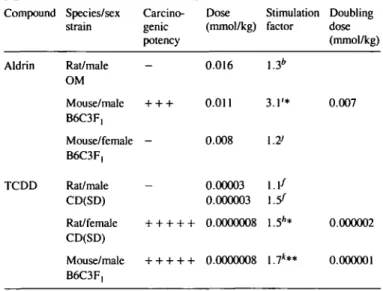
Species and sex differences
ALD induces liver tumors in male mice, but not in female mice
or in rats (21b). The assay was able to reproduce this difference.
DNA synthesis was stimulated only in male mice (Table III).
Here, ALD was a potent stimulator with a doubling dose of
0.007 mmol/kg.
TCDD is a carcinogen for male mice and female rats, but not
for male rats (22a). Again, TCDD was negative in male rat liver,
but was a very potent stimulator of DNA synthesis in male mice
with a DD of 10~
6mmol/kg and in female rats with a DD of
2 X 10~
6mmol/kg (Table DT). TCDD, measured in mice, was
the most potent stimulator of all compounds tested. TCDD had
been tested before in rats and was found to be negative for male
and female rats (23). The discrepancy could be explained by the
Table HI. Correlation of carcinogenic potency with stimulation of liver DNA synthesis; compounds with different carcinogenic potency in different species and sexes
Compound Species/sex Carcino- Dose Stimulation Doubling strain genie (mmol/kg) factor dose
potency (mmol/kg) Aldrin TCDD Rat/male OM Mouse/male B6C3F, Mouse/female B6C3F, Rat/male CD(SD) Rat/female CD(SD) Mouse/ male 0.016 + + + 0.011 0.008 0.00003 0.000003 + + + + + 0.0000008 + + + + + 0.0000008 1.3* 3.1'* \.7> 1.1/ l.5f 1.5** 1.7*** 0.007 0.000002 0.000001 *0.1 > P > 0.05; **P < 0.05 (Mest). b.f,h,ij.kThe. r e|a t e d c o n t r o l g r o u p ( T a b l e v i ) .
Table FV. Correlation of carcinogenic potency with stimulation of liver DNA synthesis; structural isomers with different carcinogenicity
Compound Species/sex Carcino- Dose Stimulation Doubling strain genie (mmol/kg) factor dose
potency (mmol/kg) [alpha]HCH Rat/male + + SIV-50 SD [gamma]HCH Rat/male SIV-50 SD 1.0 0.1 0.01 1.0 0.1 0.01 3 . 4 " " 1.7"** 1.4" 0.8" 0.9° 1.3" 0.2 *0.1 > P > 0.05; **P < 0.05 (Mest). "The related control group (Table VI).
fact that these authors measured DNA synthesis 3 5 - 3 6 h after
TCDD administration, whereas we measured after 24 h, which
was found to coincide with the peak DNA synthesis in our system.
Also, the dose used by these authors was 40 times larger than
ours and it is possible that a potential toxicity interfered with the
induction of DNA synthesis.
Structural isomers
It was investigated whether the test system was able to reflect
the differences in carcinogenicity of structurally related
com-pounds. Table IV summarizes the results obtained with the
struc-tural isomers [alpha]- and [gamma]HCH. Whereas [gamma]HCH
has no carcinogenic activity in rats (21a), the isomer
[alpha]-HCH is carcinogenic (18b,c). Only the carcinogenic [alpha][alpha]-HCH
isomer stimulated DNA synthesis with a DD of 0.2 mmol/kg.
The non-carcinogenic isomer [gamma] HCH did not have an
ef-fect on DNA synthesis.
The results obtained with a second pair of structurally related
compounds are shown in Table V. DEHP is a liver carcinogen
in rats (22c), while DEHA has no such effects in rats (22b). Both
compounds were able to stimulate DNA synthesis with DD's of
0.5 mmol/kg for DEHP and 0.7 mmol/kg for DEHA. DEHA
was the only compound tested so far where liver tumor
induc-tion and stimulainduc-tion of DNA synthesis were not correlated.
Table V. Correlation of carcinogenic potency with stimulation of liver DNA synthesis; structurally related compounds
Compound Species/sex Carcino- Dose Stimulation Doubling strain genie (mmol/kg) factor dose
potency (mmol/kg) DEHA DEHP Rat/male F-344 Rat/male F-344 3.78 1.73 10.5'** 0.7 7.8'** 0.5 *0.1 > P > 0.05; **P < 0.05 (Mest). T h e related control group (Table VI).
Control experiments
Bioavailability of radiolabelled TdR. All results are based on
measurements of the specific activity of liver DNA (d.p.m./mg)
arising from the incorporation of methyl radiolabelled thymidine
after oral gavage. Exhalation of
I4CO
2as a consequence of
en-zymatic thymidine degradation was routinely checked in order
to assure that the bioavailability of the radiolabelled DNA
precur-sor was uniform amongst the animals. On an average, 16 ± 4%
of the administered radioactivity in rats and 43 ± 5% in mice
was recovered as
I4CO
2within 3 h.
Autoradiography. To prove that an increase in DNA radioactivity
was correlated with an increase in dividing hepatocytes, a parallel
experiment with an autoradiographic evaluation was conducted
with 0.05 mmol/kg DDT. The fraction of cells in S-phase was
0.51%, control liver showed 0.15%. The SF by determination
of the specific activity of the DNA and by autoradiographic
analysis were therefore on a similar level.
Histology. Literature data show that regenerative DNA synthesis
in rat liver after single administration of a necrogenic dose of
CC1
4does not start until 30 h after the administration (24). A
separation between adaptive (= promotive?) and regenerative
liver DNA synthesis therefore seems possible. Nevertheless, in
order to exclude the possibility that an increase in DNA
radio-activity was only the result of regenerative processes after cell
death induced by the test compounds, histological preparations
of the livers were examined for necrotic regions after treatment
with DDT, TAA, ALD, TCDD, DEHA and DEHP. An increase
in necroses was never found.
Interindividual variability. In all experiments there was a large
variability of the TdRII for equally treated animals. The
stan-dard deviation was on average about 50% of the mean value.
The variability was not treatment-related, being also found in
the controls. Table VI summarizes the mean values and
stan-dard deviations of the TdRIIs of all control groups. The means
range from 2680 to 5540 in rats and from 850 to 1310 in mice.
It was therefore necessary to relate the results of each
experi-ment to its own control group. Unsuccessful attempts to reduce
the variability will be discussed.
Discussion
It has been known for a long time that many xenobiotics induce
liver growth. The topic has been comprehensively reviewed by
Schulte-Hermann (25,26). The role of liver growth in the
pro-cess of carcinogenesis has been studied primarily on a mechanistic
and cellular level and relatively little effort has been made to
evaluate this response of the liver for toxicological purposes. In
our assays, liver weights were not increased above control. This
lack of an observable effect is most probably due to the short
Table VI. Group (index) A. Rats a b c d e
f
8 h B. Mice i j k Incorporation of [l4C]TdR Mean value of incorpora-tion index 2680 4370 5540 5080 4600 4270 4280 3460 850 1130 1310 into liver Absolute standard deviation 980 2570 4360 3880 1860 1730 3130 430 580 640 260DNA of untreated control groups n 4 4 4 4 4 4 4 4 3 3 4 Relative standard deviation (%) 37 59 79 76 40 41 73 12 67 56 20
period of time between the administration of the test compound
and the isolation of the liver DNA.
We have tried to characterize the liver's response in a
quan-titative way by comparing the doses required for various
com-pounds to double the control level of DNA synthesis. Our results
indicated in most cases not only a qualitative but also a
quan-titative correlation between the carcinogenic potency and the
DNA synthesis stimulating potency (expressed as the DD) of
car-cinogens which do not act by DNA binding. Compounds with
a high carcinogenic potency were active at lower dose levels to
stimulate DNA synthesis than compounds with a lower
poten-cy. DNA-binding carcinogens did not stimulate DNA synthesis
at all. Therefore the stimulation test could represent a useful tool
for detecting a number of carcinogens which are missed by the
routine genotoxicity test systems.
Furthermore, we checked whether compounds that induce liver
tumors only in one rodent species, or only in one sex would
stimulate DNA synthesis in the susceptible but not in the
resis-tant case. For the compounds investigated in this study, it was
possible to pinpoint mouse- or rat-specific carcinogens and
sex-specific carcinogens. A difference in carcinogenicity of
structurally-related compounds was reflected by the assay with
the HCH isomers but not with the plasticizers DEHP and
DEHA. Further work is necessary to elucidate this 'false positive'
situation.
Attempts to reduce the interindividual variability
A large interindividual variability sometimes unacceptably
reduc-ed the level of significance of an effect. Various means were
tested to reduce this drawback:(l) [
l4C]methanol was used
in-stead of [
14C]thymidine to label all four nucleotides via the
carbon-1 pool; (2) only brothers were taken in a treatment group,
to have genetically similar animals; (3) food was available for
15 h instead of 5 h in order to reduce the stress of hunger;
(4) 'internal standardization': at day 0 at 8 a.m., the animals were
administered [
JH]TdR, to determine the level of DNA synthesis
before treatment with the test compound. Twenty-four hours later
the animals were given the test compound. After additional 24 h,
they received [
l4C]TdR, to determine the level of DNA
syn-thesis after the treatment with the test compound. On the basis
of the
3H- and the
l4C-counts in each liver DNA every animal
served as its own control. Unfortunately, none of these procedures
resulted in a reduction of the variability (data not shown).
Combination with short-term tests for genotoxicity and future
developments
The assay proposed here seems to be useful to recognize some
of those carcinogens which are notoriously missed by tests for
genotoxicity. The concept of the doubling dose (i.e. the single
dose of a compound required to double the control level of DNA
synthesis) might even allow a quantitative evaluation. Parodi and
co-workers have shown that a combination of two short-term tests
leads to an improvement of the predictivity if both tests can be
evaluated quantitatively and if both tests correlate to some
ex-tent with carcinogenic potency. The improvement will be
max-imal if the two tests recognize different classes of carcinogens,
i.e. do not correlate with each other (27). A preliminary analysis
of the present data showed that a combination of the DD with
a quantitative short-term test for genotoxicity, for instance with
a test measuring covalent DNA binding in vivo (1) indeed
markedly improved the prediction of the potency of a carcinogen
on the basis of a combination of two short-term tests (28).
The results show that a single administration of the test
com-pound is sufficient to induce an observable effect and to provide
a surprisingly good correlation with carcinogenic potency. It
might be expected that a repeated application would reflect the
real situation even better. It is also imaginable that this could
reduce the interindividual variability, because fluctuations from
day to day would be averaged out. On the other hand, the costs
for performing the test would be greater than with a single
ap-plication.
With the compounds investigated so far, only one false positive
and no false negative result was found. This is an astonishingly
good result. It is probable, however, that false negative results
sometimes will also be found, because no short-term test can be
expected to reflect all aspects and stages of carcinogenesis by
chemicals. It could, for instance, be imagined that a compound
leads to an increased formation of peroxides and oxygen radicals
able to damage DNA. This indirect genotoxicity would go
unobserved in a DNA binding study and possibly also in a test
on DNA synthesis, although it was recently reported that
hydro-peroxides of fatty acids stimulated DNA synthesis in rat colon
(29). It will then be necessary to search for the biological
activi-ty responsible in that specific situation for the tumors induced,
and to try to develop a corresponding short-term test. This would
finally allow the replacement of the long-term bioassay for
car-cinogenicity by a battery of test systems with complementary
scope. The mechanistic information available from such a
com-bination of assays would also be of value for extrapolations to
man.
Acknowledgements
This work was supported by the Krebsliga des Kantons Zurich.
References
1. Lutz.W.K. (1979) In vivo covalent binding of organic chemicals to DNA as a quantitative indicator in the process of chemical carcinogenesis. Mulal. Res., 65, 289-356.
2. Williams.G.M. (1981) The detection of genotoxic chemicals in the hepatocyte primary culture/DNA repair test. Gann Monogr. Cancer Res., 27, 45—55. 3. McCann,J., Choi.E., Yamasaki,E. and Ames.B.N. (1975) Detection of car-cinogens as mutagens in the Salmonella/microsome test: assay of 300 chemicals./Voc. Nail. Acad. Sci. USA, 72, 5135-5139.
4. Ashby.J., de Serres.F.J., Draper,M., Ishidate.M., Margolin.B.H., Mat-ter,B.E. and Shelby,M.D. (eds) (1985) Evaluation of Short-term Tests for Carcinogens. Elsevier Science Publishers, Amsterdam.
5. Armuth,V. and Berenblum.I. (1972) Systemic promoting action of phorbol in liver and lung carcinogenesis in AKR mice. Cancer Res., 32, 2259—2262. 6. Peraino.C, Fry,R.J.M., Staffeldt.E. and Christopher,J.P. (1975) Comparative enhancing effects of phenobarbital, amobarbital, diphenylhydantoin, and dichlorodiphenyltrichloroethane on 2-acetylaminofluorene-induced hepatic tumorigenesis in the rat. Cancer Res., 35, 2884-2890.
7. Goldsworthy.T.L. and Pitot.H.C. (1985) An approach to the development of a short-term whole animal bioassay to distinguish initiating agents (in-complete carcinogens), promoting agents, (in-complete carcinogens, and non-carcinogens in rat liver. J. Toxicol. Environ. Health, 16, 389-402. 8. Abanobi,S.E., Lombardi.B. and Shinozuka.H. (1982) Stimulation of DNA
synthesis and cell proliferation in the liver of rats fed a choline-devoid diet and their suppression by phenobarbital. Cancer Res., 42, 412—415. 9. Colbum.N.C, Wendel.E.J. and Abruzzo,G. (1981) Dissociation of
mitogenesis and late-stage promotion of tumor cell phenotype by phorbol esters: mitogen-resistant variants are sensitive to promotion. Proc. Natl. Acad. Sci. USA, 78, 6912-6916.
10. Cayama,E., Tsuda,H., Sarma.D.S.R. and Farber.E. (1978) Initiation of chemical carcinogenesis requires cell proliferation. Nature, 275, 60—62. 11. Columbano,A., Rajalakshmi,S. and Sarma,D.S.R. (1981) Requirement of
cell proliferation for the initiation of liver carcinogenesis as assayed by three different procedures. Cancer Res., 41, 2079-2083.
12. Craddock,V.M. (1976) Cell proliferation and experimental liver cancer. In Cameron,H.M., Linsel.D.A. and Warwick,G.P. (eds), Liver Cell Cancer. Elsevier/North Holland Biomedical Press, Amsterdam, pp. 153-201. 13. Kinzel.V., Fiirstenberger.G., Loehrke,H. and Marks,F. (1986) Three-stage
tumorigenesis in mouse skin: DNA synthesis as a prerequisite for the con-version stage induced by TPA prior to initiation. Carcinogenesis, 7, 779-782. 14. Scribner,J.D. and Siiss.R. (1978) Tumor initiation and promotion. Int. Rev.
Exp. Pathol, 18, 137-198.
15. Schulte-Hermann.R. and Landgraf,H. (1974) Circadian rhythm of cell pro-liferation in rat liver: synchronisation by feeding habits. Z. Naturforsch., 29c, 421-424.
16. Viviani.A. and Lutz.W.K. (1978) Modulation of the binding of the carcinogen benzo[a]pyrene to rat liver DNA in vivo by selective induction of microsomal and nuclear aryl hydrocarbon hydroxylase activity. Cancer Res., 38, 4640-4644.
17. Gold,L.S., Sawyer.C.B., Magaw.R., Backman,G.M., de Veciana,M., Levin-son,R., Hooper.N.K., Havender.W.R., Bernstein,L., Peto,R., Pike,M.C. and Ames,B.N. (1984) A carcinogenic potency database of the standardized result of animal bioassays. Environ. Hllh Perspect., 58, 9 - 3 1 9 . 18.1ARC Monographs on the Evaluation of the Carcinogenic Risk of Chemicals
to Humans. International Agency for Research on Cancer, Lyon. a: Vol. 1, 1972; b: Vol. 5, 1974; c: Vol. 20, 1979; d: Vol. 24, 1980; e: Vol. 29, 1982. 19. Rossi,L., Ravera,M., Repetti,G. and Santi,L. (1977) Long-term adminstra-tion of DDT or phenobarbital-Na in Wistar rats. Int. J. Cancer, 19, 179-185. 20. Dasgupta,A., Chatterjee.R. and Chowdhury,J.R. (1981)
Thioacetamide-induced hepatocarcinoma in rat. Oncology, 38, 249—253.
21. National Cancer Institute (NCI), Carcinogenesis, Technical Report Series. Bioassays for Possible Carcinogenicity. US Department of Health, Educa-tion, and Welfare, Public Health Service, National Institutes of Health, a: Vol. 14, 1977; b: Vol. 21, 1978.
22. National Toxicology Program (NTP), Carcinogenesis, Technical Report Series. Carcinogenesis Bioassays. US Department of Health and Human Services, Public Health Service, National Institutes of Health, a: Vol. 209, 1982; b: Vol. 212, 1982; c: Vol. 217, 1982.
23. Christian,B.J. and Peterson.R.E. (1983) Effects of 2,3,7,8-tetrachlorodi-benzo-p-dioxin on [3H]thymidine incorporation into rat liver deoxyribonucleic acid. Toxicology, 28, 133 — 146.
24. Doerfler,J. (1982) Einfluss von Hepatomitogenen und Ernahrung auf die DNS-Synthese und Monooxygenaseaktivitat in der Rattenleber nach Tetrachlorkohlenstoffvergiftung. Inaugural-Dissertation, Institut fur Toxikologie und Pharmakologie der Universitat Marburg/Lahn.
25. Schulte-Hermann.R. (1974) Induction of liver growth by xenobiotic compounds and other stimuli. CRC Crit. Rev. Toxicol., 3, 9 7 - 1 5 8 .
26. Schulte-Hermann,R. (1985) Tumor promotion in the liver. Archs. Toxicol., 57, 147-158.
27. Parodi.S., Taningher.M., Boero.P. and Santi,L. (1982) Quantitative correla-tions amongst alkaline DNA fragmentation, DNA covalent binding, mutagenicity in the Ames test and carcinogenicity for 21 compounds. Mutat. Res., 93, 1-24.
28. Lutz,W.K., Busser,M.-T. and Sagelsdorff,P. (1984) Potency of carcinogens derived from covalent DNA binding and stimulation of DNA synthesis in rat liver. Toxicol. Pathol., 12, 106-111.
29. Bull.A.W., Nigro.N.D., Golembieski,W.A., Crissman^.D. and Mamett.L.J. (1984) In vivo stimulation of DNA synthesis and induction of ornithine
decar-boxylase in rat colon by fatty acid hydroperoxides, autoxidation products of unsaturated fatty acids. Cancer Res., 44, 4924-4928.

![Table VI. Group (index) A. Rats a b c d e f 8 h B. Mice i j k Incorporation of [ l4 C]TdRMean valueof incorpora-tion index2680437055405080460042704280346085011301310 into liverAbsolutestandarddeviation980257043603880186017303130430580640260](https://thumb-eu.123doks.com/thumbv2/123doknet/14929507.664627/4.930.62.449.107.383/table-group-index-incorporation-tdrmean-valueof-incorpora-liverabsolutestandarddeviation.webp)