Compartmentalization of Cells Bearing "Rheumatic" Cell Surface Antigens
in Peripheral Blood and Tonsils in Rheumatic Heart Disease
E. D. Gray, W. E. Regelmann, Z. Abdin, A. EI Kholy, S. Zaher, R. Kamel, M. Mansour, L. Miller, P. Ferrieri, J. B. Zabriskie, and D. Braun
From the Departments of Pediatrics, Biochemistry, and Laboratory Medicine/Pathology, University of Minnesota Medical School, Minneapolis, Minnesota; the Free Children's Rheumatic Heart Center, Child Health Institute; the Egyptian Organization for Biological and Vaccine Production; the Department of Otolaryngology, Cairo University, Cairo, Egypt; the Rockefeller University, New York, New York; and the Pharmaceuticals Research Department, Ciba-Geigy, Basel, Switzerland
Monoclonal antibodies that recognize "rheumatic" antigens of peripheral blood non-T cells were used to study the compartmentalization of such cells in peripheral blood and tonsils of individuals with rheumatic heart disease (RHD) and suitable control subjects. The peripheral blood of most (710/0)of the 42 individuals with RHD contained cells reacting with monoclonal antibody 83S19.23 or 256S.l0, whereas these cells were present in only 17% of the 41 control subjects(P<.02). However, none of 21 individuals with RHD had such cells in their tonsils, although they were present in the tonsils of 50% of the 40 control subjects(P< .03). These results may reflect a failure in RHD of organ-specific homing of cells with the epitopes recognized by the antibodies. The presence of these cells in tonsils may be important in the immune response to streptococcal pharyngeal infection, and their absence in RHD may be involved in the unusual immune responses characteristic of this disease.
Elements of the cell surfaces of some peripheral blood mononuclear cells from individuals with a his-tory of rheumatic fever (RF) appear to be antigeni-cally distinct from those of the majority of the popu-lation [1]. This difference has been defined with human alloantisera that react with non-T cells (those not possessing a receptor for sheep red blood cells [SRBCs]) of >750/0 of individuals who have had documented RF. Monoclonal antibodies with simi-lar specificities have been developed that also allow the definition of "rheumatic" antigens on non-T cells [2, 3] in a similar proportion of individuals having had RF. Previous studies of the distribution of these antigens have examined their presence on the mononuclear cells of peripheral blood. The present
Received for publication 2 May 1986, and in revised form 11 August 1986.
Informed consent was obtained from the individuals under-going tonsillectomy, who donated peripheral blood and tonsillar tissue.
This study was supported by research grant HL-30058 from the National Institutes of Health.
We thank Z. Tavakoli, M. Verstegen, and Dr. Saied Morsy for technical assistance.
Please address requests for reprints to Dr. E. D. Gray, Box 296, University of Minnesota Medical School, 420 Delaware Street S. E., Minneapolis, Minnesota 55455.
247
study was directed at their distribution in both pe-ripheral blood and tonsils in individuals having in-active rheumatic heart disease (RHD) compared with those having recurrent or chronic tonsillitis.
Subjects and Methods
The individuals with RHD and control subjects were residents of Cairo. RHD was diagnosed by careful history and physical examination. None of the indi-viduals with RHD had heart failure or showed signs of rheumatic activity (i.e., fever or elevated erythro-cyte sedimentation rate) within a year of the study, and none showed obvious signs of nutritional defi-ciency.
Detailed history and physical examination of the control subjects excluded past or present RF or RHD. The average ± SD age of those with RHD was 11.4± 2.5 years and of the control subjects, 9.8
± 4.8 years (difference not significant). Samples of peripheral blood and tonsillar tissue were obtained from individuals undergoing tonsillectomy on the advice of their physician. The indication cited for tonsillectomy was recurrent or chronic tonsillitis. The subjects were afebrile and had normal erythrocyte sedimentation rates. The individuals with RHD had
248
approximately two incidents of pharyngitis during the year before study, whereas the control subjects had six incidents. Each tonsil was cultured and ex-amined for the presence of group AStreptococcus. The recovery of this organism was infrequent and not different in the RHO and control groups. This finding was consistent with the attending physicians' reports that all of the volunteers had taken penicil-lin or sulfonamides for at least one month before tonsillectomy. Some of the individuals with RHD either had already undergone tonsillectomy or were not scheduled for this operation, so only blood was studied from this group.
Mononuclear cells were isolated from blood and tonsils by gradient centrifugation. Mononuclear cells were rosetted with SRBCs, and T cells were separated from non-T cells by gradient centrifugation as pre-viously described [4, 5]. The blood was diluted with an equal volume of Eagle's MEM, layered on Ficoll-Paque" (Pharmacia, Uppsala, Sweden), and cen-trifuged at 650 g for 30 min. The mononuclear cells were removed from the gradient interface and washed twice with MEM, and a portion was subjected to ro-sette depletion for removal of T lymphocytes. The tonsils were minced with scissors and shaken for 1 min with MEM. Mononuclear cells were isolated from the resulting cell suspension by the same method used with blood. T cell depletion was ac-complished by SRBC rosetting. The mononuclear cells from blood and tonsils were resuspended in MEM containing 20070 fetal calf serum (previously adsorbed with SRBCs) and then incubated for 5 min
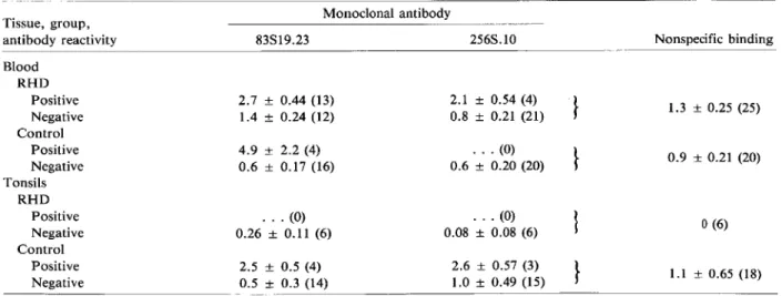
Table 1. Immunofluorescence assay of "rheumatic" cells.
Gray et al.
at 37 C with an equal volume of 5070 SRBCs in 20070 adsorbed fetal calf serum. After incubation the cell suspension was layered on Ficoll-Paque and cen-trifuged as described above. The mononuclear cells were removed, washed twice, and used for cell-marking studies. These nonrosetting cells are referred to as non-T cells. The non-T population isolated by these methods included 50070 B lymphocytes, 30070 monocytes, and <3070 residual T cells. This distribu-tion was not determined for each preparadistribu-tion.
The monoclonal antibodies used in the study were 83S19.23 and 256S.10. They were produced in re-sponse to immunization of mice with non-T cells from peripheral blood of individuals with a history of RF and were prepared in the Pharmaceuticals Re-search Department, Ciba-Geigy, Ltd. (Basel, Swit-zerland). These antibodies have specificity similar to that of the human alloantisera previously shown to react with the B cells of individuals with a history of RF [1, 2].
The study was conducted in two annual visits to Cairo. In one year the cell surfaces were stained by an indirect immunofluorescence technique, and in the other year an immunoperoxidase technique was used.
Indirect immunofluorescence staining was done by a method similar to that described by Abramson et al. [6]. For each non-T sample of tonsil and blood, three aliquots were stained: monoclonal antibodies 83S19.23 and 256S.10 and a control for determina-tion of nonspecific adsorpdetermina-tion. The slides were care-fully labeled with a code and read in a blinded
man-Tissue, group, Monoclonal antibody
antibody reactivity 83S19.23 256S.1O
Blood RHD Positive 2.7 ± 0.44 (13) 2.1 ± 0.54 (4)
'}
Negative 1.4 ± 0.24 (12) 0.8 ± 0.21 (21) Control Positive 4.9 ± 2.2 (4) . . . (0)}
Negative 0.6 ± 0.17 (16) 0.6 ± 0.20 (20) Tonsils RHD Positive . . . (0) . . . (0)}
Negative 0.26 ± 0.11 (6) 0.08 ± 0.08 (6) Control Positive 2.5 ± 0.5 (4) 2.6 ± 0.57 (3)}
Negative 0.5 ± 0.3 (14) 1.0 ± 0.49 (15) Nonspecific binding 1.3 ± 0.25 (25) 0.9 ±0.21 (20)o
(6) 1.1 ± 0.65 (18) NOTE. Data are mean ± SE percentages of positive cells (no. of individuals).Table 2. Immunoperoxidase assay of "rheumatic" cells.
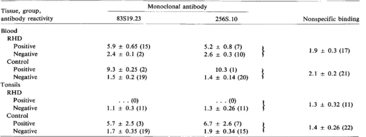
Tissue, group, Monoclonal antibody
antibody reactivity 83S19.23 256S.1O
Blood RHD Positive 5.9 ± 0.65 (15) 5.2 ± 0.8 (7)
}
Negative 2.4 ± 0.1 (2) 2.6 ± 0.3 (10) Control Positive 9.3 ± 0.25 (2) 10.3 (1)}
Negative 1.5 ± 0.2 (19) 1.4 ± 0.14 (20) Tonsils RHD Positive . . . (0) . . . (0)}
Negative 1.1±0.3(11) 1.3 ± 0.26 (11) Control Positive 5.7 ± 2.5 (3) 6.7 ± 2.6 (7)}
Negative 1.7 ± 0.35 (19) 1.9 ± 0.34 (15)NOTE. Data are mean ± SE percentages of positive cells (no. of individuals).
Nonspecific binding
1.9 ± 0.3 (17)
2.1 ± 0.2 (21)
1.3 ± 0.32 (11)
1.4 ± 0.26 (22)
nero An individual was regarded as "positive" for a given monoclonal antibody if the proportion of cells binding the monoclonal antibody was significantly (P
<
.05 by'1}analysis) greater than the proportion of cells binding the serum control. Sufficient num-bers of cells were counted so that the probability«(3)of not detecting a difference of~1070was <.10 (range of cells counted, 200-1,800).
A summary of the results obtained with the im-munofluorescence method is shown in table 1.The data represent mean values of the staining results and illustrate the background staining obtained with this method and the differences between positive and negative groups. The technique detects the binding of monoclonal antibodies to only a small fraction of non-T cells, but the group of positive individuals clearly have a higher proportion of these cells than do the negative groups, which are indistinguishable from the nonspecific serum control. The percentages of labeled cells in the positive RHD groups are not significantly different from those in the positive con-trol groups.
The immunofluorescence method requires that slides be read soon after preparation, and so that this limitation on the study of increased numbers of samples was overcome, an immunoperoxidase staining method was developed. Immunoperoxidase staining was accomplished by the avidin-biotin com-plex method with a kit from Vector Laboratories (Burlingame, Calif) [7, 8]. The non-T cells were spread on carefully labeled glass slides, and a circle
rv2em in diameter was inscribed around the cells,
which were allowed to dry overnight. The slides were immersed in95070ethanol for 5min, allowed to dry, and then immersed in PBS (pH 7.4) for 10 min. The slides were blotted dry around the circles containing the cells, 30J..lI of diluted horse serum was added to the circles, and the slides were incubated at room tem-perature ("-123 C) in a humid chamber for 30 min so that nonspecific binding of immunoglobulin was blocked. They were then immersed in PBS for 10 min and blotted, and 30J..lI of diluted monoclonal anti-body was added. After incubation for 30 min the slides were washed again in PBS and blotted, and 30 J..lI of biotin-labeled antiserum to mouse im-munoglobulin was added. After incubation for 30 min and a wash in PBS, 30 J..lI of biotin-avidin-peroxidase solution was added and incubated for 30 min. After a PBS wash and blotting, 50 J..lI of di-aminobenzidine-hydrogen peroxide substrate was added, and the slides were incubated for 10 min. They were then washed in distilled water and dried. Con-trol slides were similarly stained without monoclonal antibody. The dried slides were transported to the University of Minnesota Medical School (Min-neapolis) and read in a blinded and coded manner. Positive cells were those with black granules around a greater part of their periphery. A slide was consid-ered positive when it had a significantly greater num-ber of positive cells than did the horse serum con-trol slide, as determined by 'Y} analysis.
Table 2 summarizes the immunoperoxidase results and illustrates the higher proportion of monoclonal antibody-binding cells in positive individuals, as
250
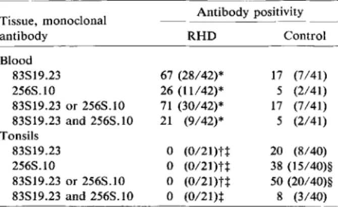
Table 3. Cell surface labeling of peripheral blood and tonsillar non-T cells.
Tissue, monoclonal Antibody positivity
antibody RHD Control Blood 83S19.23 67 (28/42)* 17 (7/41) 256S.1O 26 (11142)* 5 (2141) 83S19.23 or 256S.1O 71 (30/42)* 17 (7/41) 83S19.23 and 256S.1O 21 (9/42)* 5 (2/41) Tonsils 83S19.23 0 (0121)tt 20 (8/40) 256S.1O 0 (0121)H 38 (15/40)§ 83S19.23 or 256S.1O 0 (0121)tt 50 (20/40)§ 83S19.23 and 256S.1O 0 (0121)t 8 (3/40) NOTE. Data are percentages (no. of subjects positive/no. tested).
* Significantly different from control subjects(P<.02).
tSignificantly different from control subjects(P<.03). t Significantly different from blood from individuals with RHO (P<.02).
§ Significantly different from blood from control subjects(P
<.002).
compared with the immunofluorescence method. With the immunoperoxidase method the proportion of labeled cells in individuals considered negative is not significantly different than the proportion in slides with nonspecific serum instead of monoclonal antibody.
Although the percentage of reactive cells detected by the two methods differed, the proportion of in-dividuals with or without these cells was not signifi-cantly different in the two years of the study. There-fore the data on individuals from the two years were pooled for analysis.
Results
Cell surface labeling of peripheral blood non-T cells. Most individuals with RHD had peripheral blood non-T cells, with antigenic structures identi-fied by the monoclonal antibodies 83S19.23 and 256S.10 (table 3). A significantly lower proportion of the control subjects had non-T cells bearing these epitopes in their peripheral blood(P
<
.02). In both the RHD and control groups, most of the positive individuals had cells reacting with antibody 83S19.23, and fewer had cells reacting with antibody 256S.10. Cells marked by this latter antibody were most frequently present in individuals with cells also marked by antibody 83S19.23. These antibodies evi-dently are to a cell surface structure commonlypres-Gray et al.
ent on the peripheral blood mononuclear cells of in-dividuals with RHD.
Cell surface labeling of tonsillar non-Tcells. In remarkable contrast to the results with peripheral blood, the non-T cells from tonsils of individuals with RHD were not labeled by antibody 83S19.23 or 256S.10 (table 3). None of the 21 subjects with RHD possessed tonsillar non-T cells with cell sur-faces identified by these antibodies. However, the tonsils of 50070 of the control subjects contained cells reactive with these antibodies. Furthermore, most of the positive subjects possessed cells marked by anti-body 256S.10 rather than antianti-body 83S19.23, as seen in peripheral blood. Clearly, in both the RHD and control groups, the populations of non-T cells pres-ent in tonsils are differpres-ent than those in peripheral blood.
Relation between blood and tonsils in individu-als. Both blood and tonsils were studied from 17 individuals with RHD and from 34 individuals with-out RHD. This approach permitted a comparison between the blood and tonsils of each of these indi-viduals for the presence or absence of a significant proportion of monoclonal antibody-labeled non-T cells. An individual's blood or tonsil was considered "positive" if it contained a significantly greater proportion of non-T cells binding either or both monoclonal antibodies than did their serum controls. Each of these individuals could therefore have posi-tive tonsillar non-T cells, posiposi-tive blood non-T cells, or positive non-T cells in both compartments or in neither compartment. Table 4 shows the unexpected and highly significant(P
<
.001) difference between the RHD group and the control group in the distri-bution of positive non-T cells between blood and ton-sils. Individuals whose blood was positive for the "rheumatic" antigens identified by these monoclonal antibodies were much more likely to be in the RHD group (P< .005,q>coefficient=
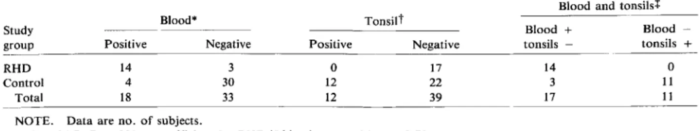
0.39; table 5).How-Table 4. Distribution of non-T cells bearing "rheumat-ic" antigens in blood and tonsils.
Study Blood + Blood + Blood - Blood-group tonsils - tonsils + tonsils - tonsils + Total
RHO 14 0 3 0 17
Control 3 1 19 11 34
NOTE. Data are no. of subjects. These results are a sum-mary of those obtained from individuals whose blood and ton-sils were both studied. The difference between the RHD and control groups was significant(X2 = 28.2, P <.001).
Table 5. Analysis of association of "rheumatic" antigen with RHD.
Blood and tonsilsf
Blood* Tonsilt
Study
group Positive Negative Positive Negative
RHO 14 3 0 17 Control 4 30 12 22 Total 18 33 12 39 Blood + tonsils -14 3 17 Blood -tonsils +
o
11 11 NOTE. Data are no. of subjects.* X2
= 24.7,P<.001; q>coefficient for RHO if blood was positive = 0.70. t X2 = 7.8, P = .005; q>coefficient for RHD if tonsils were negative = 0.39.
t X2 = 18.1, P<.001; q>coefficient for RHO if blood was positive and tonsils were negative
= 0.80.
ever, those individuals whose blood non-T cells bore the "rheumatic" antigens and whose tonsillar non-T cells did not were the most likely to be in the RHO group. Those individuals whose tonsillar cells were positive and whose blood did not contain positive cells were most likely to be in the group without RHO (P
<
.001,q> coefficient = 0.80; table 5).Discussion
The presence of particular antigens on the surface of non-T cells in individuals with RF or RHD has been observed in populations as disparate as those in India, the United States, and Egypt [3, 9]. These cell surface structures are characteristically found in most individuals with this form of disease after strep-tococcal infection. Their molecular characteristics and function are not well defined, but as they may represent expression of la-like genes [1], they might play a role in the altered immune responses as-sociated with RF and RHD [4, 5, 10]. The present study does not permit conclusions regarding the functional role of these non-T cells bearing "rheu-matic" antigens. However, other studies have shown that non-T cells are a requirement for a T celllym-phoproliferative response to some group A strep-tococcal products [5]. Moreover, subjects with RHO from a similar population have an altered function of blood and tonsillar non-T cells that results in lower T cell proliferation. These individuals also have lower relative rates of T suppressor/cytotoxic cell (T8) proliferation and higher T4/T8 cell ratios after stim-ulation of their tonsillar cells by streptococcal blasto-gen A [11]. The relation between these altered im-mune responses and the "rheumatic" non-T cells may be involved in the pathogenesis of RHD.
In the population studied here, the presence of non-T cells bearing the "rheumatic" antigens in the
blood and their absence in the same individual's ton-sils were most strongly associated with RHD. In con-trast, the absence of non-T cells bearing the "rheu-matic" antigens in the blood and their presence in the same individual's tonsils were most strongly as-sociated with an absence of RHO. These relations were observed even though the control group expe-rienced more pharyngitis than did the RHO group. Because both groups showed no acute signs of in-flammation, it seems likely that the differences in compartmentalization were not due to acute changes in the tissue distribution of non-T cells. Possibly the differences were due to an underlying genetic differ-ence alone or in combination with environmental factors. The current study groups were selected be-cause both groups had ample time (mean age, 10 years) and exposure that made repeated pharyngitis due to group A streptococci very likely. This situa-tion was important for identificasitua-tion of the associ-ation of potential immunocyte markers with RHO because streptococcal pharyngeal infections are necessary for the expression of a propensity to RHO. The quantitation of RHO risk associated with these monoclonal antibody markers will be best defined by a prospective study from infancy in populations at high risk for RHO.
One of the most important and apparently abso-lute determinants for the development of RF and RHO after group A streptococcal infection is that the infection take place in the upper respiratory tract [12]. Among the local factors that must be considered to explain this observation is the role of the tonsils and related pharyngeal lymphoid tissue.Itis possible that the tonsils may be the focus of immunologic aber-rations that lead to the development of disease after streptococcal infection. However, the composition of the freshly isolated mononuclear cell populations with respect to B cells, total T cells, or helper and
252
suppressor T cell subsets is not significantly altered in the tonsils of individuals with RHD [11].
The homing of intestine-associated B lymphocytes to the mammary glands during pregnancy is well documented [13-15]. This homing is most likely de-pendent on B cell surface characteristics, with those B lymphocytes bearing antibody specific for anti-gens delivered to the maternal intestine and emigrat-ing to the breast. Itis possible that individuals are put at increased risk for the development of RF and RHD if appropriate non-T cells have not homed to the pharyngeal lymphoid tissue before or during epi-sodes of group A streptococcal pharyngitis.
References
1. Patarroyo ME, Winchester RJ, Vejerano A, Gibofsky A, Cha-lem F, Zabriskie JB, Kunkel HG. Association of a B-cell alloantigen with susceptibility to rheumatic fever. Nature 1979;278:173-4
2. Zabriskie JB, Lavenchy D, Williams RC Jr, Fu SM, Yeadon CA, Fotino M, Braun DG. Rheumatic fever-associated B cell alloantigens as identified by monoclonal antibodies. Arthritis Rheum 1985;28:1047-51
3. Williams RC Jr, Raizada V, Prakash K, Sharma KB, Anand I, Ganguly NK, Zabriskie JB. Studies of streptococcal membrane antigen-binding cells in acute rheumatic fever. J Lab Clin Med 1985;105:531-6
4. Gray ED, Wannamaker LW, Ayoub EM, El Kholy A, Abdin ZH. Cellular immune responses to extracellular streptococ-cal products in rheumatic heart disease. J Clin Invest 1981;68:665-71
5. Gray ED, Regelmann WE, Wannamaker LW, El Kholy A, Abdin ZH. Functional alterations in non T cells in rheu-matic heart disease. Clin Exp Immunol 1982;49:488-92 6. Abramson CS, Kersey JH, LeBien TW. A monoclonal anti-body (BA-l) reactive with cells of human B lymphocyte lineage. J Immunol 1981;126:83-8
7. Hsu S-M, Raine L, Fanger H. Use of avidin-biotin-peroxidase
Gray et al.
complex (ABC) in immunoperoxidase techniques: a com-parison between ABC and unlabeled antibody (PAP) procedures. J Histochem Cytochem 1981;29:577-80 8. Sandhaus LM, Gajl-Peczalska KJ, Brunning RD.
Im-munophenotyping of leukaemia: an immunoperoxidase method using air-dried smears. Br J Haematol 1984;56: 131-8
9. Gray ED, Regelmann WE, Zabriskie JB, El Kholy A, Abdin Z, Kamel RMH, Zaher SW, Braun DG. "Rheumatic" cell surface antigens in peripheral blood and tonsils in rheu-matic heart disease. In: Kimura Y, Kotami S, Shiokawa Y, eds. Recent advances in streptococci and streptococcal diseases. Proceedings of the IXth Lancefield International Symposium on Streptococci and Streptococcal Diseases held in September, 1984. Berkshire, England: Reedbooks, 1985:337-8
10. Read SE, Fischetti VA, Utermohlen V, Falk RE, Zabriskie JB. Cellular reactivity studies to streptococcal antigens. Migration inhibition studies in patients with streptococ-cal infections and rheumatic fever. J Clin Invest 1974;54: 439-50
11. Regelmann WE, Gray ED, Wannamaker LW, LeBien TW, Mansour M, El Kholy A, Abdin Z. Lymphocyte subpopu-lations in rheumatic heart disease. In: Kimura Y, Kotami S, Shiokawa Y, eds. Recent advances in streptococci and streptococcal diseases. Proceedings of the IXth Lancefield International Symposium on Streptococci and Streptococ-cal Diseases held in September, 1984. Berkshire, England: Reedbooks, 1985:338-9
12.Wannamaker LW.The chain that links the heart to the throat. Circulation 1973;48:9-18
13. Goldblum RM, Ahlstedt S, Carlsson B, Hanson LA, Jodal U, Lidin-Janson G, Sohl-Akerlund A. Antibody-forming cells in human colostrum after oral immunisation. Nature 1975;257:797-9
14.Jalkanen ST, Butcher EC. In vitro analysis of the homing properties of human lymphocytes: developmental regula-tion of funcregula-tional receptors for high endothelial venules. Blood 1985;66:577-82
15.Wade AW, Szewczuk MR. Aging, idiotype repertoire shifts, and compartmentalization of the mucosal-associated lym-phoid system. Adv Immunol 1984;36:143-88