Cerebral Cortex June 2008;18:1233--1238 doi:10.1093/cercor/bhm170
Advance Access publication October 12, 2007
FEATURE ARTICLE
Famous Faces Activate Contextual
Associations in the Parahippocampal
Cortex
Moshe Bar
1, Elissa Aminoff
1and Alumit Ishai
21
Martinos Center at Massachusetts General Hospital, Harvard
Medical School, Charlestown, MA 02129, USA and
2Institute of
Neuroradiology, University of Zurich, 8057, Switzerland
The parahippocampal cortex (PHC) has been traditionally
impli-cated both in place processing and in episodic memory. How could
the same cortical region mediate these cognitive functions that
seem quite different? We have recently proposed that the PHC
should be seen as more generally mediating contextual associative
processing, which is required for both navigation and memory. We
therefore predicted that any associative objects should activate the
PHC. To test this generalization, we investigated the extent to
which common stimuli that are nonspatial by nature, namely faces,
activate the PHC, although their perception is typically associated
with other cortical structures. Specifically, we compared the
activation elicited by famous faces, which are highly associated
with rich pictorial and contextual information (e.g., Tom Cruise) and
are not associated with a specific place, with activation elicited by
unfamiliar faces. Consistent with our prediction, contrasting
famous with unfamiliar faces revealed significant activation within
the PHC. Taken collectively, these findings indicate that the PHC
should be regarded as mediating contextual associations in general
and not necessarily spatial or episodic information.
Keywords: associations, context, face processing, parahippocampal
cortex
Introduction
The parahippocampal cortex (PHC) in the medial temporal
lobe has been involved in the representation and processing of
spatial, place-related information (Aguirre et al. 1996; Maguire
et al. 1997; Epstein and Kanwisher 1998; Bohbot et al. 2000;
Mellet et al. 2000; Levy et al. 2001), and a significant subregion
within the PHC has been termed ‘‘parahippocampal place area’’
(PPA) (Epstein and Kanwisher 1998). In the community that
studies memory, however, the PHC has been widely known to
subserve episodic memory (Gabrieli et al. 1997; Brewer et al.
1998; Wagner et al. 1998; Schacter and Wagner 1999; Davachi
et al. 2003; Ranganath et al. 2004; Squire et al. 2004). Therefore,
it has previously been unclear how could the same region
mediate seemingly different cognitive functions (Fig. 1).
Although spatial processing functions are generally different
from episodic memory, they rely on similar elements. We have
recently proposed that these shared elements are provided by
contextual associations (Bar and Aminoff 2003; Bar 2004;
Aminoff et al. 2007) and showed that the PHC is differentially
activated for a wide range of familiar and newly learned
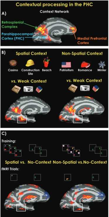
contextual associations (Fig. 2).
In our initial experiments (Bar and Aminoff 2003), we
compared the functional magnetic resonance imaging (fMRI)
activation elicited when participants viewed pictures of
individual objects that are highly associated with a certain
context (e.g., a cowboy hat) with the activation elicited by
individual objects that were not strongly associated with any
context (e.g., a camera). Contextual associations differentially
activated a network including the PHC, retrosplenial complex,
and parts of the medial prefrontal cortex (Fig. 2A). Activation
within this network has since been found to be robust and
highly replicable (Bar and Aminoff 2003; Bar 2004; Aminoff
et al. 2007; Diana et al. forthcoming). Standardized localizer
information can be found at http://barlab.mgh.harvard.edu/
ContextLocalizer.htm.
Given that the PHC activation elicited by strongly contextual
objects seemed to overlap with the activation reported in the
PPA (Epstein and Kanwisher 1998; Epstein et al. 1999), it was
unclear whether our activation was mainly due to contextual
associations or the result of an indirect activation of specific
places, which in turn activated the PPA. We therefore
conducted a second study where the highly contextual objects
were clearly separated for spatial contextual objects (e.g.,
a construction hat) and highly contextual objects that are
associated with nonspatial contexts (e.g., cupid). Both
con-ditions activated the same contextual network (Fig. 2B).
Interestingly, we found segregation in the PHC between the
activation elicited by spatial and nonspatial contexts, whereby
spatial contexts activate primarily the posterior part of the PHC
and the nonspatial contexts activate a more anterior, adjacent
part of the PHC (Bar and Aminoff 2003).
In a third study (Aminoff et al. 2007), we created novel
objects and relations, such that participants did not have any
subjective associations and memories with the new stimuli
beyond those on which they were trained prior to scanning.
The findings of this experiment (see Fig. 2c) corroborated our
previous findings and thus added support to our proposal that
the PHC is involved in both spatial and nonspatial contextual
associations.
Associations are required to link landmarks to places in
spatial processes, and contextual associations are needed to
glue the constituents of an episode in memory. Consequently,
by modifying the ascription of a role to the PHC to be
mediating contextual associative representations and
pro-cessing in general, findings from various experiments can be
naturally reconciled. A critical test of our hypothesis is
whether complex stimuli with contextual but not spatial
associations would also activate the PHC. Therefore, we
compared here PHC activation elicited by famous faces, which
evoke a large amount of contextual associations that are not
particularly spatial, with the PHC activation elicited by
un-familiar faces.
Face perception is a highly developed visual skill in humans,
which is mediated by activation in a distributed neural system
that encompasses visual, limbic, and prefrontal regions (Ishai
Ó 2007 The Authorset al. 2005; Fairhall and Ishai 2007). Specifically, the cortical
network that mediates face perception includes the fusiform
gyrus, an extrastriate region that processes the identification of
individuals (Kanwisher et al. 1997; Ishai et al. 1999; Grill-Spector
et al. 2004); the superior temporal sulcus (Hoffman and Haxby
2000; Puce et al. 2003); the amygdala and insula (Breiter et al.
1996; Ishai et al. 2004); the inferior frontal gyrus, where
semantic aspects are processed (Leveroni et al. 2000; Ishai
et al. 2002); and regions of the reward circuitry, including the
nucleus accumbens and orbitofrontal cortex (Aharon et al. 2001;
O’Doherty et al. 2003; Kranz and Ishai 2006; Ishai 2007). In most
previous studies, the response to faces was contrasted with
assorted common objects to localize the fusiform face area (FFA)
(e.g., Kanwisher et al. 1997) or with scrambled faces to localize
the underlying network (e.g., Ishai et al. 2002; Ishai et al. 2004).
Familiar (e.g., famous) faces elicit enhanced activation in
various face-responsive regions (Ishai et al. 2005). In addition,
however, famous faces are associated with rich amount of
contextual and pictorial information, without being associated
with particular spatial information (e.g., Angelina Jolie’s face
elicits associations about her appearance in Mr. and Mrs.
Smith, her liaison with Brad Pitt, her adopted children, and her
humanitarian work in Africa). Therefore, famous faces are
optimal for testing our critical generalization and for
contrast-ing the 2 hypotheses: If the PHC contains a place-specific
module (PPA), then it should not be particularly responsive to
faces that have no pronounced spatial associations. However, if,
as we propose, the PHC more generally mediates contextual
Figure 2. Summary of our previous studies of contextual associations. (A) Medial activation for the contrast between strong contextually associative objects and weakly contextual objects. This statistical map was derived from averaging together this strong versus weak contrast from 6 different experiments, of which various tasks were used, with a total of 68 participants (Bar et al. 2007). (B) A demonstration that both objects strongly related to specific spatial context, or a place, and objects strongly related to a nonspatial context (e.g., romance) significantly activate the PHC when compared with weak contextual objects (Bar and Aminoff 2003). (C) A further demonstration that the PHC processes contextual associations. Participants were trained to learn both spatial and nonspatial associations between novel shapes. At test, participants were scanned when only viewing 1 shape at a time. Statistical maps show the comparison of viewing shapes associated with a spatial (left) or a nonspatial (right) context compared with no-context shapes (Aminoff et al. 2007). Figure 1. Spatial and episodic processing in the PHC. A comparison of Talairachcoordinates from the spatial processing literature (17 studies) and the episodic memory literature (17 studies). Spatial processing (numbers marked in red): (1) Epstein et al. (1999), (2) Epstein et al. (2003), (3) Levy et al. (2001), (4) Goh et al. (2004), (5) Gorno-Tempini and Price (2001), (6) O’Craven and Kanwisher (2000), (7) Janzen and van Turennout (2004), (8) Mellet et al. (2000), (9) Maguire et al. (1997), (10) Rosenbaum et al. (2004), (11) Shelton and Gabrieli (2002), (12) Sugiura et al. (2005), (13) Yi and Chun (2005), (14) Steeves et al. (2004), (15) Goel et al. (2004), (16) Suzuki et al. (2005), and (17) Burgess et al. (2001). Episodic memory (numbers marked in yellow): (1) Wagner et al. (1998), (2) Medford et al. (2005), (3) Sommer et al. (2005), (4) Morcom et al. (2003), (5) Davachi et al. (2003), (6) Kirchhoff et al. (2000), (7) Casasanto et al. (2002); (8) Brewer et al. (1998), (9) Reber et al. (2002), (10) Takahashi et al. (2002), (11) Dobbins et al. (2003), (12) Henke et al. (1999), (13) Yonelinas et al. (2001), (14) Pihlajamaki et al. (2003); Kirwan and Stark (2004), (16) Tsukiura et al. (2002), and (17) Ranganath et al. (2003). Reproduced with permission from Aminoff et al. (2007).
associations, it would exhibit differential activation for famous
compared with unfamiliar faces because of the contextual
associations they evoke.
Before we proceed, it might be worth elaborating on what
we mean by ‘‘contextual associations.’’ Our working definition
is that items are contextually associated with each other if they
typically coappear in the environment around us or their
corresponding representations tend to be coactivated.
There-fore, a desk and a chair are contextually associated because
they tend to appear together, but a cupid and a heart-shaped
chocolate box are also contextually associated with each other,
even if they do not typically physically appear next to each
other. Furthermore, the strength of the associations, rather
than their number, is the primary dimension that determines
whether a certain visual stimulus is considered strongly or
weakly contextually associative. For example, a flyswatter is
associated only with a fly. A refrigerator, on the other hand, is
associated with many objects that tend to share its context.
Both a flyswatter and a refrigerator are considered strongly
contextual objects, which is also reflected in the consensus
observed in participants’ responses (Bar and Aminoff 2003).
Objects that we consider weakly contextual objects (e.g.,
a camera or a bottle of water), on the other hand, are associated
with many objects, but only weakly, and most importantly,
these associates do not share the same context. There are
many such strong associations for the famous faces, whereas
they are nonexistent for the unfamiliar faces in our critical
comparisons.
Materials and Methods
The methods described here pertain exclusively to the face study, which is also the study for which we present new results here. For detailed descriptions of the material and methods used in the studies of contextual processing with objects and shapes, see Bar and Aminoff (2003); Aminoff et al. (2007); and Bar et al. (2007).
Participants
Ten healthy participants (5 males, mean age 25 years) with normal vision participated in the study. All participants gave written informed consent in accordance with protocols approved by the University Hospital of Zurich.
Stimuli and Tasks
Participants were presented with grayscale photographs of unfamiliar neutral and emotional (fearful and happy) faces and neutral famous faces. The famous faces consisted of contemporary Hollywood celebrities, taken from the database of Ishai et al. (2002). Phase-scrambled versions of these faces were used as visual baseline (Ishai et al. 2005). Each stimulus was presented for 3 s. Each run included 3 alternating epochs of scrambled faces (24 s) and faces (36 s). Four runs were collected per each participant, and the order of stimulus formats was randomized. In each block, 12 faces were presented, namely 36 faces per run and a total of 144 faces in the experiment. Stimuli were generated using Presentation (http://www.neurobs.com, version 9.13) and were projected with a magnetically shielded LCD video projector onto a translucent screen. The subject was instructed to view the faces attentively.
Data Acquisition
Data were collected using a 3T Philips Intera whole-body MR scanner. Functional data were obtained from 39 transverse slices covering the whole brain with a spatial resolution of 2.332.333 mm (acquisition matrix 96396), using a sensitivity-encoded single-shot gradient-echo planar sequence. Images were acquired with field of view=220 mm,
time repetition [TR]=3000 ms, time echo [TE]=35 ms, h=82° and with a SENSE acceleration factor of 2.0 (Pruessmann et al. 1999). High-resolution spoiled gradient recalled echo structural images were obtained with 1 3 1 3 1-mm spatial resolution (acquisition matrix 2243224), TE =2.30 ms, TR= 20 ms, h= 20°. These T1-weighted
images provided detailed anatomical information for the region of interest analysis.
Data Analysis
Data were analyzed using SPM5 software (http://www.fil.ion.ucl.ac.uk/ spm/). All volumes were slice time corrected, realigned to the first volume, corrected for motion artifacts, mean adjusted by proportional scaling, normalized into standard stereotactic space (template provided by the Montreal Neurological Institute [MNI]), and smoothed using a 5-mm full-width-at-half-maximum Gaussian kernel. The time series were high-pass filtered to eliminate low-frequency components (filter width 128 s) and adjusted for systematic differences across trials. The statistical analysis was based on a conventional general linear model (Friston, Holmes, Poline, et al. 1995; Friston, Holmes, Worsley, et al. 1995). To test our hypothesis, famous faces were contrasted with unfamiliar faces (for the main effect analysis see Ishai et al. 2005). Montreal Neurological Institute coordinates were converted to the normalized space of the brain atlas (Talairach and Tournoux 1988).
Results
We report here novel findings showing that famous faces also
activate the PHC. The data from object studies with which
these results overlap have been published previously and are
overviewed in the Introduction.
When contrasted with scrambled faces, face stimuli elicit
significant activation within multiple visual, limbic, and
pre-frontal regions (Fig. 3). To contrast our contextual association
hypothesis with the PPA hypothesis, we reanalyzed the data set
of Ishai et al. (2005) and directly compared famous faces with
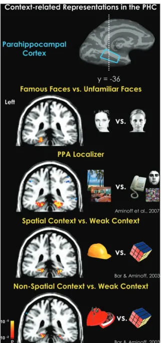
unfamiliar faces. Critically, the PHC region (mean Talairach
coordinates: x
=–20, y
=–36, z
=–10) responded significantly
more strongly to famous than unfamiliar faces (P
<0.001) (Fig.
4). All face stimuli were equated for size and other low-level
features, and indeed, given that the faces in the 2 conditions did
not significantly differ in any dimension other than the
information associated with the famous faces, we did not
observe significant activation in any other face-responsive
region. Additionally, in the postscan debriefing, all participants
reported that when they viewed the famous faces, they
spontaneously remembered other pictures of these celebrities
and information they knew about them from the tabloids and
their recent movies. In contrast, viewing unfamiliar faces did
not elicit any associations or recall of prior knowledge about
these faces. In summary, when compared with unfamiliar faces,
for which no prior knowledge exists, viewing famous
celebrities evokes rich contextual associations that resulted
in the PHC activation.
To demonstrate the generalization we are proposing here,
Figure 4 shows a comparison of old and new contrasts
involving different stimuli, tasks, and conditions: the statistical
maps and differential activation evoked by scenes, objects, and
faces. Across all 4 contrasts, it is clear that stimuli with rich
contextual associations activate the PHC significantly,
regard-less of whether these associations are related to places or not.
Overall, this remarkable compatibility between the activations
elicited by various objects and famous faces provides significant
support to our proposal that the PHC mediates contextual
associations in general, rather than spatial associations proper.
Discussion
To reconcile the various cognitive functions that elicit
activation in the PHC, we have proposed that this region
should be considered as mediating contextual associations in
general, without committing to a specific type of associations
(Bar and Aminoff 2003; Aminoff et al. 2007; Bar et al.
forthcoming). The new findings reported here, that famous
faces activate the PHC, support a critical prediction that was
derived from this proposal and provide complementary
evidence that allows a generalization of this proposal.
Specifically, we found that when faces are highly associative,
they activate areas involved in contextual associations in the
PHC, although faces, when compared with assorted common
objects, evoke activation predominantly in the FFA (Kanwisher
et al. 1997; Gorno-Tempini and Price 2001; Grill-Spector et al.
2004). There have been some reports showing that famous faces
activate the PHC (Sergent et al. 1992; Leveroni et al. 2000;
Trauner et al. 2004; Pourtois et al. 2005). Although these previous
reports did not make a reference to context and associations,
they support our findings and overarching proposal.
Faces that are personally familiar differ from famous faces in
terms of emotional attachment and intimate knowledge about
biographical information. When personally familiar faces are
compared with famous faces, stronger activation is observed in
the fusiform gyrus, posterior superior temporal sulcus,
poste-rior
cingulate/precuneus,
and
the
paracingulate
cortex
(Gobbini et al. 2004). Activation in these regions, previously
associated with theory of mind (Gallagher and Frith 2003) and
the brain’s ‘‘default’’ network, suggests that viewing personally
familiar faces elicits spontaneous retrieval of social and personal
knowledge associated with close friends and family members.
This is in accordance with our previous observation of a major
overlap between the cortical network mediating contextual
associations and the default network (Bar et al. 2007). Taken
collectively, these findings suggest that the response to faces is
modulated not only by the degree of visual familiarity but
importantly by the concomitant associations involved.
It is of interest that although most of the famous faces used
in our study were also attractive faces, the comparison with the
unfamiliar faces did not reveal enhanced activation in the
orbitofrontal cortex. Our previous studies have shown that
attractive faces evoke stronger activation in the orbitofrontal
cortex (Kranz and Ishai 2006; Ishai 2007). Therefore, our
current findings suggest that beauty was implicit to the
spontaneous retrieval of contextual associations associated
with these famous faces.
We did not observe stronger activation to famous than
unfamiliar faces in other regions of the contextual processing
network, namely the retrosplenial complex and the medial
prefrontal cortex. This lack of activation could be explained in
terms of cognitive demands. Tasks that involve recognition,
generation of mental images, and retrieval from episodic
memory are more likely to evoke activation within these
regions than passive viewing (Ishai et al. 2000; Ishai et al. 2002;
Bar and Aminoff 2003; Yago and Ishai 2006; Aminoff et al. 2007;
Bar et al. 2007; Aminoff et al., forthcoming).
Figure 3. When various face stimuli (e.g., unfamiliar and famous faces) are contrasted with scrambled faces, activation is found within a distributed cortical network that includes visual, limbic, and prefrontal regions. Coronal sections, taken from a representative subject, illustrate activation within the inferior occipital gyrus (IOG); fusiform gyrus (FG), superior temporal sulcus (STS); amygdala; inferior frontal gyrus (IFG); and orbitofrontal cortex (OFC). Coordinates are in the Talairach space. Adapted from Ishai et al. (2005).
Figure 4. PHC activations by places, spatial context, nonspatial context, and famous faces. Across all contrasts, highly associative stimuli differentially and significantly activated the PHC, regardless of nature of the stimulus and the type of associations (Bar and Aminoff 2003; Aminoff et al. 2007).
Finally, our current findings revealed an intriguing
hemi-spheric asymmetry. As can be seen in Figure 4, nonspatial
information seems to elicit stronger activation in the left
hemisphere, whereas spatial information seems to elicit
stronger activation in the right hemisphere. Although face
stimuli evoke stronger activation in the right hemisphere (Ishai
et al. 2005), contrasting famous faces with unfamiliar ones
resulted in a significant cluster in the left PHC. These findings
are consistent with prior studies of visual imagery of faces, in
which stronger activation was found in the left hemisphere
(Ishai et al. 2000; Ishai et al. 2002). Given that imagery and
contextual activation share the property of activating
repre-sentations of information that is not physically present, this
convergence supports our proposal that viewing famous faces
elicits contextual associations, possibly mediated by semantic
knowledge stored in the left hemisphere. Future studies will
determine the extent to which this hemispheric asymmetry
can be generalized.
In summary, using faces, we have extended our previous
stud-ies (Bar and Aminoff 2003; Bar 2004; Aminoff et al. 2007; Bar et al.
2007) and provided additional strong support to our proposal
that the operation of the PHC, including the PPA within it, is
best explained as mediating contextual associations.
Funding
National Institute of Neurological Disorders and Stroke
(NS044319 and NS050615); Dart Scholar Award to M.B.;
NRSA-T32MH070328
to
E.A.;
NCRR-P41RR14075;
Swiss
National Science Foundation (3200B0-105278); Swiss National
Center for Competence in Research:
Neural Plasticity and
Repair grant to A.I.
Notes
We thank S. Fairhall for assistance with data analysis and D. Greve for help with the analysis tools. Conflict of Interest: None declared.
Address correspondence to email: [email protected].
References
Aguirre GK, Detre JA, Alsop DC, D’Esposito M. 1996. The para-hippocampus subserves topographical learning in man. Cereb Cortex. 6:823--829.
Aharon I, Etcoff N, Ariely D, Chabris CF, O’Connor E, Breiter HC. 2001. Beautiful faces have variable reward value: fMRI and behavioral evidence. Neuron. 32:537--551.
Aminoff E, Gronau N, Bar M. 2007. The parahippocampal cortex mediates spatial and non-spatial associations. Cereb Cortex. 27:1493--1503.
Aminoff E, Schacter DL, Bar M. Forthcoming. The cortical under-pinnings of context-based memory distortion.
Bar M. 2004. Visual objects in context. Nat Rev Neurosci. 5:617--629. Bar M, Aminoff E. 2003. Cortical analysis of visual context. Neuron.
38:347--358.
Bar M, Aminoff E, Mason M, Fenske M. 2007. The units of thought. Hippocampus. 17:420--428.
Bar M, Aminoff E, Schacter DL. Forthcoming. Scenes unseen: the parahippocampal cortex subserves contextual associations, not scenes per se.
Bohbot VD, Allen JJ, Nadel L. 2000. Memory deficits characterized by patterns of lesions to the hippocampus and parahippocampal cortex. Ann N Y Acad Sci. 911:355--368.
Breiter HC, Etcoff NL, Whalen PJ, Kennedy WA, Rauch SL, Buckner RL, Strauss MM, Hyman SE, Rosen BR. 1996. Response and habituation of the human amygdala during visual processing of facial expression. Neuron. 17:875--887.
Brewer JB, Zhao Z, Desmond JE, Glover GH, Gabrieli JD. 1998. Making memories: brain activity that predicts how well visual experience will be remembered. Science. 281:1185--1187.
Burgess N, Maguire EA, Spiers HJ, O’Keefe J. 2001. A temporoparietal and prefrontal network for retrieving the spatial context of lifelike events. Neuroimage. 14:439--453.
Casasanto DJ, Killgore WD, Maldjian JA, Glosser G, Alsop DC, Cooke AM, Grossman M, Detre JA. 2002. Neural correlates of successful and unsuccessful verbal memory encoding. Brain Lang. 80:287--295. Diana RA, Yonelinas AP, Ranganath C. Forthcoming. High-resolution
multi-voxel pattern analysis of category selectivity in parahippo-campal cortex.
Davachi L, Mitchell J, Wagner A. 2003. Multiple routes to memory: distinct medial temporal lobe processes build item and source memories. Proc Natl Acad Sci USA. 100:2157--2162.
Dobbins IG, Rice HJ, Wagner AD, Schacter DL. 2003. Memory orientation and success: separable neurocognitive components underlying episodic recognition. Neuropsychologia. 41:318--333. Epstein R, Graham KS, Downing PE. 2003. Viewpoint-specific scene
rep-resentations in human parahippocampal cortex. Neuron. 37:865--876. Epstein R, Harris A, Stanley D, Kanwisher N. 1999. The parahippocampal place area: recognition, navigation, or encoding? Neuron. 23:115--125. Epstein R, Kanwisher N. 1998. A cortical representation of the local
visual environment. Nature. 392:598--601.
Fairhall SL, Ishai A. 2007. Effective connectivity within the distributed cortical network for face perception. Cereb Cortex. 17:2400--2406. Friston KJ, Holmes AP, Poline JB, Grasby PJ, Williams SC, Frackowiak RS, Turner R. 1995. Analysis of fMRI time-series revisited. Neuroimage. 2:45--53.
Friston KJ, Holmes AP, Worsley KJ, Poline JB, Frith CD, Frackowiak RS. 1995. Statistical parametric maps in functional imaging: a general linear approach. Hum Brain Mapp. 2:189--210.
Gabrieli JD, Brewer JB, Desmond JE, Glover GH. 1997. Separate neural bases of two fundamental memory processes in the human medial temporal lobe. Science. 276:264--266.
Gallagher HL, Frith CD. 2003. Functional imaging of ‘‘theory of mind’’. Trends Cogn Sci. 7:77--83.
Gobbini MI, Leibenluft E, Santiago N, Haxby JV. 2004. Social and emotional attachment in the neural representation of faces.[erratum appears in Neuroimage. 2006;32:1484]. Neuroimage. 22:1628--1635. Goel V, Makale M, Grafman J. 2004. The hippocampal system mediates logical reasoning about familiar spatial environments. J Cogn Neurosci. 16:654--664.
Goh JO, Siong SC, Park D, Gutchess A, Hebrank A, Chee MW. 2004. Cortical areas involved in object, background, and object-background processing revealed with functional magnetic reso-nance adaptation. J Neurosci. 24:10223--10228.
Gorno-Tempini ML, Price CJ. 2001. Identification of famous faces and buildings: a functional neuroimaging study of semantically unique items. Brain. 124:2087--2097.
Grill-Spector K, Knouf N, Kanwisher N. 2004. The fusiform face area subserves face perception, not generic within-category identifica-tion. Nat Neurosci. 7:555--562.
Henke K, Weber B, Kneifel S, Wieser HG, Buck A. 1999. Human hippocampus associates information in memory. Proc Natl Acad Sci USA. 96:5884--5889.
Hoffman EA, Haxby JV. 2000. Distinct representations of eye gaze and identity in the distributed human neural system for face perception. Nat Neurosci. 3:80--84.
Ishai A. 2007. Sex, beauty and the orbitofrontal cortex. Int J Psychophysiol. 63:181--185.
Ishai A, Haxby JV, Ungerleider LG. 2002. Visual imagery of famous faces: effects of memory and attention revealed by fMRI. Neuroimage. 17:1729--1741.
Ishai A, Pessoa L, Bikle PC, Ungerleider LG. 2004. Repetition suppression of faces is modulated by emotion. Proc Natl Acad Sci USA. 101:9827--9832.
Ishai A, Schmidt CF, Boesiger P. 2005. Face perception is mediated by a distributed cortical network. Brain Res Bull. 67:87--93.
Ishai A, Ungerleider LG, Haxby JV. 2000. Distributed neural systems for the generation of visual images. Neuron. 28:979--990.
Ishai A, Ungerleider LG, Martin A, Schouten JL, Haxby JV. 1999. Distributed representation of objects in the human ventral visual pathway. Proc Natl Acad Sci USA. 96:9379--9384.
Janzen G, van Turennout M. 2004. Selective neural representation of objects relevant for navigation. Nat Neurosci. 7:673--677.
Kanwisher N, McDermott J, Chun MM. 1997. The fusiform face area: a module in human extrastriate cortex specialized for face perception. J Neurosci. 17:4302--4311.
Kirchhoff BA, Wagner AD, Maril A, Stern CE. 2000. Prefrontal-temporal circuitry for episodic encoding and subsequent memory. J Neurosci. 20:6173--6180.
Kirwan CB, Stark CE. 2004. Medial temporal lobe activation during encod-ing and retrieval of novel face-name pairs. Hippocampus. 14:919--930. Kranz F, Ishai A. 2006. Face perception is modulated by sexual
preference. Curr Biol. 16:63--68.
Leveroni CL, Seidenberg M, Mayer AR, Mead LA, Binder JR, Rao SM. 2000. Neural systems underlying the recognition of familiar and newly learned faces. J Neurosci. 20:878--886.
Levy I, Hasson U, Avidan G, Hendler T, Malach R. 2001. Center-periphery organization of human object areas. Nat Neurosci. 4:533--539. Maguire EA, Frackowiak RS, Frith CD. 1997. Recalling routes around
London: activation of the right hippocampus in taxi drivers. J Neurosci. 17:7103--7110.
Medford N, Phillips ML, Brierley B, Brammer M, Bullmore ET, David AS. 2005. Emotional memory: separating content and context. Psychiatr Res. 138:247--258.
Mellet E, Briscogne S, Tzourio-Mazoyer N, Ghaem O, Petit L, Zago L, Etard O, Berthoz A, Mazoyer B, Denis M. 2000. Neural correlates of topographic mental exploration: the impact of route versus survey perspective learning. Neuroimage. 12:588--600.
Morcom AM, Good CD, Frackowiak RS, Rugg MD. 2003. Age effects on the neural correlates of successful memory encoding. Brain. 126:213--229.
O’Craven KM, Kanwisher N. 2000. Mental imagery of faces and places activates corresponding stimulus-specific brain regions. J Cogn Neurosci. 12:1013--1023.
O’Doherty J, Winston J, Critchley H, Perrett D, Burt DM, Dolan RJ. 2003. Beauty in a smile: the role of medial orbitofrontal cortex in facial attractiveness. Neuropsychologia. 41:147--155.
Pihlajamaki M, Tanila H, Hanninen T, Kononen M, Mikkonen M, Jalkanen V, Partanen K, Aronen HJ, Soininen H. 2003. Encoding of novel picture pairs activates the perirhinal cortex: an fMRI study. Hippocampus. 13:67--80.
Pourtois G, Schwartz S, Seghier ML, Lazeyras F, Vuilleumier P. 2005. View-independent coding of face identity in frontal and temporal cortices is modulated by familiarity: an event-related fMRI study. Neuroimage. 24:1214--1224.
Pruessmann KP, Weiger M, Scheidegger MB, Boesiger P. 1999. SENSE: sensitivity encoding for fast MRI. Magn Reson Med. 42:952--962. Puce A, Syngeniotis A, Thompson JC, Abbott DF, Wheaton KJ, Castiello U.
2003. The human temporal lobe integrates facial form and motion: evidence from fMRI and ERP studies. Neuroimage. 19:861--869. Ranganath C, Johnson MK, D’Esposito M. 2003. Prefrontal activity
associated with working memory and episodic long-term memory. Neuropsychologia. 41:378--389.
Ranganath C, Yonelinas AP, Cohen MX, Dy CJ, Tom SM, D’Esposito M. 2004. Dissociable correlates of recollection and familiarity within the medial temporal lobes. Neuropsychologia. 42:2--13.
Reber PJ, Wong EC, Buxton RB. 2002. Encoding activity in the medial temporal lobe examined with anatomically constrained fMRI analysis. Hippocampus. 12:363--376.
Rosenbaum RS, Ziegler M, Winocur G, Grady CL, Moscovitch M. 2004. ‘‘I have often walked down this street before’’: fMRI studies on the hippocampus and other structures during mental navigation of an old environment. Hippocampus. 14:826--835.
Schacter DL, Wagner AD. 1999. Medial temporal lobe activations in fMRI and PET studies of episodic encoding and retrieval. Hippocampus. 9:7--24.
Sergent J, Ohta S, MacDonald B. 1992. Functional neuroanatomy of face and object processing. A positron emission tomography study. Brain. 115:15--36.
Shelton AL, Gabrieli JD. 2002. Neural correlates of encoding space from route and survey perspectives. J Neurosci. 22:2711--2717. Sommer T, Rose M, Weiller C, Buchel C. 2005. Contributions of
occipital, parietal and parahippocampal cortex to encoding of object-location associations. Neuropsychologia. 43:732--743. Squire LR, Stark CE, Clark RE. 2004. The medial temporal lobe. Annu
Rev Neurosci. 27:279--306.
Steeves JK, Humphrey GK, Culham JC, Menon RS, Milner AD, Goodale MA. 2004. Behavioral and neuroimaging evidence for a contribution of color and texture information to scene classifica-tion in a patient with visual form agnosia. J Cogn Neurosci. 16: 955--965.
Sugiura M, Shah NJ, Zilles K, Fink GR. 2005. Cortical representations of personally familiar objects and places: functional organization of the human posterior cingulate cortex. J Cogn Neurosci. 17: 183--198.
Suzuki M, Tsukiura T, Matsue Y, Yamadori A, Fujii T. 2005. Dissociable brain activations during the retrieval of different kinds of spatial context memory. Neuroimage. 25:993--1001.
Takahashi E, Ohki K, Miyashita Y. 2002. The role of the para-hippocampal gyrus in source memory for external and internal events. Neuroreport. 13:1951--1956.
Talairach J, Tournoux P. 1988. Co-planar stereotaxic atlas of the human brain. New York: Thieme Medical Publishing.
Trauner P, Dieti T, Staedtgen M, Mecklinger A, Grunwald T, Elger CE, Kurthen M. 2004. Recognition of famous faces in the medial temporal lobe. Neurology. 63:1203--1208.
Tsukiura T, Fujii T, Takahashi T, Xiao R, Sugiura M, Okuda J, Iijima T, Yamadori A. 2002. Medial temporal lobe activation during context-dependent relational processes in episodic retrieval: an fMRI study. functional magnetic resonance imaging. Hum Brain Mapp. 17:203--213.
Wagner AD, Schacter DL, Rotte M, Koutstaal W, Maril A, Dale AM, Rosen BR, Buckner RL. 1998. Building memories: remembering and forgetting of verbal experiences as predicted by brain activity. Science. 281:1188--1191.
Yago E, Ishai A. 2006. Recognition memory is modulated by visual similarity. Neuroimage. 31:807--817.
Yi DJ, Chun MM. 2005. Attentional modulation of learning-related repetition attenuation effects in human parahippocampal cortex. J Neurosci. 25:3593--3600.
Yonelinas AP, Hopfinger JB, Buonocore MH, Kroll NE, Baynes K. 2001. Hippocampal, parahippocampal and occipital-temporal contribu-tions to associative and item recognition memory: an fMRI study. NeuroReport. 12:359--363.