Different techniques of distal aortic repair in acute type A dissection:
impact on late aortic morphology and reoperation
1B. Nguyen
a, M. Mu¨ller
b, B. Kipfer
a, P. Berdat
a, B. Walpoth
a, U. Althaus
a, T. Carrel
a ,*
aClinic for Thoracic and Cardiovascular Surgery, University Hospital, CH-3010 Berne, Switzerland b
Department of Radiology, University Hospital, CH-3010 Berne, Switzerland
Received 28 September 1998; received in revised form 23 December 1998; accepted 8 January 1999
Abstract
Objective: To compare three different techniques of distal aortic repair in acute type A (de Bakey type I) aortic dissection and to evaluate
their impact on the late morphology of the aortic arch and descending aorta and on the incidence of reoperation. Methods: From 65 patients operated on due to an acute type A aortic dissection between 1989 and 1993, 54 long-term survivors underwent clinical and radiologic follow-up examination after a mean postoperative interval of 62±16 months. The surgical techniques of distal aortic reconstruction included closed repair using Teflon felt reinforcement under moderate hypothermic cardiopulmonary bypass (n=20) and open repair in deep hypothermic circulatory arrest using either Teflon felt reinforcement (n=16) or gelatin-resorcin-formaldehyde (GRF) glue (n=18) to readapt the dissected aortic layers. In all patients, MR imaging was performed on a 1.5-T whole body imaging system for the evaluation of the morphology and function of the heart, aorta and supraaortic branches. Results: Overall hospital mortality following surgical repair of type A aortic dissection was 15.4% during this time period. The highest rate of persistent false lumen perfusion (17/20, 85%) and presence of an intimal flap in the aortic arch (13/20, 65%) was observed in patients following closed repair of acute ascending aortic dissection, whereas the lowest rate of such findings was demonstrated in patients who had undergone open distal aortic repair using biological glue (false lumen perfusion 10/18, 55% and intimal flap in the arch 2/18, 11%). Redo-surgery was significantly reduced in the open repair group using GRF glue (1/18, 5.5%) as compared with the Teflon felt repair group (3/16, 18%) and the closed repair group (6/20, 30%).
Conclusions: In patients with acute type A dissection, open distal aortic repair using GRF-glue favourably influences both (1) the severity
of late morphologic alterations in the downstream aorta and (2) the incidence of reoperation.1999 Elsevier Science B.V. All rights reserved.
Keywords: Aortic dissection; Surgical repair; Magnetic resonance imaging; Morphology of the distal aorta
1. Introduction
Long-term follow-up of patients who have suffered from acute aortic dissection and survived operative repair is still unsatisfactory, compared with the late follow-up of patients who undergo myocardial revascularization or valvular sur-gery. In the recent literature, several reports have
demon-strated superior early and late results after open distal repair of the ascending aorta with the use of fibrin sealant or glu-taraldehyde-resorcine-formol (GRF)-glue, while others could not confirm this observation [1–6]. Although long-term evaluation of the morphology of the downstream aorta following repair of acute type A dissection has been eval-uated by several groups, the role of the technique of distal aortic repair on the incidence of reoperation has not yet been clearly elucidated. In this study, we compared three differ-ent techniques of distal aortic repair in acute type A (de Bakey type I) aortic dissection and evaluated their impact on the late morphology of the aortic arch and descending aorta and on the incidence of reoperation.
1010-7940/99/$ - see front matter 1999 Elsevier Science B.V. All rights reserved. P I I : S 1 0 1 0 - 7 9 4 0 ( 9 9 ) 0 0 0 3 6 - 6
* Corresponding author. Tel.: +41-31-632-2375; fax: +41-31-382-0279; e-mail: [email protected]
1Presented at the 12th Annual Meeting of the European Association of Cardio-thoracic Surgery, Brussels, Belgium, September, 20–23, 1998.
2. Methods
Between 1989 and 1993, 65 patients underwent surgery due to an acute type A aortic dissection in our institution. Out of them, 54 patients (suffering from DeBakey type I dissection) who had survived surgical repair underwent clin-ical and radiologic follow-up examination after a mean
postoperative interval of 62±16 months. All patients
underwent surgery using cardiopulmonary bypass con-ducted after external iliac artery and right atrial cannulation and antegrade cold blood cardioplegia for myocardial pro-tection. For the purposes of the study, only patients present-ing with the most classical location of the intimal tear in the ascending aorta were included. Patients with retrograde type A dissection [7] and those with a primary longitudinal tear in the aortic arch were excluded. The surgical techniques of distal aortic reconstruction included closed repair (aorta cross-clamped) using Teflon felt reinforcement under
mod-erate hypothermic (28–30°C) cardiopulmonary bypass
(n=20) and open repair (aorta de-cross-clamped) during
a brief period of deep hypothermic (18–22°C) circulatory
arrest using either Teflon felt reinforcement (n=16) or
gelatin-resorcin-formalin (GRF) glue (n=18) to readapt
the dissected aortic layers. Some extension of the repair into the aortic arch was performed in 20 of the 34 patients operated under deep hypothermic circulatory arrest, while a classical hemi-arch repair was performed in ten patients. No sutures were used to close the false lumen distally. In cases where GRF-glue application was used, antegrade re-institu-tion of cardiopulmonary bypass after circulatory arrest was performed through direct cannulation of the prosthesis or more recently through a side-arm of the ascending aortic
vascular prosthesis (Vaskuteky anteflow, SulzerMedica,
Winterthur, CH).
All patients were interviewed to establish intercurrent problems with their aorta, actual symptoms and medicamen-tous treatment. MR imaging was performed on a 1.5-T whole body imaging system (VISION, Siemens Medical Systems, Erlangen, Germany). A circularly polarized body coil was used with the subject in the supine position. Three ECG leads were attached to the anterior chest wall over the heart. Cardiac and respiratory motion induced artefacts were reduced or eliminated by the use of acquisitions gated to the R-wave and breath hold imaging. The morphology and function of the heart, aorta and supraaortic branches were evaluated by use of a T1-weighted turbo spin-echo sequence, a T2-weighted inversion recovery turbo spin-echo sequence and a gradient spin-echo cine sequence. First a dark blood (flowing blood dark) half-Fourier T2-weighted
turbo spin-echo sequence (matrix=128×256, seven
sec-tions per breath hold, section thickness 6 mm) in the coronal plane and then dark blood high resolution single slice, seg-mented T1-, T2-weighted, and inversion recovery turbo
spin-echo sequences (turbo factor 33, matrix=128×256,
one section per breath hold, section thickness 5 mm) tar-geted to specific regions were acquired in the oblique
sagit-tal plane parallel to the aortic arch and perpendicular to the aorta. For functional imaging a bright blood (flowing blood bright) single-slice segmented fast low angle shot (FLASH)
sequence (TR =100, TE=4.8 ms, flip angle=20°, nine
phase-encoding steps per segment, matrix =128 ×256,
section thickness 6 mm) was applied in the same regions mentioned above. In each patient, the vascular prosthesis (especially the proximal and distal anastomoses) and any segment of the native aorta were evaluated.
Statistical analysis was performed using the StatView Programm (Los Angeles, CA). Comparison between the
group was performed with the chi2 test and a P-value of
less than 0.05 was considered as statistically significant.
3. Results
Overall hospital mortality was 15.4% (10/65 patients) during this time period. It was 13.1% (3/23) following closed repair, 15% (3/20) after open repair with Teflon-felt reinforcement and 18.1% (4/22) after open repair using GRF-glue. One additional patient died during the fol-low-up interval.
From the 54 patients who underwent radiological fol-low-up, 38 (70.3%) had some demonstrable blood flow in the distal false channel and 16 patients (29.7%) were found to have complete obliteration of the false lumen. The high-est rate of persistent false lumen perfusion (17/20, 85%) frequently associated with the presence of an intimal flap in the aortic arch (13/20, 65%) was observed in patients fol-lowing closed repair of acute ascending aortic dissection, whereas the lowest rate of such findings was demonstrated in patients who had undergone open distal aortic repair using biologic glue (false lumen perfusion 10/18, 55% and intimal flap in the arch 2/18, 11%). Redo surgery was significantly reduced in the open repair group using GRF glue (1/18, 5.5%) as compared with the Teflon felt repair group (3/16, 18%) and the closed repair group (6/20, 30%) (Table 1). Some interesting findings are presented in Figs. 1–5.
Reasons for redo-surgery were expanding diameter of the false lumen in the majority of patients. All patients were asymptomatic when redo-surgery was found to be indicated. From ten redo-operations, six were directed to the replace-ment of the aortic arch using the elephant trunk technique (four after closed repair, two after open Teflon-felt repair), whereas four patients underwent replacement of the des-cending (three patients) or thoraco-abdominal aorta (one patient). The mean interval between initial ascending aortic repair and re-operation was 4.5 years (ranging from 1 to 6 years). Five reoperations were performed before actual MR follow-up and five were found to be indicated on the basis of the findings of the MR examination. All re-interventions could be performed on an elective timing. There was no hospital mortality in this small group of re-operated patients.
4. Discussion
The present series – looking at three different techniques of distal aortic repair during surgery due to an acute type A aortic dissection – did not allow to demonstrate any differ-ence in the early outcome: mortality was comparable in the three groups of patients.
Long-term follow-up after operated acute type A dissec-tion is usually characterized by complicadissec-tions from persis-tent blood flow in the distal false lumen [8,9]. A papersis-tent distal false lumen with demonstrable blood flow has been found in as high as 80% of the patients following replacement of the ascending aorta [10]. The mechanisms through which the false channel remains patent may be due to a constant blood flow to one or several major aortic branches that commu-nicate or originate from the false channel, which might be fed by distal entry sites [8]. The major problem inherent to persistent distal false lumen perfusion is increasing
dilata-tion of the false channel with a potential for late aortic rupture.
Svensson found that residual dilatation of the aorta after repair of aortic dissection was a significant risk factor for late aortic rupture and that patients with dilated aorta usually have double lumina without thrombosis [8]. His data did not show that the risk of rupture was greater when a double lumen was present and the aorta was not dilated. In their observation, Erbel et al., showed that com-plete obliteration of the false lumen is rare in the remaining dissected aorta [11]. The risk of reoperation or rupture was thought to be higher for patients with communication between the true and false lumina or with no thrombus for-mation in the false lumen [12]. Unfortunately, the majority of studies dealing with persistent false lumen perfusion after repaired type A dissection do not analyze the influence of operative technique on this potentially dangerous finding.
In the present series, a significant reduction of patent distal false channel was observed after open distal repair using GRF-glue when compared with the closed repair tech-nique. In contrast, Barron et al., found that their incidence of persistent distal false lumen was exactly comparable with other series using various open techniques and that neither open repair, GRF-glue nor extension of the repair into the arch contributed to reduce the incidence of persistent distal false lumen [5]. Only few groups were able to demonstrate that surgical intervention reduces the incidence of patent distal false lumen [4,13]. Antegrade reperfusion following circulatory arrest might improve immediate healing process of the glued aortic layers at the level of the distal anasto-mosis and also contribute to diminish distal false lumen perfusion.
The advantages of biologic GRF-glue are numerous and have been outlined in several previous reports [14]. The main advantages of glue are the reinforcement of the native aortic tissue and the closure of small tears at the level of the anastomosis with the vascular prosthesis.
The location of the intimal tear in the aortic arch has been considered to be a reason to extend surgery into the aortic arch by many authors [15,16]. Looking at the results obtained in this series, we believe that repair of the proximal aortic arch by gluing the dissected layers and confection of the distal anastomosis with some extension in the concavity
Table 1
The summary of the observations made during magnetic resonance imaging, comparing morphology of the distal aorta and reoperation rate in function of the technique of distal aortic repair
Closed repair Open repair
Teflon felt GRF-glue
Persistent perfused false lumen 17/20 (85%) *,° 11/16 (68%) 10/18 (55%)
Intimal flap (aortic arch) 13/20 (65%)a,b 7/16 (43%)c 2/18 (11%)
Distal aneurysm (>5.5 cm) 4/20 (20%)b 2/16 (13%) 2/18 (11%)
Redo-surgery 6/20 (30%)a,b 3/16 (18%)c 1/18 (5.5%)
a
Closed versus open Teflon;bclosed versus open GRF-glue;copen Teflon versus open GRF-glue. P-value, 0.05.
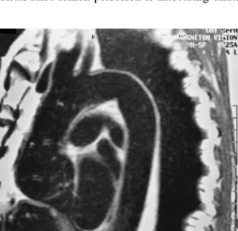
Fig. 1. A MR image obtained in a 62-year-old man 4 years after surgical repair of a type A aortic dissection using GRF-glue. The oblique sagittal T1-weighted view demonstrates a perfect morphology of the entire thor-acic aorta with a slight dilatation of the aortic root (4.4 cm). Echocardio-graphy did not show any aortic regurgitation.
of the aortic arch, followed by antegrade reperfusion through the vascular prosthesis represent an valuable option. In our experience, obliteration of the intimal flap at least at the level of the aortic arch was obtained in more than 80% of the cases, as compared with an incidence of 43% persisting intimal flap in the arch following repair with Teflon felt only and 65% when the closed method was used. This might be due to the fact that re-entries are infrequently localized in the aortic arch, as compared with the proximal descending aorta. Therefore, only few re-interventions were needed on the aortic arch after open distal repair with GRF-glue. At the moment, we are not able to demonstrate any advantage of antegrade rewarming on early or late outcome. However, we strongly believe that this additional refinement might con-tribute to decrease the incidence of significant morphologic
changes at the level of the aortic arch, since the cannula is advanced into the proximal descending aorta.
The incidence of redo-surgery in this series was similar to that reported in previous works [17]. Interestingly, all patients could be operated on a elective schedule (six of them only after the actual MR-scan) and there was no mor-tality in this small series of complex reoperations.
Magnetic resonance imaging has been recognized recently as the optimal method for follow-up of surgically treated type A acute aortic dissection [18–20]. In the chronic phase of the disease, sensitivity and specificity have been described as high as 96–100%, compared with somewhat lower values for contrast enhanced computed tomography or echocardiography [21,22]. MRI is superior
Fig. 2. T1-weighted image with a limited flap in the concavity of the distal aortic arch but without persistent false lumen in the downstream aorta.
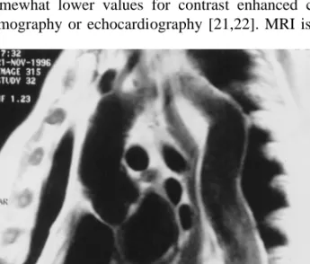
Fig. 3. Coronal MR image in a 73-year old patient who underwent supra-coronary graft replacement of the ascending aorta 8 years before. The examination shows a dissecting aneurysm originating from the distal ana-stomosis performed with cross-clamped aorta and Teflon-felt support. The dissection involves the innominate artery.
Fig. 4. The axial MR image demonstrates the dissection in the enlarged aortic arch with patent true and false lumen.
Fig. 5. Saggital MR image obtained in a 63-year-old patient who under-went supracoronary graft repair using an open distal anastomosis technique and GRF-glue 4 years before. The picture shows a normal morphology of the aortic root and no sign of persisting dissection in the downstream aorta but a tortuous segment in the distal part of the descending aorta.
to CT since it does not require injection of contrast medium or ionizing radiation, it allows multiplane scanning and demonstrates flowing blood. MRI provides excellent infor-mation about distal aneurysm forinfor-mation, pattern of blood flow within the true and the false channels and allows in certain cases to distinguish aortic branch malperfusion.
Our current follow-up protocol includes a basic MRI examination before hospital discharge in every patient and then every 6 months: all patients are referred to our institu-tional consultation for thoracic aortic disease. Depending on the evolution of the disease in the remnant aorta, the interval between scans may be reduced to 3 months or extended to 1 year.
5. Conclusions
In patients with acute type A dissection, open distal aortic repair using GRF-glue favourably influences both (1) the severity of late morphologic alterations in the downstream aorta (the most beneficial effect being observed at the level of the aortic arch) and (2) the incidence of reoperation. Magnetic resonance imaging provides excellent information about the remnant native thoraco-abdominal aorta. Repeti-tive examinations are necessary to improve the long-term survival, since the incidence of significant pathological find-ings is high.
References
[1] Westaby S. Management of aortic dissection. Curr Opinion Cardiol 1995;10:505–510.
[2] Se´guin JR, Picard E, Frapier JM, Chaptal PA. Repair of the aortic arch with fibrin glue in type A aortic dissection. J Card Surg 1994;9:734–739.
[3] Se´guin JR, Frapier JM, Colson P, Chaptal PA. Fibrin sealant improves surgical results of type A aortic dissection. Ann Thorac Surg 1991;52:745–749.
[4] Pecher P, Schumacher B, Stegmann T. Verbesserte Langzeitergeb-nisse in der Chirurgie der akuten Aortendissektion durch Einsatz des GRF-Klebers. Z Herz-, Thorax- und Gefa¨sschir 1996;10:275–281. [5] Barron DJ, Livesey SA, Brown IW, Delaney DJ, Lamb RK, Monro JL. Twenty-year follow-up of acute type A dissection: the incidence and extent of distal aortic disease using magnetic resonance imaging. J Card Surg 1997;12:147–159.
[6] Kipfer B, Striffeler H, Gersbach P, Mohadjer A, Gerber B, Schu¨pbach P, Althaus U. Surgery for acute ascending aortic dissec-tion: closed versus open distal aortic repair. Eur J Cardio-thorac Surg 1995;9:248–252.
[7] Carrel T, Pasic M, Vogt P, von Segesser L, Linka A, Ritter M, Jenni R, Turina M. Retrograde aortic dissection: a diagnostic and thera-peutic challenge. Eur J Cardio-thorac Surg 1993;7:146–152. [8] Svensson LG, Crawford ES. Cardiovascular and vascular disease of
the aorta. Philadelphia: Saunders, 1997:42–77.
[9] Bachet J, Termingnon JL, Goudot B, Dreyfus G, Piquois A, Brodaty D, Dubois C, Delentdecker P, Guilmet D. Late reoperations in patients with aortic dissection. J Card Surg 1994;9:740–746. [10] Moore NR, Parry AJ, Trottman-Dickenson B, Pillai R, Westaby S.
Fate of the native aorta after repair of acute type A aortic dissection. A magnetic resonance imaging study. Heart 1996;75:62–66.
[11] Erbel R, Oelert H, Meyer J. Effect of medical and surgical therapy on aortic dissection evaluated by transesophageal echocardiography: implications for prognosis and therapy. Circulation 1993;87:1604– 1615.
[12] Khandheria B. Aortic dissection: the last frontier. Circulation 1993;87:1765–1768.
[13] Ergin MA, Philipps RA, Galla JD, Lansman SL, Mendelson DS, Quintana CS, Griepp RB. Significance of distal false lumen after type A dissection repair. Ann Thorac Surg 1994;57:820–825. [14] Borst HG. Surgical treatment of aortic dissection. New York:
Churchill–Livingstone, 1996:117–122.
[15] Kazui T, Kimura N, Yamada O, Komatsu S. Total arch graft repla-cement in patients with acute type A aortic dissection. Ann Thorac Surg 1994;58:1462–1468.
[16] Crawford ES, Kirklin JW, Naftel DC, Svensson LG, Coselli J, Safi HJ. Surgery for acute dissection of the ascending aorta: should the arch be included?. J Thorac Cardiovasc Surg 1992;104:46–59. [17] Carrel T, Pasic M, Tkebuchava T, Jenni R, Turina M. Reoperations
after operation on the thoracic aorta; etiology, surgical technique and prevention. Ann Thorac Surg 1993;56:259–269.
[18] Gaubert JY, Moulin G, Mesana T, Chagnaud C, Caus T, Delannoy L, Blin D, Bartoli JM, Kasbarian M. Type A dissection of the thoracic aorta: use of MR imaging for long-term follow-up. Radiology 1995;196:363–369.
[19] Kersting-Sommerhoff BA, Higgins CB, White RD, Sommerhoff CP, Lipton MJ. Aortic dissection: sensitivity and specificity of MR imaging. Radiology 1988;166:651–655.
[20] Petasnick JP. Radiologic evaluation of aortic dissection. Radiology 1991;180:297–305.
[21] Masani ND, Banning AP, Jones RA, Ruttley MS, Fraser AG. Follow-up of chronic thoracic aortic dissection: comparison of transesopha-geal echocardiography and magnetic resonance imaging. Am Heart J 1996;131:1156–1163.
[22] Nienaber CA, von Kodolitsch Y, Brockhoff CJ, Koschyk DH, Spielmann RP. Comparison of conventional and transesophageal echocardiography with magnetic resonance imaging for anatomical mapping of thoracic aortic dissection. Int J Card Imaging 1994;10:1– 14.
Appendix A. Conference discussion
Dr R. Dion (Brussels, Belgium): What is your attitude with regard to
the aortic arch when you operate on a dissection type I? Are you system-atically inspecting the inside of the aortic arch even if you do a closed approach? If you don’t, in the closed approach, you may leave behind an intimal flap or a reentry, which might influence the outcome.
Dr Carrel: That’s truly correct. This study was a retrospective study
comparing three different techniques done during three different time periods. But actually since more than 5 years at my institution, only open distal repair is performed. The group of closed distal repair was the first group done between 1989 and 1990.
As I mentioned, in those 34 patients with open distal repair, some extension in the arch by gluing quite far away (distally) from the site of the anastomosis was performed in a majority and a classical Hemiarch repair was done in 10 of 34. So it probably means that with very good gluing and a limited extension of the anastomosis in the concavity of the arch, you might eliminate a majority of the problem, in the arch at least. But you still see something between 55 and 75% of false lumen perfusion at the level of the proximal segment of the descending aorta, since there you have also the first reentries.
Dr J. Bachet (Suresnes, France): There is, I think, a missing group in
your experience, that is, the closed distal anastomosis with the GRF glue. It is indeed in fashion now to perform open anastomosis. But I wonder if this is compulsory in any patients and if we are not trading some
compli-cations to others. As a matter of fact, performing the open anastomosis means, in one way or another, to perform arch surgery. Indeed, it means that to protect the brain you have to go down in temperature, you have to make circulatory arrest, and by doing so you are threatening your patient with some possible neurologic trouble.
After more than 210 acute dissections with the GRF glue, I believe (and I know that Stephen Westaby totally disagrees with me) that open anasto-mosis is not compulsory at all. If the tear is on the ascending aorta, just put on a clamp and replace the ascending aorta after checking the arch.
On the other hand, what I believe is of most importance, is to reperfuse the patient antegradely through the prosthesis and not through the femoral artery when one resumes the cardiopulmonary bypass after the replace-ment of the aorta has been completed. Can you please comreplace-ment on that?
Dr Carrel: Starting with the last question, I think this is one of the most
important factors, because during the initial experience when we glued the distal anastomosis and reperfused retrogradely, we saw in some cases a subadventitial hematoma caused by a splitting of the fresh-glued layers, and I think that it doesn’t make sense to redissect your fresh-glued ana-stomosis. So that is certainly one of the main issues to prevent any pro-blems at the level of the distal anastomosis.
Concerning the second problem, I do not agree completely with gluing the dissection and closed repair. Usually now we do not clamp the aorta during cooling: there are many reasons for that. We had some experience where we had the impression that the repair was technically perfect but the patient didn’t wake up like we were expecting, and probably the cross-clamping during the cooling period was responsible for this. In fact if you don’t have any major reentry in the descending aorta or in the arch during retrograde perfusion, you might compromise the true or the false lumen by cross-clamping the cranial portion of the ascending aorta, since the major reentry for unproblematic perfusion might be the primary tear in the ascending aorta, clamping the aorta might really compromise perfusion of the supraaortic branches.
So now we first cool down and then open the aorta without cross-clamping the aorta and always perform the distal repair first and then reinstitute perfusion through the prosthesis, and most recently through the Anteflow (Vaskntek, Sulzer, Winterthur, CH) vascular prosthesis within the already sewn side arm of the prosthetic graft.
Dr A. Moulijn (Antwerp, Belgium): Some time ago we had a similar
experience like yours: an acute retrograde dissection appeared after can-nulation of the common femoral artery in a patient operated for an aneur-ysm of the ascending aorta.
However the patient survived, after this event in the following cases we changed towards the cannulation of the subclavian artery by using a 8 mm Dacron artery graft anastomosed end to side the subclavien artery who on his turn was connected with the arterial line of the CPB.
We found it to not be a very demanding procedure and successful in every case.
We think it is a tremendous way of perfusing this patient safely and antegradely.
Mr S. Westaby (Oxford, UK): You looked at all those groups. Was there
an increase in operative mortality in the closed over the open patients?
Dr Carrel: Surprisingly, one colleague of our group had published early
outcome 3 years before and the comparison of open versus closed repair showed a slightly superior mortality in the open group, a fact that would support the findings of Mr. Bachet. The worst outcome was interpretated as a result of more extensive reconstruction, probably. Presently the compar-ison is no more valuable because we always perform open repair and closed repair has been abandoned.