This paper is available online free of all access charges (see http://jxb.oxfordjournals.org/open_access.html for further details)
RESEARCH PAPER
The co-chaperone p23 controls root development through
the modulation of auxin distribution in the Arabidopsis root
meristem
Stefano D’Alessandro1, Serena Golin1, Christian S. Hardtke2, Fiorella Lo Schiavo1, and Michela Zottini1
1 Department of Biology, University of Padova, Via U. Bassi 58/B, I-35131 Padova, Italy 2 University of Lausanne - Biophore Building, DBMV CH-1015 Lausanne, Switzerland To whom correspondence should be addressed. E-mail: [email protected]
Received 11 May 2015; Revised 11 June 2015; Accepted 12 June 2015 Editor: Angus Murphy
Abstract
Homologues of the p23 co-chaperone of HSP90 are present in all eukaryotes, suggesting conserved functions for this protein throughout evolution. Although p23 has been extensively studied in animal systems, little is known about its function in plants. In the present study, the functional characterization of the two isoforms of p23 in Arabidopsis
thaliana is reported, suggesting a key role of p23 in the regulation of root development. Arabidopsis p23 mutants, for
either form, show a short root length phenotype with a reduced meristem length. In the root meristem a low auxin level associated with a smaller auxin gradient was observed. A decrease in the expression levels of PIN FORMED PROTEIN (PIN)1, PIN3, and PIN7, contextually to an inefficient polar localization of PIN1, was detected. Collectively these results suggest that both Arabidopsis p23 isoforms are required for root growth, in particular in the maintenance of the root meristem, where the proteins are located.
Key words: Arabidopsis, auxin, p23-chaperone, pin formed protein (PIN), polar auxin transport, root growth.
Introduction
Root growth and development are fundamental processes for the life of plants, controlling water uptake, nutrient acquisi-tion, anchorage to soil, and secondary metabolite synthesis and storage (Saini et al., 2013). Post-embryonic growth and development are controlled through the meristems, which generate differentiating cells that shape adult plant structures (Moubayidin et al., 2009). Meristem maintenance is con-trolled through a finely regulated orchestration of hormones and signalling molecules, among which auxins are indeed the master regulators of root development (Saini et al., 2013). Auxin is virtually involved in all phases of plant growth and development, and auxin signalling is regulated at the level of
biosynthesis, transport, and perception (Saini et al., 2013). From the sites of biosynthesis (Ljung et al., 2005), auxin is actively transported via the combination of two routes involv-ing the long-distance transport through mature phloem and cell-to-cell polar transport (PAT), mediated through specific influx/efflux carriers (Vieten et al., 2007; Geisler et al., 2014). AUX1 and LAX (LIKE-AUX1) are major auxin importers, whereas PIN (PIN FORMED PROTEIN) and ABCB (ATP-BINDING CASSETTE B) are the main families of auxin exporters (Geisler and Murphy, 2006; Verrier et al., 2008;
Krecek et al., 2009). At the site of action, auxin perception is mediated through members of the TIR1 (TRANSPORT
This is an Open Access article distributed under the terms of the Creative Commons Attribution License (http://creativecommons.org/licenses/by/3.0/), which permits unrestricted reuse, distribution, and reproduction in any medium, provided the original work is properly cited.
INHIBITOR RESPONSE 1)/AFB family. TIR1 is an F-box subunit of the ubiquitin ligase complex SCFTIR1, which
inter-acts with and ubiquitinates Aux/IAA proteins (AUXIN/ INDOLE-3-ACETIC ACID) (Gray et al., 2001). Aux/IAA proteins are auxin response inhibitors that interact and block ARF (AUXIN RESPONSE FACTOR) transcription factors (Mockaitis and Estelle, 2008). Indole-3-acetic acid (IAA) is the most abundant natural auxin, and the apoplastic acidic environment facilitates the protonation of IAA to IAAH, which diffuses into cells. Due to the chemical nature of aux-ins, PIN and ABCB exporters are the primary control ele-ments of PAT (Geisler and Murphy, 2006). Auxin signalling continuously crosses pathways with other hormones and sig-nalling molecules in a complex chain of feedback controls (Dello Ioio et al., 2008; Overvoorde et al., 2010; Saini et al., 2013). In particular, auxins and cytokinins are the major players of a well-known crosstalk mechanism that regulates root meristem maintenance and consequent root growth (Moubayidin et al., 2009). Cytokinin–auxin crosstalk occurs at the transport level of auxin signalling through a regulatory circuit converging on the type-B ARR1 (ARABIDOPSIS RESPONSE REGULATOR 1) transcription factor and con-sequently on the SHY2 (SHORT HYPOCHOTHYL 2) pro-tein, which negatively regulates the expression of PIN genes (Dello Ioio et al., 2008).
The small acidic protein, p23, has been identified in animal systems as an HSP90 co-chaperone (Johnson and Toft, 1994), which stabilizes the active conformation of the progesterone-receptor HSP90 complex after binding to the ATP-bound form of HSP90 and lowering the ATPase activity rate of HSP90 (Chadli et al., 2000; Ali et al., 2006; Li et al., 2012). Two homologues of p23 have been identified in Arabidopsis: p23-1 (At4g02450) is 241 amino acids in length with a mass of 25.47 kDa, whereas p23-2 (At3g03773) is 150 amino acids in length with a mass of 17.4 kDa (Zhang et al., 2010). Each iso-form shares 27% and 25% amino acid identities with human p23, respectively, and shows 38–60% identity with other plant p23 homologues. The difference in length of p23-1 and p23-2 reflects a glycine rich (MG/GA) segment of 70 amino acids in the C-terminal region of p23-1, whose function is not yet understood. Both plant p23 isoforms and the chimeric pro-tein p23-1-d (lacking the glycine rich tail) bind to HSP90 in vitro; however, unlike their animal counterpart, these proteins do not reduce the chaperone ATPase activity rate (Zhang
et al., 2010).
Although Arabidopsis p23 isoforms have been the object of previous biochemical characterization studies (Zhang et al., 2010; Tosoni et al., 2011), the physiological role of these proteins remains elusive. These analyses revealed that p23 proteins are chiefly located in the root meristem, where they intervene in the regulation of root growth and development.
Material and methods
Plant materials and growth conditions
All experiments were performed using Arabidopsis thaliana ecotype Columbia (Col-0). The mutant seedlings for p23-1 (SAIL 245_ H06), p23-2 (SALK_003076), and arr1-4 (SALK_042196) were
obtained from the European Arabidopsis Stock Centre (NASC). T-DNA insertions were verified through PCR (primers are listed in Supplementary Table S1 available at JXB online). From the p23 single mutants, a double mutant p23-1×p23-2 (dKO) was generated. The plant seeds were surface sterilized in 70% EtOH and 0.05% Triton X-100, followed by 100% EtOH. The seeds were sown onto square Petri dishes containing one-half-MS medium supplemented with 0.5 g/l MES-KOH, pH 5.7, 0.8% Plant Agar (Duchefa), and 1% Sucrose, stratified for 2 d at 4 °C in the dark, and placed vertically in a growth chamber under a long daylight period (16 h light/ 8 h dark) using 150 μmol m−2 s−1 at 22 °C. The translational reporter plants, pp23-1:p23-1-GUS and pp23-2:p23-2-GUS, were generated after transforming the single mutants p23-1 or p23-2 with Agrobacterium harbouring a construct containing the entire sequence of the p23-1 or p23-2 genes, starting from base pair −540 or −3191, respectively, fused to the β-Glucuronidase reporter gene (UID-A; the primers are reported in Supplementary Table S1) and subcloned into the pGreen 0029 binary vector (Hellens et al., 2000). Complemented lines p23-1 T 35S:p23-1 and p23-2.1 T 35S:p23-2 were generated after trans-forming the single mutants p23-1 or p23-2 with Agrobacterium har-bouring a construct containing the coding sequence (CDS) of p23-1 or p23-2 under the control of the CaMV 35S constitutive promoter (primers for CDS cloning are reported in Supplementary Table S1) and subcloned into the pGreen 0029 binary vector (Hellens et al., 2000). The reporter lines DR5:GUS, DII-Venus, PIN1-GFP, PIN7-GFP, or the mutant line arr1-4, were crossed with the dKO line, and the F2 homozygous lines were used for subsequent analyses.
RNA isolation and qRT-PCR
Seven days after germination (dag), the roots and shoots of wild-type and mutant seedlings were harvested separately for subsequent anal-yses. Total RNA was extracted from plant samples using TRIzol® Reagent (Invitrogen) according to the manufacturer’s instruc-tions. First-strand cDNA synthesis was performed using 1 μg of RNA, oligo(dT) primers, and SuperScript-II Reverse Transcriptase (Invitrogen) according to the manufacturer’s instructions. The primers for qRT-PCR (quantitative real-time PCR) are shown in
Supplementary Table S1. The expression levels of each gene were normalized to the expression level of the housekeeping genes
Actin-2 (ACTActin-2; At3g18780) or elf1α (At5g60390), as indicated.
Root growth assay
The plant seeds from the different genotypes were sown onto solid growth medium, and vertically grown as described above. Plates were scanned using a flatbed scanner every day from the fourth to the 10th dag. The roots were analysed at different developmental stages through image analysis (Fiji – ImageJ bundle software). The experiments were performed at least three times and each sample comprised 25 seedlings.
For N-1-naphtylphtalamic acid (NPA) and auxin treatments, 5-d-old seedlings were transferred to growth medium supplemented with different concentrations of NPA, IAA, or 1-Naphthaleneacetic acid (NAA; Sigma-Aldrich). After 5 d of treatment, the primary root length was measured as described above.
β-Glucuronidase histochemical assay
Histochemical staining was performed at different developmental stages. Samples were analysed for β-glucuronidase activity. The samples were incubated for variable times (1 h to overnight) at 37 °C in the reaction medium (2 mM X-Gluc, 0.05% Triton X-100, 5 mM K3Fe(CN)6, 5 mM K4Fe(CN)6 3× H2O, 10 mM EDTA, and 50 mM sodium phosphate buffer, pH 7.0). The seedlings were mounted on chloral hydrate solution. The images were captured using a micro-scope (Leica 5000B) with Normarsky correction (DIC). The experi-ments were performed at least in triplicate, and each sample set comprised 10 seedlings.
Confocal microscopy
Seven-day-old seedlings were mounted in a drop of 2% (20 μg/ml) propidium iodide (PI; Sigma-Aldrich) solution, on a microscope slide, and the images were acquired using a LEICA SP5 laser scan-ning confocal imaging system. The excitation and emission wave-lengths are reported in the image captions. High-definition images were acquired (1024 × 1024, 25× objective) and analysed using the Fiji – ImageJ bundle software (http://fiji.sc/Fiji). The experiments were performed at least in triplicate, and each sample set comprised 10 seedlings.
Statistics
All experiments were performed at least in triplicate, and the images represent typical examples. The values are represented as the means ± standard deviation. The statistical significance was demonstrated using Student’s t-test.
Results
p23-1 and p23-2 knockout mutants show shorter roots
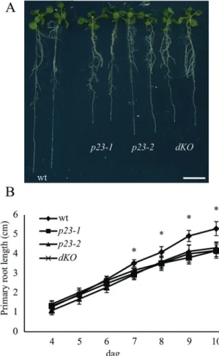
To investigate the functional role of p23 proteins in Arabidopsis, knockout mutant lines of the two isoforms were isolated and phenotypically characterized. Although the aerial part of the plant showed no altered traits throughout the plant development, root growth was strongly impaired in single and double (dKO) mutants (Fig. 1A). In particular, in 10-d-old single mutant seedlings, the primary root length was approxi-mately 70% that of the wild-type (wt) (wt=5.3 ± 0.36 cm; p23-1=4.17 ± 0.15 cm; and p23-2=4.31 ± 0.15 cm) (Fig. 1B). In mutant lines, the reduction in root growth was evident and significant from the seventh dag compared with that of the wild-type (Fig. 1B). The short-root phenotype, detectable only at an advance stage of development, suggests that the p23 mutants are likely impaired in meristem maintenance rather than in the early post-embryonic stages of root growth.
Double knockout mutants were generated after crossing p23-1 and p23-2 (dKO). The latter provides information for determining whether these two proteins play a role in the same pathway controlling root growth and to evaluate the contribution of each isoform to this process. The primary root length of the dKO line is comparable with that of both single knockout mutants (dKO=4.17 ± 0.2 cm), as shown in
Fig. 1A and B, thus showing no additive effect of the two mutations. This behaviour suggests that although not redun-dant, the two proteins intervene in the same process regulat-ing root growth.
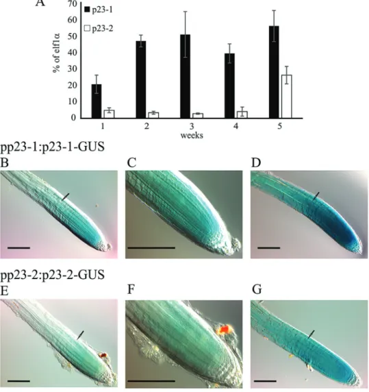
Localization and expression profile of p23-1 and p23-2 To evaluate the expression profile of the two genes, the transcripts of p23-1 and p23-2 were analysed through qRT-PCR during plant development (1–5-week-old plants). As reported in Fig. 2A, both isoforms are expressed at all growth stages. p23-1 is the most expressed isoform ranging from 20–50% of elf1α expression, whereas p23-2 expres-sion ranges from 2–5% of elf1α expression. This result demonstrates a 10-fold higher expression of the long
isoform than that of the short isoform, during the first 4 weeks and suggests that p23-1, although showing a similar phenotype in the roots, is the major p23 isoform present in plants.
The expression and localization of p23-1 and p23-2 was determined at cellular level in planta. The short isoform p23-2 was previously described as nuclear and cytosolic (Tosoni
et al., 2011). To analyse the subcellular localization of the long isoform p23-1, stably transformed Arabidopsis plants expressing the protein under the control of the constitutive viral promoter CaMV 35S were generated. In these plants, the fusion protein p23-1, tagged at the C-terminus with YFP, was expressed in all tissues. These transgenic lines showed florescence throughout the plant body. The analysis of differ-ent plant tissues using confocal laser microscopy defined the subcellular localization of the chimeric protein. p23-1-YFP was detectable in leaves of 10-d-old seedlings (Supplementary Fig. S1 available at JXB online) with a cytosolic and nuclear localization and similarly in all analysed tissues.
Fig. 1. p23 knockout mutants show shorter roots compared with the
wild-type. (A) Twelve-day-old Arabidopsis seedlings of wild-type (wt), p23-1, p23-2, and double knockout (dKO) background. p23 single knockout mutants and the double knockout mutant show shorter roots as compared with the wild-type. (B) Quantification of the primary root length of the different backgrounds: wt, p23-1, p23-2, and dKO from the fourth to the 10th dag. The short-root phenotype is characterized by a slower root growth appreciable from the seventh dag (*P<0.01, Student’s t-test). Scale bar = 1 cm. (A colour version of this figure is available at JXB online.)
To examine the expression and localization of the two pro-teins in the entire plant, stable transformed Arabidopsis plants expressing the β-glucuronidase gene (UID-A) fused either to p23-1 or p23-2 were generated. The reporter allowed defining the localization of the two proteins through a histochemical assay. Several transgenic lines were obtained and seedlings at different developmental stages were analysed.
In 3–10-d-old seedlings, the presence of the chimeric pro-teins is detectable in the root meristematic zone, where they strongly concentrate (Fig. 2B, C–F). Four hours of stain-ing let the signal appear in the vascular tissue of the roots (Fig. 2D, G) and after 16 h the reporter was detected in the entire root and in the vasculature of the leaves (data not shown). It is worthwhile to highlight that the two proteins are highly expressed in the root meristem and the signal drops at the level of the first full-elongated cell (black arrows,
Fig. 2B–G). Furthermore, the difference in the expression level of the two isoforms, observed by qRT-PCR (Fig. 2A), was confirmed by the histochemical assay. In fact, although p23-1-GUS is detectable in the root meristem after 5 min
of staining, p23-2-GUS shows comparable levels only after 30 min.
p23 mutants show a reduced number of dividing cells in the root meristem
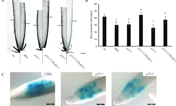
The structure of the primary root was accurately analysed in wild-type and mutant lines by confocal microscopy. For this purpose, root width, meristem length, and cell number, in the zone between the quiescent centre and the first corti-cal elongating cell, were measured (Moubayidin et al., 2013). These analyses revealed that roots of the different genotypes have a comparable width, whereas a significant difference was observed in the length of the meristematic zone (Fig. 3A). As shown in Fig. 3B, both single and dKO lines showed a reduced number of cortical cells in the root meristem compared with the wild-type (wt=41.8 ± 3.2; p23-1.1=31 ± 4; p23-2.1=26 ± 4.8; dKO=30 ± 4.8). To verify the causality between the deficiency of p23 proteins and short-root phenotype, stable transformed lines overexpressing p23-1-HA or p23-2-HA were generated in
Fig. 2. Transcript levels of p23-1 and p23-2 and protein localization in planta. (A) Quantitative real-time analyses of the transcript levels of p23-1 and p23-2 in Arabidopsis plants, grown in soil from 1–5 weeks. Data are reported as percentage of elf1α transcript levels. (B–G) Histochemical analyses of the translational reporter of p23-1 and p23-2. (B, E) pp23-1:p23-1-GUS and pp23-2:p23-2-GUS Arabidopsis staining (30 min) concentrates in the root meristem and drops above the first full-elongated cell (arrow). (C, F) Magnification of the root meristem. (D, G) Four hours of GUS staining reveals that both p23 proteins appear also in the upper part of the root. Scale bar = 100 μm. (A colour version of this figure is available at JXB online.)
the p23-1 (p23-1 T p23-1-HA) or p23-2 (p23-2 T p23-2-HA) background, respectively. As reported in Fig. 3A and B, the overexpression of p23 proteins could complement the respective mutant lines both in terms of meristem length and cortical cell number (p23-1 T p23-1=44.3 ± 3.8; p23-2 T p23-2=37.40 ± 4.7).
To assess whether the reduction in meristem cell number could reflect the loss of meristematic-cell-division potential, the expression of pCyclinB1:GUS (a broadly used marker to indicate the G2‐to‐M phase of the cell cycle) was monitored (Colón-Carmona et al., 1999) in wild-type, and p23-1 and p23-2 single mutants, to visualize root meristem cells in the G2–M phase. As shown in Fig. 3C, the pCyclinB1:GUS in both p23-1 and p23-2 backgrounds showed a strong reduc-tion in the intensity and extent of GUS staining as compared with the wild-type background (CTRL). This result demon-strated that cell division is impaired in the root meristem of both the single mutant lines, further suggesting a common pathway for the two isoforms in regulating root growth. Auxin distribution is altered in p23 mutants
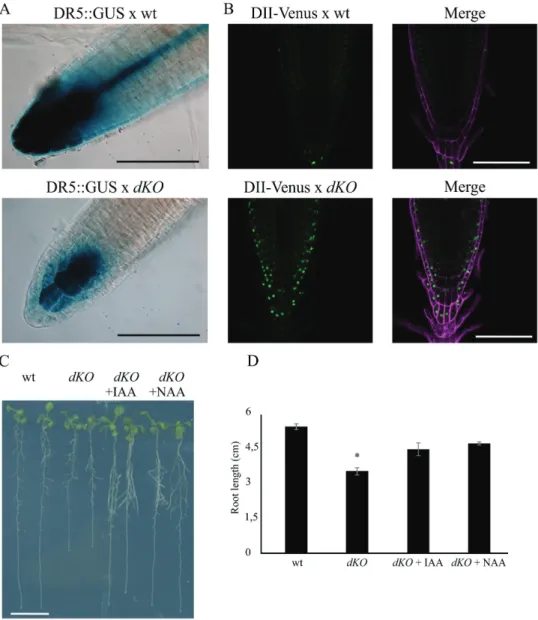
A p23 double knockout mutant harbouring the DR5::GUS construct was generated to evaluate whether auxin distri-bution was altered. DR5 is a widely used auxin molecular marker (Ulmasov et al., 1997) that allows the detection of cellular responses to auxin using a histochemical assay. As shown in Fig. 4A, strong DR5 activity was detected in the wild-type primary root (upper panel), particularly at the level of columella cells and the quiescent centre (QC), whereas lower expression was observed in dKO plants (lower panel).
To obtain a more detailed analysis of auxin distribution in the meristem at the cellular level, the DII-Venus auxin responsive marker line was crossed with the dKO line, lead-ing to a more direct detection system for auxin in the mutant (Brunoud et al., 2012). Analyses of DII-Venus fluorescence using laser scanning confocal microscopy (Fig. 4B) showed that DII-Venus fluorescence was higher in the root meristem of the dKO line (lower panels) than in wild-type plants (upper panels). In particular, higher levels of fluorescence were detected in the root cap, epidermis, and columella cells—tis-sues that are typically characterized by auxin accumulation (Krecek et al., 2009). However, the fluorescent signal was not detectable at the level of the quiescent centre in both wild-type lines, as expected, and p23 mutants, suggesting that this mutation does not compromise auxin levels in the QC. Indeed, the expression of ASB1 (ANTHRANILATE SYNTHASE BETA SUBUNIT 1) and YUC6 (YUCCA 6) in the shoot of dKO plants was similar to that of wild-type plants, whereas slightly enhanced expression was observed in the roots (Supplementary Fig. S2 available at JXB online). This result suggests that alterations in auxin levels in the roots do not depend on the lower biosynthetic rate of the hormone.
Furthermore, it was observed that in dKO mutants treated at 5 dag with 10 nM of either IAA or NAA, the wild-type primary root length was rescued (Fig. 4C, D), thus directly associating the short-root phenotype with low auxin con-tent at the root level. These results suggest that alterations in auxin sensing can be excluded. Moreover, both IAA and NAA restored normal root growth, suggesting that the dKO line is not altered in auxin influx.
Fig. 3. p23 mutant lines show a reduced number of cells in the root meristematic zone. (A) PI stained root of 7-d-old seedlings of the different lines.
Excitation and emission wavelengths for confocal acquisition are 488 nm and 600–650 nm, respectively. The arrows show the upper limit of the meristematic zone. Scale bar = 50 μm. (B) Meristem cell number in the different genetic backgrounds. p23 mutant lines show a reduced number of cortical cells in the root meristematic zone, while complemented lines show no difference as compared with the wild-type (aP<0.01 compared with the
wt, bP<0.01 compared with the background line; both Student’s t-test). (C) Histochemical analysis of the CyclinB1:GUS reporter in the wild-type (CTRL),
PINs expression level and localization is altered in p23 double mutants
As auxin sensing and biosynthesis are not the primary cause in determining the reduction of intracellular auxin levels in the dKO root, auxin transport was analysed to determine whether this process could cause the short-root phenotype of p23 mutants.
The directed cell-to-cell distribution of auxin is achieved through a system of auxin influx and efflux transporters, and auxin fluxes can be predicted based on the asymmetric plasma membrane distribution of PINs determining PAT (Bailly
et al., 2008). PIN transcription is enhanced through auxin and diminished by cytokinin via SHY2 (Dello Ioio et al., 2008). In addition, PIN activity is finely controlled through post-translational modifications, such as phosphorylation by PID (PINOID) and D6PK (D6 PROTEIN KINASE)
(Zourelidou et al., 2014), which also control the polar subcel-lular localization of these proteins (Michniewicz et al., 2007;
Marhavý et al., 2014).
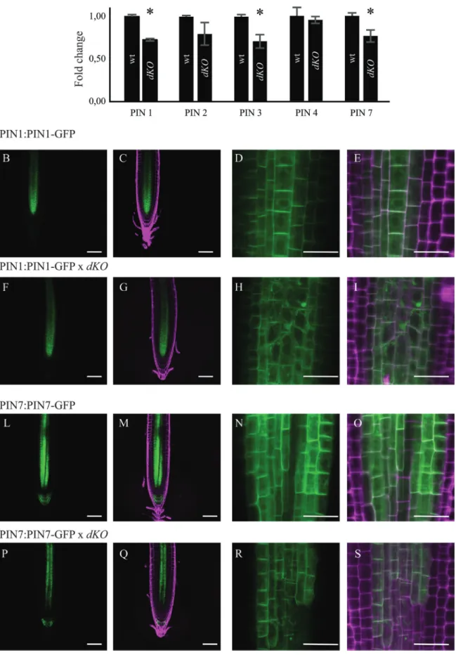
The transcription levels of the major PAT protein were assayed, analysing the gene expression level of PIN1, PIN2, PIN3, PIN4, and PIN7 using qRT-PCR analysis. The expres-sion of PIN1, PIN3, and PIN7 was approximately 75% in the dKO compared with that of the wild-type (Fig. 5A). PIN proteins undergo regulation at both transcriptional and post-transcriptional levels; thus, the translational report-ers of PIN1 and PIN7, the main acropetal auxin transport mediators, were analysed in the dKO background. As shown in Fig. 5B, the impaired level of expression of these two car-riers is reflected by their translational reporters, as observed in 7-d-old dKO seedlings. Particularly, PIN1-GFP and PIN7-GFP reporters confirmed the reduced expression of these proteins in the root tip of dKO plants, as appreciable from
Fig. 4. Auxin distribution is altered in the root meristem of the dKO. (A) GUS expression pattern of DR5:GUS in dKO background (lower panel) is weaker
than in DR5:GUS (upper panel). Scale bar = 100 μm. (B) The green fluorescence of DII-Venus shows much higher levels in the dKO background (lower panel) compared with the wild-type (upper panel), in particular in the columella cells and in the epidermis. Venus Ex: 488 nm, Em: 520–540 nm; PI Ex: 488 nm, Em: 600–650 nm. Scale bar = 100 μm. (C) dKO short-root phenotype is rescued by application of 10 nM IAA or NAA to 5-d-old seedlings and analysed after 5 d. Scale bar = 1 cm. (D) Quantification of the primary root length of wild-type and dKO lines under control condition or upon auxin treatment (*P<0.01, Student’s t-test). (A colour version of this figure is available at JXB online.)
Fig. 5. PIN expression levels, and PIN1 and PIN7 localization in the dKO. (A) qRT-PCR of PIN1, 2, 3, 4, and 7; data were normalized to ACT2 and
shown as fold change on wild-type (*P<0.05, Student’s t-test). (B) Confocal Laser Scanning images of PIN1 (B–E), PIN1×dKO (F–I) or PIN7 (L–O) and PIN7×dKO (P–S) showing altered expression and localization of PIN1 and PIN7 in the mutant line. GFP Ex: 488 nm, Em: 500–520 nm; PI Ex: 488 nm, Em: 600–650 nm. Scale bars (B, C, F, G, L, M, P, Q) = 50 μm; (D, E, H, I, N, O, R, S) = 10 μm. (A colour version of this figure is available at JXB online.)
the quantification of the GFP fluorescence in Supplementary Fig. S3 (available at JXB online). In addition, increased PIN1-GFP internalization into subcellular compartments of dKO plants was detected compared with that of the wild-type plants (Fig. 5D, E, H, I). However, alterations in the periclinal/anticlinal distribution of PIN1 and PIN7 were not observed in dKO mutants. Moreover, PIN7-GFP was absent in the stele cells near the stem cell niche (Fig. 5R, S). These results demonstrate that alterations in PIN expression and localization are indeed the most likely cause of lower auxin levels in p23 mutants, thus linking p23 activity to the main-tenance of a correct PAT in the root meristem. In support of this hypothesis, a partial resistance of p23 mutants to treat-ment with NPA (Supplementary Fig. S4 available at JXB online), a well-known inhibitor of PAT (Bailly et al., 2008), was observed. In fact, while low concentration of NPA short-ened the primary root of the wild-type, a similar effect was detected in the dKO only with NPA concentration >1 μM. Rescue of short-root phenotype in the triple mutant p23-1×p23-2×arr1-4
Root meristem maintenance and continuous root growth are guaranteed through a strict equilibrium between cell division and cell differentiation, which is finely controlled through plant hormone crosstalk. In this context cytokinin and auxin play key roles (Dello Ioio et al., 2008; Moubayidin et al., 2010; Overvoorde et al., 2010; Depuydt and Hardtke, 2011;
Sankar et al., 2011).
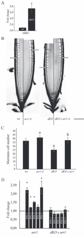
For this reason, the expression of ARR1—a key player in cytokinin signalling—was evaluated to test the status of cytokinin signalling in the p23 double knockout mutant. As shown in Fig. 6A, ARR1 transcripts were markedly upregu-lated in the dKO background compared with wild-type plants (7.4-fold higher). This result suggests that p23 proteins act as negative regulators of the cytokinin signalling pathway, and, at the same time, it supports a role of ARR1 in the short-root phenotype of p23 mutants. To evaluate these hypotheses, the triple mutant line p23-1×p23-2×arr1-4 (dKO×arr1-4) was generated. Laser scanning confocal analysis of the PI-stained root meristem (Fig. 6B, C) showed an average meristem size of 280 µm and an average cortical cell number of 37 in the wild-type line. The p23 dKO mutant showed a reduced meris-tem length and cell number (200 µm and 25 cells, respectively), whereas the single arr1-4 mutant showed both increased mer-istem length and mermer-istematic cell number (305 µm and 41 cells), as previously described (Dello Ioio et al., 2007). The triple knockout mutant dKO×arr1-4 rescued the phenotype of the dKO, showing a meristem length and cortical cell num-ber indistinguishable from those of wild-type (280 µm and 38 cells). In addition, the arr1-4 mutant was not completely epi-static on the dKO, confirming that although restoring a more equilibrated auxin/cytokinin ratio by lowering cytokinin sig-nalling, auxin levels are not sufficient to support the meristem activity present in the single arr1-4 mutant.
As the dKO×arr1-4 showed a wild-type-like meristem length, the expression of PINs in this line was tested. As shown in Fig. 6D, the arr1-4 mutant showed increased
Fig. 6. Rescue of short-root phenotype in the triple mutant p23-1× p23-2×arr1-4. (A) qRT-PCR of ARR1; data were normalized to ACT2 and shown as fold change on wild-type (*P<0.05, Student’s t-test).
(B) PI-stained root of 7-d-old seedlings of the different lines. Excitation and emission wavelength for confocal acquisition are 488 nm and 600–650 nm, respectively. The arrows show the upper limit of the meristematic zone. Scale bar = 50 μm. (C) Meristem cell number in the different genetic background. dKO mutant shows a reduced number of cortical cells in the root meristematic zone, while arr1-4 shows an increased number of cortical cells. The triple mutant dKO×arr1-4 shows no difference as compared with the wild-type (aP<0.01 compared with the wild-type, bP<0.01 compared with the dKO; both Student’s t-test). (D) qRT-PCR of
PIN1, 2, 3, 4, and 7; data were normalized to ACT2 expression and shown as fold change on wild-type (black line) (*P<0.05, Student’s t-test). (A colour version of this figure is available at JXB online.)
expression levels of PIN1, PIN2, PIN3, and PIN7, as previ-ously described (Dello Ioio et al., 2008) and the triple mutant dKO×arr1-4 recovered PIN expression levels comparable with those of the wild-type. This result supports the promi-nent role of PIN expression levels in the phenotype of p23 mutants, highlighting, at the same time, an indirect role of p23 proteins on PIN expression.
Discussion
In the present study, experimental evidence on the role of the p23 co-chaperone as a novel component of the protein net-work regulating post-embryonic root development has been reported.
Upon seed germination, the root apical meristem grows as cell division prevails over differentiation, reaching a final size at approximately 5 dag, when a fixed meristem cell num-ber is established and meristem maintenance is guaranteed through a balance between the rate of cell differentiation and the rate of new cell generation (Blilou et al., 2005; Dello Ioio
et al., 2007, 2008; Moubayidin et al., 2009; Perilli et al., 2012;
Pacifici et al., 2015). Arabidopsis p23 mutants show impaired root development due to a reduced number of cells in the root meristematic zone, where the two p23 isoforms localize. Using two specific auxin responsive markers (DR5:GUS and DII-Venus), a lower level of auxin accumulation was observed in the root tip of the mutant lines than in the wild-type. It was also shown that the short-root phenotype could be rescued both in single and double knockout mutants by supplying auxin in the growth medium (Supplementary Fig. S5 avail-able at JXB online). Based on these results, a constraint at the level of auxin biosynthesis or perception can be excluded, whereas impairment in auxin distribution, reflecting altera-tions at the level of auxin PAT, was implied and subsequently detected. Indeed, not only were the main components of acropetal PAT—PIN1, PIN3, and PIN7—transcriptionally down-regulated but the localization patterns of PIN1-GFP and PIN7-GFP proteins were also modified. Particularly, PIN1-GFP remains, at least in part, accumulated in intra-cellular compartments and does not correctly localize to the basal membranes, whereas PIN7 showed a limited localiza-tion pattern in the root of the p23 double mutant compared with wild-type. This result suggests that altered PAT is the most likely cause of the reduced meristem length of p23 mutants. In fact, as consequence of the compromised PAT, a narrowed auxin gradient is established in the root meristem of p23 mutants, associated with a shift of the auxin maxi-mum towards the root cap (Grieneisen et al., 2007), together with a reduction in its intensity.
To obtain a more complete picture of hormone signalling regulating meristem maintenance, the expression of ARR1—a key component of cytokinin signalling—was evaluated and an increased expression in the mutant compared with wild-type plants was observed. ARR1, in turn, is induced through ASB1 (Moubayidin et al., 2013), whose expression was slightly induced in the mutant. These results indicate that cytokinin signalling is enhanced in p23 mutant lines whereas the auxin
signalling pathway is reduced. In addition, it was observed that a reduction in cytokinin signalling through the reduction of ARR1 in the dKO mutant generates a wild-type-like phenotype. This triple mutant (dKO×arr1-4) shows a meristem length and a number of meristematic cortical cells similar to the wild-type, but still reduced compared with that of the arr1-4 mutant (Dello Ioio et al., 2007), thus revealing the pathway in which p23 oper-ates. Indeed, this result provides genetic evidence that p23 pri-marily acts on auxin distribution in the root meristem, where these proteins are specifically expressed (Fig. 2B–G). In addi-tion, PIN expression in the triple mutant was tested and PIN levels were found to recover to wild-type values. Taken together, these results confirm that the altered expression and localiza-tion of PIN proteins in the p23 double knockout mutant are the primary cause of the reduced meristem length and of the fewer number of meristematic cortical cells. Furthermore, the enhanced level of ARR1 expression could likely be the cause of PINs detriment in p23 mutants, and supports a role of p23 proteins in meristem maintenance a few days after germination, when ARR1 becomes the principal cytokinin effector (Pacifici
et al., 2015). At the same time, the recovery of PIN expression level in the triple mutant excludes a direct role of p23 proteins on PIN levels, while its role in PIN trafficking remains to be eluci-dated. Notably, p23 binds to HSP90 (Zhang et al., 2010), which shows a specific expression at the level of the root (Krishna and Gloor, 2001). Moreover, HSP90 has been demonstrated to interact and regulate TWD1, and consequently ABCB trans-porters activity, a family of genes involved in extracellular auxin transport that associate with PINs (Pérez-Pérez et al., 2004;
Blakeslee et al., 2007; Wang et al., 2013). In this context, the hypothesis is presented that the absence of p23 could compro-mise the stability of the HSP90 complex, causing alterations in the TWD1-ABCB system, and thus on PAT, leading to reduced auxin levels in the root meristem, altered PIN expression and, as a consequence, a short-root phenotype.
Taken together, these results are consistent with increasing experimental evidence indicating that although cytokinin con-trols the cell differentiation rate, acting specifically at the tran-sition zone, the graded distribution of auxin is pivotal for the dynamic regulation of root meristem size (Pacifici et al., 2015).
In conclusion, it is proposed that p23 sustains meristem maintenance, playing a key role in post-embryonic root growth via the regulation of auxin signalling and conse-quently the preservation of a balanced rate of cell differentia-tion and division at the transidifferentia-tion zone.
Supplementary data
Supplementary data are available at JXB online.
Table S1. Primers used in genotyping, cloning, and qRT-PCR.
Figure S1. p23-1-YFP shows nuclear and cytoplasmic sub-cellular localization.
Figure S2. Auxin biosynthesis is not altered in the shoot of dKO.
Figure S3. PIN1-GFP and PIN7-GFP fluorescence quanti-fication in the wild-type and dKO backgrounds.
Figure S4. dKO is partially insensitive to inhibition of auxin PAT.
Figure S5. Exogenous IAA rescues the short-root pheno-type of p23 mutants.
Funding
This work was supported by the University of Padova [PRAT 2012, CPDA122838] and CARIPARO Foundation to SG.
Acknowledgements
We are thankful to Sabrina Sabatini for providing us with PIN1-GFP, PIN7-GFP, and Arabidopsis reporter lines, and for critical reading of the manu-script. We thank Alex Costa for his valuable comments and critical reading of the manuscript.
References
Ali MMU, Roe SM, Vaughan CK, Meyer P, Panaretou B, Piper PW, Prodromou C, Pearl LH. 2006. Crystal structure of an Hsp90–
nucleotide–p23/Sba1 closed chaperone complex. Nature 440,
1013–1017.
Bailly A, Sovero V, Vincenzetti V, Santelia D, Bartnik D, Koenig BW, Mancuso S, Martinoia E, Geisler M. 2008. Modulation of
P-glycoproteins by auxin transport inhibitors is mediated by interaction with immunophilins. Journal of Biological Chemistry 283, 21817–21826. Blakeslee JJ, Bandyopadhyay A, Lee OR, et al. 2007. Interactions
among PIN-FORMED and P-glycoprotein auxin transporters in Arabidopsis. The Plant cell 19, 131–147.
Blilou I, Xu J, Wildwater M, Willemsen V, Paponov I, Friml J, Heidstra R, Aida M, Palme K, Scheres B. 2005. The PIN auxin efflux
facilitator network controls growth and patterning in Arabidopsis roots. Nature 433, 39–44.
Brunoud G, Wells DM, Oliva M, et al. 2012. A novel sensor to map
auxin response and distribution at high spatio-temporal resolution. Nature
482, 103–106.
Chadli A, Bouhouche I, Sullivan W, Stensgard B, McMahon N, Catelli MG, Toft DO. 2000. Dimerization and N-terminal domain proximity
underlie the function of the molecular chaperone heat shock protein 90. Proceedings of the National Academy of Sciences of the United States of America 97, 12524–12529.
Colón-Carmona A, You R, Haimovitch-Gal T, Doerner P. 1999.
Spatio-temporal analysis of mitotic activity with a labile cyclin-GUS fusion protein. Plant Journal 20, 503–508.
Depuydt S, Hardtke CS. 2011. Hormone signalling crosstalk in plant
growth regulation. Current Biology 21, R365–R373.
Geisler M, Murphy AS. 2006. The ABC of auxin transport: The role of
p-glycoproteins in plant development. FEBS Letters 580, 1094–1102. Geisler M, Wang B, Zhu J. 2014. Auxin transport during root
gravitropism: Transporters and techniques. Plant Biology 16, 50–57. Gray WM, Kepinski S, Rouse D, Leyser O, Estelle M. 2001. Auxin
regulates SCF(TIR1)-dependent degradation of AUX/IAA proteins. Nature
414, 271–276.
Grieneisen VA, Xu J, Marée AFM, Hogeweg P, Scheres B. 2007.
Auxin transport is sufficient to generate a maximum and gradient guiding root growth. Nature 449, 1008–1013.
Hellens RP, Edwards EA, Leyland NR, Bean S, Mullineaux PM.
2000. pGreen: a versatile and flexible binary Ti vector for Agrobacterium-mediated plant transformation. Plant molecular biology 42, 819–832. Dello Ioio R, Linhares FS, Scacchi E, Casamitjana-Martinez E, Heidstra R, Costantino P, Sabatini S. 2007. Cytokinins Determine
Arabidopsis Root-Meristem Size by Controlling Cell Differentiation. Current Biology 17, 678–682.
Dello Ioio R, Nakamura K, Moubayidin L, Perilli S, Taniguchi M, Morita MT, Aoyama T, Costantino P, Sabatini S. 2008. A genetic
framework for the control of cell division and differentiation in the root meristem. Science (New York, N.Y.) 322, 1380–1384.
Johnson JL, Toft DO. 1994. A novel chaperone complex for steroid
receptors involving heat shock proteins, immunophilins, and p23. The Journal of biological chemistry 269, 24989–24993.
Krecek P, Skupa P, Libus J, Naramoto S, Tejos R, Friml J, Zazímalová E. 2009. The PIN-FORMED (PIN) protein family of auxin
transporters. Genome biology 10, 249.
Krishna P, Gloor G. 2001. The Hsp90 family of proteins in Arabidopsis
thaliana. Cell stress & chaperones 6, 238–246.
Li J, Soroka J, Buchner J. 2012. The Hsp90 chaperone machinery:
Conformational dynamics and regulation by co-chaperones. Biochimica et Biophysica Acta - Molecular Cell Research 1823, 624–635.
Ljung K, Hull AK, Celenza J, Yamada M, Estelle M, Normanly J, Sandberg G. 2005. Sites and regulation of auxin biosynthesis in
Arabidopsis roots. The Plant cell 17, 1090–1104.
Marhavý P, Duclercq J, Weller B, Feraru E, Bielach A, Offringa R, Friml J, Schwechheimer C, Murphy A, Benková E. 2014. Cytokinin
controls polarity of PIN1-dependent auxin transport during lateral root organogenesis. Current biology : CB 24, 1031–1037.
Michniewicz M, Zago MK, Abas L, et al. 2007. Antagonistic Regulation
of PIN Phosphorylation by PP2A and PINOID Directs Auxin Flux. Cell 130,
1044–1056.
Mockaitis K, Estelle M. 2008. Auxin receptors and plant development: a
new signaling paradigm. Annual review of cell and developmental biology
24, 55–80.
Moubayidin L, Di Mambro R, Sabatini S. 2009. Cytokinin-auxin
crosstalk. Trends in Plant Science 14, 557–562.
Moubayidin L, Perilli S, Dello Ioio R, Di Mambro R, Costantino P, Sabatini S. 2010. The rate of cell differentiation controls the arabidopsis
root meristem growth phase. Current Biology 20, 1138–1143. Moubayidin L, Di Mambro R, Sozzani R, et al. 2013. Spatial
coordination between stem cell activity and cell differentiation in the root meristem. Developmental cell 26, 405–15.
Overvoorde P, Fukaki H, Beeckman T. 2010. Auxin control of root
development. Cold Spring Harbor perspectives in biology 2, 1–16. Pacifici E, Polverari L, Sabatini S. 2015. Plant hormone cross-talk : the
pivot of root growth. Journal of Experimental Botany 66, 1113–1121. Pérez-Pérez JM, Ponce MR, Micol JL. 2004. The ULTRACURVATA2
gene of Arabidopsis encodes an FK506-binding protein involved in auxin and brassinosteroid signaling. Plant physiology 134, 101–117.
Perilli S, Di Mambro R, Sabatini S. 2012. Growth and development of
the root apical meristem. Current Opinion in Plant Biology 15, 17–23. Saini S, Sharma I, Kaur N, Pati PK. 2013. Auxin: A master regulator in
plant root development. Plant Cell Reports 32, 741–757.
Sankar M, Osmont KS, Rolcik J, Gujas B, Tarkowska D, Strnad M, Xenarios I, Hardtke CS. 2011. A qualitative continuous model of cellular
auxin and brassinosteroid signaling and their crosstalk. Bioinformatics 27,
1404–1412.
Tosoni K, Costa A, Sarno S, D’Alessandro S, Sparla F, Pinna L a., Zottini M, Ruzzene M. 2011. The p23 co-chaperone protein is a novel
substrate of CK2 in Arabidopsis. Molecular and Cellular Biochemistry 356,
245–254.
Ulmasov T, Murfett J, Hagen G, Guilfoyle TJ. 1997. Creation of a
Highly Active Synthetic AuxRE. Society 9, 1963–1971.
Verrier PJ, Bird D, Burla B, et al. 2008. Plant ABC proteins--a unified
nomenclature and updated inventory. Trends in plant science 13, 151–159. Vieten A, Sauer M, Brewer PB, Friml J. 2007. Molecular and cellular
aspects of auxin-transport-mediated development. Trends in plant science
12, 160–168.
Wang B, Bailly A, Zwiewka M, Henrichs S, Azzarello E, Mancuso S, Maeshima M, Friml J, Schulz A, Geisler M. 2013. Arabidopsis
TWISTED DWARF1 functionally interacts with auxin exporter ABCB1 on the root plasma membrane. Plant Cell 25, 202–214.
Zhang Z, Sullivan W, Felts SJ, Prasad BD, Toft DO, Krishna P. 2010.
Characterization of plant p23-like proteins for their co-chaperone activities. Cell Stress and Chaperones 15, 703–715.
Zourelidou M, Absmanner B, Weller B, et al. 2014. Auxin efflux by
PIN-FORMED proteins is activated by two different protein kinases, D6 PROTEIN KINASE and PINOID. eLife 3, e02860.