Synovial sarcomas usually metastasize after >5 years:
a multicenter retrospective analysis with minimum
follow-up of 10 years for survivors
A. H. Krieg
1*, F. Hefti
1, B. M. Speth
1, G. Jundt
2, L. Guillou
3, U. G. Exner
4, A. R. von Hochstetter
5,
M. D. Cserhati
6, B. Fuchs
7, E. Mouhsine
8, A. Kaelin
9, F. M. Klenke
10& K. A. Siebenrock
101
Department of Paediatric Orthopaedics, Children’s University Hospital (UKBB) Basel;2
Institute of Pathology, University Hospital Basel (USB), Basel;3
Department of Orthopaedic Surgery, University Hospital Bern, Bern;4
Orthopaedic Center, Zurich;5
Pathology Institute Enge, Zurich;6
Orthopaedic Practice, Zurich;7
Department of Orthopaedic Surgery, University Hospital Balgrist, Zurich;8
Department of Traumatology and Orthopaedics, University Hospital Vaudois (CHUV), Lausanne;
9
Department of Paediatric Orthopaedics, University Hospital Geneva, Geneva;10
Department of Orthopaedic Surgery, University Hospital Bern, Bern, Switzerland
Received 19 March 2010; revised 31 May 2010; accepted 2 June 2010
Background:
Synovial sarcoma (SS) is a malignant soft tissue sarcoma with a poor prognosis because of late local recurrence and distant metastases. To our knowledge, no studies have minimum follow-up of 10 years that evaluate long-term outcomes for survivors.Patients and methods:
Data on 62 patients who had been treated for SS from 1968 to 1999 were studied retrospectively in a multicenter study. Mean follow-up of living patients was 17.2 years and of dead patients 7.7 years.Results:
Mean age at diagnosis was 35.4 years (range 6–82 years). Overall survival was 38.7%. The 5-year survival was 74.2%; 10-year survival was 61.2%; and 15-year survival was 46.5%. Fifteen patients (24%) died of disease after 10 years of follow-up. Local recurrence occurred after a mean of 3.6 years (range 0.5–14.9 years) and metastases at a mean of 5.7 years (range 0.5–16.3 years). Only four patients were treated technically correctly with a planned biopsy followed by a wide resection or amputation. Factors associated with significantly worse prognosis included larger tumor size, metastases at the time of diagnosis, high-grade histology, trunk-related disease, and lack of wide resection as primary surgical treatment.Conclusions:
In SS, metastases develop late with high mortality. Patients with SS should be followed for>10 years.Key words:
follow-up study, metastasis, sarcoma, sarcoma/surgery, soft tissue neoplasms, synovial sarcomaintroduction
Synovial sarcoma (SS) is a high-grade, malignant soft tissue
sarcoma accounting for 5%–10% of soft tissue sarcomas [1–3].
After rhabdomyosarcoma, SS is the most common soft tissue
sarcoma in children, adolescents, and young adults [1]. The
term ‘SS’ is derived from the morphological similarity to the
embryonic synovialis [2, 3] and is often misinterpreted to mean
that the tumor originates from synovial tissue, which is not the
case [3–5]. SS has been proposed to originate from myogenic
cell lines [5] and occurs in soft tissues almost anywhere in the
body, most frequently in the lower (62%) and upper (21%)
extremities [6, 7]. Histologically, these tumors are classified as
biphasic, monophasic (purely epithelioid or fibroblastic), or
poorly differentiated [8].
No consensus has been reached regarding important
prognostic factors. Some studies report tumor grade as the
most important prognostic indicator, while others regard all SS
as high grade and do not differentiate between grade 2
and grade 3 tumors [9, 10]. The prognostic impact of SYT–SSX
fusion type continues to be a matter of debate [10–13].
Two large multi-institutional series reported conflicting
results regarding the predictive role of SYT–SSX fusion type
[9, 10].
SS is associated with local recurrence and distant metastases.
Metastases occur in 50%–70% of cases. Since these tumors
grow slowly, they have a high incidence of late metastases [1],
as reflected in the difference between 5-year and 10-year
survival [14]. Slow tumor growth and the apparent
harmlessness of symptoms often lead to late referral to a tertiary
referral center. Consequently, diagnosis and therapy are
delayed, and inadequate surgery further reduces the
effectiveness of therapy.
The current standard treatment is wide resection followed by
polychemotherapy with or without irradiation [6, 15–17].
Regional lymph nodes also should be removed [18].
Neoadjuvant chemotherapy is a matter of debate. Initial
surgical treatment with adequate surgical margins by surgeons
experienced with sarcomas, preferably at specialized centers,
original
article
*Correspondence to: Dr A. H. Krieg, Department of Paediatric Orthopaedic, University Children’s Hospital (UKBB), PO Box, 4005 Basel, Switzerland. Tel:+41-61-6855734; Fax:+41-61-6855006; E-mail: [email protected]
should be considered to improve local control, outcome, and
survival [16].
To our knowledge, currently no long-term study of the
outcome of SS has been published. Although individual cases
with longer follow-up have been reported, no study has
a defined minimum follow-up of 10 years. Here, we
investigated the extent to which individual clinical
tumor-specific factors as well as surgical approach affect the outcome
of patients with SS with at least 10-year follow-up.
patients and methods
Sixty-two patients (26 men and 36 women) treated from 1968 to 1999 in the Swiss tumor centers of Basel, Bern, Geneva, Lausanne, and Zu¨rich were included in this study with approval of the ethical review board. Written consent was obtained for participation in the study, which was conducted under the guidance of the Orthopaedic Department of the Children’s University Hospital in Basel (AHK, BMS, and FH) with the participation of the Institutes of Pathology in Basel (GJ), Lausanne (LG), Zurich (ARvH), the Department of Orthopedic Surgery in Bern (FMK and KAS), University Hospital Balgrist Zurich (GUE and BF), Orthopedic University Hospital Lausanne (EM), and the Pediatric Orthopedic Department of the Children’s University Hospital in Geneva (AK).
Patient and tumor data were collected from records of the participating hospitals and pathological institutes and through clinical and radiological follow-up examinations. All living and deceased patients with histological diagnosis of SS with known treatment modalities of the primary tumor and follow-up of at least 10 years (diagnosed before 1999) were included. The median follow-up of all patients was 11.4 years [range 0.3–27.6 years, interquartile range (IQR) 5.0–16.3 years]; that of living patients was 17.2 years (range 10.1–27.6 years, IQR 12.4–21.8 years); and that of dead patients was 7.7 years (range 0.3–19.6 years, IQR 2.6–11.3 years). Surviving patients without regular oncological follow-up (n = 10) were invited for clinical examination with magnetic resonance imaging (MRI) of the original tumor site, chest X-ray, and, in case of amputation, sonography of the regional lymph nodes.
Retrieved information included age at diagnosis, sex, tumor localization, presence of metastases at diagnosis, tumor size (£5 versus >5 cm), histological subtype (biphasic versus monophasic), histological tumor grade (according to the Fe`de`ration Nationale des Centres de Lutte Contre le Cancer (FNCLCC) grading system) [19, 20], fusion type (SYT–SSX1 versus SYT–SSX2), treatment modalities, and tumor margins.
Tumors were classified as limb based and trunk related. The exact size of the primary tumor was available for 59 patients. In two patients, only the tumor size category of <5 cm was available. In one patient, information regarding the size of the primary tumor was not available. In addition, according to the tumor stage at diagnosis, SS was categorized as localized or metastatic disease.
The histological typing and subtyping was carried out with hematoxylin-and eosin-stained slides according to the 2002 World Health Organization classification for bone and soft tissue tumors [8]. Histological specimens were reinvestigated by two pathologists (LG and GJ). All tumors with reference to glandular structures were, regardless of the amount of glandular tissue, classified as a biphasic SS, as well as those with predominantly epithelial structures. Monophasic SS showed the predominant presence of spindle cells, round cells, or a combination of both. Poorly differentiated tumors showed a high proportion of cellularity, high-grade nuclear features, numerous mitoses (10/10 high-power fields), and partly necrotic portions [8]. Mitotic activity and tumor necrosis were used to classify tumors according to the current FNCLCC grading system as previously described [11, 21]. In cases in which paraffin blocks were available (n = 43), these were submitted for molecular analysis.
Forty-three cases were analyzed for SYT–SSX fusion type at the University Institute of Pathology of Lausanne using reverse transcriptase– PCR as previously described [11, 22]. Nineteen cases were excluded from this analysis because histological specimens were unavailable of which 13 had been previously destroyed.
Surgical treatment was defined as technically correct if the biopsy was followed by a wide resection or amputation (adequate treatment). Nonplanned wide resection (without biopsy) was considered adequate but technically incorrect, and simple excisions or marginal resections were considered inadequate. Patients with metastases at diagnosis (n = 4) were excluded from the analysis of technically correct/incorrect local treatment because of their predisposal toward an adverse outcome independent of local therapy to the primary tumor.
For data input and all numerical and graphical evaluations, we used the statistical software package, SPSS (Statistical Product and Services Solutions, version 17.0; SPSS Inc., Chicago, IL). In the statistical analysis, the above-mentioned variables were examined with regard to their prognostic significance. The description of steady end points used the median, first and third quartile, and minimum and maximum. The description was based on categorical end points of absolute and relative frequencies. The method of Kaplan and Meier was used for survival analysis [23]. In addition to overall survival (OS), we analyzed local recurrence-free survival (LRFS) and metastases-free survival (MFS) as a function of various clinical parameters. Comparisons were tested for statistical significance using the log-rank test [24]. The origin for the calculation of OS, LRFS, and MFS was defined as the time of histological diagnosis. The interval for LRFS was the time between diagnosis and local recurrence. MFS covered the period between diagnosis and occurrence of metastases. For MFS, the first occurrence of metastases regardless of location was defined as an event. In patients with metastatic disease at diagnosis, tumor stage had a greater influence than other prognostic factors. Therefore, these patients (n = 9) were not included in the statistical analysis of LRFS and MFS. Patients with other causes of death were censored at the time of death. The results of significance tests were expressed in P values, with P < 0.05 indicating statistical significance.
results
Patient and tumor data are summarized in Table 1. Mean age at
diagnosis was 34.5 years (range 6–82 years). At the time of last
follow-up, 24 patients (39%) were alive and 38 patients (61%)
were deceased of which 2 died of nontumoral causes (stroke
and aspiration pneumonia) 50 and 57 months after diagnosis.
At the time of last follow-up, 22 patients showed no evidence of
disease and 2 patients were alive with tumor. Fifteen patients
(24%) died of disease after 10 years of follow-up.
Of 47 patients with primary tumors of the limbs, 12 (25%)
had tumors of the upper extremity and 35 (75%) of the lower
extremity. All trunk-related SS (n = 15, 24%) were tumors of
the body wall. There were no visceral SS. Patients with
metastatic disease at diagnosis (n = 9) had significantly worse
outcome than patients with localized disease (n = 53) (P <
0.001; Figure 1).
Metastases occurred in 29 patients (47%), and median time
to occurrence was 4.5 years (mean 5.9 years, range 0.5–16.3
years, IQR 2.4–8.1 years). Distant metastases were mainly
located not only in the lungs (79%) but also in the regional
lymph nodes (11%) and chest wall and abdomen (7%). In one
case, metastases were located in the kidney and pancreas, and in
another case, in the brain and lungs.
Local recurrence occurred in 29 patients (47%). The average
time to local recurrence was 4.1 years (range 0.5–14.9 years,
IQR 1.0–7.2 years). In eight patients (28%), local recurrence
occurred after >5 years, with as many cases (14%) occurring
between 5 and 10 years and after >10 years (14%).
Information regarding all treatment modalities was available
for 61 of 62 patients. Of these, 20 patients (33%) had surgical
treatment only, 7 patients (11%) had surgery and radiotherapy,
14 patients (23%) had surgery and chemotherapy, and 21
patients (33%) received all three treatment modalities. In terms
of adjuvant radiation, 28 patients (45%) received some form of
radiation therapy. Four patients received preoperative radiation
with a median dose of 50 Gy (range 40–60 Gy), and 24 patients
received postoperative radiation with a median dose of 56.8 Gy
(range 45–64 Gy). All patients with postoperative radiation had
the therapy because of insufficient margins or after resurgery
because of intralesional resection. Thirty-five patients (56.4%)
received chemotherapy at some time in the course of treatment.
The most common regimen was doxorubicin and ifosfamide in
24 of 35 patients (69%), in 3 of those combined with other
additional drugs. The other 11 patients different regimen with
diverse combinations of drugs were used from the different
oncological teams.
In 19 patients (31%), a biopsy was obtained in the referral
center; in 41 patients (66%), treatment occurred at an outside
facility before referral; and for 2 patients (3%), no information
about the place of primary treatment was available. Almost
half of tumors (n = 26, 42%) were marginally resected. In 17
cases (27%), resection was intralesional. In 10 patients (16%),
biopsy was followed by wide resection and in 9 cases (15%), by
amputation as a primary procedure. For one patient, no
information was available on the type of first resection. For
only four patients (7%), complete diagnostic and primary
treatment was made in a referral center.
Survival analysis results are summarized in Tables 1 and 2.
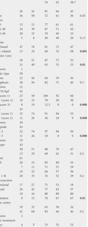
The 5-year survival was 74.2% (66%); 10-year, 61.2% (66%);
and 15-year, 46.5% (67%) (Figure 2). OS was 38.7%. Mean
MFS (n = 53) was 11 years (range 0.5–27.6 years, IQR 4.2–16.2
years). The 5-year MFS was 72% (66%) and 10-year MFS, 60%
(67%).
The majority of patients were 20–40 years old (39%). While
OS appeared to decrease with age, there was no significant
relationship between age and OS (P = 0.13), LRFS (P = 0.2), or
MFS (P = 0.9). There was no significant difference in OS
in patients of pediatric age (<18 years) (n = 12 in comparison
to adults) (P = 0.182). In these patients, 5-year survival
was 75% (614%) and 10-year survival was 58% (614%)
compared with adult patients 72% (66%) and 58% (67%).
There was also no significant difference in MFS (P = 0.887)
and LRFS (P = 0.321). In addition, there was no significant
relation between sex and OS (P = 0.45), LRFS (P = 0.1), and
MFS (P = 0.1).
In patients with limb-based tumors or trunk-related SS,
5-year survival rates were higher than 10-5-year survival rates.
Patients with tumors in the extremities had significantly better
OS than patients whose tumors were located on the trunk (P =
0.001; Figure 3). There was a significant trend for better
outcomes in limb-based SS based on local recurrence (P = 0.06)
or distant metastases (P = 0.07).
Table 1. Patient data and OS for 62 patients with synovial sarcoma
All patients OS (%) n % 5 Year 10 Year OS P All 76 63 38.7 Sex Male 26 42 81 66 42 Female 36 58 72 61 36 0.45 Age, years 0 £ 20 13 21 77 61 61 > 20 £ 40 24 39 87 71 42 > 40 £ 60 20 32 70 60 25 >60 5 8 40 40 20 0.13 Tumor site Limb based 47 76 81 72 47 Trunk related 15 24 60 33 13 0.001 Tumor size (cm) £5 30 51 87 71 55 >5 31 49 63 53 23 0.01 Unknown 1 Histologic type 50 Biphasic 22 44 64 59 36 Monophasic 28 56 82 71 46 0.5 Unknown 12 Mitoses/10 hpf 43 0–9 (score 1) 25 58 100 92 60 10–19 (score 2) 10 23 70 50 30 >19 (score 3) 8 19 12.5 0 0 0.000 Necrosis 43 £50% (score 1) 32 74 91 84 53 >50% (score 2) 11 26 36 18 9 0.000 Unknown 20 Tumor grade 43 Grade 2 32 74 97 84 56 Grade 3 11 26 18 0 0 0.000 Unknown 19 Fusion type 43 SSX1 30 71 80 70 47 SSX2 13 29 69 62 31 0.2 Treatment 61 Only S 20 33 85 80 55 S + R 7 11 71 57 29 S + C 14 23 64 57 36 S + C + R 20 33 76 52 29 0.2 Type of resection Intralesional 17 27 71 53 18 Marginal 26 42 77 62 27 Wide 10 16 80 80 80 Amputation 9 15 78 67 67 0.01 Biopsy in center Yes 19 32 63 58 26 No 41 68 83 66 46 0.2 Unknown 2 Adequate treatment Correct 4 7 75 75 75 Not correct 54 93 81 67 39 0.2 Excluded 4
Significant P-values are given in bold.
C, chemotherapy; hpf, high-power field; OS, overall survival; R, radiotherapy; S, surgery.
Patients with tumors of diameters >5 cm had significantly
worse OS (P = 0.01; Figure 4) and LRFS (P = 0.04) than those
with tumors £5 cm in diameter. However, distant metastases
were not significantly associated with tumor size (P = 0.1).
Information on the histological subtype was available for 50
patients, and 28 SS (56%) were monophasic and 22 (44%) were
biphasic. The 5-year and 10-year survival rates did not differ
significantly between patients with monophasic or biophasic SS
(P = 0.5), and there was no significant relationship between
histological subtype and OS (P = 0.5), local recurrence (P =
0.8), or distant metastases (P = 0.5).
Data on fusion type was available for 46 patients. In 27
patients (58.7%), the tumor was positive for SSX1 and in 19
patients (41.3%) for SSX2. The 5-year and 10-year survival
rates did not significantly differ with fusion type nor was the
association of fusion type with OS (P = 0.2), local recurrence (P
= 0.053), and distant metastases statistically significant (P =
0.1).
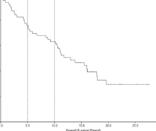
Compared with patients with grade 2 tumors, patients with
grade 3 tumors had significantly poorer prognosis in terms of
OS (P < 0.001) (Figure 5), local recurrence (P = 0.02), and
distant metastases (P < 0.001).
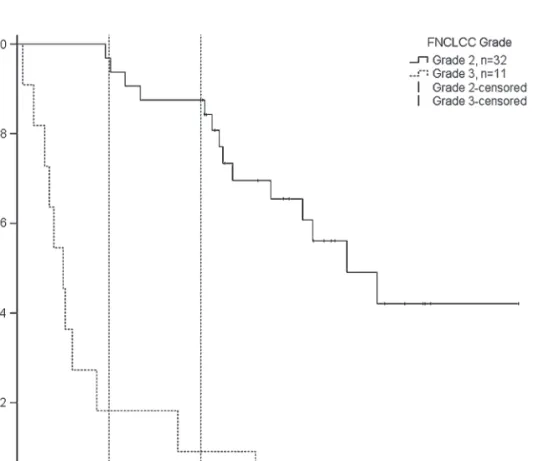
There was a very high rate of technically incorrectly treated
patients (93%; n = 54). Patients who underwent wide resection
(n = 10) had a significantly better prognosis in terms of OS (P =
0.04; Figure 6) and local recurrence (P = 0.001) than those who
did not (n = 52). Patients with wide resection had
a significantly better OS (P = 0.038) than those with marginal
resection combined with radiotherapy (n = 12). Patients with
intralesional resections compared with those with other surgical
procedures had a significantly poorer prognosis regarding OS
(P = 0.021), local recurrence (P = 0.055), and distant metastases
(P = 0.021).
The comparison of the subgroups of patients who underwent
adjuvant treatment in addition to surgery (i.e. surgery and
radiotherapy, surgery and chemotherapy, surgery and
radiochemotherapy) showed no significant difference in OS (P
= 0.24). Patients treated with surgery only (and therefore in
a better prognostic group) had a significantly better outcome in
terms of OS (P = 0.055; Figure 7) and distant metastases (P =
0.012) than those who received adjuvant treatment.
discussion
The aim of this study was to identify prognostic factors and
evaluate surgical treatment in terms of their influence on the
long-term outcome of SS. Other than solitary cases, no published
studies have a minimum follow-up of 10 years for survivors;
therefore, we chose extended follow-up as the most important
inclusion criterion for our study. This is especially important
because of the slow growth of SS and because metastases and
local recurrence are known to occur very late. Various prognostic
factors including age, tumor size [11, 14, 25–28], surgical
margins [11, 14], histological subtype [10, 11], tumor grade [11,
29], and fusion type [10, 11] have been identified in previous
studies. The relative influence of these factors, however, is
controversial. Only tumor size (>5 cm) is consistently associated
with a negative outcome [6, 9, 11, 14, 30, 31].
Our study has several limitations. It is retrospective, and the
patient group consisted of a very heterogenous population of
Table 2. Local recurrence and distant metastases in patients with synovial sarcoma
All patients Local recurrence (%) (n = 29) Distant metastasesa(%) (n = 29)
n % 5 Year 10 Year P 5 Year 10 Year P
All patients 36 45 28 40
Sex
Male 26 42 25 30 18 23
Female 36 58 44 56 0.1 36 53 0.1
Age at diagnosis, years
0 £ 20 13 21 31 31 39 39 > 20 £ 40 24 39 25 41 30 39 > 40 £ 60 20 32 48 56 21 53 >60 5 8 75 0.2 0 50 0.9 Tumor site Limb based 47 76 31 39 24 34 Trunk related 15 24 56 71 0.056 45 71 0.07 Tumor size (cm) £5 31 51 27 31 24 35 £5 30 49 49 64 0.04 35 49 0.1 Unknown 1 Histologic type 50 Biphasic 22 44 26 33 33 39 Monophasic 28 56 30 38 0.8 13 27 0.5 Unknown 12 Mitoses/10 hpf 43 0–9 (score 1) 25 58 20 29 12 21 10–19 (score 2) 10 23 21 21 33 47 >19 (score 3) 8 19 83 83 0.000 75 100 0.000 Necrosis 43 £50% (score 1) 32 74 28 32 16 30 <50% (score 2) 11 26 43 56 0.4 67 67 0.04 Unknown 19 Tumor grade Grade 2 32 74 22 29 16 26 Grade 3 11 26 45 – 0.02 67 0.00 Unknown 19 Fusion type 43 SSX1 30 71 25 25 19 27 SSX2 13 29 40 60 0.053 37 55 0.1 Treatment 62 Only S 20 32 28 40 11 11 S + R 7 11 33 67 20 57 S + C 14 23 39 49 45 64 S + C + R 21 34 43 43 0.955 37 54 0.06 Type of resection Intralesional 17 27 54 67 33 62 Marginal 26 42 45 57 29 42 Wide 10 16 0 0 20 20 Amputation 9 15 14 0.000 29 29 0.05 Biopsy in center Yes 19 32 23 40 31 52 No 41 68 42 48 0.3 28 37 0.2 Unknown 2 Adequate treatment
Correct 4 7 None None 15 15
Not correct 54 93 37 47 0.087 29 42 0.4
Excluded 4
aPatients with distant metastases are those without primary metastases.
Significant P-values are given in bold.
Figure 2. Overall survival at 5 years was 74.2% (66%); at 10 years, 61.2% (66%); and at 15 years, 46.5% (67%).
Figure 4. Overall survival according to tumor size (£5 versus >5 cm), P = 0.013
Figure 6. Overall survival according to the type of resection. In comparison with other types of resection, overall survival in patients with wide resection was significantly better (P = 0.04) and overall survival in patients with intralesional resection was significantly worse (P = 0.021).
Figure 7. Patients who were treated with surgery only had a significantly better prognosis in terms of overall survival (P = 0.055) than those who received adjuvant treatment.
which many were treated outside of a tertiary referral center
before their referral. Through its multicenter design, there was
no agreement on surgical and adjuvant treatment modalities.
The patients early in the series were diagnosed by means of
histological examination without staging through modern
radiologic methods such as MRI or computed tomography and
without molecular verification of SYT–SSX type. For analysis of
tumor size, we chose largest diameter instead of tumor volume
due to a lack of available data for calculation of volume. Eight
patients with minimum follow-up of 10 years were not
available for follow-up examination because they had either
moved to other countries or could not be found.
In the literature, 5-year survival ranges from 25% to 75% and
10-year survival from 11% to 63% [6, 10, 11, 14, 25, 26, 32–36].
In these studies, 10-year survival was extrapolated, whereas in
our study with minimum follow-up of 10 years, it represents
a true estimate. In the present study, 5-year survival was 75.8%
and 10-year survival was 62.9%, which is in accordance with
the literature. The 15-year survival was 46.5%. The difference
between 10-year and 15-year survival reflects the fact that
metastases in SS often occur very late, even beyond 10 years
(n = 5), and furthermore suggests that for these patients,
clinical follow-up of 5 or 10 years is insufficient.
Metastases at diagnosis were a significant negative prognostic
factor in our study (15% of patients, n = 9). The literature
reports metastases at diagnosis in 11%–14% of cases [10, 11,
37] and a significant correlation between metastases at
diagnosis and survival [10, 11, 37].
Tumor size >5 cm was a strong prognostic factor with
a negative influence on OS (P = 0.01) in our study. For tumor
size, we chose cut-off values of £5 cm and >5 cm since these
achieved the best prognostic value. ten Heuvel et al. [37] and
Ladanyi et al. [10] used the same cut-off values and also found
a significant correlation between tumor size and survival
(P = 0.0214 and 0.04, respectively).
In our study, neither histological subtype (P = 0.5) nor fusion
type (P = 0.2) significantly influenced OS. The impact of fusion
type on prognosis is controversial. In addition to its diagnostic
meaning, a prognostic value has been ascribed to fusion type. In
a pilot study, Kawai et al. [12] showed that patients with
tumors of type SYT–SSX1 had significantly shorter MFS than
those with tumors of type SYT–SSX2. These preliminary results
were confirmed by the same authors in a large multicenter
study with 243 patients [10, 12]. In accordance with our results,
the relationship between fusion type and survival was not
confirmed in several other studies [11, 29, 37].
Most local recurrences result from inadequate primary
surgical treatment [17]. In our series, 42% of tumors (n = 26)
were resected marginally and 27% (n = 17) of patients had
intralesional resections. Only 31% of patients (n = 19) were
adequately treated with a wide resection or amputation. Only
four patients were correctly treated without delay after biopsy.
Patients who had wide or radical resection had a significantly
better outcome in terms of OS (P = 0.04) and local recurrence
(P = .001). Those patients in whom surgery was intralesional
had a significantly poorer outcome in terms of OS (P = 0.021),
local recurrences (P = 0.055), and distant metastases
(P = 0.021). The better outcome in patients who had surgery
only compared with those with adjuvant chemotherapy/
radiotherapy in addition might represent the fact that, in those
cases, the margins were usually clear and therefore additional
chemotherapy and/or radiotherapy was not considered
necessary.
The fact that two-thirds of patients in our study were
primarily treated outside a tertiary referral center is an
important problem in our country. The large number of
patients (n = 54) who underwent inadequate treatment initially
is in contrast to the current recommendation that these patients
need coordinated interdisciplinary treatment that is only
possible in tumor centers.
conclusion
Significant adverse prognostic factors for SS included larger
tumor size, metastases at diagnosis, high histological grade,
trunk-related disease, and intralesional or marginal surgery.
Sex, histological subtype, and SYT–SSX fusion type were not
significant prognostic factors. The high proportion of SS with
technically incorrect treatment emphasizes the
recommendation that the entire treatment should be carried
out in an interdisciplinary tumor center from the beginning.
Based on the high rate of local recurrence and metastases even
after 5 and 10 years, we suggest that patients with SS be
followed for >10 years. For SSs, patient education and yearly
follow-up for even >10 years with thorough history and
physical examination seem advisable to detect the common late
recurrences at an early stage, when treatment might still be
feasible. By reducing morbidity and limiting the extent of
secondary treatment, this may also be a cost-effective strategy.
Routine surveillance imaging is only of significant benefit if the
risk for asymptomatic recurrence is high or if other factors
make clinical assessment difficult.
funding
Suzy Ru¨ckert Foundation, Basel, Switzerland.
acknowledgements
We thank S. Korn, for her support with the statistical analysis
and BioScience Writers, Houston Texas, for the editing of the
manuscript. Also we would like to thank M. Paulussen,
Department of Oncology, University of Basel, for his useful
comments on the manuscript.
disclosure
The authors declare no conflict of interest.
references
1. Weiss S, Goldblum J, Publishing M. Enzinger and Weiss’s Soft Tissue Tumors. St Louis, MI: Mosby 2001.
2. Milchgrub S, Ghandur-Mnaymneh L, Dorfman HD, Albores-Saavedra J. Synovial sarcoma with extensive osteoid and bone formation. Am J Surg Pathol 1993; 17: 357–363.
3. Miettinen M, Virtanen I. Synovial sarcoma—a misnomer. Am J Pathol 1984; 117: 18–25.
4. Smith ME, Fisher C, Wilkinson LS, Edwards JC. Synovial sarcoma lack synovial differentiation. Histopathology 1995; 26: 279–281.
5. Haldar M, Hancock JD, Coffin CM et al. A conditional mouse model of synovial sarcoma: insights into a myogenic origin. Cancer Cell 2007; 11: 375–388. 6. Lewis JJ, Antonescu CR, Leung DH et al. Synovial sarcoma: a multivariate
analysis of prognostic factors in 112 patients with primary localized tumors of the extremity. J Clin Oncol 2000; 18: 2087–2094.
7. Okcu MF, Munsell M, Treuner J et al. Synovial sarcoma of childhood and adolescence: a multicenter, multivariate analysis of outcome. J Clin Oncol 2003; 21: 1602–1611.
8. Fletcher CDM, Unni KK, Mertens F. World Health Organization Classification of Tumours: Pathology and Genetics of Tumours of Soft Tissue and Bone. Lyon, France: IARC Press 2002.
9. Ferrari A, Gronchi A, Casanova M et al. Synovial sarcoma: a retrospective analysis of 271 patients of all ages treated at a single institution. Cancer 2004; 101: 627–634.
10. Ladanyi M, Antonescu CR, Leung DH et al. Impact of SYT-SSX fusion type on the clinical behavior of synovial sarcoma: a multi-institutional retrospective study of 243 patients. Cancer Res 2002; 62: 135–140.
11. Guillou L, Benhattar J, Bonichon F et al. Histologic grade, but not SYT-SSX fusion type, is an important prognostic factor in patients with synovial sarcoma: a multicenter, retrospective analysis. J Clin Oncol 2004; 22: 4040–4050. 12. Kawai A, Woodruff J, Healey JH et al. SYT-SSX gene fusion as a determinant of
morphology and prognosis in synovial sarcoma. N Engl J Med 1998; 338: 153–160.
13. Nilsson G, Skytting B, Xie Y et al. The SYT-SSX1 variant of synovial sarcoma is associated with a high rate of tumor cell proliferation and poor clinical outcome. Cancer Res 1999; 59: 3180–3184.
14. Singer S, Baldini EH, Demetri GD et al. Synovial sarcoma: prognostic significance of tumor size, margin of resection, and mitotic activity for survival. J Clin Oncol 1996; 14: 1201–1208.
15. Pisters PW, O’Sullivan B, Maki RG. Evidence-based recommendations for local therapy for soft tissue sarcomas. J Clin Oncol 2007; 25: 1003–1008. 16. Sakabe T, Murata H, Konishi E et al. Evaluation of clinical outcomes and
prognostic factors for synovial sarcoma arising from the extremities. Med Sci Monit 2008; 14: CR305–C310.
17. Goodlad JR, Fletcher CD, Smith MA. Surgical resection of primary soft-tissue sarcoma. Incidence of residual tumour in 95 patients needing re-excision after local resection. J Bone Joint Surg Br 1996; 78: 658–661.
18. Maduekwe UN, Hornicek FJ, Springfield DS et al. Role of sentinel lymph node biopsy in the staging of synovial, epithelioid, and clear cell sarcomas. Ann Surg Oncol 2009; 16: 1356–1363.
19. Coindre JM. Histologic grading of adult soft tissue sarcomas. Verh Dtsch Ges Pathol 1998; 82: 59–63.
20. Coindre JM, Terrier P, Guillou L et al. Predictive value of grade for metastasis development in the main histologic types of adult soft tissue sarcomas: a study of 1240 patients from the French Federation of Cancer Centers Sarcoma Group. Cancer 2001; 91: 1914–1926.
21. Guillou L, Coindre JM, Bonichon F et al. Comparative study of the National Cancer Institute and French Federation of Cancer Centers Sarcoma Group
grading systems in a population of 410 adult patients with soft tissue sarcoma. J Clin Oncol 1997; 15: 350–362.
22. Guillou L, Coindre J, Gallagher G et al. Detection of the synovial sarcoma translocation t(X;18) (SYT;SSX) in paraffin-embedded tissues using reverse transcriptase-polymerase chain reaction: a reliable and powerful diagnostic tool for pathologists. A molecular analysis of 221 mesenchymal tumors fixed in different fixatives. Hum Pathol 2001; 32: 105–112.
23. Kaplan EL, Meier P. Nonparametric estimations for incomplete Observations. J Am Stat Assoc 1958; 53: 457–481.
24. Peto R, Pike MC, Armitage P et al. Design and analysis of randomized clinical trials requiring prolonged observation of each patient. II. analysis and examples. Br J Cancer 1977; 35: 1–39.
25. Brodsky JT, Burt ME, Hajdu SI et al. Tendosynovial sarcoma. Clinicopathologic features, treatment, and prognosis. Cancer 1992; 70: 484–489.
26. Oda Y, Hashimoto H, Tsuneyoshi M, Takeshita S. Survival in synovial sarcoma. A multivariate study of prognostic factors with special emphasis on the comparison between early death and long-term survival. Am J Surg Pathol 1993; 17: 35–44.
27. Okcu MF, Despa S, Choroszy M et al. Synovial sarcoma in children and adolescents: thirty three years of experience with multimodal therapy. Med Pediatr Oncol 2001; 37: 90–96.
28. Ladenstein R, Treuner J, Koscielniak E et al. Synovial sarcoma of childhood and adolescence. Report of the German CWS-81 study. Cancer 1993; 71: 3647–3655.
29. Takenaka S, Ueda T, Naka N et al. Prognostic implication of SYT-SSX fusion type in synovial sarcoma: a multi-institutional retrospective analysis in Japan. Oncol Rep 2008; 19: 467–476.
30. Spillane AJ, A’Hern R, Judson IR et al. Synovial sarcoma: a clinicopathologic, staging, and prognostic assessment. J Clin Oncol 2000; 18: 3794–3803. 31. Trassard M, Le Doussal V, Hacene K et al. Prognostic factors in localized primary
synovial sarcoma: a multicenter study of 128 adult patients. J Clin Oncol 2001; 19: 525–534.
32. Cadman NL, Soule EH, Kelly PJ. Synovial sarcoma; an analysis of 34 tumors. Cancer 1965; 18: 613–627.
33. Wright PH, Sim FH, Soule EH, Taylor WF. Synovial sarcoma. J Bone Joint Surg Am 1982; 64: 112–122.
34. Mullen JR, Zagars GK. Synovial sarcoma outcome following conservation surgery and radiotherapy. Radiother Oncol 1994; 33: 23–30.
35. Bergh P, Meis-Kindblom JM, Gherlinzoni F et al. Synovial sarcoma: identification of low and high risk groups. Cancer 1999; 85: 2596–2607.
36. Oda Y, Hashimoto H, Takeshita S, Tsuneyoshi M. The prognostic value of immunohistochemical staining for proliferating cell nuclear antigen in synovial sarcoma. Cancer 1993; 72: 478–485.
37. ten Heuvel SE, Hoekstra HJ, Bastiaannet E, Suurmeijer AJ. The classic prognostic factors tumor stage, tumor size, and tumor grade are the strongest predictors of outcome in synovial sarcoma: no role for SSX fusion type or ezrin expression. Appl Immunohistochem Mol Morphol 2009; 17: 189–195.