Glycobiology vol. 1. no. 1 pp. 71-82, 1990
Purification and properties of arylmannosidases from mung bean seedlings and soybean
cells
Irena Pastuszak, G.P. Kaushal, K.A. Wall, Y.T. Pan, A. Sturm2 and A.D. Elbein1
Department of Biochemistry, University of Texas Health Science Center, San Antonio, TX 78284, USA and 2Fnednch Miescher Institute, Basel, Switzerland
'To whom correspondence should be addressed
Two arylmannosidases (signified as A and B) were purified to homogeneity from soluble and microsomal fractions of mung bean seedlings. Arylmannosidase A from the microsomes appeared the same on native gels and on SDS gels as soluble arylmannosidase A, the same was true for arylmannosidase B. Sedimentation velocity studies indicated that both enzymes were homogeneous, and that arylmannosidase A had a molecular mass of 237 kd while B had a molecular mass of 243 kd. Arylmannosidase A showed two major protein bands on SDS gels with molecular masses of 60 and 55 kd, and minor bands of 79, 39 and 35 kd. All of these bands were N-linked since they were susceptible to digestion by endo-glucosaminidase H. In addition, at least the major bands could be detected by Western blots with antibody raised against the xylose moiety of N-linked plant oligosaccharides, and they could also be labeled in soybean suspension cells with [2-3H]mannose. Arylmannosidase B showed three major bands with molecular masses of 72, 55 and 45 kd, and minor bands of 42 and 39 kd. With the possible exception of the 45 and 42 kd bands, all of these bands are glycoproteins. Arylmannosidases A and B showed somewhat different kin-etics in terms of mannose release from high-mannose oligo-saccharides, but they were equally susceptible to inhibition by swainsonine and mannostatin A. Polyclonal antibody raised against the arylmannosidase B cross-reacted equally well with arylmannosidase A from mung bean seedlings and with arylmannosidase from soybean cells. However, monoclonal antibody against mung bean arylmannosidase A was much less effective against arylmannosidase B. Antibody was used to examine the biosynthesis and structure of the carbohydrate chains of arylmannosidase in soybean cells grown in |2-3H|mannose. Treatment of the purified enzyme with Endo H released - 5 0 % of the radioactivity, and these labeled oligosaccharides were of the high-mannose type, i.e. mostly Man9GlcNAc. The precipitated protein isolated from the Endo H treatment still contained 50% of the radioactivity, and this was present in modified structures that probably contain xylose residues.
Key words: Mung beans/amannosidases/glycoproteins/-soybean-a-mannosidases/xylose-containing N-linked glyco-proteins
Introduction
During the course of purification of the glycoprotein processing mannosidase II from mung bean seedlings
(Kaushal et al., 1990), we detected two other peaks of a-mannosidase activity on DEAE-cellulose columns that also reacted with the /j-nitrophenyl-a-D-mannopyranoside substrate. In order to determine the relationship of these activities to mannosidase II and to the processing of N-linked oligosaccharides in general, we decided to purify these enzymes to homogeneity so that we could determine their structures and properties. Plant cells, like animal cells, have two different classes of a-mannosidases which differ in their pH optima and in their substrate specificities (Forsee, 1985; Szumilo et al., 1986a). One class is the glycoprotein processing a-mannosidases, mannosidase I (Szumilo et al., 1986a) and mannosidase II (Kaushal et al., 1990), that are involved in trimming mannose residues from high-mannose oligosaccharides of the N-linked glycoproteins (Kornfeld and Kornfeld, 1985). These enzymes have pH optima of ~6.5, reside in the endoplasmic reticulum or Golgi ap-paratus and act on Man9(GlcNAc)2-proteins, or
GlcNAc-Man5(GlcNAc)2-proteins (Tulsiani et al., 1982a).
Manno-sidase II will also utilize p-nitrophenyl-a-D-manno-pyranoside as a substrate, while mannosidase I is inactive on this substrate (Kaushal et al., 1990). These various enzymes can also be distinguished by their susceptibility to various glycosidase inhibitors (Elbein, 1988).
The second class of mannosidases comprises the aryl-a-mannosidases that have pH optima of ~ 3.5—5.0, show rather broad aglycone specificities, including/7-nitrophenyl-a-mannopyranoside, and are usually associated with vacu-oles or protein bodies of plants (McGee and Murray, 1985) or lysosomes of animals (Opheim and Touster, 1978). These enzymes are probably involved in catabolism and turnover of N-linked glycoproteins (Neely and Beevers, 1980). Aryl-mannosidases that are associated with the plant cell wall (Greve and Ordin, 1977; Van der Wilden and Chrispeels, 1983) have also been described. Since the cell wall is considered to be involved in lytic activity, a similar glyco-protein degradative function could be postulated for cell wall mannosidases (Greve and Ordin, 1977).
In this report we have purified 2 aryl-a-mannosidases from the microsomal and soluble fractions of mung bean seedlings. Polyclonal antibodies prepared against either of these enzymes cross-reacts with the other protein, as well as with arylmannosidase activity isolated from the soluble fraction of mung bean seedlings. The antibodies also cross-react with proteins from suspension cultured soybean cells. A monoclonal antibody raised against one of the mung bean enzymes, however, only reacts with that native enzyme and reacts poorly with the other native mannosidase. Various properties of the two aryl-a-mannosidases are presented in order to compare them to each other and to the known processing mannosidases. Having determined the various properties of these two arylmannosidases, it seems likely that they are protein body or vacuolar enzymes, and are solubilized by disruption of these organelles during cell
I.Pastuszak et al.
breakage. The small amount of enzyme found associated with the microsomal pellet is probably due to enzymes that are in the process of being synthesized or trimmed in the endoplasmic reticulum or Golgi apparatus.
Results
Purification of aryl-a-mannosidases
Two different aryl-a-mannosidases (i.e. enzymes that use/j-nitrophenyl-a-D-mannopyranoside as substrate) were separated from each other by chromatography of the mung bean extract on DEAE -cellulose (see below), and each of these enzymes was purified to homogeneity by the pro-cedures described below. Although much of the purification shown here was done with the microsomal fraction, later studies showed that these enzymes were also in the cyto-plasmic fraction, and their purification could be performed with the same methodology.
Ammonium sulfate precipitation. Solid ammonium sulfate was added slowly with stirring to 5 1 of supernatant liquid from the solubilization step to bring the mixture to 75% saturation of ammonium sulfate. The mixture was allowed to stand for 30 min in ice and the precipitate was isolated by centrifugation at 7000 g for 30 min. The supernatant liquid was discarded and the pellet was dissolved in ~400 ml of 10 mM HEPES buffer, pH 7.2, containing 5% glycerol, and dialyzed against the same buffer.
DEAE-cellulose chromatography. The dialysate from the ammonium sulfate precipitation (400 ml) was applied to a 3.7 x 42 cm column of DE-52 that had been equilibrated with 10 mM HEPES buffer, pH 7.2. The column was washed well with the equilibration buffer and was then eluted with a 0-300 mM gradient elution of NaCl in the same buffer. The elution profiles of several different glyco-sidases that are present in the mung bean extract are shown
in Figure 1. Two different peaks of arylmannosidase activity emerged earliest from this column (at ~ 50—100 mM salt), followed by the peak of mannosidase II activity (~ 150 mM salt) and then a peak of glucosidase II activity (~ 250 mM salt). Since the two peaks of arylmannosidase activity were not well separated and in some cases emerged as a shoulder and a main peak, we pooled the two peaks together (e.g. fractions 150-190 on this column) and separated them at the next purification step (i.e. phosphocellulose).
Chromatography on phosphocellulose. The pooled aryl-mannosidase peak from DEAE-cellulose was dialyzed over-night against 10 mM acetate buffer, pH 5.2. It was then applied to a 1.9 x 10 cm column of cellulose phosphate that was prepared and equilibrated in acetate buffer, pH 5.2. The column was washed well with buffer and then eluted with a linear gradient of 0-200 mM NaCl in the same buffer. As shown in Figure 2, this gradient separated the two peaks of aryl-a-mannosidase activity. The first peak, which was labeled arylmannosidase A, emerged at ~ 50-75 mM NaCl, while the second peak, labeled arylmannosidase B, emerged at ~ 100-150 mM NaCl. These two peaks were pooled individually and each was purified to homogeneity as described below.
Hydroxyapatite chromatography. The pooled fractions from the phosphocellulose column (either peak A or peak B) were dialyzed overnight against 10 mM phosphate buffer, pH 7.1. The dialyzed preparations were applied to a 1.6 x 8.5 cm column of hydroxyapatite that had been equilibrated with 10 mM phosphate buffer, pH 7.1. The column was washed well with the equilibration buffer and was then eluted with a linear gradient of 10-250 mM phosphate buffer, pH 7.1. Arylmannosidase A activity emerged from the column at 50-120 mM phosphate buffer whereas arylmannosidase B emerged at 80-200 mM phosphate buffer (data not shown).
5
0.8 >• " 0 . 4 Glc : / / • -ARYL-MANNOSIDASE PROTEIN MANn
I ,-•• 20 "60 80 100 120 140 160 180 200 2 FRACTION NUMBER 240 260Fig. 1. Purification of arylmannosidases by DEAE-cellulose chromatography. The solubilized (or cytosolic) enzyme fraction prepared from mung bean microsomes was applied to a DE-52 column that had been equilibrated with 10 mM HEPES buffer containing 10% glycerol and 0.1% Triton X-100. The column (3.7 x 45 cm) was washed with Tris buffer and eluted with 800 ml of a linear gradient of 0-0 3 M NaCl in the same buffer. Two peaks of arylmannosidase activity emerged from this column at ~ 50-100 mM salt, followed by a peak of mannosidase II (~ 150 mM salt) and then a peak of glucosidase II (~250 mM salt). Active fractions were pooled for the next purification step.
Purification and properties of arvlmannosidases
Purification using Concanavalin A-Sepharose. The active
fractions from the hydroxyapatite column were pooled, concentrated to ~ 10 ml on the Amicon filtration apparatus and then mixed with 2 ml of a suspension of Concanavalin A-Sepharose (15 mg/ml). The mixture was allowed to stand at 0°C for 3 h with occasional stirring and was then transferred to a small glass column. The column was washed with 200 mM NaCl in 10 mM phosphate buffer, pH 7.1,
E
o 10
d6
>. 0.8 Ul Nz
0.4 GRADIENT ARYL MAN A PROTEIN . • - • /ARYL-* MANB 20 40 60 80 100 120 140 FRACTION NUMBERFig. 2. Separation of arylmannosidase A from arylmannosidase B by cellulose phosphate chromatography. The column was prepared and equilibrated as described in the text. The bound enzymes were eluted with a 0-0.2 M linear gradient of NaCl. Fractions of 4 ml were collected and assayed for aylmannosidase activity as described in the text. Active fractions were pooled for the next purification step.
also containing 1 mM CaCl2 and 1 mM MgCl2. Then 0.3 M
a-methylmannoside was applied to the columns, and the columns were stoppered and stored in the cold for ~ 5 h while in contact with the eluent. The elution was then continued with additional amounts of a-methylmannoside being passed through the column. Fractions were collected and those fractions showing a-mannosidase activity were pooled and dialyzed overnight against 10 mM HEPES buffer, pH 7.1.
Chromatography on columns of Sephacryl S-300. The
dialyzed eluate from the Concanavalin A-Sepharose was concentrated on an Amicon filter and the concentrated fraction was applied to a 1.5 x 150 cm column of Sephacryl S-300 that had been equilibrated in 10 mM HEPES buffer, pH 7.1. As shown in Figure 3, the enzymatic activity was eluted in a sharp, symmetrical peak with this same buffer.
Purification by polyacrylamide gel electrophoresis.
Prepara-tive non-denaturing polyacrylamide gel electrophoresis was done at 4°C in 7% acrylamide, pH 6.8, as described (Orr et
al., 1972). A 0.2 ml sample (100 /xg protein) was applied to
each of 18 tube gels, and electrophoresis was carried out at a current of 2 mA/tube. The active band was localized by incubating one of the gels in the presence of 2 mM 4-methylumbelliferyl-a-mannoside in 50 mM acetate buffer, pH 4.5, for 2-5 min. The gel was then examined under UV light to localize the enzyme activity. The corresponding area was sliced from the remaining gels and eluted into 10 mM HEPES buffer, pH 7.1.
Using the above procedures, both the aryl-a-mannosidase A and aryl-a-mannosidase B were purified ~ 1000-fold, with a recovery of ~ 4 % . These data are summarized in Table I for each step of the purification procedure. In this table the purification of the arylmannosidases found in the
2.4 E c O ' (O.D . A IVIT Y : AC T NZYM I 2.0 1.6 1.2 0.8 0.4 ENZYME 10 20 30 40 60 70 80 90 FRACTION NUMBER
Fig. 3. Purification of arylmannosidase B on a Sephacryl S-300 column. The enzyme preparation obtained from the Concanavalin A-Sepharose
column was applied to the Sephacryl S-300 column. The column was run at a flow rate of 15 ml/h and fractions of 2 ml were collected. Each fraction was examined for enzyme activity and for the amount of protein. Fractions containing arylmannosidase activity were pooled and purified further, as described in the text.
I.Pastuszak et al.
Table I. Purification procedure
Step (mg) 1. Supernatant 2. Ammonium sulfate precipitation 3 DE-52 4. Cellulose phosphate A B 5. HAP arylman. A arylman. B 6. Con A arylman. A arylman. B 7. Sephacryl S-300 A B 8. Polyacrylamide gel electrophoresis A B
for cytoplasmic arylmannosidases (A and B)
Protein 12000 10000 1560 40 90 10.5 17. 3.1 5. 2 1 2.4 0.49 0.44 Total (fold) 284.5 275.8 269. 114.8 122. 84.9 61.8 45.9 43.9 41.3 33.4 13.36 11.8 Activity Sp. act (%) 0.024 0.027 0.172 2.87 1.35 8.086 3.635 14.8 8.78 19.7 13 92 27.26 26.8 Purification 1.12 7.16 119.58 56.25 336.92 151.45 616.66 365.83 820 83 580.0 1135.8 1116.6 Yield 100 96.9 94.5 40.3 42.9 29 8 21 7 16 1 15.4 14.5 11 7 4.7 4.1
soluble fraction rather than in the microsomal fraction is presented, since much more of the enzymes were found in soluble form. However, the purification data and properties were similar for both soluble and microsomal forms. In fact, it seems likely that these mannosidases are actually protein body enzymes that are released upon cell breakage, and the microsomal fraction may represent mannosidases in the process of being synthesized and trimmed in the endoplasmic reticulum and Golgi apparatus of the cell.
The purity of each of the arylmannosidases (both soluble and microsomal) was assessed by native gel electrophoresis and by sedimentation velocity studies. Figure 4 shows that both arylmannosidase A and arylmannosidase B gave single bands when subjected to native gel electrophoresis and staining with Coomassie Blue, and a mixture of the two enzymes gave the expected two bands. In addition, when the enzymes were examined by sedimentation equilibrium in a Model E ultracentrifuge, a straight line relationship was observed for each enzyme, indicating that each was homo-geneous. The molecular mass of aryl-a-mannosidase A was estimated to be 237000 and that of aryl-a-mannosidase B to be 243000. It is unlikely that arylmannosidase A could be a proteolytic product of arylmannosidase B since the two enzymes are still isolated in about the same ratios when various protease inhibitors are included in the purification buffers (e.g. leupeptin, pepstatin, PMSF and aprotinin). In addition, one would not expect a single product after protease treatment, as is seen in Figure 4.
The two mannosidases were also examined by SDS gel electrophoresis to compare their subunit composition, as shown in Figure 5. In this case, the two enzymes appear to be somewhat different, although they apparently do have some subunits in common. Thus, as seen in lane A, aryl-a-mannosidase A shows two major subunits, with mol.wts of ~59.5 and 55.5 kd. There were also smaller amounts of four or five other protein bands, which had mol.wts of 72, 39, 35, 32 and 24 kd. It is not clear whether these other bands are the result of proteolysis, whether they come from contamination of the A enzyme by the B enzyme or whether
they are integral parts of the enzyme. Lane B in Figure 5 shows that each of these subunits is a glycoprotein and contains a high-mannose (or hybrid) type of N-linked oligosaccharide, since the mobility of each band was in-creased by digestion with endoglucosaminidase H. This increase in migration rate suggests the loss of ~2 kd, which would indicate the removal of one oligosaccharide chain.
In order to determine whether arylmannosidases have complex or modified oligosaccharides in addition to the high-mannose chains, arylmannosidase A was treated with trifluoromethanesulfonic acid (TFMS) to remove all of the carbohydrate, and the protein was run on gels as shown in Figure 5b. Lane 2 shows the migration of the original subunits of arylmannosidase A whereas lane 3 is after treatment with endo H, and lane 4 after TFMS treatment. It can be seen that removal of all the carbohydrate resulted in faster moving protein bands, indicating that each subunit contained at least one modified oligosaccharide in addition to the high-mannose structure. Further evidence for this is seen in Figure 12.
When aryl-a-mannosidase B was subjected to SDS gel electrophoresis it gave three major bands, with estimated mol.wts of 72, 55.5 and 45.4 kd, and minor bands of 42.5 and 39 kd. In this case, the 72, 55.5 and 39 kd subunits were N-linked glycoproteins, as shown by their increased mobility after digestion with endoglucosaminidase H, but the 45.5 and 42.5 kd bands were resistant to Endo H, indicating that they do not contain high-mannose or hybrid types of oligosaccharides. They could, of course, still be N-linked glycoproteins with Endo H-resistant chains. At this stage, the relationship of arylmannosidase A to arylmannosidase B is not clear, nor is the origin of each of the protein bands seen in this figure.
The purified enzymes were quite stable, either stored in an ice bucket or frozen at -20°C, as long as the storage buffer contained 10% glycerol and 0.5 mM dithiothreitol. Thus, the enzymes have been kept in the frozen state for several months with no detectable loss of activity. However, when either enzyme was stored in the cold for a week in
Purification and properties of arylmannosidases S A B C D S E F
97.4—
66.2-
42.731
-
21.5-B
1
4 5
Fig. 4. Native gel electrophoresis of the purified arylmannosidases A
and B. Proteins were applied to the gels and run as described in the text. Protein was detected by staining with Coomassie Blue. Lanes are as follows: lane I, arylmannosidase A; lane 2, arylmannosidase B; lane 3, mixture of arylmannosidase A and arylmannosidase B.
buffer below pH 5.5, it lost considerable activity and the extent of loss increased as the pH was lowered. Thus, at pH 4.5 ~ 5 0 % of the activity was lost in 1 week, whereas at pH 4.0 > 8 5 % of the activity was gone in 1 week (data not shown).
Properties of the purified arylmannosidases
The release of mannose from /j-nitrophenyl-a-D-manno-pyranoside was linear with time for up to 60 min and with the amount of protein up to ~ 15 ug of purified enzyme (with either arylmannosidase A or B; data not shown). The pH optimum for both arylmannosidase A and
aryl-Fig. 5. (A) SDS gel electrophoresis of purified arylmannosidases A and
B. The purified enzymes were subjected to SDS gel electrophoresis, either before (lanes A, C and E) or after (lanes B, D and F) digestion with Endo H. Proteins were stained with Coomassie Blue. Lanes A and B are from arylmannosidase A; lanes C and D from arylmannosidase B; lanes E and F from jack bean a-mannosidase. Standards shown in lane S are as follows' phosphorylase b, 97.4 kd; bovine serum albumin, 66.2 kd; ovalbumin, 42.6 kd; carbonic anhydrase, 31 kd; trypsin inhibitor, 21.5 kd. (B) Gel electrophoresis of arylmannosidase A after release of oligosaccharides by hydrolysis with TFMS (lane 4). Lanes 1 and 5 are standards; lane 2 is untreated arylmannosidase A; lane 3 is enzyme treated with endo H; lane 4 is enzyme treated with TFMS.
I.Pastuszak el al.
•••'& i i 9
0.2 0.4 0.6 0.8
AMOUNT OF INHIBITOR (Hg)
Fig. 6. Effect of various processing inhibitors on the activity of arylmannosidases. Various amounts of the inhibitors were added as indicated in the figure. The reaction mixtures were as described in the text, with p-nitrophenyl-a-D-mannopyranoside as the
substrate. The release of />-nitrophenol was measured at 410 nm. The experiment shown here was done with arylmannosidase A, but arylmannosidase B gave essentially the same results.
mannosidase B was somewhat broad, between 3.5 and 5.0, shown using either citrate-phosphate buffer or acetate buffer. However, activity rapidly declined above pH 5.0 and below 3.5 (data not shown). The effect of concentration of the substrate, /7-nitrophenyl-a-D-mannopyranoside, on the activity of arylmannosidases A and B was determined. Enzyme activity increased with increasing substrate con-centration up to ~8 mM and then slowly leveled off at higher concentrations of substrate. When the data were plotted by the method of Lineweaver and Burk, the Km
for /7-nitrophenyl-a-D-mannopyranoside was calculated to be 3.3 for either enzyme.
We also compared the reactivity of arylmannosidase A to that of arylmannosidase B in terms of release of mannose units from a mannose-labeled Man9GlcNAc substrate. At
low concentrations of enzyme, arylmannosidase A could re-lease most of the mannose from this substrate, and gave rise mostly to Man-/#-GlcNAc and free mannose over a 24 h incubation. However, with arylmannosidase B (using an amount of enzyme giving the same reactivity towards p-nitrophenyl-a-mannopyranoside as used with A) there were still considerable amounts of Man4GlcNAc and
Man3GlcNAc, even after 25 h of incubation, suggesting
that the B enzyme probably acts more slowly on a 1,3- and al,6-mannosidic linkages than the A enzyme (data not shown). Detailed kinetic studies with these enzymes have not yet been done but will be of interest to understand their mechanism of catalysis. \ MANE i» '. -JACKBEAN 0 10 20 30 40 50 60 70 " 140 AMOUNT OF ANTISERUM (Hg)
Fig. 7. Effect of polyclonal antibody against arylmannosidase B on the immunoprecipitation of various mannosidases. Mannosidases prepared from mung bean seedlings, cultured soybean cells or jack beans (commercially obtained) were incubated with various amounts of anti-mannosidase B antibody. After 2-3 h of incubation, S. aureus was added. The suspension was kept at 4°C for 1.5 h with occasional stirring. The mixture was centrifuged for 2 min and the enzymatic activity remaining in the supernatant liquid determined as described in the text.
Effect of various mannosidase inhibitors on the arylmannosidases
We examined the effects of various inhibitors of manno-sidase I and mannomanno-sidase II on the purified mung bean arylmannosidases, as shown in Figure 6. It can be seen that the inhibitors of mannosidase II and jack bean a-manno-sidase, i.e. swainsonine (Tulsiani et al., 1982b; Kang and Elbein, 1983) and mannostatin (Tropea et al., 1990), were very good inhibitors of the mung bean arylmannosidases, whereas inhibitors of mannosidase I, i.e. deoxymanno-jirimycin (Fuhrmann et al., 1984) and kifunensine (Elbein et al., 1990), were inactive towards the arylmannosidases. Clearly the best inhibitor was mannostatin which showed an IC50 of ~ 1 x 10~7 M for this enzyme when
/?-nitrophenyl-a-D-mannopyranoside was used as the substrate. Swain-sonine was somewhat less effective and gave an IC50 of ~ 5
x 10-7M.
Preparation of antibody against arylmannosidases A and arylmannosidase B
Purified arylmannosidase B, as seen in Figure 4, was injected into a rabbit for the preparation of polyclonal antibody. This antibody was then tested to determine its specificity towards several different enzyme preparations and extracts
Purification and properties of arylmannosidases ARYL-MAN B 2 0 -ARYL-MAN A A— o 0 200 400 600 1000 AMOUNT OF ANTIBODY (pg)
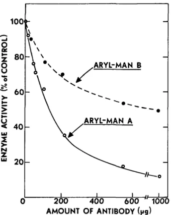
Fig. 8. Effect of monoclonal antibody against arylmannosidase A on the immunoprecipitation of arylmannosidases A and B from solution. Arylmannosidases A and B were incubated with various amounts of monoclonal antibody for several hours and then 5. aureus was added. After an incubation, with occasional stirring for several hours, the precipitate was removed by centrifugation and the supernatant liquid assayed for mannosidase activity.
from various organisms. These results are shown in Figure 7. In this experiment various amounts of the antibody were incubated with the indicated extract or enzyme preparation, and then S. aureus was added to precipitate the antigen-antibody complex. After centrifugation to remove the precipitated antigen, the supernatant liquid was assayed for remaining arylmannosidase activity using />-nitrophenyl-a-D-mannopyranoside as substrate. It can be seen that the polyclonal antibody prepared against arylmannosidase B was equally as effective against the purified arylmannosidase A, and was also very effective against arylmannosidase activity in extracts from suspension-cultured soybean cells. Interestingly enough, the antibody also reacted with a commercial preparation of jack bean a-mannosidase, although it was not nearly as effective towards this enzyme as it was on the mung bean enzymes. The SDS gels of jack bean a-mannosidase shown in Figure 5 do indicate that this enzymes shares at least one common subunit with the mung bean arylmannosidases. The arylmannosidase B antibody did not react with either of the purified mung bean processing mannosidases, mannosidase I (Szumilo el al., 1986a) and mannosidase II (Kaushal et al., 1990), indicating that these Golgi mannosidases are distinct from the aryl-mannosidases.
We also prepared a monoclonal antibody against aryl-mannosidase A by injecting this enzyme into a mouse and screening hybridomas for their ability to produce an anti-body that would precipitate arylmannosidase A from sol-ution. Figure 8 shows the results obtained with the
mono-clonal antibody tested against purified arylmannosidases A and B. While the antibody was quite effective at precipitating arylmannosidase A from solution (in the presence of S.
aureus), it was much less effective against arylmannosidase
B, although it still had some activity towards this protein. This is further evidence that arylmannosidases A and B are distinct proteins although they must share some common epitope(s). On the other hand, the monoclonal antibody had no activity towards jack bean a-mannosidase, or against mannosidases I and II. This monoclonal antibody could not be used for immunoblotting the arylmannosidases on SDS gels since it apparently does not recognize the denatured proteins. It can, however, be used to detect the proteins on native gels.
Characterization of the carbohydrate structure of the soybean arylmannosidases
Since the polyclonal antibodies directed against the mung bean arylmannosidases (A and B) also cross-reacted with protein(s) from extracts of suspension cultured soybean cells, we used this antibody to isolate [3H]-mannose-labeled
arylmannosidase from cultured soybean cells in order to determine the carbohydrate structure of these enzymes. Thus, soybean cells were grown in the presence of
[2-3H]mannose for ~ 24 h, as previously described (Hori and
Elbein, 1983), and cell-free extracts were prepared. The arylmannosidase was partially purified by DEAE-cellulose and hydroxylapatite chromatography, and the labeled aryl-mannosidase was then immunoprecipitated with the poly-clonal antibody. The labeled immunoprecipitate was sus-pended in 0.5% SDS containing 5 raM /?-mercaptoethanol and heated at 100°C for 5 min. After cooling and diluting the mixture to 0.1% SDS, it was incubated with endo-glucosaminidase H to release high-mannose or hybrid types of N-linked oligosaccharides. The reaction was stopped by the addition of trichloroacetic acid (TCA) to a final con-centration of 5%, and the protein was removed by centri-fugation. The supernatant liquid, containing the Endo H-released oligosaccharides, was removed and saved, and the precipitated protein was washed several times with 50% methanol to remove the TCA. This protein was digested exhaustively with pronase to obtain glycopeptides repre-senting the Endo H-resistant oligosaccharides. The amount of radioactivity in the oligosaccharides released by Endo H was ~55250c.p.m., compared with 62 300 c.p.m. remaining with the protein after Endo H and released by pronase digestion of the precipitate. Assuming these are three mannose residues in the Endo H resistant oligosaccharides as compared with six to eight mannose residues in the Endo H-sensitive structures, the ratio of resistant chains to sensitive chains is ~ 2 or 3:1.
The Endo H-released oligosaccharides were chromato-graphed on a long, calibrated column of Biogel P-4, as seen in Figure 9. The major radioactive peak corresponded to a hexose8_9GlcNAc, while the next major peak was in the
position of hexose7GlcNAc, with smaller peaks
correspond-ing to hexose6GlcNAc and GlCjMan9GlcNAc. The lower
profile of Figure 9 demonstrates that the oligosaccharide mixture was almost completely susceptible to digestion by jack beana-mannosidase, indicating that these oligo-saccharides were essentially all high-mannose structures.
I.Pastuszak et al. a.
3
a1'
M M £NDO H SENSITIVE OLIGOSACCHARIDES OLIGOSACCHARIDES TREAT WITH a-MANNOSIDASE 120 130 140 150 160 170 180 190 FRACTION NUMBERFig. 9. Characterization of the oligosacchandes of soybean arylmannosidase released by digestion with endoglucosaminidase H. Soybean cells were labeled with [2-3H]mannose and arylmannosidase was isolated by chromatography on DEAE-cellulose and then by immunoprecipitation. The radiolabeled immunoprecipitate was treated with Endo H and the reaction was stopped by the addition of TCA. The precipitate was removed by centrifugation and saved to isolate Endo H-resistant glycopeptides and the supernatant liquid extracted with ethyl ether to remove TCA and applied to a column of Biogel P-4 (upper profile) Fractions were collected and assayed for their
radioactive content. The entire radioactive peak (fractions 120-145) was pooled, digested exhaustively with a-mannosidase and
rechromatographed on the Biogel P-4 column (lower profile). The arrows indicate the various ohgosaccharide standards used to calibrate the column; G3 = Glc3Man9GlcNAc; M9 = Man8GlcNAc; M5 =
Man5GlcNAc, M = mannose.
This was also confirmed by chromatography on columns of Concanavalin A-Sepharose, as shown in Figure 10. It can be seen by the upper profile that essentially all of the radioactivity bound tightly to the Concanavalin A column and required 500 mM-a-methylmannoside to be released. This is typical behavior for high mannose oligosaccharides (Cummings and Kornfeld, 1982).
The glycopeptides released by pronase digestion were also chromatographed on the Biogel P-4 columns, as seen in Figure 11. In this case the radioactivity emerged in three peaks, labeled A, B and C (upper profile). The major peak, designated peak B, emerged near the Man9GlcNAc standard
indicating a mol.wt of ~ 1800. The next largest peak emerged before the Man5GlcNAc standard, suggesting a
mol.wt of ~ 1300. A small peak of radioactivity also emerged near the Glc3Man9GlcNAc standard. Each of
these peaks was pooled separately and treated exhaustively with jack bean a-mannosidase. The lower profile in Figure 11 shows the results obtained with glycopeptide B. It can be
10.0 7.5 5.0
1"
a. o >-> u 2 8 a ENDO H SENSITIVE - OLIGOSACCHARIDES «<-MM <V*tt-o-o-o-+-<>-o-t/%-o t o-o-o-o-o-f «<-MG «<-MM ENDOHKESISTENT GLYCOPEPTIDES 20 30 FRACTION NUMBER 40 50Fig. 10. Characterization of oligosaccharides and glycopeptides by
chromatography on Concanavalin A—Sepharose The radiolabeled oligosaccharides or glycopeptides from Biogel P-4 were
chromatographed on 2 ml columns of Concanavalin A-Sepharose. Unbound material was removed in the buffer wash (20 mM Tris-HCl buffer, pH 7.2, containing 200 mM NaCl, 1 mM MnCl2, 1 mM MgCl2,
and 1 mM CaCl2). The columns were then eluted with 10 mM
a-methylglucoside to remove biantennary and some hybrid structures, and then with 200 mM ct-methylmannoside to elute high-mannose and hybrid chains. The upper profile is that from the Endo H-sensitive oligosaccharides of Figure 9, while the lower profile is that from the Endo H-resistant glycopeptides of Figure 11.
seen that the enzyme caused the release of some radioactive mannose, as shown by the slow moving peak running with the mannose standard. In addition, the migration of the remaining oligosaccharide peak was shifted to a slower moving area, suggesting the loss of one hexose. Similar results were obtained with the other two glycopeptide peaks, i.e. a release of a small amount of radioactive mannose and a shift in migration that would be indicative of the loss of one hexose (data not shown). Thus the Endo H-resistant peaks appear to have one accessible mannose for the jack bean a-mannosidase to release.
Demonstration of xylose in the arylmannosidase submits Since an antibody is available that specifically recognizes the xylose on the N-linked oligosaccharides (Lauriere et al., 1989), we used this antibody to determine whether the various subunits of arylmannosidase A and B contained xylose. The results of this experiment are shown in Figure 12. Arylmannosidase A and B, from both mung bean seedlings and suspension cultured soybean cells, were sub-jected to SDS gel electrophoresis, and the proteins were transferred to Immobilon and treated with either the polyclonal antibody against mannosidase B (gels on the right) or with the xylose-recognizing antibody (gels on left).
Purification and properties of arylmannosidases M9 * M ENDO HRESISTENT GLYCOPEPTIDES GLYCOPEPTIDE B TREAT WITH X-MANNOSIDASE 120 130 140 150 160 170 FRACTION NUMBER 180 190
Fig. 11. Partial characterization of the Endo H-resistant glycopeptides. The glycopeptides released from the TCA-precipitated protein were chromatographed on a 1 x 150 cm column of Biogel P-4. Aliquots of each fraction were removed for the determination of radioactivity (upper profile) and individual peaks pooled for further analysis. In the lower profile glycopeptide B was treated exhaustively with jack bean a-mannosidase and the digest rechromatographed on the Biogel column.
The antibody binding proteins were then visualized by reacting with peroxidase-labeled anti-rabbit antibody. It can be seen by comparing the gels that each of the subunits of both arylmannosidase A and arylmannosidase B reacted with both the polyclonal antibody and with the xylose-recognizing antibody. Since with one or two exceptions, each of the subunits was also susceptible to Endo H, it seems likely that each of these proteins has at least 2 oligosaccharide chains, one of which is high-mannose and the other a modified xylose-containing structure. It should be pointed out that it is possible that our polyclonal antibody also contains antibodies that recognize xylose or other carbohydrate epitopes.
Discussion
Plant cells contain a number of different a-mannosidase activities, which apparently have varied functions. The mannosidases whose function is best understood are those involved in glycoprotein processing mannosidases I (Szumilo et al., 1986a) and II (Kaushal et al., 1990), which are located in the Golgi complex and serve to remove a-linked mannose residues from N-linked oligosaccharides in order to produce various modified structures (Kornfeld and Kornfeld, 1985). In addition, there are a number of less specific a-mannosidases in plant cells that are generally referred to as
XYLOSE ANTIBODY POLYCLONAL ANTIBODY MUNG B A SOY B A MUNG B A SOY B A %• -••>-.
Fig. 12. Comparison of Western blots of arylmannosidases A and B using anti-arylmannosidase B polyclonal antibody and antibody directed against the xylose portion of the modified oligosaccharides. Purified arylmannosidases A and B were subjected to SDS gel electrophoresis and the proteins transferred to Immobilon and treated with the indicated antibodies as described in the text.
arylmannosidases because they can be assayed with the synthetic substrate, /7-nitrophenyl-a-mannopyranoside (McGee and Murray, 1985). The function of these enzymes is less clear, although they are synthesized in the ER, transported through the Golgi and packaged into the protein bodies (Van der Wilden and Chrispeels, 1983; Faye et al., 1988). We assume that the two arylmannosidases described here are protein body or vacuolar enzymes. We are in the process of doing enzyme localization studies on these proteins using immunocytochemical methods to determine where the bulk of the enzymes are located in soybean cells. In some cases the arylmannosidases have been found to be closely associated with the cell wall, and extensive washing with a buffer of low ionic strength did not remove an appreciable amount of the activity (Greve and Ordin, 1977). Arylmannosidase was also detected in the vacuoles isolated from protoplasts of cells of tobacco cell-suspension culture, tulip petals and pineapple leaves (Boiler and Kende, 1979). The lytic function of vacuoles suggests an analogy with animal lysosomes (Nichimura and Beevers, 1978). Thus, one would suspect that the vacuolar mannosidases are involved in turnover of N-linked glycoproteins, just like the lysosomal mannosidases of animal cells. However, the function of the cell wall mannosidases is not so clear. They could be involved in a protective function to prevent attachment or invasion of pathogens, or they could be artifacts of growing cells in culture; or, since cell walls have been postulated to have a lytic function, they could be involved in such a role (Greve and Ordin, 1977). In animal cells, a portion of the lysosomal enzymes that are normally targeted from the endoplasmic reticulum to the lysosomes are apparently mistargeted to the cell surface and ultimately are found in the medium (Kornfeld, 1990).
I.Pastuszak et al.
During the course of our studies on the purification and properties of the processing mannosidases in mung bean seedlings, we detected two arylmannosidases in extracts of this plant. These two enzymes were purified to homogeneity and their structures and properties were examined. The native enzymes were similar in mol.wt to that from M. sativa (216000-226000) (De Prijker et al., 1974), and some of the subunits were also similar in size to those from M. sativa. However, the mol.wts of arylmannosidases from plants have been reported to be as high as 630000 in A. sativa (Greve and Ordin, 1977), and as low as 170000 in soybean (Saita et al., 1971). Essentially all of the plant arylmannosidases appear to be glycoproteins and are prob-ably of the N-linked type, although no glucosamine was detected in the enzymes from Phaseolus vulgaris (Paus,
1977).
Since we used antibody against the mung bean mannosidases and this antibody cross-reacted with aryl-mannosidase activity from suspension-cultured soybean cells, we could use it to isolate biosynthetically radiolabeled enzyme, in order to study the formation and structure of the carbohydrate portion of these molecules. After labeling soybean cells for 24 h in [2-3H]mannose, the enzyme was
isolated by immunoprecipitation and treated with endo-glucosaminidase H. This enzyme released ~50% of the radioactivity as high mannose oligosaccharides of the Man9_7(GlcNAc)2 type. The remaining 50% of the
radio-activity was released as glycopeptides when the Endo treated protein was digested with pronase. This Endo H-resistant glycopeptide(s) did not bind to Concanavalin A and was only slightly susceptible to a-mannosidase, prob-ably losing only one mannose by this treatment. Although we do not know the exact structure of these modified chains, it is likely that they are the xylose-containing structures that give rise to immunoblotting of each subunit by the xylose antibody. Further biosynthetic studies, as well as some structural characterization, will be necessary to determine the number of oligosaccharide chains on each subunit and how many of them carry xylose. In addition, such studies should provide information on when and where xylose is added and help us to understand its role in the function of these enzymes.
Some biosynthetic studies in plants have provided valu-able information on N-linked glycosylation of proteins. For example, the biosynthesis of phaseolin, a reserve glyco-protein stored in glyco-protein bodies of P. vulgaris seeds, has been examined in various subcellular compartments using phaseolin antibodies. Four precursor polypeptides of phase-olin were detected in the endoplasmic reticulum when the developing cotyledons were labeled with [3H]amino acids,
[3H]glucosamine and [3H]mannose, indicating that
glyco-sylation of phaseolin occurs in the endoplasmic reticulum (Bollini et al., 1983). Additional in vitro experiments using a wheatgerm translation system and isolated polysomes and ER membranes demonstrated that glycosylation is a co-translational event (Bollini et al., 1983). Similar biosynthetic experiments were performed in the case of the lectin, phytohemagglutinin, a tetrameric glycoprotein of P. vulgaris. In this case, each of the four polypeptide chains is cotranslationally glycosylated at two different sites. Only high-mannose chains are present in the proteins in the ER but one of the high-mannose chains in each is modified in the Golgi body, where fucose and GlcNAc residues are
80
incorporated before the glycoprotein is transferred to pro-tein bodies (Vitale and Chrispeels, 1984). Nevertheless, the role of carbohydrate in these proteins is still not known. Since xylose is a unique substituent found only in plants and some lower animals, its presence suggests some specific function. Further studies on the biosynthesis and targeting of these mannosidases, as well as studies on the alteration of carbohydrate structure with various processing inhibitors, should help us to understand the role of N-linked oligo-saccharides.
Materials and methods
Materials
[6-3H]Galactose (20 Ci/mmol) and [2-3H]mannose (15 Ci/mmol) were
purchased from American Radiolabeled Chemicals or from New England Nuclear, Inc. Nitrocellulose, Biobeads SM-2, Horseradish peroxidase-conjugated goat anti-rabbit IgG (H + L) antibody and rabbit anti-mouse IgG (H + L), Biogel P-4 (200-400 mesh), hydroxyapatite (Biogel HT), acrylamide, bis-acrylamide, sodium dodecylsulfate and mol.wt standards were obtained from Biorad. Concanavalin A-Sepharose, 4-chloro-l-naphthol, dithiothreitol, leupeptin, 1,10-phenanthroline, phenylmethyl-sulfonyl fluoride (PMSF), />-nitrophenyl-<z-D-mannopyranoside and other p-nitrophenylglycosides were from Sigma Chemical Co. Glycopeptidase F and Slaphylococcus aureus were purchased from Boehringer Co. Endo-/?-iV-acetylglucosaminidase H was from ICN Immunobiologicals, Immobilon P membranes from Millipore and DEAE cellulose (DE-52) from Whatman Chemical Separation, Ltd. Castanospermine was isolated from the seeds of
Castanospermum auslrale as previously described (Hohenschutz et al.,
1981) and swainsonine was isolated from the leaves of Astragalus
lentiginosus (Molyneux and James, 1982). Deoxymannojirimycin was
purchased from Genzyme, and mannostatin was generously supplied by Dr T.Aoyagi, Microbial Chemistry Research Foundation, Tokyo, Japan. All other chemicals were obtained from reliable chemical sources and were of the best grade available.
Preparation of membrane fraction from mung bean seedlings
Mung beans were soaked in tap water overnight at 25°C, spread on moist paper towels and placed in the dark for 2 or 3 days for germination. The seedlings were picked by hand and placed in ice. One kilogram of seedlings were blended in 500 ml of 50 mM HEPES buffer, pH 7.4, containing 0.25 M sucrose, 0.5 mM dithiothreitol, 1 mM EDTA and 0.5% poly-vinylpyrrolidone, for 10 s (three times) in a Waring blendor. The resulting suspension was filtered through eight layers of cheesecloth, and the filtrate was centrifuged at 3000 g for 10 mm to remove whole cells and large particles. The supernatant liquid from this centrifugation was then centri-fuged at 100 000 g for 45 mm to isolate the membrane fraction. Aryl-mannosidase activity was found in the soluble (cytoplasmic) fraction and in the microsomal fraction, but much more activity was in the soluble form. The enzymes (both A and B) were purified from both sources and compared.
Solubilizalion of aryl-a-mannostdases
The membrane pellet was washed with 50 mM HEPES buffer, pH 7.4, containing 0.1% Triton X-100, and was then centrifuged at 100000 g for 45 min. The resulting pellet was resuspended in HEPES buffer, pH 7.4, containing 5% glycerol, 0.5 mM PMSF and 1.5% Triton X-100, and homogenized for 15 min in a Dounce homogenizer. The suspension was subjected to ultracentrifugation as before, and the supernatant liquid, containing the solubilized mannosidase activity, was removed and saved. The residue was re-extracted with the same solubilization buffer, and after centrifugation the supernatant liquid was pooled with the first supernatant.
Assay for aryl-a-mannosidase activity
The arylmannosidase was assayed by using />-nitrophenyl-a-D-manno-pyranoside as the substrate. The assay mixture contained 50 mM sodium acetate buffer, pH 4.5, and 5 mM p-nitrophenyl-a-mannoside, in a final volume of 0.2 ml. The incubation was at 37°C for varying periods of
Purification and properties of arylmannosidases
time, and the reaction was stopped by the addition of 2.5 ml of 0.4 M glycine buffer, pH 10.4. The amount of p-nitrophenol was measured by its absorbance at 410 nm. In some experiments, the purified enzyme was tested with various radioactive mannose-containing oligosaccharides to determine whether it would release mannose from these substrates. [3H]Mannose-labeled Man9GlcNAc was prepared by incubating influenza
virus-infected Madin Darby canine kidney (MDCK) cells with deoxy-mannojirimycin, to inhibit mannosidase I (Fuhrmann et al, 1984), and labeling the glycoproteins with [2-3H]mannose (Elbein et al, 1984). The
isolated virus was digested with pronase to produce glycopeptides and the isolated glycopeptides were treated with endo-ZJ-JV-acetylglucosaminidase H to produce the Man9GlcNAc. This substrate was purified by
chromato-graphy on a long (1.5 x 150 cm), calibrated column of Biogel P-4. [3H]Mannose-labeled GlcNAc-Man5GlcNAc was prepared by incubating
the Man9GlcNAc with partially purified mannosidase I (Szumilo et al.,
1986a) to produce Man5GlcNAc, which was purified by gel filtration. The
Man5GlcNAc was then incubated with a partially purifed GlcNAc
transferase I (Szumilo et al., 1986b) in the presence of UDP-GlcNAc to attach a GlcNAc to the 3-linked mannose branch (Harpaz and Schachter, 1980, Oppenheimer and Hill, 1981). Assay mixtures for these substrates usually contained 100 mM MES buffer, pH 6.0, 0 . 1 % Triton X-100, 3500 c.p.m. of GlcNAc-Man5GlcNAc or Man9GlcNAc, 5 mM CaCl2 when
mannosidase I was assayed and various amounts of enzyme, all in a final volume of 0.2 ml (Szumilo et al., 1986a; Kaushal et al, 1990). After incubation for the appropriate period of time, the reactions were stopped and deproteinized by the addition of 2.5% phosphotungstic acid and 5% trichloroacetic acid, and the release of free mannose was determined by a Concanavahn A-Sepharose binding assay (Szumilo and Elbein, 1985).
Polyacrylamide gel electrophoresis
Preparative polyacrylamide gel electrophoresis was performed at 4°C in tube gels containing 7% acrylamide (Orr et al, 1972), using a TEA (triethanolamine)-TES{Ar
-[tris(hydroxymethyl)methyl]-2-aminoethanesul-fonic acid} buffer system. The pHs of the stacking and resolving gels were maintained at 5.8 and 6.8 respectively. The tube gel apparatus and the buffers were cooled before use. SDS gel electrophoresis was performed according to the method of Laemmli (1970) in 10% gels. Gels were stained for protein with 0.05% Coomassie Blue in 10% acetic acid containing 20% 2-propanol, and destained in a mixture of 10% 2-propanol and 10% acetic acid.
Enzymatic digestions
To obtain information about the carbohydrate structure of the aryl-mannosidases, the radiolabeled mannosidases were treated with various glycosidases to see what effects these enzymes would have on carbohydrate structure. For treatment with jack bean a-mannosidase, the oligo-saccharides (or the intact glycoproteins) were incubated in 50 mM acetate bufTer, pH 4.5, containing 1250 mU of jack bean a-mannosidase and 1 mM ZnCl2 for 24 hours under a toluene atmosphere. For endo-fl-N-acetylglucosaminidase H digestions, the protein samples were denatured in 0.5% SDS at 100°C for 5 min. The samples were diluted 10 times with 50 mM citrate buffer, pH 6.0, in order to dilute the SDS concentration to 0.05%, and 10 mU of Endo H were added. Incubations were at 37°C for 24 h under a toluene atmosphere. Products of these digestions were examined by gel filtration on columns of Biogel P-4, or by SDS gel electrophoresis.
Other methods
Protein concentration was measured by a Coomassie Blue procedure (Bradford, 1976). Oligosaccharides were separated and partially charac-terized on 1.5 x 150 cm columns of Biogel P-4 (200-400 mesh) that had been calibrated with various oligosaccharide standards, including GlCjManjGlcNAc, Man9GlcNAc, Man5GlcNAc, GlcNAc-Man6GlcNAc
and Man3GlcNAc. Columns were equilibrated and run in 0.5% acetic acid
at room temperature.
The mol.wt of the native a. nannosidases was determined by gel filtration on columns of Sephacryl S-300 and also by sedimentation velocity studies, whereas the mol.wt of the subunits was measured by SDS gel electrophoresis. A number of mol.wt standards were run on the column, including apoferritin (Mr 443000), ^-amylase (Mr 200000), alcohol
dehydrogenase (Mr 150000), bovine serum albumin (Mr 66000) and
carbonic anhydrase (Mr 29000).
Preparation and assay of antibody against arylmannosidase B
Polyclonal antibody against purified aryl-a-mannosidase B was prepared as follows: 25-30 /tg of protein in 0.5 ml of phosphate-buffered saline was emulsified with an equal volume of Freund's complete adjuvant, and the mixture was injected at multiple sites along the back of a female rabbit. After 4 weeks a second injection of 25 /tg of enzyme, emulsified with Freund's incomplete adjuvant, was given. A third booster of another 25 fig of arylmannosidase emulsified with Freund's incomplete adjuvant was given after another 4 weeks. Two weeks after this final injection, the rabbit was bled and the serum was collected and stored in aliquots at -80°C.
For monoclonal antibody production, a BALB/c mouse was immunized as above with arylmannosidase A. Three days after an intravenous boost in saline, the mouse was killed and the spleen cells were fused with Sp2/o hybridoma cells using 50% polyethylene glycol as described (Harlow and Lane, 1988). Following selection and growth, specific antibody in the supernatants was assayed by an antibody capture ELISA assay (Harlow and Lane, 1988) using arylmannosidase A as the antigen and horseradish peroxidase-conjugated rabbit anti-mouse Ig. Clones that were positive for binding were further analyzed by the immunoprecipitation assay described below. For further experiments, the monoclonal antibody selected, 1A, was purified by ammonium sulfate precipitation and dialysis against phosphate buffered saline.
Antibody was tested for its reactivity with arylmannosidases by the precipitation assay The antibody was diluted in phosphate-buffered saline and various amounts of this antibody solution were incubated with the purified enzyme in Eppendorf tubes at 4°C for 3-4 h. Control tubes were also done with preimmune serum. At the end of this incubation, 20 ul of a suspension of fixed Staphylococcus aureus (1 g of cells in 10 ml of H20)
was added and the mixture was allowed to stand for 1.5 h at 4°C. The mixture was then centrifuged to remove the precipitated antibody (and antigen) and the enzyme activity in the supernatant fluid was tested. The control tubes served to show that preimmune serum did not contain inhibitory factors towards this enzyme and did not remove it from solution.
For the immunoblot analysis, the enzyme was resolved by SDS gel electrophoresis and proteins were electrophoretically transferred to nitro-cellulose paper as described (Towbin el al, 1979). Subsequent blocking of nonspecific binding sites on the blot, antibody binding and further detection of the immunoreactive spots was done as described (Harlow and Lane, 1988).
Acknowledgements
This work was supported by a grant from the Robert A.Welch Foundation and grant DK 21800 from the National Institutes of Health. We would like to thank Dr Neal Robinson and Linda Talbert for performing the sedimentation velocity studies, and Mike Mitchell and Pat Schwartz for helpful suggestions and assistance with some of the experiments.
References
Boller,T. and Kende,H. (1979) Hydrolytic enzymes in the central vacuole of plant ceils. Plant Physiol, 63, 1123-1132.
Bollihi.R., Vitale,A. and Chrispeels.MJ. (1983) In vivo and in vitro processing of seed reserve protein in the endoplasmic reticulum; evidence for two glycosylation steps. J. Cell Biol, 96, 999-1007.
Bradford,M.M. (1976) A rapid and sensitive method for the quantitation of microgram quantities of protein utilizing the principle of protein dye binding. Anal. Biochem., 72, 248-254.
Cummings.R.D. and Kornfeld,S. (1982) Fractionation of asparagine-linked oligosaccharides by serial lectin affinity chromatography. /. Biol.
Chem., 257, 11235-11240.
De Prijker,J., Vervoort.A. and De Bruyne,C.K. (1974) Purification and properties of a-D-mannosidase from Mendicago saliva L. Eur. J.
Bwchem., 47, 561-566.
Elbein.A.D. (1988) Glycoprotein processing and glycoprotein processing inhibitors. Plant Physiol, 87, 291-295.
Elbein,A.D., Legler,G., Tlusty.A., McDowell.W. and Schwartz.R.T. (1984) The effect of deoxymannojirimycin on the processing of the influenza viral hemagglutinin. Arch. Biochem. Biophys., 235, 579-588.
Elbein,A.D., Tropea,J.E. and Kaushal,G.P. (1990) Kifunensine, a potent inhibitor of the glycoprotein processing mannosidase I. J. Biol Chem., in press.
I.Pastuszak et al.
(1988) Transport and posttranslational processing of the vacuolar enzyme a-mannosidase in jack-bean cotyledons. Ptanta, 271-282. Forsee,W.T. (1985) Characterization of microsomal and cytosolic
a-1,2-mannosidases from mung bean hypocotyls. Arch. Biochem. Biophvs., 242, 48-57.
Fuhrmann.U., Bause,E., Legler,G. and Ploegh,H. (1984) Novel manno-sidase inhibitor blocking conversion of high-mannose to complex oligosaccharides. Nature, 307, 755-758.
Greve,L.C. and Ordin,L (1977) Isolation and purification of an a-mannosidase from coleoptiles of Avena saliva. Plant Physiol., 60. 478-481.
Harlow,E. and Lane,D. (1988) Antibodies: a Laboratory Manual. Cold Spring Harbor Laboratory Press, Cold Spring Harbor, NY
Harpaz,N. and Schachter,H. (1980) Control of glycoprotein synthesis: bovine colostrum UDP-yV-acetylglucosamine a-D[IPS]-mannoside fi-2-N-acetylglucosaminyl transferase I; separation from UDP-./V-acetyl-glucosamine a-D[IPS]-mannoside-/?-2-./V-acetylglucosaminyl transferase II. Partial purification and substrate specificity. J. Biol. Chem., 255, 4884-^893.
Hohenschutz,L.D., Bell,E.A., Jewess,P.Y., Leworthy.D.D. and Pryce,R.Y. (1981) Castanospermine, a 1,6,7,8-tetrahydroxyoctahydroindolizidine alkaloid from seeds of Castanospermum australe. Phvlochemislrv, 20, 811-814.
Hori,H. and Elbein,A.D. (1983) Processing of N-linked oligosaccharides in soybean cultured cells. Arch. Biochem Biophys., 220, 415-425. Kang.M.S. and Elbein.A.D. (1983) Mechanism of inhibition ofjackbean
a-mannosidase by swainsonine. Plant Physiol, 71, 551-554.
Kaushal,G.P., Szumilo,T., Pastuszak,I. and Elbein.A.D. (1990) Puri-fication to homogeneity and properties ofmannosidase II from mung bean seedlings. Biochemistry, 29, 2168-2176.
Kornfeld,S (1990) Lysosomal enzyme targeting. Biochem. Soc. Trans , 18, 367-374.
Kornfeld,R. and Kornfeld.S. (1985) Assembly of asparagine-linked oligo-saccharides. Ann. Rev Biochem., 54, 631-634.
Laemmli,U.K. (1970) Cleavage of structural proteins during the assembly of the head of bacteriophage T4. Nature, 111, 680-685.
Lauriere,M., Lauriere,C, Chrispeels,M.J., Johnson,K.D. and Sturm.A. (1989) Characterization of a xylose-specific antiserum that reacts with the complex asparagine-linked glycans of extracellular and vacuolar glycoproteins. Plant Physiol., 90, 1182-1188.
McGee.C.M. and Murray,D.R. (1985) Localization of acid glycosidases in protein bodies of pea cotyledons. J. Plant Physiol., 120, 1-8 Molyneux.R J. and James,L.F. (1982) Loco intoxication' indolizidine
alkaloids of spotted locoweed (Astragalus lentiginosus). Science, 216, 190-191.
Neely,R.S. and Beevers.L. (1980) Glycosidases from cotyledons of Pisum
sativum. J. Exp. Bot., 31, 299-312.
Nichimura.H. and Beevers,H. (1978) Hydrolases in vacuoles from castor bean endosperm. Plant Physiol., 62. 44-48.
Opheim.D.J. and Touster,O. (1978) Lysosomal a-D-mannosidases of rat liver. Purification and comparison with the Golgi and cytosolic a-D-mannosidases. J Biol. Chem., 253, 1017-1023.
Oppenheimer,C.L. and Hill,R.L. (1981) Purification and characterization of a rabbit liver a-l,3-mannoside /?-1,2-/V-acetylglucosaminyl trans-ferase. J. Biol. Chem., 256, 799-804.
Orr,M.D., Blakley.R.L. and Panagou.D. (1972). Discontinuous buffer systems for analytical and preparative electrophoresis of enzymes on acrylamide gels. Anal. Biochem., 45, 68-85.
Paus.E. (1977) a-Marinosidase from Phaseolus vulgaris. Composition and structural properties. Eur. J. Biochem.. 73, 155—161.
Saita.M., Ikenaka.T. and Matushima,Y. (1971) Isolation and character-ization of a-D-mannosidase from soybean. J. Biochem., 70, 827-833. Szumilo,T. and Elbein,A.D. (1985) A simple and reliable assay for
glycoprotein processing. Anal. Biochem., 151, 32—40.
Szumilo.T, Kaushal.G.P., Hori.H and Elbein.A.D. (1986a) Purification and properties of a glycoprotein processing a-mannosidase from mung bean seedlings. Plant Physiol., 81. 383-389.
Szumilo,T., Kaushal.G.P. and Elbein.A.D. (1986b) Demonstration of GlcNAc transferase I in plants. Biochem. Biophys. Res. Commun., 134,
1395-1403.
Towbin,H.. Staehelin.T. and Gordon,J. (1979) Electrophoretic transfer of proteins from polyacrylamide gels to nitrocellulose sheets. Procedure and some applications. Proc. Nail. Acad. Sci. USA, 76, 4350-4354. Tropea.J.E., Kaushal.G.P., Pastuszak.I.. Mitchell,M.. Aoyagi,T. and
El-bein.A.D. (1990) Mannostatin A. a new glycoprotein processing in-hibitor. Biochemistry, in press.
Tulsiani.D P.R., Hubbard.S.C, Robbins.P.W. and Touster.O. (1982a) a-D-Mannosidases of rat liver Golgi membranes: Mannosidase II is the GlcnAc-Man5-cleaving enzyme in glycoprotein biosynthesis and mannosidase 1A and 1B are the enzymes converting Man9 precursors to Man5 intermediates. J. Biol. Chem., 257, 3660-3667.
Tulsiani.D.P.R., Harris,!".M. and Touster.O. (1982b) Swainsonine inhibits the biosynthesis of complex glycoproteins by inhibition of Golgi mannosidase II. J. Biol. Chem., 257, 7936-7939.
Van der Wilden.W. and Chrispeels.M.J. (1983) Characterization of the isozymes of a-mannosidase located in the cell wall, protein bodies and endoplasmic reticulum of Phaseolus vulgaris cotyledons. Plant Physiol, 71, 82-87.
Vitale,A. and Chrispeels,M.J. (1984)Transient /V-acetylglucosamine in the biosynthesis of phytohemagglutinin: attachment in the Golgi apparatus and removal in protein bodies. J. Cell. Biol., 99, 133-140.