Artificial Teeth: Dental Biofilm Analysis on a Chip
byRaymond H. W. Lam
B. Eng. Automation and Computer-aided Engineering Chinese University of Hong Kong, 2003
and
M. Phil. Automation and Computer-aided Engineering Chinese University of Hong Kong, 2005
Submitted to the Department of Mechanical Engineering in Partial Fulfillment of the Requirements for the Degree of
Doctor of Philosophy in Mechanical Engineering at the
Massachusetts Institute of Technology
ARCHIVES
MASSACHUSETTS INSTITUTE
NOV'
0 4 2010L R
September 2010
( 2010 Massachusetts Institute of Technology All rights reserved
Signature of A uthor ...
Depo
Certified by ...
rtment rMechanical Engineering
September 1, 2010 .. . . .. . . Todd Thorsen Visiting Associate Professor of Mechanical Engineering Thesis Supervisor A ccepted by ...
David E. Hardt Graduate Officer Department of Mechanical Engineering
Artificial Teeth: Dental Biofilm Analysis on a Chip
byRaymond H. W. Lam
Submitted to the Department of Mechanical Engineering on September 1, 2010 in Partial Fulfillment of the Requirements for the Degree of Doctor of Philosophy in Mechanical Engineering
ABSTRACT
In this thesis, an "artificial teeth" microfluidic device is developed that provides unprecedented control over the conditions required to simulate the growth of complex dental biofilm. Dental plaque formation is not only a precursor to tooth decay, but also induces more serious systemic health problems such as cardiovascular disease, pre-term labor, and diabetes. Therefore, understanding the conditions promoting colonization and subsequent biofilm development involving complex bacteria coaggregation is particularly important. The requirement of the continuous culture and analysis systems for large quantities of growth media and reagents has pushed the move toward microfluidics - the miniaturization and chip-based control of fluidic operations.
Microfluidic oxygenation is necessary to regulate the cellular gas condition of culture medium, especially for mixed population biofilms consisting of both anaerobic and aerobic cell populations. A double-layer gas perfusion network structure fabricated above the cell culture regions is developed for culture oxygenation. Throughout the modeling and analysis of the oxygen transfer in microfluidic oxygenators, design strategies for such devices are proposed for different configurations. Various designs of oxygen-nitrogen mixer networks providing parallel oxygenation with differential or tunable oxygen concentrations are described and verified experimentally to test the corresponding applicability in microbiological culture.
The microfluidic "artificial teeth" platform, integrated with the microfluidic oxygenators, functions as an effective and inexpensive analysis tool to dynamically adjust critical growth parameters such as bacteria population, growth medium composition, medium flow rate and dissolved oxygen levels. The first single-chamber "artificial tooth" chip is developed for long-term dental biofilm culture with better medium handling, such as mixing, humidification and automated growth medium replentishment. This device is also
compatible with different analysis techniques using optical microscopy in order to determine the biofilm thickness, the ratio between viable and dead cells, and the visualization of spatial distribution of different dental bacteria in the biofilm.
Furthermore, the single-chamber design is extended to a device containing up to 128 chambers. This "artificial teeth" chip is developed to achieve high-throughput parallel
biofilm culture and analysis with a matrix of different growth conditions that can contribute to the quantitative studies of the physiology of dental biofilms. The artificial teeth device is
applied to investigate the response of two key dental bacteria, Streptococci sp. and Fusobacterium nucleatum, in the biofilm under different microenvironments, including their growth under different gas conditions and their adherence properties with different sucrose concentrations. This work demonstrates a successful application of microfluidics to long-term biofilm culture applications.
Thesis Supervisor: Todd Thorsen
ACKNOWLEDGMENT
I would like to express my most sincere gratitude to my supervisor Todd Thorsen, who has given me the invaluable opportunity to work in his group since I was only a first year undergraduate student. I also thank him for his assistance and kindly consideration over these years, especially when I had difficulties in my research work. I am so grateful that Todd has educated me a lot with not only the academic knowledge, but also the appropriate attitude to solve problems.
I sincerely thank Professor Roger Kamm, Professor Rajeev Ram and Professor Roman Stocker for serving as my graduate committee members, providing me many contributing comments in the thesis meeting with their patience, and also their revisions of this manuscript.
I thank Croucher Foundation for providing me a scholorship with including full tuition fee and living expenses of my first three years of studies.
Many thanks go to all my close colleagues in the Thorsen's group, and in Hatsopolous Microfluids Laboratory. I was indebted to them throughout either formal or casual discussions. Particularly, I would like to thank Dr. Zhanhui Wang for his teaching of the hands-on cell biology skills, and Dr. Min-Cheol Kim for his assistance on computational analysis techniques. I would like also to thank Dr. John Paul Urbanski for his efforts on managing almost everything in our lab. Hearty thanks to my friends Marcos, Denvid Lau, Chung Tin and Dave Kuo for sharing my worries and happiness throughout my university life in Boston. I also thank Professor David Hardt, Leslie Regan and Sean Buhrmester in the department of Mechanical Engineering for their unreplaceable assistance on the administrative and technical issues.
Finally, a big thank you to my parents, Kam Por Lam and Chun Lung Ip, and my brothers, Alan and Josh. I believe they will know how much I thank them when they read this. I also thank my experiences in these years that pushed me to grow up and provided me many unique and unforgettable memories.
Raymond Lam
Cambridge, Massachusetts 2010
CONTENTS
Abstract 3 Acknowledgements 5 CHAPTER 1 Introduction 9 1.1 Research Overview 9 1.2 Objectives 11 1.3 Thesis Outline 12 CHAPTER 2 Background Information 152.1 Dental Biofilm Formation 15
2.2 Existing Artificial Mouth Systems 17
2.3 Microfluidics and Microbioreactors 18
2.4 Specification for In Vitro Dentral Biofilm Culture 19
CHAPTER 3
Microfluidic Oxygenation 23
3.1 Basic Design of Oxygenator 23
3.2 Validation of Velocity Profile 24
3.3 Gas Mixing Microchannel 26
3.4 Polymeric Oxygen Sensor 29
CHAPTER 4
Oxygen Transfer in Oxygenator 37
4.1 Governing Equations for the Microfluidic Oxygenator 37
4.2 Finite Difference Solution of Oxygen Transfer 39
4.3 Oxygen Pressure Drop along Cell Layer 42
4.4 Diffusion Time on Oxygen Transfer 45
4.5 Effective Channel Length 48
CHAPTER 5
Generation of Differential and Tunable
Oxygen Levels 51
5.1 Oxygen Gradient Generator Using Cascade Mixer Arrays 51 5.2 Oxygen Gradient Generator Using Parallel Mixers 58
5.3 Tunable Oxygen-Nitrogen Mixer 59
5.4 Application to Cell Culture 61
CHAPTER 6
Design of Microbioreactor for Long-Term
Culture 67
6.1 Humidity Control Using Water Jacket 67
6.2 Modified Oxygenator Design Parameters 69
6.3 Medium Replacement Approaches 70
6.4 Implementation of Long-Term Culture 75
CHAPTER 7
Artificial Tooth on a Chip 81
7.1 Bacterial Adhesion and Substrate Selection 81
7.2 Artificial Tooth Design and Operation 83
7.3 Peristaltic Mixer in Culture Chamber 85
7.4 Growth Characteristics 95
7.5 Fluorescence In Situ Hybridization on a Chip 97
CHAPTER 8
Artificial Teeth on a Chip 103
8.1 Microfluidic Via-Holes for Microchannel Networks 103
8.2 Device Design and Operation 106
8.3 Adhesion Enhancement of Streptococci by Sucrose 113
8.4 Dental Biofilm Culture under Different Gas Conditions 114
CHAPTER 9
Conclusions 117
References 123
Appendix A: Oxygenated Culture on Mammalian Cells 123
CHAPTER
ONE
INTRODUCTION
Dental plaque has a complicated formation process involving multiple microorganisms and extracellular matrix, including protein, lipids and long-chain polysaccharides. Conceptually, plaque development can be viewed as a complex biological co-aggregation of microbes, which grow and evolve in a symbiotic environment that is sensitive not only to the local biofilm composition, but also the surrounding oral environment. The development of oral microbial communities involves competition as well as cooperation among more than 500 species1'2. Understanding the conditions that promote colonization and subsequent biofilm development is particularly important, as dental plaque formation not only acts as a precursor to tooth decay, but also induces more serious systemic health problems such as cardiovascular disease, pre-term labor, and diabetes especially when left untreated. Dental bacteria that have entered the bloodstream through bleeding gums can set off a cascade of events that can cause serious systemic problems. Chronic gum infection has been linked to the development of clogged arteries and blood clots, putting people at an increased risk of heart attack or stroke.
Under proper physiological conditions (e.g. nutrients and oxygen concentrations), the primary adherents in biofilms form micro-colonies provide the foundation for subsequent co-colonization with other species. As biofilm develops, bacteria secrete extracellular polymeric substances that act as a strong glue to enhance the mechanical stability of the biofilm and shield its inhabitants from adverse environmental changes. Complex dental biofilm, commonly referred as plaque, requires strong mechanical scraping for removal, and, once established, are not affected by phenol-based mouthwashes such as Listerine. Classic studies3 have outlined two stages in such microbial adhesion: (1) the reversible sorption,
which is an instantaneous attraction of micro-organisms to tooth surfaces in the early colonization stage, and (2) the following irreversible sorption, which involves biologically specific reactions providing firm adhesion to the surfaces during biofilm development. Many coaggregation adhesins have been identified on the cell surfaces of dental plaque bacteria and the majority of such coaggregation adhesins identified so far are synthesized by Streptococci species and Actinomyces naeslundii5.
Dental biofilms have been extensively studied and characterized through in vitro laboratory experiments, which investigate the co-colonization of bacteria on artificial matrices. In vitro microcosms involving culture of the mixed oral flora in a biofilm reflect
plaque behaviors and appear to be a realistic approach to some applications. To reproduce the dental biofilm formation, local environmental conditions for in vitro cell culture should be tightly regulated. However, it is non-trivial to create in vitro systems for simulating oral conditions. Minor variations in the composition or the ionic strength of the suspending medium may alter the adhesive properties in the system. With regard to temperature, pH
fluctuations and atmospheric conditions, most early dental plaque model systems failed to mimic the oral environment mainly because they lacked consistent reproducibility6. In the
past thirty years, a number of the model systems have been reported and applied to some specific applications6. The multiple artificial mouth (MAM) system developed by Sissons et al.
was employed for the long-term growth of five plaque samples sharing the same simulated oral environment generated by computer-controlled facilities7. As a platform for biofilm
culture, MAM is probably the best existing option among the various developed systems. However, its inflexible hardware settings and incapability on growth detection make it
sub-optimal as a surrogate dental biofilm culture system.
The challenges of maintaining and operating continuous bioreactors or culture systems, including the requirement for large quantities of growth media and reagents, have pushed the move toward miniaturization and chip-based control. Moreover, the considerable diffusion time, induced by macro-size length scale, limits the gas (e.g. oxygen and carbon dioxide) transfer capacity, of conventional bioreactors. Stirred tanks in bioreactors are an effective way to enhance gas and liquid transfer in media; however, their operation is often incompatible with many culture applications, especially for ones that require a steady microbial environment.
Microfluidics, consisting of molded or machined micon-scale channels to manipulate nanoliter sample volumes, is an interesting technology to scale down and potentially automate culture-based platforms. With the ability to run dozens to hundreds of assays in parallel in a low-cost, disposable polymeric chip, microfluidics has the potential to revolutionize the ability to perform procedures such as high-throughput drug screening, biological and chemical sensing, and genetic analysis. The ability to process samples at nanoliter volumes significantly reduces dosage requirements for testing, cutting down on both cost and waste. The length scale of microfluidic systems increases the speed of common experimental techniques, such as protein and DNA electrophoresis, by orders of magnitude. As a tool for cellular culture, microfluidics is a tool that enables the bench researchers to spatiotemporally regulate the environment around cell populations at different scales, ranging from single cells to thousands/millions of cells.
Silicone rubber-based chemostats8-8, bioreactors9-12, and other microfluidic
platforms-14 containing multiple cell chambers have been successfully applied in the microbial cell
culture applications in recent years. These microfluidic devices, consisting of optically-transparent poly-dimethylsiloxane (PDMS), were fabricated using a casting process from silicon wafer molds containing photoresists with positive-relief channel patterns. The fabrication is based on both standard lithographic techniques19 for a single layer of channel
networks, and multilayer soft lithography20, which uses stacked 2D networks of microchannels to add functional valves and pumps on chip, for example.
PDMS is an ideal structural material for cell culture, as it can be easily molded with soft lithography and exhibits high gas permeability. In miniaturizing culture systems with
nicrofluidic chips, the volume of the culture chambers can be reduced from from the mL to nL scale. As microfluidic devices only require a minimal amount of media and culture samples, biological waste can be greatly reduced. The pneumatic valving mechanism20 for PDMS microfluidic chips enables gas regulation, nutrient/chemicals insertion, and waste removal on a chamber-by-chamber basis. Using simple programming interfaces (e.g. Java and C++), single chamber regulation can be easily achieved, providing a higher level of spatiotemporal culture control that can not be obtained using bulk culture tools.
While previous studies of dental biofilm have enhanced our understanding of adhesion and organization properties, setting up a matrix of experimental conditions to mimic the microenvironments present in human mouth remains a laborious process. In this thesis, the microfluidic "artificial teeth" platform has been developed for the culture of dental bacterial isolates to study in vitro biofilm formation on saliva-coated substrates contained within individual microchambers. The multilayer elastomeric microfluidic devices support the spatiotemporal control of multiple parameters, such as nutrients concentration, oxygen level and microorganism composition. The corresponding throughput has obtained significant improvement in chamber density (>102 chambers) vs. existing state-of-the-art biofilm model
systems (<10 chambers).
1-2 OBJECTIVES
The overall goal of this thesis research was to develop a microfluidics-based platform to support the ongoing need to study biofilm formation that leads to plaque development and subsequent tooth decay in the mouth. The artificial teeth devices fabricated contain up to 128 culture chambers, enabling the analysis of biofilm culture dynamics under environmental changes mimicking the oral cavity, including fluctuations in sucrose and oxygen concentrations. The proposed multilayered microfluidic platform uses a series of elastomeric microchambers for bacterial colonization and subsequent biofilm formation. The platform provides the ability to dynamically adjust critical parameters such as bacteria stains, growth medium composition, medium flow rate and dissolved oxygen level. With the functionality of controllable input oxygen concentration, solution flow rate and supplied nutrients, this microfluidic platform functions as a very powerful, inexpensive tool to study differential biofilm formation under a matrix of environmental conditions.
The specific aims of the research outlined in the thesis are:
1. To develop a high-throughput chemostat-type dental biofilm system, which can perform growth characterization and be further applied to general biofilm analyses with no/minimal modifications;
2. To provide a digitally-controllable environment (steady or transient) for the co-culture of multiple bacterial species;
3. To study how the variations in oral environment, such as sucrose and oxygen levels, affect the dental biofilm formation;
4. To evaluate the growth process of biofilm, formation especially in the early stage, including the ratio of viable/dead cells and the evolution of the biofilm thickness as it matures; and
Overall, the work contained in this thesis provides a roadmap for the development of a microfluidic platform for the multiplexed culture and analysis of a model dental biofim. Having a microlfuidic tool to study the complex and dynamic environmental conditions driving dental biofilm formation, the platform opens the door for future follow-up work, including screening mechanisms to disrupt the biofilm formation through techniques such as
small molecule inhibitors and surface chemistry modifications.
1-3 THESIS OUTLINE
This thesis is divided into two principal research thrusts: (1) Development and modeling microfluidic oxygenation systems on a chip, and (2) "artificial teeth" devices for in
vitro dental biofilm development and analysis. The following chapter first describes about the fundamental technologies and concepts, including also the literature reviews.
The works on microfluidic oxygenation are split into three chapters. Chapter 3 introduces the concept and basic structure of oxygenator, as well as the related measurement techniques. Chapter 4 analyzes the oxygen transfer in oxygenator and outlines the device design strategies. Chapter 5 can be viewed as the extension of oxygenation to generate differential and tunable dissolved oxygen levels. Chapter 6 discusses about the issues on long-term culture using microfluidic devices, including the regular medium replacement and term oxygenation approaches. It also includes verification experiments on the long-term culture of several dental bacteria. Chapters 7 and 8 focus on the design and operations of "artificial teeth" devices. Chapter 7 first describes the design and analysis techniques of the device unit for dental biofilm culture - the single chamber "artificial tooth" chip. Following this chapter, two versions (32-chamber and 128-chamber) of multiple chamber "artificial teeth" devices are described in chapter 8. This chapter also includes the results on parallel dental biofilm culture and analyses for different cellular environments. Finally, the thesis is summarized in chapter 9. Potential applications and further development are also proposed in this chapter.
REFERENCES
1. Paster, B.
J.;
Boches, S. K.; GalvinJ.
L.; Ericson R. E.; Lau, C. N.;Levanos, V. A.; Sahasrabudhe, A.; Dewhirst, F. E.J.
Bacteriol. 2001, 183, 3770-3783.2. Kroes, I.; Lepp, P. W.; Relman, D. A.; Proc. Natl. Acad. Sc. USA 1999, 96,
14547-14552.
3. Marshall, K. C.; Stout, R.; Mitchell, R.
J.
General Microbiol. 1971, 68, 337-348.4. Kolenbrander, P. E. Ann. Review Microbiol. 2000, 54, 413-437.
5. Kolenbrander, P. E.; Andersen, R. N.; Kazmerzak, K. M.; Palmer,
J.
R.J.
SGMSymposium 2000, 59, 65-85.
7. Sissons, C. H.; Cutress, T. W.; Hoffman, M. P.; Wakefield,
J.
S.J.
Dent. Res. 1991, 70,1409-1416.
8. Sia, S. K.; Whitesides, G. M. Electrophoresis 2003, 24, 3563-3576.
9. Balagadde, F. K.; You, L.; Hansen, C. L.; Arnold, F. H.; Quake, S. R. Science 2005,
309, 137-140.
10. Groisman, A.; Lobo, C.; Cho, H.; Campbell,
J.
K.; Dufour, Y. S.; Stevens, A. M.; Levchenko, A. Nature Methods 2005, 2, 685-689.11. Zhang, Z.; Boccazzi, P.; Choi, H.-G.; Perozziello, G.; Sinskey, A.
J.;
Jensen, K. F. Lab Chip 2006, 6, 906-913.12. Lee, P.
J.;
Hung, P.J.;
Rao, V. M.; Lee, L. P. Biotechnol. Bioeng. 2006, 94, 5-14. 13. Lee, H. L. T.; Boccazzi, P.; Ram, R.J.;
Sinskey, A.J.
Lab Ch0 2006, 6, 1229-1235. 14. Szita, N.; Boccazzi, P.; Zhang, Z.; Boyle, P.; Sinskey, A.J.;
Jensen, K. F. Lab Chip2005, 5, 819-826.
15. Zenzotto, A.; Szita, N.; Boccazzi, P.; Lessard, P.; Sinskey, A.
J.;
Jensen, K. F. Biotechnol. Bioeng. 2004, 87, 243-254.16. Tourovskaia, A.; F.-Masot, X.; Folch, A. Lab Chip 2005, 5, 14-19.
17. Gomez-Sjoberg, R.; Leyrat, A. A.; Pirone, D. M.; Chen, C. S.; Quake, S. R. Anal. Chem. 2007, 79, 8557-8563.
18. Brischwein, M.; Motrescu, E. R.; Cabala, E.; Otto, A. M.; Grothe, H.; Wolf, B. Lab Chip 2003, 5, 234-240.
19. Xia, Younan; Whitesides, G. M. Angewandte Chemie 1998, 37, 550-575.
20. Unger, M. A.; Chou, H.-P.; Thorsen, T.; Scherer, A.; Quake, S. R. Science 2000, 288,
CHAPTER
TWO
BACKGROUND INFORMATION
2-1 DENTAL BIOFILM FORMATION
Dental biofilm is best characterized as a complex multispecies mat of bacteria adhering to and growing on tooth surfaces (Fig. 2.1). It often develops with the aid of polymers secreted from the wide variety of dental bacteria, including two major colonizing groups. The early colonizers, principally spherical coccal bacteria like Streptococci, dominate the biofilm over the first 24 hours. The relative number of viable micro-organisms is lower in the early (<4 hr) (vs. the late (>24 hr)) phase of plaque formation, probably due to the exposure of salivary antimicrobial factors against early colonizers1. As an early colonizer, Streptococcus sp. adheres to the teeth and acts as a template that defines the specificity of subsequent microbial colonization in early plaque formation2. As the biofilm ages, filamentous bacteria like Actinomyces sp. and Fusobacterium sp. co-colonize with the Streptococcus
sp. and give the biofilm a pronounced corn-cob-like morphology3. Eventually, high levels of
gram-negative anaerobic bacteria join the biofilm, fermenting sugars and creating noxious by-products, as found in halitosis.
Van Loosdrecht et al. schematically illustrated microbial adhesion to tooth surfaces as a four-stage sequence4, and suggested that the four stages may be studied separately in a laboratory environment. The first adhesion stage involves the initial transport of a bacterium to the tooth surface. Random contact may occur through Brownian motions, sedimentation of micro-organisms, liquid flow, or active movement to the micro-organisms. Afterwards, the initial and reversible adsorption is established by the interplay between attractive van der Waals' forces and repulsive electrostatic interaction. These interactions are influenced by factors such as the pH and ionic strength of the suspension medium. The adhesion of early colonizers initiates dental plaque formation. Streptococci first bind to various components in the pellicle coated on tooth surface, including praline-rich proteins and enzymes such as oc-amylase. Biologically-specific microscopic characteristics of the pellicle-covered tooth and microbial surfaces become determinant for the attachment. Streptococcus sp. is found to be the only genus of oral bacteria that demonstrates extensive intrageneric co-aggregation as well as intergeneric co-aggregation, 6. The ability to bind to other early colonizers and small molecules (e.g. sugars and peptides) on the tooth surfaces provide an advantage that Streptococci exploit in establishing early dental plaque. S. gordonii and S. ganguis were shown to
be primary colonizers that promote adhesion to tooth surfaces. Actinomyces sp., such as A. naeslundii, then attaches to Streptococci and form the major primary colonized community of the tooth surface. The interactions between Streptococci and Actinomyces, and the anchoring tooth substrate provide the scaffolding for the early biofilm community7. Once the initial colonizers have attached to the surface, cell-cell communication comes into play8-10. F. nucleatum is extremely important in this stage by co-aggregating with all of the early colonizers and the late colonizersn-13, such as P. gingivalis and P. intemedia. (However the later colonizers generally do not co-aggregate with each other.) In the absence of F. nucleatum, many other secondary colonizers cannot become part of the dental plaque community14. Additionally, anaerobic secondary colonizers cannot survive in the planktonic state unless coaggregated to F. nucleatum15. In the last stage, surface colonization and biofilm formation continues until a saturated state is achieved, where the thickness is a function of both the biofilm composition and the local environmental resources (e.g. oxygen and nutrients).
Figure 2.1. Diagrammatic representation of the human oral bacterial accretion on tooth surfaces (adapted from ref. 16).
... --- ----
-2-2 EXISTING ARTIFICIAL MOUTH SYSTEMS
Many dental biofilm culture systems, often called "artificial mouths", with the control of multiple environmental factors have been developed over the past thirty years 7 for various aspects on the biofilm analysis and characterization. In some basic applications, flow cells with saliva-coated surfaces are particularly useful for short-term studies of biofilm formation and observation18-20. In terms of "artificial mouth" systems, Sissons has summarized the recent model systems into four major categories (Table 2.1): chemostat-based systems, growth rate-controlled biofilm fermenters (GRBFs), constant-depth film fermenters (CDFFs) and multiple artificial mouth (MAM) models.21
The chemostat-based system22-25 supports the growth of microorganisms in a
physiological steady state. It consists of a continuous-flow culture chamber. Culture medium is removed while fresh medium is being applied during operation. The steady growth rate of bacteria can be controlled by varying the medium flow rate. GRBFs26,27 were developed to regulate the development rate of biofilm. They can be viewed as the extended version of chemostat-based systems with real-time cell counting capability. The growth of cell communities were regulated by a limited medium supply depending on the transient growth rate estimated by the number of freshly divided daughter cells at the medium outlet. The CDFF27,28 provides a set of multiple culture regions with a defined allowable height. When
the dental biofilm grows beyond the target height, the excessive portion of biofilm surface will be swept off by a Teflon blade. Such a culture environment is not realistic mimic of the human cavity as the bacteria removal by sweeping generates an extremely high shear over the entire community, and may cause serious mechanical damage, even on the inner part of biofilm. The MAM29 3' was specifically designed to mimic the human oral environment with computer control of (i) three or more independent simulated oral fluid supply, and (ii) data acquisition for continuous measurement (e.g. pH) up to two weeks.21 However, its inflexible hardware settings and incapability of detecting biofilm growth indicate that there is room for improvement in dental biofilm systems.
Table 2.1. Summary of dental biofilm systems
Chemostat-based sys. GRBF CDFF MAM
Throughput Single Single <10 <10
Culture duration -hrs - days ~hrs - days ~days ~weeks
Shear Low Low High Low
Flow controllability Possible Possible No No
Thickness Free Free Defined Free
Growth measurement No, but possible Yes No No Environmental Temp., pH, nutrients Temp., pH, Temp., pH, Temp., pH,
control/detection nutrients nutrients gas, nutrients The chemostat-based system, while a potential candidate to handle the high-throughput biofilm research, is constrained by the need to provide continuous flow, which impedes primary colonization. This problem can be solved by adopting a programmable flow control scheme that provides a more stable liquid environment and enables control of the flow rate
and direction. With an optimized configuration of the dynamic flow control, such a system can provide adequate nutrient supply, and at the same time, negligible shear and a static nicroenvironment for cell attachment. The modified chemostat-based approach is also scalable, enabling parallel culture processes in multiple chambers can also be controlled by a
shared medium supply. For growth measurement, chemostat-based systems can be manufactured with an optical transparent material, with dynamic biofilm analysis monitored using optical microscopy.
2-3 MICROFLUIDICS FOR MICROBIOLOGY RESEARCH
Research efforts in microfluidics - an enabling technology to perform operations on fluid using sub-microliter volumes - have rapidly expanded in terms of both applied and theoretical applications over past two decades. 32 Microfluidic device engineering has become a cross-disciplinary effort, often involving different aspects of science and engineering (e.g. optics, biology, chemistry, mechanics and material science)33,34. Some of the major advantages of the device miniaturization are the reduction in biological and chemical wastes production, precise fluid handling, faster analysis and molecular-based detection, and the capability to carry out parallel or high-throughput operations.35
Microfluidic devices can handle clearly handle small volume of fluids, extending down to attoliter-scale volumes in the case of droplets, which can be applied a large variety of biological solutions (i.e. enzymes, nucleic acids, cells). Such devices offer excellent benchtop controllability and provide a tool to manipulate molecules or cells in micro-channels/chambers with dimensions on the order of -10 - 100 tm. Some examples of applied microfluidic research in microbiology include the quantification of bacteria in environmental samples36, toxin detection in food samples37, saliva-based detection of
infectious diseases38, and characterization of bacterial transport parameters39. Microfluidic devices have also been used to precisely control the in vitro biofilm environment. In particular, a device was developed to study the biofilm morphology forming under shear by imposing different flow velocities.40 An integrated microfluidic chip has also been used for monitoring cell culture, including the monitoring of culture density over time and the probing cellular functions at a single-cell level.41
The implementation of traditional laboratory work with microfluidic chips is often called "micro total analysis systems (TAS)" or "lab-on-a-chip (LOC)", and the biocompatibility and biological/chemical stability of the device material is a serious issue. While silicon-based microdevices manufactured with methods developed in the semiconductor industry42 were some of the earliest examples of microfluidics-based research,
there has been a shift over the last decade towards polymer-based devices, using materials such as Teflon, thermoset polyesters, silicon elastomer photoresist, SU-8 photoresist, poly-dimethylsiloxane (PDMS) and poly-methylmethacrylate (PMMA) .43-46
While there have been many good examples of microfluidic tools introduced for cell typing, primarily using modified immunoassay protocols, organism identification using nucleic-acid based probes, while potentially faster and easier to develop for a specific strain, is still challenging.47Nucleic-acid based assays, using techniques such as fluorescence in situ
hybridization (FISH), have broad applications that extend beyond typing, including gene expression monitoring in individual cells48, and the chromosomal analysis for mammalian cells4 9. The optimization of FISH assays for biofim analysis is particularly interesting, as
probes targeting the highly-conserved 16s rRNA, for example, can be used to identify species variants within a film.
2-4 SPECIFICATIONS FOR IN VITRO DENTAL BIOFILM CULTURE
A microfluidic dental biofilm system developed to culture dental bacterial isolates and study dental biofilm formation in vitro should be scalable, gas permeable to enable dissolved oxygen levels to be modified to support the culture of both aerobic and anaerobic sp., temperature-regulated, and optically clear to dynamically monitor biofilm formation and growth.Using multilayer soft lithography (MLS) and PDMS rubber, a scalable microfluidic device architecture can be readily developed for parallel operation, creating a miniaturized version of the chemostat-based biofilm culture system with dozens to hundreds of chambers. MLS provides a facile fabrication method to incorporate integrated functional elastomeric
components into microfluidic devices for cell biology (e.g. pneumatic microvalves, micropumps and multiplexors53,54) that enable not only sample compartmentalization, but also control of nutrient delivery and waste removal. As an example of a proof-of-concept microfluidic device with a scalable architecture for biofilm analysis, an "artificial teeth" device with up to 128 chambers was developed (with its target specifications outlined in Table 2.2). Each culture chamber has sufficient capacity and depth (>150 tm) to accommodate the salivary film (pellicle) (~10 m 50) and dental biofilm (~80 Lm51,52). The microfluidic platform functions as a very powerful, inexpensive tool to study differential biofilm formation under a matrix of environmental conditions, and establishes an important baseline for other similar in vitro platforms that provide a tailored application-specific environment for cell culture and analysis.
Gas regulation is also a critical feature in a dental biofilm chip, of PDMS is also a good choice for cell-based microfluidic devices, as both the composition of a dental biofilm and its growth rate depend on the level of dissolved oxygen (DO) in the surrounding saliva. In human oral cavity, the volumetric ratio of oxygen in daytime is around 16 (exhaled) - 20 % (inbreathed)55-57. While in a deep sleep stage, the tongue often relaxes and naturally falls towards the back of throat. Such action prevents substantial amount of oxygen to reach the back of the mouth during overnight hours and so it induces a microaerobic or even anaerobic environment. However, most of the reported dental biofilm systems provide solely an aerobic6,58,59 or anaerobic environment29,60. To improve on prior efforts, and create
a more realistic model system for biofilm growth, the artificial teeth chip described in this thesis enables dynamic tuning of the DO environment by exploiting the gas permeable nature of the PDMS substrate (as presented in Chapters 3 - 5).
Temperature regulation is essential for a microfluidic in vitro dental biofilm system, as parameters such as cell growth and dissolved oxygen levels ultimately depend on temperature. For work in this thesis, temperature regulation was carried out using a
commercial regulated plexiglass enclosure (Weather StationTM, Olympus) that fits over a
microscope stage, for microscopes. Because the temperature in oral cavity is steady over time, analyzing the bacterial growth behavior under different local temperature was not the major interest in this work. In case a tunable temperature control scheme is needed for particular applications, embedded micro-heaters can be fabricated underneath the artificial teeth chip. Table 2.2. Specifications of the proposed artificial teeth platform.
Requirement Reasons Culture duration Throughput Chamber si.Ze Medium insertion Mediumflow rate Substrate material Temperature Sucrose concentration DO content -Days >102 Height: >150 urm Diameter: >>height Continuous or regular replacement
0 - 0.01 uL/min with accurate
control
Compatible with salivary protein deposition
-37 *C with negligible variation
0 - 10 %, changeable
0 - 21 %, changeable
Allow enough time for biofilm development Enable parallel culture with different
combinations of environmental factors, including sucrose and DO concentrations, to study the adaptability of biofilm upon growth environment Provide enough capacity for biofilm and a minimal volume to reduce biological waste Supply sufficient nutrient content and emulate saliva excretion
Match the in vivo salivary flow rate (maximum
flow rate of -5.6 mL/min6l in -150 mL62 oral
cavity)
Initiate dental bacteria attachment, and so the biofilm formation
Mimic the same condition as oral environment Simulate the daily diet
Simulate the oral environment in daytime
(aerobic) and nighttime (microaerobic/anaerobic)
<5 mPa Maintain a nearly static fluid condition as on in
vivo tooth surface
Capable of measuring biofilm thickness, and the spatial
distributions of species and living/dead cells (optional)
Provide information for further analyses of biofilm content/healthiness, as well as the evaluation of the artificial teeth platform, in terms of the appropriate DO and sucrose regulation
Finally, optical transparency (and low fluorescence background) of the microfluidic substrate is desirable for both biofilm growth analysis and cell labeling studies. PDMS has excellent transparency and background characteristics, enabling the dynamic monitoring of the biofilm dynamics using conventional optical microscopy platforms.
REFERENCES
1. Weiger, R.; Netuschil, L.; van Ohle, C.; Brecx, M. Proc. NOF/CED Mtg. 1993, 152. Shear stress
2. Nyvad, B.; Kilian, M. Caries Res. 1990, 24, 267-272. 3. Lisgarten, M. A. Arch. Oral Biol. 1973, 18, 651-656.
4. van Loosdrecht, M. C. M.; Lyklema,
J.;
Norde, W.; Zehnder, A.J.
B. Microbiol. Rev.1990, 54, 75-87.
5. Kolenbrander, P. E. Ann. Rev. Microbiol. 2000, 54, 413-437.
6. Kolenbrander, P. E.; Andersen, R. N.; Moore, L. V. Applied Enviro. Microbiol. 1990,
56, 3890-3894.
7. Sandberg, A. L. Infect. Immun. 1995, 63, 2625-2631.
8. Bloomquist, C. G.; Lundebrek, R.; McClintock, K.; Resch, D.; Dunny, G.; Reilly, B. E. J. Dent. Res. 1994, 73, 1599.
9. Bloomquist, C. G.; Reilly, B. E.; Liljemark, W. F. J. Bacteriol. 1996, 178, 1172-1177.
10. Liljemark, W. F.; Bloomquist, C. G.; Reilly, B. E.; Bemards, C.
J.;
Townsend, D. W.Adv. Dent. Res. 1997, 11, 13-14.
11. Andersen, R. N.; Ganeshkumar, N.; Kolenbrander, P. E. OralMicrobiol, Immun. 1998,
13, 51-54.
12. Kolenbraner, P. E. Dental Plaque Revisited: Oral Biofilms in Health and Disease 1999,
Bioline Press, 171-186.
13. Kolenbrander, P. E.; Andersen, R. N.; Moore, L. V. Infect. Immun. 1989, 57,
3194-3203.
14. Bradshaw, D.
J.
Infect. Immun. 1998, 66, 4729-4732.15. Diaz, P. I. Microbiol. 2002, 148, 467-472.
16. Kolenbrander, P. E.; Andersen, R. N.; Blehert, D. S. Egland P. G. Microbiol. Molecular Biol. Rev. 2002, 66, 486-505.
17. Tang, G.; Yip, H.-K.; Cutress, T. W.; Samaranayake, L. P. J. Dent. 2003, 31, 161-171. 18. Foster,
J.
S.; Kolenbrander, P. E.; Hansen, M. C.; Kolenbrander, P. E. Infect. Immun.2001, 69, 5794-5804.
19. Bussher, H.
J.;
van der Mei, H. C. Methods Enjmol. 1995, 253, 455-477.20. Palmer Jr., R.
J.;
Kazmerzak, K.; Hansen, M. C.; Kolenbrander, P. E. Infect. Immun.2001, 69, 5794-5804.
21. Sissons, C. H. Adv. Dent. Res. 1997, 11, 110-126.
22. Marsh, P. D.; Bradshaw, D.
J.
Int. Dent.J.
1993, 43, 399-406.23. Bowden, G. H. W.; Li, Y. H. Adv. Dent. Res. 1997, 11, 81-99.
24. Li, Y. H.; Bowden, G. H. Oral Microbiol. Immunol. 1994, 9, 1-11. 25. Li, Y. H.; Bowden, G. H. J. Dent. Res. 1994, 73, 11615-1626.
26. P. Gilbert, Das, J. Foley, I. Adv. Dent. Res. 1997, 38, 119-126.
27. Gilbert, P.; Allison, D. G.; Evans, D.
J.;
Handley P. S.; Brown, M. R. W. App. Environ. Microbiol. 1989, 55, 1308-1311.28. Kinniment, S. L.; Wimpenny,
J.
W. T.; Adams, D.; Marsh, P. D. Microbiol. 1996, 142, 631-638.29. Sissons, C. H.; Cutress, T. W.; Hoffman, M. P.; Wakefield,
J.
St.J. J.
Dent. Res. 1991,70, 1409-1416.
30. Sissons, C. H.; Cutress, T. W.; Faulds, G.; Wong, L. Arch. OralBiol. 1991 70,
1409-1416.
32. Gomez, R.; Bashir, R.; Sarikaya, A. Biomed. Microdevices 2001, 3, 201-209.
33. Barber, R. W. Emerson, D. R. Microfluid. Nanofluid. 2008, 4, 171-191. 34. Weile,
J.;
Knabbe, C. Anal. Bioanal. Chem. 2009, 394, 731-742.35. Ertl, P.; Emrich, C. A.; Singhal, P.; Mathies, R. A. Anal. Chem. 2004, 76, 3749-3755. 36. Sakamoto, C.; Yamaguchi, N.; Nasu, M. App. Environ. Microbiol. 2005, 71,
1117-1121.
37. Palchetti, H.; Mascini, M. Anal. Bioanal. Chem. 2008, 391, 455-471.
38. Starke, E. M.; Smoot,
J.
C.; Wu,J.
H.; Liu, W. T. Ann. N. Y. Acad. Sci. 2007, 1098, 345-361.39. Ahmed, T.; Stocker, R. Biophys.
J.
2008, 95, 4481-4493.40. Lee, J.-H.; Kaplan,
J.
B.; Lee, W. Y.; Biomed. Microdevice 2008, 10, 489-498. 41. Bao, N.; Wang,J.;
Lu, C.; Anal. Bioanal. Chem. 2008, 391, 933-942.42. Vilkner, T.; Janasek, D.; Manz, A. Anal. Chem. 2004, 76, 3373-3385.
43. Ionescu-Zanetti, C.; Shaw, R. M.; Seo,
J.;
Jan, Y. N.; Jan, L. Y.; Lee, L. P. Proc. Nat. Acad. Sci. U.S.A. 2005, 102, 9112-9117.44. Becker, H.; Ghrtner, C. Anal. Bioanal. Chem. 2008, 390, 89-111. 45. Mukhopadhyay, R. Anal. Chem. 2007, 9, 3248-3253.
46. Duffy, D. C.; McDonald,
J.
C.; Schueller, 0.J.
A.; Whitesides, G. M. Anal. Chem. 1998, 70, 4974-4984.47. Saleh-Lakha, S.; Trevors,
J.
T.J.
Microbiol. Methods 2010, MIMET-03380. 48. Marcus,J.
S.; Anderson, N. F.; Quake, S. R. Anal. Chem. 2006, 78, 3084-3089.49. Sieben, V.
J.;
Debes-Marun, C. S.; Pilarski, L. M.; Backhouse, C.J.
Lab Chz 2008, 8, 2151-215650. Watanabe, S; Dawes, C. J. Dent. Res. 1989, 69, 1150-1153.
51. Rolland, S. L.; McCabe,
J.
F.; Robinson, C.; Walls, A. W. G. Euro.J.
Oral Si. 2006, 114, 243-249.52. Auschill, T. M.; Hellwig, E.; Sculean, A.; Hein, N.; Arweiler, N. B. Clin. OralInvest.
2004, 8, 97-101.
53. Xia, Younan; Whitesides, G. M. Angewandte Chemie 1998, 37, 550-575.
54. Unger, M. A.; Chou, H.-P.; Thorsen, T.; Scherer, A.; Quake, S. R. Science 2000, 288, 113-116.
55. http://www.breezecare.com/infobb/morningbreath.html
56. http://encarta.msn.com/encnet/features/columns/?article=bnmorningbreath 57. http://www.wisegeek.com/what-is-morning-breath.htm
58. Hodgson, A. E.; Nelson, S. M.; Brown, M. R. W.; Gilbert, P.
J.
App. Bacteriol. 1995, 79, 87-93.59. Dibdin, G. H.; Shellis, R. P.; Wilson, C. M.
J.
Appl. Bacteriol. 1976, 40, 261-268. 60. McBain, A.J.;
Sissons, C.; Ledder, R. G.; Sreenivasan, P. K.; De Visio, W.; Gilbert,P.
J.
App!. Microbiol. 2005, 98, 624-634.61. Mariko, K.; Yoshinobu, M.; Yoshinori, T.; Kazuo, T.
J.
Tokyo Dent. College Soc. 2001,101, 48-56.
62. Lida-Kondo, C.; Yoshino, N.; Kurabayashi, T.; Mataki, S.; Hasegawa, M.; Kurosaki, N. J. Med. Dent. Science 2006, 53, 119-126.
CHAPTER
THREE
MICROFLUIDIC OXYGENATION
3-1 BASIC DESIGN OF OXYGENATOR
Monitoring and controlling the dissolved oxygen (DO) concentration in medium are critical for biological culture and tissue engineering applications. Cellular growth, especially biofilm formation, involves the complex correlations of growth environment- 2 and cell-cell communications among cellular species3-4. For cellular growth analysis, including the single cells/small cell cluster5 monitoring, precise control of the cellular environment is clearly desirable. Several microscale silicone-based chemostats6-8, bioreactors9-12, and other microfluidic platforms13-14 containing multiple cell chambers have been developed for this purpose. Such platforms were engineered to provide moderate to long-term control (on the order of hours to days) of the microenvironment, including elements such as temperature, pH value, dissolved gas concentration, nutrient delivery and waste removal. Because of the excellent reproducibility of soft lithography15, the structural material choice of many microfluidic platforms is polydimethylsiloxane (PDMS), which is biocompatible and has an oxygen diffusivity (DO2-PDMS ~ 6 X 10⁻⁵ cm2s-1)16 on the same order as water at standard
temperature and pressure (STP) (20 'C and 101.325 kPa)17.
Several oxygenation methods have been recently reported that regulate medium DO using integrated microfluidic gas channels8 ,18-21. Oxygenation can be achieved through an electrode array to regulate the local oxygen levels along a series of culture chambers22. In such approach, oxygen was generated using water electrolysis in a channel fabricated below the medium channel. While very effective, the method utilized a complex fabrication protocol and required precise control of the electrolysis process. Controlling local DO levels in PDMS microfluidic devices can be also achieved by flowing oxygen through dedicated gas microchannels that are in close proximity to the fluid-filled microchannels. Using conventional soft lithography methods1 5,2 3, separation distances between gas and fluid microchannels on the order of tens of microns can readily be achieved. A double-layer gas perfusion network structure fabricated above the cell culture region was designed for parallel mammalian cell culture24. Using a continuous oxygen supply, identical DO levels were maintained within an array of wells via passive gas diffusion.
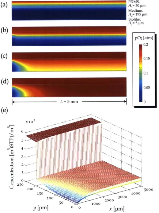
Due to the low aspect ratio (height to width) of the microchannels fabricated by multilayer soft lithography, the mass transfer for the double-layer microfluidic oxygenator
can be modeled as 2-D geometry as shown in Fig. 3.1. The oxygenator contains two channel layers with an upper oxygen reservoir over the cell culture chamber (height: H2). A thin PDMS membrane (thickness: H3) separated the channel layers, allowing only diffusion for the gas transfer across the membrane. The PDMS substrate is bonded on a glass slide to close the medium channel, so the bottom surface of medium channel is oxygen-impermeable. During cell culture, a layer of biofilm (thickness: H1) can form on the lower surface along channel over continuous/regular supply of culture medium. Under steady fluid flow with very low Reynolds number, there is a locally fully-developed velocity profile u(y) along the
cell culture chamber.
PDMS
Gas supply 0. Gas Reservoir
H3 PDMS
H u(y) Medium
H1 Cell Layer
I x Glass
Figure 3.1. Basic Design of microfluidic oxygenator.
The PDMS membrane should be sufficiently thin to obtain a short diffusion time. However, it should also provide sufficient stiffness against the deformation by gas pressure or hydraulic pressure of medium, setting a constraint on the membrane thickness. For a long oxygenation channel, the maximum possible defection of membrane h can be approximated as a simply supported beam model (in the channel width direction) under an equivalent pressure difference:
5W|4AP
S = (3.1)
32E H
where Ep,, (= 750 kPa) is the Young's modulus of PDMS; Wi, and H are the width and thickness of membrane; and AP is the pressure difference between input gas and medium. For an oxygenation channel with W = 100 pim and Hb= 20 ptm, a culture channel can be designed with thickness 20 ptm with negligible membrane deflection (-1 pm) under the operation with gas pressure -0.5 - 1.5 kPa and medium flowrate
-0.01
d/min (equivalent to hydraulic pressure -1 kPa).3-2 VALIDATION OF VELOCITY PROFILE
The velocity profile of medium mentioned in the previous section can be modeled as the planar Poiseuille flow. Validation of the flow profile in useful in the analysis of the molecular diffusion of oxygen from the gas channel to cell layer in the developing region.
.. ... ... ... ... ... ... ... ... . ... .... ...
Considering the Navier-Stokes equations of the medium layer with a chamber height (100 jim - 800 jim) much smaller than its length (3 mm) and width (3 mm), Reynolds number (Re << 1) is very small (<< 1) under an imposed medium velocity of <1 mm/s. It is assumed that the rate of change of the medium flowrate is sufficiently small such that the quasi-steady assumption holds. In the ideal case, the velocity profile u(y) is parabolic along the vertical direction, described by
u(y -h)= = 9 (Hy -792),
forH1 < <g H2 (3.2)
WHl
where Q is the flow rate of culture medium, Wis the chamber width and H,= H2- H1 is the
effective chamber height.
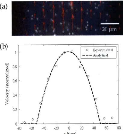
Particle Image Velocimetry (PIV) was applied to validate the steady velocity field along the effective region of microchannel (H1 <y < H2 in Fig. 3.1). In the experiment, fluorescent
beads (Molecular Probes' yellow-green-fluorescent Fluospheres beads (F8852), 1 jim diameter, Invitrogen) were first seeded with distilled water (Q = 1 jiL/min at Re = 0.003) in a microchannel without a cell layer (H1
=
0 jim, H, 500 jim and W= 100 jim) fabricatedby soft lithography. A relatively high aspect ratio
(
5) was used to generate a parabolic velocity profile with repect to channel width, which is equivalent to the velocity profile with respect to channel height direction of microchannel with aspect ratio 0.2 (H, = 100 jim and W = 500 m). Hence, this experiment can also validate the developed velocity profile u(y) along the medium channel (aspect ratio: <0.2) in Fig. 3.1. A series of microscopic images was then photographed with a GFP fluorescence filter at a fixed spot on the channel. The mean local fluidic velocity of each image sub-region, segmented with fixed length (in pixel) along width and height, was calculated by the cross-correlation of image intensity. The detailed calculation procedures are described as follows 25:1. Consider a series of consecutive grayscale images of fluorescent particles Fk(iJ),
Fk(i,j),fork= 1,2, ... ,N+ 1;i= 1,2, ... ,X;j= 1,2, ... , Y (3.3) where k is the image index, N + 1 is the number of images, X and Y are the image width
and height in pixel, respectively.
2. Since particles seldom exist at a particular image pixel over a long period of image capture, the background intensity I(ij) can be estimated by
1N+1
I(i,Ij) Fk(i, j), for i= 1, 2, ... , X;j= 1, 2, ... , Y. (3.4)
N+ 1 k=1
3. Removing background noise, obtain fk(i, j) by sampling the updated intensity in an segmented region with lower left corner (X,, Y,) and upper right corner (X, + X/N, Y,
+ Y/N), where N, and N are the number of segments along image width and height, respectively:
fk(i,
j)
= Fk (X, -1+i,Y, -1+1)- I(X, -1+iY, -1+j),f4 C ora1, 2, ... N + 1; i=1, 2, ... , X/Nx;j= 1, 2, ... , Y/Ny. (3.5) 4. Calculate the cross-correlation of each image sub-region <k(u, P) by
X/N, Y/N
GDk(U,V)= I Ifkai, -f,.(i+u, j+v), for k = 1, 2, ... , N. (3.6)
i=1 j=1
The peak location of <(u, v) is the estimate of mean velocity in a particular image region [pixel/(time between two consecutive images)], calculated by
1N
(D (u, P) = -L (u, v). (3.7)
Nk=1
The result (Fig. 3.2a,b) shows that the medium velocity basically follows a parabolic profile, as expected.
(a)
I /0/
I I I I I I I I I 01I Expeimental _ --- Analytral %0 -80 -60 -40 -20 0 20 40 60 80 :: [p m]Figure 3.2. (a) Measured velocity field using PIV, and (b) its comparison with the analytical parabolic profile (u(z)/u(W/2)) along channel width (z-) direction.
3-3 GAS MIXING MICROCHANNEL
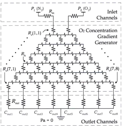
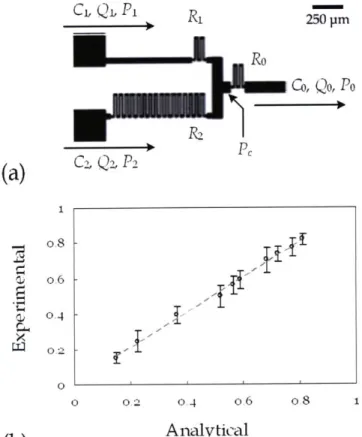
The double-layer oxygenator effectively regulates the medium DO, emphasizing the importance of the design of the gas exchange system. The preparation of gases with a particular concentration of oxygen can be achieved by mixing pure nitrogen and oxygen in different ratios. To scale down the process to a chip level operation, the basic microfluidic gas mixing element was designed as a microchannel structure composed of two inlet
(b)
N Q 4.-. 0.8 -0.6 -0.4-. -.
...
f)channels and one outlet channel as shown in Fig. 3.3a. By designing sufficiently long inlet channels, nitrogen and oxygen are locally fully mixed along them. The validity of such assumption is supported by the low scaled Peclet number, defined by the ratio between convection and diffusion, in the gas microchannels, i.e. Pe*
=
UW/D-W/L << 1, with diffusion dominating over convective fluxes. For a gas mixer having two inlets with different flow rates and oxygen concentrations, the corresponding oxygen concentration C after mixing can be estimated based on the conservation of mass:C = (3.8)
where
Q1
and Q2 are the flow rates of the channel inlets; and C1 and C2 are thecorresponding oxygen concentrations.
Practically speaking, the flow rates of gases along channels are regulated by the supply pressure of the gases at inlets (P1 and P2) and outlet (P). For given inlet and outlet pressure,
the flow rates along each channel can be predefined by the fluidic resistance along inlet (R1
and R2) and outlet (R.) channels, given by
Q= (3.9)
Q2= 2 and (3.10)
Q
= R "(3.11)where
Qo
is the flow rate of outlet channel, and P, is the gage pressure at the channel connection defined byP / R-1 + P2 /R2 + P, /R",3.2
1/R± +1/R 2+1/ R,
(3.12) In microchannels with length scale -100 tm flowed with gases at velocity -1 ms-1, the corresponding scaled Reynolds number (Re*
=
eUL/,u -H2/Lz), which is defined as the ratiobetween inertial and viscous effects, are typically in the range -10-3 - 10-1. Under dominating viscous effect, the velocity profile of gas flow is locally fully developed and the fluidic resistance R for a straight rectangular microchannel can be estimated as
1 WIH 192H tanh[(2n+1);W/2H]
1
R 12pL ; , o (2n + 1)(where , is the viscosity of fluid along channel; L, W and H are the channel length, width and height, respectively.
The folded channels were designed to be much narrower (-20 rim) than the channels for flow connection (-100 jim). So, the resistance of folded channels dominates over other sections in the mixer, and the overall resistance can be regulated by the length of folded channels. Eq. 3.13 indicates that the fluidic resistance is determined by channel dimensions and the viscosity of flowing gas mixture. Due to the different viscosity between N2and 02,
such viscosity of gas mixture p,, depending on the volumetric ratio of oxygen and nitrogen, can be approximated as
1
p ~ (3.14)
C02 / 02 +CN 2 /YN2
where C02 and Cw2 (= 1 - C62) are the volumetric concentration of oxygen and nitrogen; 02
(= 2 x 10⁻⁴ cm/s) and pm (= 1.8 x 10⁻⁴ cm/s) are the viscosity of oxygen and nitrogen, respectively.
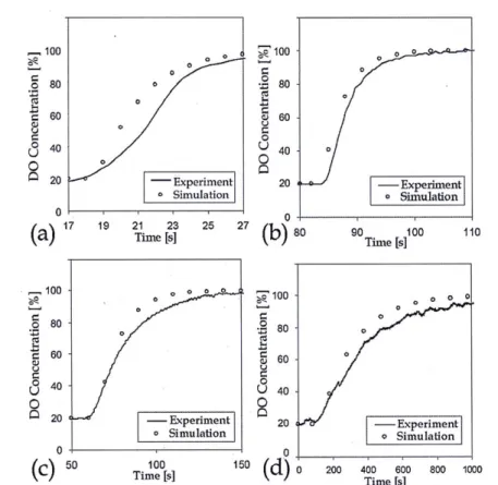
The performance of mixer was validated by a series of experiments using the fabricating mixing channel with different inlet resistances (Ri and R2). Pure nitrogen and oxygen gases were applied at the inlets with the same pressure (Pi = P2 = 1 kPa), while the outlet is connected to an oxygen sensor. (Details of the oxygen measurement are discussed in section 3.4.) Result (Fig. 3.3b) shows that the experimental oxygen levels correlated well with the expected values calculated by Eqs. 3.8 - 3.14.
Cl.
Q1.
P1 1_ 250 pm Ro Co, Qo, Po(a)
C2, Q2, P2 1 OS C) o.6 -0.4 02 -0 0 0.2 0.4 o.6 o.8 1(b)
AnalyticalFigure 3.3. (a) The structure of microfluidic gas mixer and (b) the result of the validation experiment. Experimental values are plotted against analytical volumetric ratios of oxygen, scaled

![Table 4.1. Diffusivity and solubility of oxygen in the dental biofilm, medium and PDMS Material Solubility [mM/atm] Diffusivity [cm 2 /s]](https://thumb-eu.123doks.com/thumbv2/123doknet/14093633.464865/38.918.194.745.798.920/table-diffusivity-solubility-oxygen-biofilm-material-solubility-diffusivity.webp)